Báo cáo y học: "Single ventricle with persistent truncus arteriosus as two rare entities in an adult patient: a case report" doc
Bạn đang xem bản rút gọn của tài liệu. Xem và tải ngay bản đầy đủ của tài liệu tại đây (768.88 KB, 6 trang )
BioMed Central
Page 1 of 6
(page number not for citation purposes)
Journal of Medical Case Reports
Open Access
Case report
Single ventricle with persistent truncus arteriosus as two rare
entities in an adult patient: a case report
Inna Porter and James Vacek*
Address: University of Kansas Hospital, Rainbow Boulevard, Kansas City, KS 66160, USA
Email: Inna Porter - ; James Vacek* -
* Corresponding author
Abstract
Introduction: Single ventricle and truncus arteriosus are both rare congenital cardiac syndromes
with limited survival. Their occurrence together is extremely uncommon and prolonged survival is
exceptionally rare. We present the case of a patient who had both of these defects with survival
to age 45.
Case presentation: We describe the vase of a 45-year-old man with the unusual occurrence of
two very rare congenital cardiac defects. He was found to have both truncus arteriosus and single
ventricle with long survival. His history, clinical course, and anatomic findings are discussed along
with the factors which may have contributed to his longevity, which is unique in the medical
literature. His management reflected the state of medical knowledge at the time when he
presented, and although alternate approaches may have been utilized if the patient presented today,
this case does indicate the efficacy of the management options available at the time and place of the
patient's contacts with the medical care system in Belarus. We discuss the findings, frequency,
classification, and management of both of these congenital defects.
Conclusion: This case demonstrates that patients with very complex congenital cardiac disease
may survive to adulthood, presenting challenges in both medical and surgical treatment. As the
management of these patients is constantly evolving, and interventional techniques are improving,
patients such as this with prolonged survival will be more common, with each case providing
insights to future treatment. Challenges in management may include prior care provided in health
care systems with limited resources.
Introduction
We present the case of a patient who was born with the
simultaneous occurrence of two congenital cardiac
defects, truncus arteriosus and single ventricle, which are
individually uncommon. The patient survived to age 45,
which has not been reported previously for a patient with
these types of defects. We discuss this patient's medical
history, physical, laboratory, and autopsy findings, and
provide a review of the individual congenital cardiac
lesions. We include a literature review for the congenital
cardiac defects as well as the results of investigation for
similar prior cases [1-19] which discusses anatomic find-
ings, occurrence, management, and outcomes.
Case presentation
A 45-year-old man presented to our hospital in Belarus
with the following history. He had been born in Belarus
after an uncomplicated pregnancy. His family history was
Published: 30 May 2008
Journal of Medical Case Reports 2008, 2:184 doi:10.1186/1752-1947-2-184
Received: 13 February 2007
Accepted: 30 May 2008
This article is available from: />© 2008 Porter and Vacek; licensee BioMed Central Ltd.
This is an Open Access article distributed under the terms of the Creative Commons Attribution License ( />),
which permits unrestricted use, distribution, and reproduction in any medium, provided the original work is properly cited.
Journal of Medical Case Reports 2008, 2:184 />Page 2 of 6
(page number not for citation purposes)
negative for congenital defects and the patient had no sib-
lings. After birth, a systolic murmur was heard along the
left sternal border leading to a referral to a pediatric cardi-
ology centre in Ukraine where the diagnosis of tetralogy of
Fallot was given based on physical examination, chest
radiographs, and an electrocardiogram. Surgery was
declined at that time. At no time in the patient's life were
chromosomal studies undertaken. The patient had no
children.
As a child, the patient had normal growth and mental
development, but marked cyanosis, weakness, clubbing,
and intolerance of moderate physical activity. The patient
was referred to a medical institute in Moscow at age 18,
after several episodes of syncope. A diagnosis of severe
pulmonary stenosis with ventricular septal defect was con-
sidered at that time. A right Blalock-Taussig shunt was per-
formed. The postoperative diagnosis was single ventricle
type BIII with severe pulmonary artery stenosis and hypo-
plasia. After surgery, the patient was treated with digoxin,
pentoxifylline, and spironolactone. The patient's condi-
tion improved significantly and he was able to walk sev-
eral blocks without significant dyspnea.
The patient's condition remained stable for the next five
years. He had shortness of breath with moderate exertion,
but he was asymptomatic at rest. At 27 years of age, the
patient reported an increase in dyspnea with minimal
exertion. He was diagnosed with thrombosis of the Bla-
lock-Taussig anastomosis and was treated with heparin
for 4 weeks. He never returned to his improved, postoper-
ative condition. He complained of palpitations, dull chest
pain at rest, episodes of shortness of breath at rest, and
abdominal pain. The patient had several documented epi-
sodes of ventricular tachycardia at age 35 years and was
successfully treated with propafenone.
At 39 years of age, the patient presented to our hospital in
Belarus. At the time of presentation, he complained of
severe cyanosis, shortness of breath with minimal exer-
tion, and chest pain. An electrocardiogram indicated sinus
tachycardia of 120 beats per minute. The QRS axis was to
the right (mean axis +130°). High QRS voltage suggestive
of ventricular hypertrophy was noted. There were also
ventricular extrasystoles in a trigeminal pattern and hori-
zontal ST-segment depression in the inferior leads. A 24-
hour cardiac monitor showed multiple episodes of non-
sustained ventricular tachycardia with subjective feelings
of palpitation and lightheadedness.
The patient was switched empirically from propafenone
to mexiletine with better control of his ventricular tachy-
cardia. His hematocrit was 58% to 64%. Transcutaneous
oxygen saturation was 75% to 85% on room air. The
patient could tolerate well most of his daily activities such
as walking for two blocks, grocery shopping, and perform-
ing minor work at home. Over the next 6 years he had
repeated hospitalizations for dyspnea, chest pain, and
near syncope. He was treated with phlebotomies, saline
and/or dextran infusions to improve viscosity, and medi-
cations including spironolactone, pentoxifylline, and
aspirin.
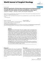
Electrocardiogram from the terminal hospitalizationFigure 1
Electrocardiogram from the terminal hospitalization. Leads are as follows: top to bottom on the left, I, II, III, aVR, aVL,
aVF; and top to bottom on the right, V1 through V6.
Journal of Medical Case Reports 2008, 2:184 />Page 3 of 6
(page number not for citation purposes)
At the time of the terminal hospitalization, physical exam-
ination was remarkable for a 3/6 systolic murmur heard at
the upper left sternal border, edema of the right ankle and
foot, and tachycardia of 140 beats per minute. The patient
was noted to experience severe shortness of breath with
even minimal exertion and was deeply cyanotic. The elec-
trocardiogram is shown in Figure 1 and demonstrated
sinus rhythm with a mild tachycardia at a rate of 110 beats
per minute, right axis deviation, and a non-specific intra-
ventricular conduction delay with diffuse ST-T changes.
Chest radiograph indicated a small pleural effusion on the
right and increased pulmonary vascularity. The heart was
moderately enlarged with prominence of the aorta. An
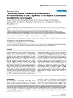
echocardiogram was performed (Figure 2). A single ventri-
cle with unremarkable atrioventricular valves was seen.
The end diastolic diameter of the ventricle was measured
as 69 mm, the posterior wall thickness was 17 to 19 mm,
and the left atrium measured 29 mm. The estimated ejec-
tion fraction was 45% to 50%. A vessel with a semilunar
valve (the truncal valve) arising from the ventricle was
seen. The cusps of the valve were hyperechogenic. Moder-
ate regurgitation was noted. No pulmonary artery was
seen. Doppler study of the lower extremities showed
thrombosis in the veins of the right calf. Heparin therapy
and intravenous fluids were initiated. The patient's condi-
tion deteriorated rapidly and several hours later he
became comatose and died.
Autopsy showed an enlarged heart that weighed 750 g,
composed of two atria with an intact septum, and a single
ventricle. The right atrium was enlarged to a diameter of
10 cm. The diameter of the left atrium was 3 cm. Both
caval veins emptied normally into the right atrium. All
four pulmonary veins entered the left atrium normally.
Both atrioventricular valves had normal anatomy, free of
vegetations. The single ventricle with left ventricular char-
acteristics had a diameter of 12 cm with a wall thickness
of 2 to 2.2 cm. A single artery, truncus arteriosus, arose
from the ventricle and arched to the left. The truncal valve
had three cusps that were moderately calcified. The coro-
nary ostia and vessels were normal. The pulmonary artery
trunk was located 3.5 cm from the origin of the truncus
and divided to form left and right pulmonary arteries. At
the hilum of the right lung the right pulmonary artery was
surgically connected to the right subclavian artery. Red-
gray masses were noted at the Blaylock-Taussig shunt
anastomosis. Below the anastomosis the right pulmonary
artery was almost completely occluded by a dark red
adherent thrombus. The ligamentum arteriosum was a
fibrous cord. Microscopically the lungs showed dilatation
of the pulmonary arterioles and alveolar capillaries. Many
bronchial and pulmonary arterioles contained recanal-
ized thrombi or emboli.
The main anatomic diagnoses were: single ventricle with
truncus arteriosus; status post Blalock-Taussig procedure;
old thromboses of the established anastomosis; hypopla-
Echocardiogram from the terminal hospitalizationFigure 2
Echocardiogram from the terminal hospitalization. Apical view showing single ventricle with two atrioventricular
valves.
Journal of Medical Case Reports 2008, 2:184 />Page 4 of 6
(page number not for citation purposes)
sia of the right pulmonary artery; congestive heart failure;
recent thrombosis of the right pulmonary artery; and deep
vein thrombophlebitis of the right calf.
Discussion
A single ventricle is defined as a heart with one ventricle
receiving inflow from two separate atrioventricular valves
or a common atrioventricular valve [1]. Single ventricle
accounts for about 1% of all cardiac anomalies with an
incidence of about 0.05 to 0.1 per 10,000 live births [2].
The cause is unknown, but it is most likely multifactorial
with a genetic predisposition.
Single ventricles may be classified based on the location of
the great arteries [3]. There may be normally related great
arteries (type I), D-transposition of the great arteries (type
II), or L-transposition (type III). The existence of pulmo-
nic stenosis or pulmonary atresia further subdivides the
types of single ventricle. A single ventricle may be accom-
panied by pulmonary atresia (type A), presence of pulmo-
nic stenosis (type B), or absence of pulmonic stenosis
(type C). Depending on the ventricular morphology, the
single ventricle can be subdivided as left ventricular type
(65% to 70%), right ventricular type (20%), or indetermi-
nate type (10% to 14%) [3]. All patients with a single ven-
tricle have some degree of hypoxemia caused by
intracardiac shunting. Clinical manifestations are usually
apparent shortly after birth. The most common findings
are dyspnea, tachycardia, cyanosis, and progressive heart
failure. Later, secondary erythrocytosis and clubbing are
usually present.
The diagnosis may be defined by echocardiography, car-
diac catheterization, and cardiac magnetic resonance
imaging. Treatment options depend on the presence of
associated defects. Infants with increased pulmonary
blood flow and pulmonary artery pressure require pulmo-
nary artery banding. This procedure helps to prevent early
death from congestive heart failure but carries a signifi-
cant surgical mortality rate. Patients with severe pulmo-
nary outflow obstruction require creation of an
aortopulmonary shunt. The Blalock-Taussig procedure is
the most commonly performed shunting operation. A
modified Fontan procedure may be performed later to
separate the pulmonary and systemic circulations. Opera-
tive mortality is about 8% to 25% and 10-year survival is
60% to 81%, depending on the pre- and postoperative
risks [4]. The median life expectancy of patients without
surgical correction is 4 to 14 years [5], although there are
descriptions of very rare cases in the literature when such
patients have survived over 40 years [2,5-8]. About 65% to
75% of patients without surgical corrections die during
the first year of life [2]. The most common causes of death
are arrhythmias, heart failure, and sudden cardiac death.
Truncus arteriosus is another rare anomaly, defined as a
single great artery that originates from the base of the
heart and gives rise to the pulmonary, systemic, and coro-
nary circulation [9-13]. A single semilunar valve is found
in truncus arteriosus. The arterial trunk can be connected
with the right ventricle, left ventricle, or override and be
symmetrically distributed over both ventricles. It has an
incidence of about 0.5 to 0.9 per 10,000 live births [9]. A
ventricular septal defect is almost always present. Several
classifications are used for this anomaly. Van Praagh [10]
classified the disorder as types A and B. In type B, there is
no association with ventricular septal defect. Type A is
subdivided as follows:
1. Type A1: partially separated pulmonary trunk.
2. Type A2: two pulmonary arteries arising directly from
the truncus arteriosus.
3. Type A3: a single pulmonary artery originating from the
arterial trunk, along with collaterals originating from the
descending aorta.
4. Type A4: significant abnormalities of the aortic arch in
association with anomalies of the ductus arteriosus.
Patients with truncus arteriosus have some degree of cya-
nosis during the first week of life. Congestive heart failure
usually occurs by a few weeks of age. Excessive pulmonary
blood flow at high pressure results in pulmonary vascular
obstructive disease by 3 months. The diagnosis of truncus
arteriosus is suspected in newborns with mild cyanosis, a
cardiac murmur, and pulmonary overcirculation. Factors
that limit pulmonary blood flow, such as pulmonary
artery stenosis or persistently elevated pulmonary vascular
resistance, may delay the appearance of symptoms. The
diagnosis is established by echocardiography and cardiac
catheterization. The only definitive treatment for this
anomaly is surgical correction. Complete repair is pre-
ferred and involves three major components: separating
the pulmonary arteries from the main truncus, closure of
the ventricular septal defect using a patch, and creating a
connection between the right ventricle and the pulmo-
nary arteries using a valve conduit, usually a homograft
pulmonary artery. Currently over 90% of children survive
repair of truncus arteriosus. Long-term survival after surgi-
cal correction is about 83% at 15 years after surgery [10].
The prognosis for patients without surgical correction is
dismal. The mortality rates are about 20% at 1 week of age
and more than 90% at 1 year [11].
Our presented case is a unique case of a single ventricle
with truncus arteriosus type A1 (Van Praagh classifica-
tion) in a patient who lived for 45 years. The reported
association of these two defects is extremely rare [6,14-
Journal of Medical Case Reports 2008, 2:184 />Page 5 of 6
(page number not for citation purposes)
19], and most of these patients die within a few weeks of
birth. As described above, the embryology of a single ven-
tricle and truncus arteriosus is different. It is thought that
the unlikely concurrence of the two unusual developmen-
tal defects within the same patient may explain the
extreme rarity of this condition.
The prolonged survival of the presented patient is also
exceptional. Only one case of long-term survival of a
patient with a single ventricle defect and truncus arterio-
sus has been reported previously [6] which was a woman
with a single ventricle and truncus arteriosus (apparently
type 4) who lived for 56 years. To the best of the authors'
knowledge there are no cases of prolonged survival
reported for a patient with a single ventricle and type 1A
truncus arteriosus. The patient in our report had a history
of pulmonary stenosis and pulmonary hypoplasia (not
typical for truncus type 1) that limited pulmonary flow
transmission of systemic pressure to the pulmonary arte-
rial system and likely improved his survival. In patients
with single ventricle who do not have significant obstruc-
tion to pulmonary arterial flow with protection of the pul-
monary vasculature, early occurrence of congestive heart
failure or pulmonary vascular occlusive disease is likely
with very limited survival unless a procedure is under-
taken to limit pulmonary flow (such as pulmonary band-
ing). If, however, severe pulmonary stenosis or atresia is
present without provision of adequate pulmonary flow by
surgical intervention with a shunt procedure, outcomes
are also very poor due to severe cyanosis. Survival is opti-
mized when an appropriate balance of systemic to pulmo-
nary flow is present either spontaneously or by surgical
intervention. In this patient, his initial degree of pulmo-
nary outflow obstruction and subsequent Blalock-Taussig
shunt fortunately provided the necessary achievement of
a balance of systemic versus pulmonary circulatory flow
that allowed prolonged survival.
With the passage of time and advances in medical knowl-
edge and experience, future cases such as this may benefit
from other types of surgical interventions, long-term full
intensity oral anticoagulation, or different approaches to
anti-arrhythmic management. For the patient in this
report, placing him on therapeutic anticoagulation after
his thrombotic event at age 27 would have been a strong
consideration. Anticoagulation in the setting of prior
thromboembolic events or significant polycythemia for a
patient such as this is very reasonable. Utilization of
thrombolytic therapy in the setting of a suspected life-
threatening thromboembolic event should also be con-
sidered, although diagnosis may be difficult.
Management of arrhythmias in patients such as this, as for
many congenital heart disease patients, remains challeng-
ing with limited data in subsets of unusual entities such as
those expressed by our patient [20]. Options for manage-
ment include typical anti-arrhythmic agents (amiodar-
one, mexilitine, beta blockers), investigational agents,
radiofrequency ablation, or implantation of cardioverter-
defibrillators. Much of the available literature on radiofre-
quency ablation in patients with congenital heart disease
relates to supraventricular arrhythmias, without clear doc-
umentation of mortality benefit, but symptomatic
improvement in some patients [21]. Due to the complex-
ity of anatomy, both intrinsic and corrected, identification
and radiofrequency ablation of arrhythmic foci may be
difficult. Utilization of an implanted device may be best
reserved for patients with prior episodes of sudden cardiac
death, documented sustained ventricular tachycardia,
unexplained syncope, and/or significantly reduced ven-
tricular function, although definitive documentation of
benefit is lacking at this point in time [22].
Conclusion
In summary, we have presented a unique case of a man
who lived for 45 years with a single ventricle with truncus
arteriosus type I. The described association of these two
defects is extremely rare and the prolonged survival of this
man is also exceptional. To the best of the authors' knowl-
edge there are no cases of prolonged survival reported in
a person with a single ventricle and type 1 truncus arteri-
osus.
Competing interests
The authors declare that they have no competing interests.
Consent
Written informed consent could not be obtained in this
case since the patient's next-of-kin were untraceable. We
believe this case report contains a worthwhile clinical les-
son which could not be as effectively made in any other
way. We expect the patient's next-of-kin not to object to
the publication since every effort has been made so that
the patient remains anonymous.
Authors' contributions
IP was involved with the patient's care at the end of his life
as well as gathering the case history, doing the majority of
the literature review, and writing the original manuscript.
JV has reviewed and revised the manuscript through sev-
eral drafts, extensively modified substantial portions of
the narrative, and performed some of the literature review.
References
1. Van Praagh R, Plett JA, Van Praagh S: Single ventricle: pathology,
embryology, terminology, and classification. Herz 1979,
4:113-150.
2. Samanek M, Voriskova M: Congenital heart disease among
815,569 children born between 1980 and 1990 and their 15-
year survival: a prospective Bohemia survival study. Pediatr
Cardiol 1999, 20:411-417.
Publish with BioMed Central and every
scientist can read your work free of charge
"BioMed Central will be the most significant development for
disseminating the results of biomedical research in our lifetime."
Sir Paul Nurse, Cancer Research UK
Your research papers will be:
available free of charge to the entire biomedical community
peer reviewed and published immediately upon acceptance
cited in PubMed and archived on PubMed Central
yours — you keep the copyright
Submit your manuscript here:
/>BioMedcentral
Journal of Medical Case Reports 2008, 2:184 />Page 6 of 6
(page number not for citation purposes)
3. Fulton DR, Freed MD: The pathology, pathophysiology, recog-
nition, and treatment of congenital heart disease. In Hurst's
The Heart 11th edition. Edited by: Fuster V, Alexander RW, O'Rourke
RA, Roberts R, King SB. Wellens HJJ: New York, NY: McGraw-Hill;
2004:1840-1842.
4. Driscoll DJ, Offord KP, Feldt RH, Schaff HV, Puga FJ, Danielson GK:
Five- to fifteen-year follow-up after Fontan operation. Circu-
lation 1992, 85:469-496.
5. Moodie DS, Ritter DG, Tajik AJ, O'Fallon WM: Long-term follow-
up in the unoperated univentricular heart. Am J Cardiol 1984,
53:1124-1128.
6. Mehta JB, Hewlett RF: Cor triloculare biauriculare: an unusual
adult heart. Br Heart J 1945, 7:41-49.
7. Carns ML, Ritchie G, Musser MJ: An unusual case of congenital
heart disease in a woman who lived for forty-four years and
six months. Am Heart J 1941, 21:522-529.
8. Ammash NM, Warnes CA: Survival into adulthood of patients
with unoperated single ventricle. Am J Cardiol 1996, 77:542-544.
9. Mair DD, Edwards WD, Julsrud PR, Seward JB, Danielson GK, Gold-
muntz E: Truncus Arteriosus. In Moss and Adam's Heart Disease in
Infants, Children and Adolescents 6th edition. Edited by: Allen HD, Gut-
gesell HP, Clark EB, Driscoll DJ. Baltimore, MD: Lippincott Williams
& Wilkins; 2001:911-923.
10. Van Praagh R: Truncus arteriosus: what is it really and how
should it be classified? Eur J Cardiothorac Surg 1987, 1:65-70.
11. Rajasinghe HA, McElhinney DB, Mohan Reddy VM, Mora BN, Hanley
FL: Long-term follow up of truncus arteriosus repaired in
infancy: a twenty-year experience. J Thorac Cardiovasc Surg 1997,
113:869-879.
12. Sirbu A, Sirbu D: Sudden death due to truncus arteriosus-a
case report. Rom J Leg Med 2003, 11:302-306.
13. Carter JB, Blieden LC, Edwards JE: Persistent truncus arteriosus:
report of survival to age of 52 years. Minn Med 1973,
56:280-282.
14. Paris YM, Bhan I, Marx GR, Rhodes J: Truncus arteriosus with a
single left ventricle: case report of a previously unrecognized
entity. Am Heart J 1997, 133:377-380.
15. Siddoway JJ, Chernish SM: Truncus arteriosus associated with
single ventricle. Am J Dis Child 1952, 84(6):706-717.
16. Shapiro SR, Ruckman RN, Kapur S, Chandra R, Galioto FM, Perry LW,
Scott LP: Single ventricle with truncus arteriosus in siblings.
Am Heart J 1981, 102:456-459.
17. Shaddy RE, McGough EC: Successful diagnosis and surgical
treatment of single ventricle, truncus arteriosus. Ann Thorac
Surg 1989, 48:298-300.
18. Shakibi JG, Aryanpur I, Nazarian I, Siassi B: Association of atriov-
entricular valve atresia with single ventricle, truncus arteri-
osus communis and transposition. A basic reorientation in
the approach to the definition of congenital heart defects.
Jpn Heart J 1978, 19:346-57.
19. Miller AJ, Prec O, Akman L, Katz LN, Gibson S: A case of congen-
ital heart disease-truncus aorticus solitarius, single ventricle,
and aberrant coronary drainage into the common ventricle.
Am Heart J 1950, 39:607-614.
20. Friedman RA: Sudden cardiac death in patients with congeni-
tal heart disease. Card Electrophysiol Rev 1997, 1/2:241-243.
21. Triedman JK, Alexander ME, Love BA, Collins KK, Berul CI, Bevilac-
qua LM, Walsh EP: Influence of patient factors and ablative
technologies on outcomes of radiofrequency ablation of
intra-atrial re-entrant tachycardia in patients with congeni-
tal heart disease. J Am Coll Cardiol 2002, 39:1827-1835.
22. Alexander ME, Cecchin F, Walsh EP, Triedman JK, Beilacqua LM,
Berul CI: Implications of implantable cardioverter defibrilla-
tor therapy in congenital heart disease and pediatrics. J Car-
diovasc Electrophysiol 2004, 15:72-76.