báo cáo khoa học:" Circulating Immune Complexes and trace elements (Copper, Iron and Selenium) as markers in oral precancer and cancer : a randomised, controlled clinical trial" pot
Bạn đang xem bản rút gọn của tài liệu. Xem và tải ngay bản đầy đủ của tài liệu tại đây (290.84 KB, 10 trang )
BioMed Central
Page 1 of 10
(page number not for citation purposes)
Head & Face Medicine
Open Access
Research
Circulating Immune Complexes and trace elements (Copper, Iron
and Selenium) as markers in oral precancer and cancer : a
randomised, controlled clinical trial
Sunali S Khanna*
1
and Freny R Karjodkar
2
Address:
1
Department of Oral Medicine and Radiology, Nair Hospital Dental College, Mumbai, India and
2
Department of Oral Medicine and
Radiology, Nair Hospital Dental College, Mumbai, India
Email: Sunali S Khanna* - ; Freny R Karjodkar -
* Corresponding author
Abstract
Aim: To evaluate the levels of circulating immune complexes, trace elements (copper, iron and
selenium) in serum of patients with oral submucous fibrosis (OSMF), oral leukoplakia (L), and oral
squamous cell carcinoma (SCC), analyze the alteration and identify the best predictors amongst
these parameters for disease occurrence and progression.
Methods: Circulating immune complexes (CIC) were estimated using 37.5% Polyethylene Glycol
6000(PEG) serum precipitation. Serum estimation of copper (Cu), Iron (Fe) and selenium (Se) was
done using the Oxalyl Dihydrazide method, Colorimetric Dipyridyl method and the Differential
Pulse Cathodic Stripping Voltametry respectively.
Results: The data analysis revealed increased circulating immune complex levels in the precancer
and cancer patients. Serum copper levels showed gradual increase from precancer to cancer
patients. However, serum iron levels were decreased significantly in the cancer group. Selenium
levels showed marked decrease in the cancer group. Among CIC, serum, copper, iron and selenium
the best predictors for the occurrence of lesions were age, serum iron, CIC, serum selenium in the
decreasing order.
Conclusion: The present study shows that these immunological and biological markers may be
associated with the pathogenesis of oral premalignant and malignant lesions and their progressions.
Concerted efforts would, therefore, help in early detection, management, and monitoring the
efficacy of treatment.
Background
Oral cancer the sixth most common cancer worldwide
continues to be the most prevalent cancer related to the
consumption of tobacco, alcohol and other carcinogenic
products[1]. While the cancer incidence remains high in
South and South East Asia (its traditional high risk areas);
parts of Central and Eastern Europe are seeing alarming
increase and now constitute the highest incidence parts of
the globe[2].
Increasing awareness on part of the providers of treat-
ment, as well as the population in general, has led to a
large proportion of patients presenting with earlier stage
of the disease.
Published: 16 October 2006
Head & Face Medicine 2006, 2:33 doi:10.1186/1746-160X-2-33
Received: 04 March 2006
Accepted: 16 October 2006
This article is available from: />© 2006 Khanna and Karjodkar; licensee BioMed Central Ltd.
This is an Open Access article distributed under the terms of the Creative Commons Attribution License ( />),
which permits unrestricted use, distribution, and reproduction in any medium, provided the original work is properly cited.
Head & Face Medicine 2006, 2:33 />Page 2 of 10
(page number not for citation purposes)
Epidemiological studies indicate that intervention at an
early stage might reduce oral carcinoma related deaths.
The discovery of immunological markers at a clinical, his-
tological and molecular level has marked the end of an era
of groping in the dark for clues to the basis of cancer. Sig-
nificant reduction in mortality can be achieved my
advances in early diagnosis and implementation of multi-
disciplinary treatment programmes leading to improve-
ment of survivorship and better quality of life.
Oral precancer and cancer
In India, oral cancer is prevalent in most areas where
tobacco related practices are observed. For development
of oral cancer, tobacco is the single greatest risk factor.
This is due to higher concentration of carcinogenic expo-
sure and failure to clean the carcinogens from the mucosal
surface. If one observes the mouths of heavy tobacco
users, the accumulation of tobacco residue may be corre-
lated with areas of the oral cavity involved [3]. Alcohol,
viruses, genetic mechanisms, candida, chronic irritation
and diet deficiency states are also implicated in the etiol-
ogy[4,5].
The development of oral cancer is a multistep process aris-
ing from pre-existing potentially malignant lesions. Leu-
koplakia is the most common precancer representing 85%
of such lesions[6]. Histologically, over 95% of oral can-
cers are squamous cell carcinomas[7,8]. It has been sug-
gested that a vast majority of oral squamous cell
carcinomas in India arise from pre- existing Leukopla-
kia[9].
Likewise, the incidence of oral submucous fibrosis
(OSMF) is increasing like an epidemic, targeting the
younger generation. The etiology for OSMF is still obscure
and a varied number of factors have been proposed. Of
these, areca nut use is the most important and persistent
finding in history taking[10].
Role of circulating immune complexes
Intensive studies have documented the role of immune
complexes as modulators of both cellular and humoral
immune response. The occurrence of circulating immune
complexes (CIC) as a marker for tumor burden and prog-
nosis in the sera of patients with oral precancer and cancer
is now well established. Recent advances in the fields of
CIC, tumor progression, drug resistance, tumor cell heter-
ogeneity and metastasis have resulted in a renewed inter-
est in the development of non- specific
immunotherapeutic modalities [11].
The overall consensus is that only a small percentage of
the detected CIC in vivo represent tumor associated anti-
gens complexed with antibodies. The bulk of CIC most
likely represent auto antibodies or the reaction to dena-
tured self proteins, microbes, normal lymphocyte, anti-
gens and nuclear antigens[12]. Antigenic make up of CIC
in cancer patients reflects the host's immune response to
a variety of often overlapping antigenic stimuli and hence
paves way for further studies[13].
Trace elements have been extensively studied in recent
years to assess whether they have any modifying effects in
the etiology of cancer. Copper, iron and selenium are
essential for numerous enzymes and therefore it is reason-
able to assume that variations in serum level of these bio-
chemical markers maybe associated with the pathogenesis
of oral cancer. The importance of these elements in cancer
was reported by Schwartz [14] which opened the door for
new diagnostic and therapeutic endeavours in many areas
of medicine and specifically in the areas of oncology.
Immunological and biochemical alterations in the serum
of such patients can help not only in the early diagnosis,
appropriate treatment but also as indicators of prognosis,
as the disease progresses.
Materials and methods
This study was carried out in Nair Hospital Dental Col-
lege, Mumbai in association with Bhabha Atomic
Research Centre and Topiwala National Medical College,
Mumbai.
Thirty patients with (OSMF/L and 30 patients with OSCC
with histopathologically proven lesions were included in
this study. For comparison thirty normal subjects were
also selected. The age group of these patients ranged from
25–70 years. The symptoms and signs of the patients were
evaluated, after through history taking [15-18].
The following investigations were carried out in Serum
obtained from 10 ml of various blood collected from the
subjects -
1) Serum CICs were determined using 3.75% Polyethyl-
ene Glycol – 6000 (PEG) serum precipitation[19].
2) Serum levels of Copper (Cu) were determined using
the Colorimetric Oxalyl Dihydrazide method[20].
3) Serum analysis of Iron (Fe) was done using colorimet-
ric Dipyridyl method[20].
4) Differential Pulse Cathodic Stripping Voltametry to
determine serum selenium (Se) [21].
Statistical methods
The data was subjected to statistical analysis using the Chi
Square Test, Standard Deviation, Student's unpaired t-test,
correlation, ANOVA and Linear Regression.
Head & Face Medicine 2006, 2:33 />Page 3 of 10
(page number not for citation purposes)
Results
Firstly, groupwise comparison of gender among all cases
was considered. In the precancer (oral submucous fibro-
sis/leukoplakia) group, females were 16.70% and males
formed 83.30% of the subjects. In the cancer group
females formed 40% and males attributed to 60% of the
subjects. In comparison with normals, the difference in
gender between the three groups was not found to be sta-
tistically significant; (p value was 0.058) indicating that
the 3 groups are comparable on the basis of gender (Table
No. 1)
Most of the individuals in the study were males who had
tobacco, areca nut chewing and associated habits.
The age (in years) range of the patients with precancerous
condition/lesion was 34.10 in the precancer group as
compared to 53.97 in cancer group and 33.65 in the nor-
mal group. The mean age in precancer and cancer group
was higher than normal and the difference was statisti-
cally significant {p value1.10E-13 (1.10 × 10
-13
)}
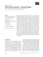
The mean CIC levels were 0.07, 0.10 and 0.03 OD
450
in
the precancer, cancer and normal group respectively.
There was a marked increased in the precancer and cancer
patients. The p value 5.67 E-08 which was statistically sig-
nificant (Table No. 1 Figure No 1 and 4).
The mean serum copper levels are 127.63, 128.27 and
116.60 μg/100 ml in the precancer, cancer and normal
group respectively. The p value was 0.012 which is statis-
tically significant (Table No 1, Figure No 2 and 4)
The mean serum iron levels are 101.13, 105.20 and
138.10 μg/100 ml in precancer, cancer and normal groups
respectively. The difference between the three groups was
found to be statistically significant (p value was 2.35E-19)
(Table No 1, Figure No 3 and 4)
The mean serum selenium content is 63.13, 51.97 and
68.04 ng/ml in precancer, cancer and normal groups
respectively. It is significantly decreased in the cancer
groups (p value was 2.35E-19) (Table No 1 and Figure No
4)
Correlation among the CIC and serum copper(Figure No
5) copper and iron, CIC and age was found to be signifi-
cant in the precancer group.
Correlation among the CIC and serum copper (Figure No.
6) and serum copper and age was found to be significant
in the cancer group. They showed a steady rise
Among age, CIC, serum copper, iron and selenium the
best predictors for the occurrence of lesion were age,
serum iron, CIC and serum selenium in the decreasing
order (Figure No. 7)
Discussion
Research emphasizes the development of generalizations,
principles or theories that will be helpful in the prediction
of future occurrences.
We would all agree that no aspect of total patient care has
been more important than the modern concepts of pre-
vention, diagnosis, treatment and their systemic relation-
ship.
The rate at which oral precancerous and cancerous lesions
are spreading like an epidemic is alarming. The prevalence
of oral precancerous lesions is much higher than that of
oral cancer and these lesions provide useful clinical mark-
ers for oral cancer.
Immunological and biochemical alterations in the sera of
such patients can help not only in early diagnosis, appro-
priate treatment but also as indicators of prognosis, as the
disease progresses.
Oral cancer is an extremely deadly disease. It comprises
approximately 2% of the total malignant tumors in West-
ern Europe and North America, but in India, upto half of
the cancers may be present in the mouth [22].
The etiology of oral squamous cell carcinomas include
various carcinogens in tobacco and related products such
as polynuclear aromatic hydrocarbons, and nitrosamines.
Alcohol, viruses, genetic mechanisms, candida, chronic
Table No. 1: Groupwise comparison of various variables among all cases.
Variables ANOVA test applied
F-value P-value Difference is-
Age 45.073 1.10E-13 Significant
CIC 20.885 5.67E-08 Significant
Cu 4.662 0.012 Significant
Fe 78.805 2.35E-19 Significant
Se 1.714 0.187 Not significant
Head & Face Medicine 2006, 2:33 />Page 4 of 10
(page number not for citation purposes)
irritation and diet deficiency states rare also implicated
[23,24].
Amongst the various precancerous lesions and conditions
known oral submucous fibrosis is gaining importance
because of the large number of case reported in the recent
years in the younger generation and because of its obscure
etiology. The incidence of malignant changes in patients
with oral submucous fibrosis ranges from 3 to 6%. Several
factors such as chillies consumption, nutritional defi-
ciency, areca nut chewing, genetic susceptibility, autoim-
munity and collagen disorders have suggested to be
involved in the pathogenesis of this condition. Currently,
areca nut chewing is considered to be most important eti-
ologic factor of oral submucous fibrosis [25].
The precancerous nature of the most common of chronic
oral mucosal lesions, leukoplakia is much better under-
stood now than at any time, since it was first brought to
professional attention by Sir James Paget 143 years ago.
Oral leukoplakia is well established as one of the very best
examples of premalignancy in man. The range of the rate
of malignant transformation of this lesion is 3% to 20%
[26].
The immunological abnormalities in patients with cancer
in the head and neck appear to be more profound than
those associated with cancers of the bronchus, breast, cer-
vix, colon or bladder (Litchenstein et al) [27]. The immu-
noglobulin deposits may represent immune (antigen-
antibody) complexes, since circulating immune com-
plexes have been detected in 75% of patients with head
and neck carcinoma (Scully et al) [28].
Majority of our study group consisted of males (66.67%)
who had tobacco, areca nut chewing and associated hab-
its. The mean age was higher in the patients suffering from
oral carcinoma.
Gross et al [29] reported that ageing is associated with a
decline in the cell mediated immunity which might pre-
dispose to oncogenesis. Circulating immune complexes
have been implicated in autoimmune diseases, neoplastic
diseases, infectious diseases caused by bacteria, viruses
and parasites. Scully C, Barkas T. et al [30] evaluated the
circulating immune complexes in patients with squamous
cell carcinoma and found them significantly raised.
Hoffken et al [31] concluded that the elevation of circulat-
ing immune complexes was attributed to change in the
levels of complement fixing and non-complement fixing
of tumour specific antibodies. This implied that it may be
possible to monitor the malignant transformation of pre-
malignant lesions. Also, emphasis should be laid on the
Illustrates marked increase in levels of CIC in precancer (OSMF/L) and cancer groupsFigure 1
Illustrates marked increase in levels of CIC in precancer (OSMF/L) and cancer groups. as comapred to normals.
0.07
0.10
0.03
0.00
0.02
0.04
0.06
0.08
0.10
0.12
Mean CIC
Precancer(OSMF/L) Cancer Normal
Group
Head & Face Medicine 2006, 2:33 />Page 5 of 10
(page number not for citation purposes)
detection of the antigenic component of the circulating
immune complexes.
Chatterjee R. and Guha [32] estimated levels of circulating
immune complexes using polyethylene glycol precipita-
tion assay; which they found to be appropriate and con-
cluded that 60% of patients with carcinoma of the buccal
mucosa had markedly higher amount of immune com-
plexes. They also noted that the amount of immune com-
plexes present in patient's sera showed no correlation with
serum level of IgG, IgA and IgM.
Balaram P et al [33] reported increased levels of circulat-
ing immune complexes in oral submucous fibrosis
patients.
In the presence study the levels of CIC show a gradual
increase in the precancer group and the cancer group is
characterized by a marked increase in levels which was sta-
tistically significant. From these results it can be hypothe-
tised that CIC represent the host's physiological and
immunological defense response in eliciting specific anti-
bodies upon exposure to most antigenic substances.
CIC deposition further leads to inflammation and tissue/
cell damage. It also leads to suppression of cell mediated
immunity and modulates the humoral response.
Circulating immune complexes are normally removed by
the mononuclear phagocytic cells. However, circulating
immune complexes formations or their defective clear-
ance under certain circumstances becomes detrimental to
the host, resulting in pathological deposition. Thus, alter-
ing the host immunological response leading to inflam-
mation and tissue injury [22].
High levels of copper in areca nut, a major etiological fac-
tor in OSMF plays an initiating role in stimulation of
fibrogenesis by up regulation of lysyl oxidase (Ma. R. H. et
al) [32] and thereby causing inhibition of degradation of
collagen. The rise in serum copper may be due to
increased turnover of ceruloplasmin (a copper carrying
globulin with essential oxidase activity) (Jaydeep et al)
[33] in the serum of carcinoma patients. Varghese et al
[34] concluded a significant reduction in serum copper in
oral cancer, OSMF and leukoplakia patients.
Margalith et al [35] suggested that role of copper ions in
biological damage is caused by superoxide radicals or
other reducing agents such as ascorbate, which reduce the
copper complex. These complexes react with hydrogen
peroxide to form hydroxyl radicals that cause damage to
protein, RNA and DNA that are not repairable by cellular
mechanisms thus initiating the malignant process
Gradual increase of copper levels from precancer to cancer as compared to normalsFigure 2
Gradual increase of copper levels from precancer to cancer as compared to normals.
127.63
128.27
116.60
108
112
116
120
124
128
132
Mean Cu level
Precancer(OSMF/L) Cancer Normal
Group
Head & Face Medicine 2006, 2:33 />Page 6 of 10
(page number not for citation purposes)
In this study, Serum levels of copper showed gradual
increase from precancer to the cancer group as compared
to normals which was statistically significant.
Serum Iron levels are considered as biochemical indica-
tors for nutritional assessment. Utilization of iron in col-
lagen synthesis [36] by the hydroxylation of proline and
lysine leads to decreased serum iron levels in OSMF
patients. In most cases clinical anemia may be a contrib-
uting factor. (Ramanathan et al) [37].
Occurrence of iron deficiency is known to present in oral
cancer. Iron is known to play a key role in the develop-
ment of hepatic fibrosis probably via oxidative stress and
lipid peroxidation [38]. Iron is also required for collagen
synthesis by enzymes in hydroxylation of proline and
lysine. This hydroxylation of proline and lysine is cata-
lyzed by proline hydroxylase and peptidyl lysine hydrox-
ylase respectively. Peptidyl proline hydroxylase requires as
co-factory molecular oxygen, ferrous iron, alpha-ketoglu-
tarate and ascorbic acid [39].
A statistically significant reduction in the serum iron level
was present in the precancer group in our study. A
decrease in the iron levels in the cancer group, but higher
than that of pre cancer groups was found to be significant.
Recently, haematological abnormalities in oral leukopla-
kia was reported by Chellacombe [39]. It was reported
that poor correlation between iron indices, tumour
parameters, serum iron and hemoglobin was probably
due to utilization of iron by bone marrow and tumours.
Ramanathan K [37] reported that oral submucous fibrosis
may be the manifestation of chronic iron deficiency ane-
mia.
There appears to be an association between the serum iron
content and oral carcinogenesis. More detailed studies on
a large data base should be instituted to elucidate the exact
role of iron.
Selenium forms the integral part of the enzyme glutath-
ione peroxidase, type I iodothyronine deiodinase, metal-
loprotein, fatty acid binding protein and selenoprotein P.
therefore selenium is considered as an antioxidant nutri-
ent and the diseases where low selenium is implicated
range from nutritional disorders like protein energy mal-
nutrition to degenerative diseases such as cancer [40].
Rajendran R [41] estimated the levels of cadmium, sele-
nium, chromium, magnesium and calcium in the sera of
patients with oral leukoplakia, oral submucous fibrosis,
squamous cell carcinoma using atomic absorption spec-
Indicates statistically significant reduction in the serum iron levels of precancer and cancer group as compared to normalsFigure 3
Indicates statistically significant reduction in the serum iron levels of precancer and cancer group as compared to normals.
101.13
105.20
138.10
0
20
40
60
80
100
120
140
Mean Iron level
Precancer(OSMF/L) Cancer Normal
Group
Head & Face Medicine 2006, 2:33 />Page 7 of 10
(page number not for citation purposes)
Groupwise comparison of CIC, copper, iron and seleniumFigure 4
Groupwise comparison of CIC, copper, iron and selenium.
0.07
0.10
0.03
127.63
128.27
116.60
101.13
105.20
138.10
63.13
51.97
68.04
0
20
40
60
80
100
120
140
Mean value
PCNPCNPCNPCN
CIC Copper ( µg %) Iron ( µg %) Selenium ( ng %)
P=Precancer gp
C=Cancer gp
N=Normal gp
Correlation between CIC and copper in the precancer groupFigure 5
Correlation between CIC and copper in the precancer group.
0.00
0.05
0.10
0.15
0.20
0.25
0.30
70 90 110 130 150 170
Copper level
CIC
Head & Face Medicine 2006, 2:33 />Page 8 of 10
(page number not for citation purposes)
trophotometry. In oral leukoplakia, significant decrease in
the serum selenium level was reported. Also oral cancer
patients showed reduced levels of selenium.
Krishnaswamy et al [42] reported decreased selenium lev-
els in both oral/oropharyngeal cancer as compared to
matched controls. Since patients in their study were at an
early stage of diagnosis, they suggested low selenium level
as a causative agent rather than a result of the disease.
Vijaykumar T [43] reported an increase in serum selenium
in oral leukoplakia and oral cancer. Various epidemiolog-
ical studies have implicated selenium as a cancer protec-
tive agent. Studies indicate that higher dietary intake of
selenium in humans may be protective.
The serum selenium concentration was found to be
decreased. The role of selenium is thus complex which can
be attributed to its protective antioxidant role.
A significant positive correlation as present between the
serum circulating immune complexes levels and copper in
the precancer group. Both parameters showed a steady
increase. There was a significant positive correlation
found between age of subjects and circulating immune
complexes, serum copper and iron levels in the cancer
group
Linear regression estimates the coefficient of the linear
equation involving one or more independent variables
that best predict the value of the dependent variable.
Applying linear regression analysis with type of lesions as
dependent variable, we identified age, serum iron, CIC
and serum levels of selenium as best predictors for the
occurrence and progression of lesions in the decreasing
order. However, gender and serum copper failed to show
any predictive value for the type of lesion. Estimation of
CIC and trace elements might help in early detection, dif-
ferential diagnosis and treatment planning of oral prema-
lignant and malignant lesions.
Conclusion
The present study highlights that circulating immune
complexes represent the host's physiological and immu-
nological response in eliciting specific antibodies upon
exposure to most antigenic substance.
High levels of copper in areca nuts, a major etiological fac-
tor in OSMF plays an initiating role in stimulation of
fibrinogenesis by up regulation of lysyl oxidase and
Correlation between CIC and copper in the cancer groupFigure 6
Correlation between CIC and copper in the cancer group.
0.00
0.02
0.04
0.06
0.08
0.10
0.12
0.14
0.16
0.18
0.20
70 80 90 100 110 120 130 140 150 160
Copper level
CIC
Head & Face Medicine 2006, 2:33 />Page 9 of 10
(page number not for citation purposes)
thereby causing inhibition of degradation of collagen and
causing its accumulation thereby causing OSMF. The rise
in serum copper may be due to increased turn over of cer-
uloplasmin in the serum of carcinoma patients.
Serum iron levels are considered as biochemical indica-
tors for nutritional assessment. Utilization of iron in col-
lagen synthesis by the hydroxylation of proline and lysine
leads to decrease serum iron levels in OSMF patients. In
most cases clinical anemia may be a contributing factor.
Inadequate intake of food due to burning sensation and
vesiculation in the oral cavity might also be an important
factor. Reduction in the serum iron level may be due to
malnutrition caused by the tumor burden in cancer
patients.
A decrease in the serum selenium level in oral carcinoma
patients can be attributed to the protective antioxidant
role in cancer. No similar study has been done on serum
levels of circulating immune complexes, trace elements,
(copper, iron and selenium) as a combination in oral pre-
cancer and cancer.
An attempt was made to assess these parameters as predic-
tors for disease occurrence and progression. We identified
age, serum iron, CIC and serum levels of selenium as best
predictors for the occurrence and progression of lesions in
the decreasing order.
It can be suggested that immunological and biochemical
assessment of oral precancer and cancer patients may help
in earlier diagnosis and/or prognosis of these lesions. This
may also serve in predicting malignant potential of the pre
malignant lesions.
These efforts maybe of value for proactive intervention of
high risk groups. (potentially malignant conditions and
lesions)
Proactive intervention might be an inconvenience,
Linear Regression Analysis with type of lesions as dependant variableFigure 7
Linear Regression Analysis with type of lesions as dependant variable.
Included Variables
Variables Entered/Removed(a)
Age
Model
Variables
Entered
Variables
Removed
Method
Gender
1
Age .
Stepwise
CIC
2
Fe .
Cu
3
CIC .
Fe
4
Se .
Se
a Dependent Variable: Lesion Groups
Model Summary
Model R R Square
Adjusted R
Square
Std. Error of
the Estimate
1
.644(a) 0.415 0.407 0.605
2
.793(b) 0.628 0.619 0.485
3
.816(c) 0.666 0.653 0.463
4
.827(d) 0.684 0.667 0.453
a Predictors: (Constant), AGE
b Predictors: (Constant), AGE, Fe
c Predictors: (Constant), AGE, Fe, CIC
d Predictors: (Constant), AGE, Fe, CIC, Se
a Predictors: (Constant), AGE
b Predictors: (Constant), AGE, Fe
c Predictors: (Constant), AGE, Fe, CIC
d Predictors: (Constant), AGE, Fe, CIC, Se
Head & Face Medicine 2006, 2:33 />Page 10 of 10
(page number not for citation purposes)
But the decision is ours,
An inconvenience rightly considered,
Or a convenience wrongly considered.
Authors' contributions
Dr. Sunali Khanna-Study concept and design, Clinical
sample and data collection, Analysis and interpretation of
data, Drafting of manuscript.
Dr. Freny Karjodkar-Critical revision of manuscript,
Administrative and material support and Overall supervi-
sion
Acknowledgements
Dr. A. V Nerurkar, Dept of Biochemistry, T.N Medical College, Mumbai,
Dr. Radha Raghunath, Environment Assessment Division, Bhabha Atomic
Research Centre, Mumbai.
Dr. K. P Sansare, Dept of Oral Medicine and Radiology, Nair Hospital Den-
tal College, Mumbai.
References
1. Daftary DK, Murti PR, Bhonsle RR, Gupta PC, Mehta FS, Pindborg JJ:
Risk factors and risk areas of the world. In Oral cancer : the detec-
tion of patients and lesions at risk Edited by: Johnson NW. Cambridge
University Press; 1991:29-63.
2. Shah J, Johsnon N, Batasakis J: Global epidemiology, oral cancer,
Martin Duntiz Group. 2003, 3:.
3. Quarri D, Adams G, Shons Alan , Browne G: Head & Neck Cancer
: Clinical decisions and management principles. 1997:219-220.
4. Deshpande VA, Jussawalla DJ: Evaluation of cancer risk in
tobacco chewers and smokers : An epidemiological assess-
ment. Cancer 1971, 28:244-252.
5. Gupta PC, Mehta FS: Comparison of carcinogenecity of Betel
quid with or without tobacco: a review. Ecology of Disease 1982,
1:213-19.
6. Bouquot JE, Whitaker SB: Oral Leukoplakia rationale for diag-
nosis and prognosis of its clinical subtypes or "phases". Quin-
tessence Int 1994, 25:133-140.
7. Chen J, Eisenberg E: Changing trends in oral cancer in United
States 1935–1985. A Connecticut Study. J Oral Maxillofac Surg
1991, 49:1152-1158.
8. Ostman J, Anneroth E: Malignant oral tumours in Sweden
1962–1989. An Epidemiological Study – Eu. J Cancer & Oral
Oncology 1995, 8:106-112.
9. Gupta PC: Leukoplakia and the incidence of oral cancer. J Oral
Pathol Med 1989, 18:11.
10. Babu S, Bhat RV, Kumar PV, Sesikaran B, Rao KV, Aruna P, Reddy PR:
A comparative clinicopathological study of OSMF in habitual
chewers of pan masala and betel quid. Clin Toxicology 1996,
34(3):.
11. Spermulli VN, Dexter DL: Human tumour cell heterogenecity
and metastasis. J Clin Oncology 1983, 1:496.
12. Salinas : Immune Complexes and human neoplasia : Review
II,Biomed. Pharmather 1983, 37:211.
13. Salinas FA, Wee KH, Silver HK: Tumour burden and antigen size
and composition of IC. In Peptides of the biological fluids
Volume 31.
Edited by: Peeters H. Pergamon Press Oxford; 1984:749-752.
14. Schwartz MK: Role of Trace elements in Cancer. Cancer
Research 1975, 35:3481-87.
15. Bhatt , Dholakia : Mast Cell density in oral submucous fibrosis.
J Ind Dent Asso 1977, 49:187-91.
16. Bhosle RB, Murti PR: Malignant transformation in OSMF over
17 year period. Comm Dent in Epidem 1985, 13:340-45.
17. Axell T, Pindborg JJ, Smith CJ, Van der waal : Oral white lesions :
Conclusions of International Symposium held in Sweden
1994. J Oral Pathol Med 1996, 25:49-54.
18. Pindborg JJ, Murti PR: OSMF as a precancerous condition. Scan
J Dent Research 1984, 92:224-229.
19. Riha I: The use of polyethyleneglycol for immune complex
detection in human sera. Immunol 1979, 16:489.
20. Gowenlock AH, Wells FE: Estimation of iron and copper. Varley's
Practical Clinical Biochemistry 6th edition. 1988:622-634.
21. Raghunath Radha : Studies on environmental levels of toxic
metals and their exposure assessment in Greater Bombay.
In Ph D Thesis Environment Assessment Division, BARC; 1996.
22. Salinas FA, Kian Wee H, Hulbert Silver K: Clinical relevance of
immune complexes associated antigen and antibody in can-
cer. In Immune complexes and human cancer Penguin Publishing Corp;
1985:62.
23. Deshpande VA, Jussawalla DJ: Evaluation of cancer risk in
tobacco chewers and smokers. Cancer 1971, 28:244-252.
24. Gupta PC, Mehta FS: Comparision of Carcinogenicity of betel
quid with and without tobacco : an epidemiological review.
Ecology of Disease 1982, 1:213-19.
25. Canniff JP, Harvey W, Harris M: OSMF – Its pathogenesis as
management. Br Dent J 1986, 10:429-34.
26. Bouquot JE: Reviewing oral Leukoplakia: Clinical concepts.
J
Am Dent Asso 1991, 122:80-82.
27. Lichtenstein : Comparison of immune derangements in
patients with different malignancies. Cancer 1990, 45:2090-5.
28. Scully Crispian : Immunologic abnormalities in oral carcinoma
and oral keratosis. J Max Fac Surg 1982:113-115.
29. Gross L: Immunological defect in the aged population and its
relationship to cancer. Cancer 1965, 18:201-6.
30. Scully C, Barkas T, Boyle P, McGregor IA: Circulating immune
complexes detected by binding on radiolabelled protein in
patients with oral cancer and oral premalignant lesions. J Clin
Lab Immunol 1982, 8:113-15.
31. Hoffken K: Circulating immune complexes in rats bearing
chemically induced tumors. Int J Cancer 1978, 22:576-582.
32. Ma RH: Increased lysyl oxidase activity in fibroblasts cultured
from OSMF. J Oral Pathol Med 1995, 24(9):407-12.
33. Jaydeep A: Serum levels of copper, zinc, iron and ceruloplas-
min in oral leukoplakia and Squamous cell carcinoma. J Exp
Clin Cancer Res 1997, 16:3.
34. Varghese , Sugathan CK, Balsubramaniyan G, Vijayakumar T: Serum
copper and zinc levels in premalignant or malignant lesions
of oral cavity. Oncology 1987, 44:224-227.
35. Margalith EJ, Schenker JG: Copper and zinc levels in normal and
malignant tissues. Cancer 1983, 52:868-872.
36. Huang S, Ling T, Wu H: Experimental study on aqueous areca
nut extracts inducing OSMF in rats. effect of mast cells on collagen
metabolism Hua Xi Kou Qiang Yi, Xve Za Zhi 1997, 15(2):94-96.
37. Ramanthan K:
OSMF – an alterative hypothesis to its cause.
Med J Malay 1981, 36(4):243-45.
38. Simpson JW: "Proteins in teeth" chapter 2 in Dental Biochemistry, Eugene
Lazzari 2nd edition. Lea & Febiger; 1976:39-46.
39. Chellacombe SJ: Int J Oral Maxillofac Surg 1986, 15:72-80.
40. Shamberger RJ: Antioxidants and Cancer-VI Selenium – sex
and age adjusted human cancer mortality. Arch Environ Health
1976, 31:231-33.
41. Rajendran : Serum levels of some trace and bulk element in
OSMF. JIDA 1992, 631(6):251-254.
42. Krishnaswamy K: A case Control Study of Selenium in Cancer.
Indian J Med Res 1993, 98(B):124-128.
43. Vijaykumar T: Circulating immune complexes as biological
marker for solid tumors. J Exp Clin Cancer Res 1986, 5:3.