báo cáo khoa học:" A new technique for mandibular osteotomy" ppt
Bạn đang xem bản rút gọn của tài liệu. Xem và tải ngay bản đầy đủ của tài liệu tại đây (2.89 MB, 8 trang )
BioMed Central
Page 1 of 8
(page number not for citation purposes)
Head & Face Medicine
Open Access
Research
A new technique for mandibular osteotomy
Edela Puricelli*
Address: School of Dentistry, Federal University of Rio Grande do Sul, Porto Alegre, RS, Brazil
Email: Edela Puricelli* -
* Corresponding author
Abstract
Sagittal split osteotomy (SSO) is a surgical technique largely employed for mandibular mobilizations
in orthognatic procedures. However, the traditional design of buccal osteotomy, located at the
junction of mandibular ramus and body, may prevent more extensive sliding between the bone
segments, particularly on the advance, laterality and verticality of the mandibular body. The author
proposes a new technical and conceptual solution, in which osteotomy is performed in a more
distal region, next to the mental formamen. Technically, the area of contact between medullary-
cancellous bone surfaces is increased, resulting in larger sliding rates among bone segments; it also
facilitates the use of rigid fixation systems, with miniplates and monocortical screws. Conceptually,
it interferes with the resistance arm of the mandible, seen as an interpotent lever of the third
gender.
Background
Osteotomies of the mandible have fundamental impor-
tance for correction of dental facial deformities (ICD
K07). Osteotomy of the condylar neck was originally
introduced by Jaboulay and Bérard in 1898 (apud Cald-
well and Letterman, 1954) [1], and received important
contributions by Babcock in 1909 [2].
Osteotomies of the mandibular ramus are currently pre-
ferred to osteotomies of the mandibular body. Their main
advantages are related to lower risk of damage to the infe-
rior alveolar neurovascular bundle, maintenance of exten-
sion of the mandibular body and no need for tooth
extraction. They also allow for better aesthetic results in
the region of the mandibular angle, through correction of
the obtuse angle which characterizes prognathism [1].
Sagittal ramus osteotomy is one of the most efficient of
these techniques [3]. The original designs for sagittal
ramus osteotomy, performed with extra-oral access and
involving a horizontal cut above the lingula, presented
problems related to the small surface of contact between
the resulting bone segments. Complications such as open
bite and pseudarthrosis were usually a consequence of the
procedures. Since the suggestion of cuts with inclined ori-
entation by Kazanjian [4], the technique received a
number of improvements. Schuchardt (apud Obwegeser)
[5] suggested cutting the medial cortical surface of the
ramus above the lingula, and the external surface 10 mm
below the first cut. Trauner and Obwegeser [6] and
Obwegeser [7] suggested that this distance should be
increased to 25 mm, allowing for a larger area of contact.
They were also responsible for the introduction of intra-
oral access for performance of the technique.
Dal Pont [8] modified Obwegeser's method with the
introduction of retromolar osteotomy. This alteration
resulted in smaller displacement of the proximal segment
due to muscle activity (jaw elevator muscles), so that the
method could be used for other anomalies besides prog-
Published: 13 March 2007
Head & Face Medicine 2007, 3:15 doi:10.1186/1746-160X-3-15
Received: 29 June 2006
Accepted: 13 March 2007
This article is available from: />© 2007 Puricelli; licensee BioMed Central Ltd.
This is an Open Access article distributed under the terms of the Creative Commons Attribution License ( />),
which permits unrestricted use, distribution, and reproduction in any medium, provided the original work is properly cited.
Head & Face Medicine 2007, 3:15 />Page 2 of 8
(page number not for citation purposes)
nathism, such as retrognathism and open bite. Retromo-
lar osteotomy was performed at the distal level of second
molar, from the external oblique line to the inferior bor-
der of the mandible. The author proposed two types of
fracture. For the sagittal type, the fracture extends to the
posterior border of the ramus, and the masseter and
medial pterygoid muscles are inserted in the proximal and
distal fragments respectively. For oblique osteotomy, the
path of medial fracture is within the mylohyoid groove,
and both muscles are inserted into the proximal fragment.
Hunsuck [9] suggested that medial osteotomy should be
extended up to the posterior region of the lingula, with no
need for involvement of the posterior border of the
ramus. Lateral osteotomy, on the other hand, according to
his suggestion was performed at the junction of the ramus
and body of the mandible.
Gallo, Moss and Gaul [10] introduced a modification to
the Dal Pont method, aimed at treating retrognathism.
According to their suggestion, vertical retromolar osteot-
omy of distal fragment starts near the external oblique
line, extending through half the distance to the basilar
region. The osteotomy tracing is turned horizontally
according to the desired orientation for mandibular
advancement, defining a step larger than the planned
advance. Vertical osteotomy is then resumed, in a more
anterior position. The area of contact between the frag-
ments is increased, allowing metal osteosynthesis in the
region of the mandibular body. Furthermore, rotation of
the proximal fragment is prevented.
Epker [11] suggested an important change to the
Obwegeser and Dal Pont method, minimizing complica-
tions such as excessive oedema, neurological complica-
tions related to the inferior alveolar bundle, hemorrhage
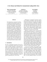
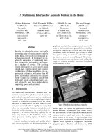
Laboratory model showing vertical osteotomies on the buccal sideFigure 1
Laboratory model showing vertical osteotomies on the buccal side.
Head & Face Medicine 2007, 3:15 />Page 3 of 8
(page number not for citation purposes)
and avascular necrosis of the segments. According to this
proposition, no blind posterior dissection and periostal
stripping of the masseteric-pterygoid sling is done. The
author suggested gentle dissection of medial tissue from
ramus just above the lingula (not extending to the poste-
rior border of the ramus) for visual inspection of the infe-
rior alveolar neurovascular bundle and elevation up to the
antegoniac incisure, without posterior extension. Osteot-
omy starts above the lingula, extending inferolaterally up
to the inferior border of the mandible, as recommended
by Hunsuck [9]. The inferior cut, on the other hand, com-
pletely involves the basilar region, which makes sagittal
split easier.
The use of different types of reciprocating saws was intro-
duced in the decade of 1980. This technology resulted in
a reduction in size of equipments and blades, allowing
their use in sagittal osteotomies of the mandible [12].
Some of the items, such as the blade for basilar cutting
developed by Wolford and Davis Jr [13], were specifically
designed for particular stages of surgery.
The methods for fixation of these bone segments evolved
from wire osteosynthesis. For rigid fixation in mandibular
sagittal split ramus osteotomy, bicortical bone screws
[14,15] and miniplates and screws [16-21] are now avail-
able.
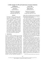
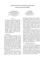
Transoperative characteristicsFigure 2
Transoperative characteristics. A. Osteotomy extending from medial aspect of the ascending ramus above the lingula, over the
oblique external line to the mesial face of the permanent first molar. This cut is then extended vertically to the lower border of
the mandible. The winding tracing is noteworthy. B. Separation of fragments after bone split. Larger extension of lateral seg-
ment and, in consequence, larger surface for bone contact. Exposition of the cruent area, including in its extension the neurov-
ascular bundle; mental nerve. C. Complete liberation of osteotomized segments, allowing ample sliding between them. The
lateral external segment of mandibular ramus and body is shown. Its extension and magnitude are noteworthy. Cruent area
clearly visible in the depth of the bone surgical wound. Mandibular body. D. Application of miniplate and screw end surgical
procedure in one of the sides. The sequence is repeated in the opposite side.
Head & Face Medicine 2007, 3:15 />Page 4 of 8
(page number not for citation purposes)
Surgical technique
The technique presented below has been in use since
1985. Performed under general anesthesia and nasotra-
cheal intubation, the access is done through mucosal inci-
sion on the mandibular ramus, extending bilaterally
below the mucogingival border beyond the mental
foramen. Elevation is accomplished with conservation of
the mental nerves. A channel retractor (Obwegeser type),
positioned above the lingula, is used for medial access to
the mandibular ramus. Two other channel retractors are
employed for elevation of the buccal tissues of the man-
dibular body and ramus. One retractor for the ramus is
placed on the temporal ridge, after partial elevation of this
muscular insertion. Langenbeck retractors may be used for
accessing the anterior region of the mandible. The areas
are sequentially accessed, on both sides.
The technique presently suggested includes, as previous
methods, medial osteotomy of the ramus, to be per-
formed above the lingula and extending slightly behind it.
An extension of sagittal osteotomy is performed on the
buccal face, in anterior direction, making the lateral cut in
the region of the mandibular body at the level of the
mesial face of the first inferior molar. It is, therefore, up to
20 mm anteriorly located as compared to current proto-
cols (Figure 1). Osteotomy is initially performed with
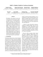
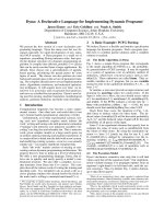
Radiographic comparisonFigure 3
Radiographic comparison. A. Lateral extra-oral radiograph for preoperative cephalometric investigation of a retrognathic
patient. Skeletal image of the retropositioned mandible can be seen in type II occlusal relationship. Soft structures show char-
acteristic deep mentolabial sulcus and small facial height. B. Postoperative lateral extra-oral radiograph. Alveolar osteotomy
can be seen from 32 to 42, associated to advancement and clockwise rotation of the mandible making up a maxilla-combined
surgery. The surgery begins at the mandible. Rigid fixation miniplates measuring 2 mm are used in an extension of six holes for
an advancement of 13 mm. On the maxilla, 1.5 mm rigid fixation may be observed. Soft tissue profile in accordance with skele-
tal results. In the naso-oro-hypopharyngeal regions, pre- and postoperative images show transversal increase of the area. This
result is supported by respiratory improvement, as clinically reported by the patient.
Head & Face Medicine 2007, 3:15 />Page 5 of 8
(page number not for citation purposes)
small spherical and cylindrical burs, following the
oblique external ridge up to the pre-determined level. The
resulting osteotomy line will determine the orientation of
the saw on the distal, more anterior direction. Distal con-
tinuity of the procedure involves a reciprocant saw, pre-
venting damage to teeth roots (Figure 2A).
For splitting, osteotomes are sequentially malleted, begin-
ning in the retromolar region with instruments oriented
to the mandibular angle. Usual care procedures to avoid
injury to the inferior alveolar bundle, particularly in the
distal region, are of fundamental importance. Among
them, maintaining lateral thickness of the proximal frag-
ment, as well as orienting instruments in a direction par-
allel to the buccal cortical, are emphasized. Opening of
the sagittal gap, from osteotomes placed on its most pos-
terior region, allows the visual inspection of all its exten-
sion and loosening of the inferior alveolar bundle, in case
it is exposed (Figure 2B, C).
After the fracture is completed, rigid internal fixation is
performed with a 2,0 mm straight miniplate and mono-
cortical screws. Its size will depend on the size of the
planned movement (Figure 2D).
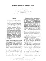
Lateral photographic studyFigure 4
Lateral photographic study. E.A., male, 22 yeas old. Main complaint: poor dental occlusion and respiratory difficulty. A. Preop-
erative profile showing characteristic facial concavity of oral breather patient with class II occlusion. B. Postoperative profile:
projection of the mentum, higher functional and skeletal balance in cervical angle and submental regions. Advancement and
rotation of the maxilla results in projections at the malar and paranasal regions. Interlabial relationship improved. Elevation of
the nasal point, resulting in more harmonious outline.
Head & Face Medicine 2007, 3:15 />Page 6 of 8
(page number not for citation purposes)
Frontal photographic study of the faceFigure 5
Frontal photographic study of the face. A, B. In the comparison of pre- and postoperative images, vertical increase of the men-
tum caused by advancement of the mandible with clockwise rotation is noteworthy. Lower incisive alveolar osteotomy, cor-
recting dental projection, enhances correction of the mentolabial sulcus and allows greater advancement of the mandible.
Better support of the median-third of face, with vertical and transversal symmetry patterns. C, D. Frontal view of smiling
patient allows observation of functionality of the lips within aesthetic patterns. The oral corridor observed in the preoperative
period was completely corrected.
Head & Face Medicine 2007, 3:15 />Page 7 of 8
(page number not for citation purposes)
Conclusion
Most studies and modifications proposed for mandibular
sagittal split ramus osteotomy have concentrated in
medial corticotomy. This is explained by the complexity
of local anatomy and incidence of atypical fractures in this
area, as well as by the frequent occurrence of neurological
complications related to the inferior alveolar nerve. Fol-
lowing this stage of technical development, studies have
concentrated in other limitations of the procedure, such
as amplitude and direction of planned movements, par-
ticularly of laterolateral and vertical advance, alternatives
for rigid fixation and stability of the results obtained.
Other studies have also reported stability of the rigid fixa-
tion in sagittal osteotomy with the use of miniplates [19-
21].
When we initiated the practice of fixation of mandibular
surgeries with miniplates [18], the design usually
employed in osteotomy was not adequate for their use. In
experimental studies, the performance of mandibular
bone cutting in a more anterior position was explored, in
a process which resulted in the present proposal. The use
of reciprocating saws facilitates the process, due to the
curved shape of osteotomy and for prevention of damage
to teeth roots
This technical proposal presents many possible advan-
tages. The area of bone contact is considerably increased,
resulting in better healing, particularly in cases of great
advance. Bone superposition is assured without interfer-
ence with the area of fixation. The mechanical resistance
decreases with anterior projection of osteotomy, lowering
the burden of osteosynthesis. This is obtained through a
2,0 mm plate and monocortical screws (5 to 7 mm),
placed in the region of the mandibular body. In this
region, intraoral access is easier (avoiding the need of
Dental occlusionFigure 6
Dental occlusion. A, B. Preoperative: Patient reported for preoperative orthodontic treatment. Class II occlusion with deep
bite, dental crowding and convergent inclination of posterior upper teeth are observed. Tooth 47 is lacking. C, D. Postopera-
tive: dental occlusion surgical and orthodontic treatments completed with correction of deep bite. Dental alignment and level-
ling improve stability of surgical results, characterized by acquisition of molar and canine occlusion keys associated with good
intercuspidation and interdigitation of the remaining dental structures.
Publish with Bio Med Central and every
scientist can read your work free of charge
"BioMed Central will be the most significant development for
disseminating the results of biomedical research in our lifetime."
Sir Paul Nurse, Cancer Research UK
Your research papers will be:
available free of charge to the entire biomedical community
peer reviewed and published immediately upon acceptance
cited in PubMed and archived on PubMed Central
yours — you keep the copyright
Submit your manuscript here:
/>BioMedcentral
Head & Face Medicine 2007, 3:15 />Page 8 of 8
(page number not for citation purposes)
transcutaneous access for insertion of screws) and the flat
bone surface facilitates adaptation of the plate. If there is
interest in its removal, application of osteosynthesis in
this area may also make it easier. In cases of simultaneous
extraction of the inferior third molars, the fixation area is
far from their alveoli and is not involved in the process.
The same happens in atypical fractures that may eventu-
ally occur, involving the basilar region of the proximal
fragment. The use of a larger miniplate will certainly give
stability to the fragments (Figures 3 to 6).
Disadvantages of the technique involve the need for larger
areas of elevation and manipulation of the mental nerve,
since in many situations fixation of the plate will be per-
formed in its proximity.
Competing interests
The author(s) declare that they have no competing inter-
ests.
Acknowledgements
All patients signed the informed consent. Thanks are due to Prof. Dr. Car-
los Eduardo Baraldi (School Dentistry-UFRGS), Isabel Pucci (Manager, Insti-
tuto Puricelli & Associados) and MS Traducoes Cientificas Ltda.
References
1. Caldwell JB, Letterman G: Vertical osteotomy in the mandibular
rami for correction of prognathism. J Oral Surg 1954,
12:185-202.
2. Babcock WW: The surgical treatment of certain deformitiesof
the jaw associated with malocclusion of the teeth. JAMA 1909,
53:833-839.
3. Wolford LM: The sagittal split ramus osteotomy as the pre-
ferred treatment for mandibular prognathism. J Oral Maxillo-
fac Surg 2000, 58:310-312.
4. Kazanjian VH: The treatment of mandibular prognayhism with
special reference to edentulous patients. Oral Surg Oral Med
Oral Pathol 1951, 4:680-688.
5. Obwegeser H: The sagittal splitting of the mandible proce-
dure. In Mandibular growth anomalies – terminology – aetiology – diag-
nosis – treatment Volume 12. Edited by: Obwegeser H. Berlin: Springer-
Verlag; 2001:359-384.
6. Trauner R, Obwegeser H: The surgical correction of mandibu-
lar prognathism and retrognathia with condideration of gen-
ioplasty. Oral Surg Oral Med Oral Pathol 1957, 10:677-689.
7. Obwegeser H: The indications for surgical correction of man-
dibular deformity by the sagittal splitting technique. Br J Oral
Surg 1964, 2:157-171.
8. Dal Pont G: Retromolar osteotomy for the correction of prog-
nathism. J Oral Surg Anesth Hosp Dent Serv 1961, 19:42-47.
9. Hunsuck EE: A modified intraoral sagittal splitting technique
for correction of mandibular prognathism. J Oral Surg 1968,
26:250-253.
10. Gallo WJ, Moss M, Gaul JV: Modification of the sagittal ramus-
split osteotomy for retroghnatia. J Oral Surg 1976, 34:178-179.
11. Epker BN: Modifications in the sagittal osteotomy of the man-
dible. J Oral Surg
1977, 35:157-159.
12. Messer EJ, Eckstein R, Nealis M, Hargis HW: Use of the micro-
reciprocating saw for mandibular saggital osteotomy. J Oral
Surg 1981, 39:381-383.
13. Wolford LM, Davis WM Jr: The mandibular inferior border
split: a modification in the sagittal split osteotomy. J Oral Max-
illofac Surg 1990, 48:92-94.
14. Jeter TS, Van Sickels JE, Dolwick MF: Modified Techniques for
internal fixation of sagital ramus osteotomies. J Oral Maxillofac
Surg 1984, 42:270-272.
15. Verdaguer Martin JJ, Escrig de Teigeiro M: Tratamento de lãs
deformidades dentofaciales clase II y clase III. In Tratado de
cirugía oral y maxilofacial Edited by: Navarro Vila C, Garcia Martin F,
Ochandiano Caicoya S. Madrid: Arán Ediciones; 2004:767-77.
16. Champy M, Lodde JP: Ostheosyntesis mandibulares: localiza-
tion de suntèses en fonction des contraintes mandibulares.
Rev Stomatol Chirurg Maxillofac 1976, 77:971-976.
17. Champy M, Lodde JP, Schmitt R, Jaeger JH, Muster D: Mandibular
osteosynthesis by miniature screwed plates via a buccal
approach. J Maxillofac Surg 1978, 6:14-21.
18. Puricelli E: Menor tempo de fixação intermaxilar nas cirurgias
do prognatismo. Rev Gaúcha Odontol 1982, 30:95-98.
19. Lee J, Piecuch JF: The sagittal ramus osteotomy: stability of fix-
ation with internal miniplates. Int J Oral Maxillofac Surg 1992,
21:326-330.
20. Scheerlinck JPO, Stoelinga PJW, Blijdorp PA, Brouns JJA, Nijs MLL:
Sagittal split advancement osteotomies stabilized with mini-
plates: a 2–5 year follow up. Int J Oral Maxillofac Surg 1994,
23:127-131.
21. Joos U: An adjustable bone fixation system for sagittal split
ramus ostectomiy: preliminary report. Br J Oral Maxillofac Surg
1999, 37:99-103.