báo cáo khoa học:" Lack of association between celiac disease and dental enamel hypoplasia in a case-control study from an Italian central region" pdf
Bạn đang xem bản rút gọn của tài liệu. Xem và tải ngay bản đầy đủ của tài liệu tại đây (379.67 KB, 6 trang )
BioMed Central
Page 1 of 6
(page number not for citation purposes)
Head & Face Medicine
Open Access
Case Study
Lack of association between celiac disease and dental enamel
hypoplasia in a case-control study from an Italian central region
Maurizio Procaccini
1
, Giuseppina Campisi*
2
, Pantaleo Bufo
3
,
Domenico Compilato
2
, Claudia Massaccesi
1
, Carlo Catassi
4
and
LorenzoLoMuzio
3
Address:
1
Istituto di Scienze Odontostomatologiche, Università Politecnica delle Marche, Italy,
2
Dip. Scienze Stomatologiche, Università di
Palermo, Italy,
3
Dip. Scienze Chirurgiche, Università di Foggia, Italy and
4
Istituto di Clinica Pediatrica, Università Politecnica delle Marche, Italy
Email: Maurizio Procaccini - ; Giuseppina Campisi* - ; Pantaleo Bufo - ;
Domenico Compilato - ; Claudia Massaccesi - ; Carlo Catassi - ;
* Corresponding author
Abstract
Background: A close correlation between celiac disease (CD) and oral lesions has been reported.
The aim of this case-control study was to assess prevalence of enamel hypoplasia, recurrent
aphthous stomatitis (RAS), dermatitis herpetiformis and atrophic glossitis in an Italian cohort of
patients with CD.
Methods: Fifty patients with CD and fifty healthy subjects (age range: 3–25 years), matched for
age, gender and geographical area, were evaluated by a single trained examiner. Diagnosis of oral
diseases was based on typical medical history and clinical features. Histopathological analysis was
performed when needed. Adequate univariate statistical analysis was performed.
Results: Enamel hypoplasia was observed in 26% cases vs 16% in controls (p > 0.2; OR = 1.8446;
95% CI = 0.6886: 4.9414). Frequency of RAS in the CD group was significantly higher (36% vs 12%;
p = 0.0091; OR = 4.125; 95% CI = 1.4725: 11.552) in CD group than that in controls (36% vs 12%).
Four cases of atrophic glossitis and 1 of dermatitis herpetiformis were found in CD patients vs 1
and none, respectively, among controls.
Conclusion: The prevalence of enamel hypoplasia was not higher in the study population than in
the control group. RAS was significantly more frequent in patients with CD.
Background
Celiac disease (CD), also known as celiac sprue or gluten-
sensitive entheropathy, can be defined as a chronic
inflammatory intestinal disease characterised by nutrient
malabsorption and improvement after the withdrawal of
gluten (found in wheat, barley) from the diet. Prevalence
of CD ranges from 1:85 to 1:300 have been reported for
CD in Western countries [1-6]. In addition to the classical
gastrointestinal presentation (diarrhoea, abdominal dis-
tension, vomiting, weight loss and pallor) CD can cause
minimal intestinal damage and weak or absent systemic
symptomatology (also known as "silent form"). In these
patients the lack of symptoms can persist for a long time,
while the biopsy of the bowel shows the typical atrophy
Published: 30 May 2007
Head & Face Medicine 2007, 3:25 doi:10.1186/1746-160X-3-25
Received: 8 November 2006
Accepted: 30 May 2007
This article is available from: />© 2007 Procaccini et al; licensee BioMed Central Ltd.
This is an Open Access article distributed under the terms of the Creative Commons Attribution License ( />),
which permits unrestricted use, distribution, and reproduction in any medium, provided the original work is properly cited.
Head & Face Medicine 2007, 3:25 />Page 2 of 6
(page number not for citation purposes)
of intestinal mucosa [7]. It is also well recognized the
association of CD with several complications, as lympho-
mas, autoimmune and degenerative nervous system dis-
eases [8-10].
The oral cavity, a part of gastrointestinal system [11], can
also be affected by several abnormalities in patients with
CD. As the mouth is very easy to examine, oral lesions can
provide a valuable clinical clue for early diagnosis of CD
[12]; in fact among the atypical aspects of CD (extra-intes-
tinals), in the international literature has been reported
some affections interesting the oral cavity, the most com-
mon are recurrent aphthous stomatitis (RAS) [13-15] and
dental enamel defects [8,13,16-21], in addition have been
described the association between CD and unspecific
forms of atrophic glossitis [22], oral manifestations of
dermatitis herpetiformis [23], Sjögren's syndrome [24,25]
and oral lichen planus [26,27]. These disorders, in
absence of a typical intestinal symptomatology, can repre-
sent useful clues for a timely diagnosis [7,22].
However, data from literature are often controversial,
probably because of different geographical origin of
patients studied and lack of adequate controls. Finally, no
studies have been performed, in CD patients of a Central
Region of Italy (Ancona, Marche, Italy)
The aim of this case-control study was to assess prevalence
of dental hard and oral soft tissues changes generally con-
sidered celiac-related (e.g. RAS, enamel hypoplasia, der-
matitis herpetiformis and atrophic glossitis) and to verify
if cases are more likely to be affected by any of the oral dis-
eases considered.
Methods
Fifty CD patients, aged between 3 and 18 years old and
living in the Region of Marche, were enrolled in the study.
CD was diagnosed at Paediatric Department of the Uni-
versity Politecnica of Marche (Ancona, Italy), and the
diagnosis of CD was based on serological tests (Ab-htTG
IgA, Ab-htTG IgG, AGA IgA, AGA IgG, EMA IgA, EMA
IgG), small-bowel biopsy during esophago-gastro-duode-
noscopy (EGDS) and histological evidence of villous atro-
phy with crypt hyperplasia and increase in intraepithelial
lymphocytes (normal, 10–30 per 100 epithelial cells),
and the disappearance of the symptoms and normaliza-
tion of serum anti-tTG and/or EMA after gluten-free-diet
(GFD) [28,29]. The control group was recruited by simple
randomization at a Primary and Secondary Public School
of Ancona, during an healthy prevention programme for
oral disease, matched one-to-one and without any signif-
icant differences with study group for geographical area,
age and gender (p > 0.2 by t-Student and chi-square test,
respectively). These young individuals neither reported
any gastrointestinal diseases and not have a family history
of CD.
Patients were examined for hard tissue changes (i.e. dental
enamel defects) and soft tissue lesions (RAS, dermatitis
herpetiformis and atrophic glossitis). Patients with CD
and healthy individuals were examined by a single
observer. Informed consent was obtained by parents who
were also asked about previous episodes of RAS affecting
child/children.
The enamel defects affecting deciduous and permanent
teeth were graded 0 to IV according to Aine's classification
[17] with a special attention to symmetric anomalies.
Soft tissues examination was carried out with conven-
tional dental chairs, artificial light, flat mirrors, monouse
probe and sterile gauzes.
With regard RAS, we registered both lesions clinically
observed and ulcerative events referred by parents or
reported by hospital clinical records. They were classified
into minor, major and herpetic aphthous ulcers [30],
according to dimension, form, localization and evolu-
tionary tendency, and also rate of occurrence was regis-
tered. Atrophic glossitis was diagnosed on the basis of
clinical features and oral mucosal lesions due to dermati-
tis herpetiformis were assessed by both clinical features
and histological/immunofluorescence studies.
Statistical analysis
Data were analyzed by means of StaView for Windows
(SAS Inc v. 5.0.1, Cary, NC, USA). To measure the associ-
ation level, Odds Ratio (OR) and the 95% corresponding
test-based Confidence Interval (CI) were calculated. T-Stu-
dent test was used to calculate significant differences
between cases and controls at baseline for ordinal varia-
bles. Chi-square test was used to assess statistical differ-
ences among categorical variables. In all of evaluations p-
values = 0.05 were considered statistically significant.
Results
Enamel alterations were observed in 13/50 (26%) sub-
jects with CD and in 8/50 (16%) controls, with a ratio
male-female of 1:2 for the celiac group and 2:1 for control
group (p > 0.2; OR = 1.8446; 95% CI = 0.6886: 4.9414).
With respect to the severity score of hypoplasia, 10/13 CD
patients showed lesions of degree 1 and 3/13 degree 2, in
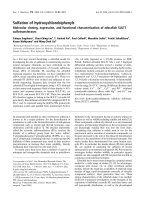
controls all were in degree 1. The grade 1 enamel defects
were generally localized on incisor surfaces (for the ante-
rior sectors) (Figure 1) and cuspid surfaces (for the poste-
rior sectors), with dimensions from 1 to 3 mm and with a
round-oval form, while that of grade 2 were on the canine
and premolar vestibular surface. The colour alterations
Head & Face Medicine 2007, 3:25 />Page 3 of 6
(page number not for citation purposes)
were white-yellowish, with clear margins, opaque and
smooth surface.
Episodes of RAS occurred in 36% of CD patients (18/50)
vs 12% of controls (6/50) (p = 0.0091; OR = 4.125; 95%
CI = 1.4725: 11.552) with a male-female ratio of 1:1 and
2:3, respectively (Figure 2). In CD patients RAS showed
greater rate of recurrence than in controls. Atrophic glossi-
tis was reported in 4 cases and one control, and dermatitis
herpetiformis in one patient with CD and none of sub-
jects without CD.
Discussion and conclusion
Recent epidemiology data showed the prevalence of CD
to approach 1% of the general population [31-34]. How-
ever, the clinical presentation of CD seems to differ from
the typical form observed in past years, as almost 50% of
the patients with newly diagnosed CD do not present with
gastrointestinal symptoms [35,36]. Thus, in order to iden-
tify the greatest number of "atypical" or "silent" CD
patients and prevent long-term complications, it has been
suggested that the clinicians should investigate those sub-
jects who present "indirect" signs of CD, such as chronic
anaemia [37], hyper-transaminasemia or hyperamy-
lasemia of unknown origin [38,39], osteoporosis [40],
autoimmune thyroid disorders [41].
As abnormalities of the oral cavity have been reported in
CD, non-invasive clinical examination of the oral cavity
can contribute to identify patients with atypical or silent
CD [13,14,17,18,42].
As regards to changes of dental tissues, we did not found
CD patients more likely to suffer from systematic and
symmetric enamel defects. Indeed, a wide range of fre-
quencies of enamel defects in CD patients has been
reported in other studies [17,43-48]; our data are in agree-
ment with other studies performed in Italy (Table 1) and
the high frequency of enamel defect found in controls, as
well as its severity, is likely to be related to environmental,
dietetic and genetic factors [46]. Further studies are war-
ranted to clarify the pathogenesis of this defect as nutri-
tional, immunologic or genetic factors (association with
the HLA DR3 allele) has been hypothesized [45,49]. With
regard to celiac patients, enamel defects have been corre-
lated to an altered phosphate-calcium metabolism and/or
formation of antibodies against the matrix of enamel
organ. The antigen correlated to class II molecules of the
MHC could prime an immunity movement against the
enamel organ, from which a mineralization disorder
could derive [18]. In addiction, there is no strong evidence
that these anomalies are correlated with the nutritional
status, vitamin D deficiency or to an excess of fluoride
incorporation. Current evidence suggests that an autoim-
mune pathogenesis is more likely, as enamel defects are
also present in autoimmune diseases, such as some poly-
endocrine syndromes [46].
With respect to oral soft lesions, we confirmed that CD
patients are likely to suffer from RAS compared with
healthy controls, especially before the gluten-free diet.
In our celiac population RAS was found in 26 % of CD
patients with an OR of 4.12 in comparison with the con-
trols. Even if a wide range of frequencies have been
reported (Table 2) our data show the highest prevalence
of RAS with respect to other Italian studies.
In agreement with Sedghizadeh et al. [14], we suggested to
consider RAS as a "risk indicator" of CD more than CD as
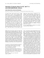
Several RAS on buccal mucosa in a CD patientFigure 2
Several RAS on buccal mucosa in a CD patient.
Symmetrical enamel hypoplasia of grade I on permanent inci-sors in a CD patientFigure 1
Symmetrical enamel hypoplasia of grade I on permanent inci-
sors in a CD patient.
Head & Face Medicine 2007, 3:25 />Page 4 of 6
(page number not for citation purposes)
a risk factor for RAS, although no definitive statement is
possible on their predictive role for CD.
In addition the term "recurrent aphthous stomatitis"
should be reserved to recurrent oral ulcer that present in
patients without systemic diseases, while ulcers that have
a clinical appearance similar to RAS, but found in patients
with systemic disorders (such as CD) should be termed
"aphthous-like ulcers" [50]. Even if the diagnostic criteria
of RAS used in this study (namely, medical history and/or
presence of detectable lesions) may represent a major lim-
itation of present research, it is well accepted that recur-
rent and episodic nature of oral ulcerations requires
medical history to be an important part of the diagnostic
process.
RAS is often associated to haematinic (iron, folate, vita-
min B12) deficiency [51,52]; since atypical or latent CD
may not manifest itself with gastrointestinal signs/symp-
toms but often with iron/folate deficiency [53-56] we sug-
gest that when patients show persistent RAS they should
be examined for haematinic deficiencies. Only if one or
more of these deficiencies are present, they should be
screened for CD.
In conclusion, our data from central Italy confirming the
higher prevalence of RAS or aphthous-like ulcers in
patients with CD validate the hypothesis of their pathoge-
netic predisposition to oral mucosal lesions more than
hard dental tissue lesions; further investigations are war-
ranted to clarify the predictive role of these lesions in
screening oligosymptomatic or asymptomatic CD.
Acknowledgements
This study was supported by Italian National Grant (PRIN, 2005) and Local
Grant (University of Palermo)
References
1. Korponay-Szabo IR, Kovacs JB, Czinner A, Goracz G, Vamos A, Szabo
T: High prevalence of silent celiac disease in preschool chil-
dren screened with IgA/IgG antiendomysium antibodies. J
Pediatr Gastroenterol Nutr 1999, 28(1):26-30.
2. Hill ID, Bhatnagar S, Cameron DJ, De Rosa S, Maki M, Russell GJ,
Troncone R: Celiac disease: Working Group Report of the
First World Congress of Pediatric Gastroenterology, Hepa-
tology, and Nutrition. J Pediatr Gastroenterol Nutr 2002, 35 Suppl
2:S78-88.
Table 2: Prevalence (%) of RAS in CD patients
Authors, years Number of CD patients Prevalence (%)
Sedghizadeh et al, 2002 [15] 61 41.0
Present study, 2007 50 36.0
Bucci et al., 2006 [19]* 72 33.3
Andersson-Wenckert 1984 [13] 19 26.3
Sood et al, 2003 [21] 96 19.8
Petrecca et al, 1994 [18] * 29 17.0
Majorana et al, 1992 [20]* 113 16.8
Lähteenoja et al, 1998 [22] 128 3.7
* = study performed among Italian individuals
Table 1: Prevalence (%) of the dental enamel defects in CD patients
Authors n CD patients Prevalence %
Aine 1996. [57] 86 96
Aine et al, 1990. [17] 40 83
Petrecca et al, 1994.* [18] 29 76
Aine et al, 1992. [58] 30 58.3
Aguirre et al,1997. [59] 137 52.5
Rasmusson et al, 2001. [8] 40 50
Balli et al, 1988.* [14] 111 34.7
Prati et al, 1987.* [60] 10 33.3
Martelossi et al., 1996. [61].* 603 32.4
Mariani et al, 1994.* [45] 84 28
Present study, 2007 50 26
Bucci et al, 2006.* [19] 72 20
Andersson-Wenckert et al, 1984. [13] 24 21
Lahteenoja et al, 1998. [22] 128 10.1
* = study performed among Italian individuals
Head & Face Medicine 2007, 3:25 />Page 5 of 6
(page number not for citation purposes)
3. Catassi C, Ratsch IM, Fabiani E, Ricci S, Bordicchia F, Pierdomenico R,
Giorgi PL: High prevalence of undiagnosed coeliac disease in
5280 Italian students screened by antigliadin antibodies. Acta
Paediatr 1995, 84(6):672-676.
4. Kolho KL, Farkkila MA, Savilahti E: Undiagnosed coeliac disease
is common in Finnish adults. Scand J Gastroenterol 1998,
33(12):1280-1283.
5. Carlsson AK, Axelsson IE, Borulf SK, Bredberg AC, Ivarsson SA:
Serological screening for celiac disease in healthy 2.5-year-
old children in Sweden. Pediatrics 2001, 107(1):42-45.
6. Not T, Horvath K, Hill ID, Partanen J, Hammed A, Magazzu G, Fasano
A: Celiac disease risk in the USA: high prevalence of antien-
domysium antibodies in healthy blood donors. Scand J Gastro-
enterol 1998, 33(5):494-498.
7. Pastore L, De Benedittis M, Petruzzi M, Tato D, Napoli C, Montagna
MT, Catassi C, Serpico R: [Importance of oral signs in the diag-
nosis of atypical forms of celiac disease]. Recenti Prog Med 2004,
95(10):482-490.
8. Rasmusson CG, Eriksson MA: Celiac disease and mineralisation
disturbances of permanent teeth. Int J Paediatr Dent 2001,
11(3):179-183.
9. Somech R, Spirer Z: Celiac disease: extraintestinal manifesta-
tions, associated diseases, and complications. Adv Pediatr 2002,
49:191-201.
10. Green PH, Fleischauer AT, Bhagat G, Goyal R, Jabri B, Neugut AI:
Risk of malignancy in patients with celiac disease. Am J Med
2003, 115(3):191-195.
11. Tomasi TB Jr LL Challacombe S, McNabb P.: Mucosal immunity:
The origin and migration patterns of cells in the secretory
system. J Allergy Clin Immunol 1980, 65(1)::12-19.
12. Wray D: Gluten-sensitive recurrent aphthous stomatitis. Dig
Dis Sci 1981, 26(8):737-740.
13. Andersson-Wenckert I, Blomquist HK, Fredrikzon B: Oral health in
coeliac disease and cow's milk protein intolerance. Swed Dent
J 1984, 8(1):
9-14.
14. Balli MP, Balli ME, Mengoli M, Balli C, Balli F: [Growth, skeletal and
dental age in chronic diarrhea in childhood]. Pediatr Med Chir
1988, 10(3):277-282.
15. Sedghizadeh PP, Shuler CF, Allen CM, Beck FM, Kalmar JR: Celiac
disease and recurrent aphthous stomatitis: a report and
review of the literature. Oral Surg Oral Med Oral Pathol Oral Radiol
Endod 2002, 94(4):474-478.
16. Aine L: Dental enamel defects and dental maturity in children
and adolescents with coeliac disease. Proc Finn Dent Soc 1986,
82 Suppl 3:1-71.
17. Aine L, Maki M, Collin P, Keyrilainen O: Dental enamel defects in
celiac disease. J Oral Pathol Med 1990, 19(6):241-245.
18. Petrecca S, Giammaria G, Giammaria AF: [Oral cavity changes in
the child with celiac disease]. Minerva Stomatol 1994,
43(4):137-140.
19. Bucci P, Carile F, Sangianantoni A, D'Angio F, Santarelli A, Lo Muzio
L: Oral aphthous ulcers and dental enamel defects in children
with coeliac disease. Acta Paediatr 2006, 95(2):203-207.
20. Majorana A, Sapelli PL, Malagoli A, Meini A, Pillan MN, Duse M, Ugazio
AG: [Celiac disease and recurrent aphthous stomatitis. The
clinical and immunogenetic aspects]. Minerva Stomatol 1992,
41(1-2):33-40.
21. Sood A, Midha V, Sood N, Malhotra V: Adult celiac disease in
northern India. Indian J Gastroenterol 2003, 22(4):124-126.
22. Lahteenoja H, Toivanen A, Viander M, Maki M, Irjala K, Raiha I, Syr-
janen S: Oral mucosal changes in coeliac patients on a gluten-
free diet. Eur J Oral Sci 1998, 106(5):899-906.
23. Economopoulou P, Laskaris G: Dermatitis herpetiformis: oral
lesions as an early manifestation. Oral Surg Oral Med Oral Pathol
1986, 62(1):77-80.
24. Ventura A, Magazzu G, Greco L: Duration of exposure to gluten
and risk for autoimmune disorders in patients with celiac dis-
ease. SIGEP Study Group for Autoimmune Disorders in
Celiac Disease. Gastroenterology 1999,
117(2):297-303.
25. Iltanen S, Collin P, Korpela M, Holm K, Partanen J, Polvi A, Maki M:
Celiac disease and markers of celiac disease latency in
patients with primary Sjogren's syndrome. Am J Gastroenterol
1999, 94(4):1042-1046.
26. Scully C, Porter SR, Eveson JW: Oral lichen planus and coeliac
disease. Lancet 1993, 341(8861):1660.
27. Bhatnagar S, Tandon N: Diagnosis of celiac disease. Indian J Pediatr
2006, 73(8):703-709.
28. van Heel DA, West J: Recent advances in coeliac disease. Gut
2006, 55(7):1037-1046.
29. Stanley HR: Aphthous lesions. Oral Surg Oral Med Oral Pathol 1972,
33(3):407-416.
30. Fasano A, Berti I, Gerarduzzi T, Not T, Colletti RB, Drago S, Elitsur
Y, Green PH, Guandalini S, Hill ID, Pietzak M, Ventura A, Thorpe M,
Kryszak D, Fornaroli F, Wasserman SS, Murray JA, Horvath K: Prev-
alence of celiac disease in at-risk and not-at-risk groups in
the United States: a large multicenter study. Arch Intern Med
2003, 163(3):286-292.
31. Tommasini A, Not T, Kiren V, Baldas V, Santon D, Trevisiol C, Berti
I, Neri E, Gerarduzzi T, Bruno I, Lenhardt A, Zamuner E, Spano A,
Crovella S, Martellossi S, Torre G, Sblattero D, Marzari R, Bradbury
A, Tamburlini G, Ventura A: Mass screening for coeliac disease
using antihuman transglutaminase antibody assay. Arch Dis
Child 2004, 89(6):512-515.
32. Maki M, Mustalahti K, Kokkonen J, Kulmala P, Haapalahti M, Kart-
tunen T, Ilonen J, Laurila K, Dahlbom I, Hansson T, Hopfl P, Knip M:
Prevalence of Celiac disease among children in Finland. N
Engl J Med 2003, 348(25):2517-2524.
33. Accomando S, Cataldo F: The global village of celiac disease. Dig
Liver Dis 2004, 36(7):492-498.
34. Maki M, Kallonen K, Lahdeaho ML, Visakorpi JK: Changing pattern
of childhood coeliac disease in Finland. Acta Paediatr Scand 1988,
77(3):408-412.
35. Pare P, Douville P, Caron D, Lagace R: Adult celiac sprue: changes
in the pattern of clinical recognition.
J Clin Gastroenterol 1988,
10(4):395-400.
36. Carroccio A, Iannitto E, Cavataio F, Montalto G, Tumminello M, Cam-
pagna P, Lipari MG, Notarbartolo A, Iacono G: Sideropenic anemia
and celiac disease: one study, two points of view. Dig Dis Sci
1998, 43(3):673-678.
37. Bardella MT, Fraquelli M, Quatrini M, Molteni N, Bianchi P, Conte D:
Prevalence of hypertransaminasemia in adult celiac patients
and effect of gluten-free diet. Hepatology 1995, 22(3):833-836.
38. Carroccio A, Di Prima L, Scalici C, Soresi M, Cefalu AB, Noto D,
Averna MR, Montalto G, Iacono G: Unexplained elevated serum
pancreatic enzymes: a reason to suspect celiac disease. Clin
Gastroenterol Hepatol 2006, 4(4):455-459.
39. Kemppainen T, Kroger H, Janatuinen E, Arnala I, Kosma VM, Pikkara-
inen P, Julkunen R, Jurvelin J, Alhava E, Uusitupa M: Osteoporosis in
adult patients with celiac disease. Bone 1999, 24(3):249-255.
40. Ventura A, Neri E, Ughi C, Leopaldi A, Citta A, Not T: Gluten-
dependent diabetes-related and thyroid-related autoanti-
bodies in patients with celiac disease. J Pediatr 2000,
137(2):263-265.
41. Meini A, Pillan MN, Plebani A, Ugazio AG, Majorana A, Sapelli PL:
High prevalence of DRW10 and DQW1 antigens in celiac
disease associated with recurrent aphthous stomatitis. Am J
Gastroenterol 1993, 88(6):972.
42. Aine L: [Dental enamel defects and dental maturity in chil-
dren and adolescents with celiac disease]. Proc Finn Dent Soc
1986, 82(4):227-229.
43. Ventura A, Martelossi S: Dental enamel defects and coeliac dis-
ease. Arch Dis Child 1997, 77(1):91.
44. Mariani P, Mazzilli MC, Margutti G, Lionetti P, Triglione P, Petronzelli
F, Ferrante E, Bonamico M: Coeliac disease, enamel defects and
HLA typing. Acta Paediatr 1994, 83(12):1272-1275.
45. Rea F, Serpico R, Pluvio R, Busciolano M, Iovene A, Femiano F, Sessa
G, Belnome G: [Dental enamel hypoplasia in a group of celiac
disease patients. Clinico-epidemiologic correlations]. Min-
erva Stomatol 1997, 46(10):517-524.
46. Ciccarelli NP DDF Greco L.: Lipoplasia dello smalto dentario
dei denti permanenti di soggetti celiaci in challenge con glu-
tine. 1993, 19/S-2:195:.
47. Mariani P FE Margutti G.: Difetti dello smalto dentario in un
gruppo di bambini e adolescenti celiaci italiani. 1993, 19/S-
2:196:.
48. Maki M, Aine L, Lipsanen V, Koskimies S: Dental enamel defects
in first-degree relatives of coeliac disease patients. Lancet
1991, 337(8744):763-764.
49. Scully C: Clinical practice. Aphthous ulceration. N Engl J Med
2006, 355(2):165-172.
Publish with Bio Med Central and every
scientist can read your work free of charge
"BioMed Central will be the most significant development for
disseminating the results of biomedical research in our lifetime."
Sir Paul Nurse, Cancer Research UK
Your research papers will be:
available free of charge to the entire biomedical community
peer reviewed and published immediately upon acceptance
cited in PubMed and archived on PubMed Central
yours — you keep the copyright
Submit your manuscript here:
/>BioMedcentral
Head & Face Medicine 2007, 3:25 />Page 6 of 6
(page number not for citation purposes)
50. Scully C, Felix DH: Oral medicine update for the dental prac-
titioner. Aphthous and other common ulcers. Br Dent J 2005,
199(5):259-264.
51. Jurge S, Kuffer R, Scully C, Porter SR: Mucosal disease series.
Number VI. Recurrent aphthous stomatitis. Oral Dis 2006,
12(1):1-21.
52. Trier JS: Celiac sprue. N Engl J Med 1991, 325(24):1709-1719.
53. Maki M, Collin P: Coeliac disease. Lancet 1997,
349(9067):1755-1759.
54. Catassi C, Fasano A: New developments in childhood celiac dis-
ease. Curr Gastroenterol Rep 2002, 4(3):238-243.
55. Green PH, Barry M, Matsutani M: Serologic tests for celiac dis-
ease. Gastroenterology 2003, 124(2):585-6; author reply 586.
56. Aine L: Coeliac-type permanent-tooth enamel defects. Ann
Med 1996, 28(1):9-12.
57. Aine L, Maki M, Reunala T: Coeliac-type dental enamel defects
in patients with dermatitis herpetiformis. Acta Derm Venereol
1992, 72(1):25-27.
58. Aguirre JM, Rodriguez R, Oribe D, Vitoria JC: Dental enamel
defects in celiac patients. Oral Surg Oral Med Oral Pathol Oral Radiol
Endod 1997, 84(6):646-650.
59. Prati C, Santopadre A, Baroni C: [Delayed eruption, enamel
hypoplasia and caries in childhood celiac disease]. Minerva Sto-
matol 1987, 36(10):749-752.
60. Martelossi S, Torre G, Zanatta M, Del Santo M, Not T, Clarich G,
Radovich F, Ventura A: Dental enamel defects and screening for
coeliac disease. Pediatr Med Chir 1996, 18(6):579-581.