báo cáo khoa học:" Scar-like lesion on dorsal nose (cellular neurothekeoma)" pdf
Bạn đang xem bản rút gọn của tài liệu. Xem và tải ngay bản đầy đủ của tài liệu tại đây (493.88 KB, 4 trang )
BioMed Central
Page 1 of 4
(page number not for citation purposes)
Head & Face Medicine
Open Access
Case report
Scar-like lesion on dorsal nose (cellular neurothekeoma)
Larissa Dorina López-Cepeda*
1
, Gisela Navarrete-Franco
2
, Josefa Novales-
Santacoloma
3
and Julio Enriquez-Merino
4
Address:
1
Consultation, Centro Dermatológico "Dr. Ladislao de la Pascua", Mexico city, Mexico,
2
Dermatopathology Department, Centro
Dermatológico "Dr. Ladislao de la Pascua", Mexico city, Mexico,
3
Dermatopathology Department, Centro Dermatológico "Dr. Ladislao de la
Pascua", Mexico city, Mexico and
4
Surgery Department, Centro Dermatológico "Dr. Ladislao de la Pascua", Mexico city, Mexico
Email: Larissa Dorina López-Cepeda* - ; Gisela Navarrete-Franco - ; Josefa Novales-
Santacoloma - ; Julio Enriquez-Merino -
* Corresponding author
Abstract
Neurothekeomas are tumors of neural differentiation and of unknown origin that occur in females
at the 2
nd
and 3
rd
decades of life. They usually affect the face with an unspecific clinical aspect. The
histological features include cellular or mixoid differentiation and immunohistochemistry can be
positive for protein s-100, vimentin and epithelilal membrane antigen (EMA).
This case report presents a 13-year-old female patient with nasal neurothekeoma of cellular variety
and strongly positive for vimentin and s-100; and negative for EMA.
Background
Neurothekeoma (NK) is a tumor of neural differentiation
first described by Harkin and Reed in 1969 [1] and later
termed "neurothekeoma" by Gallager and Helwig in 1980
[2]. Being extremely rare (1 of 4000 biopsies in interna-
tional reports) [1], it is hitologically constituted by cellu-
lar proliferation arranged in lobules or fascicles immersed
in an amorphous matrix in the dermis and rarely in sub-
cutaneous tissue [3].
The origin of this tumor is unknown. Some authors con-
sider it as Schwann cells, [4] while others state that
perineural tissue or supporting structures of peripheric
nerves, fibroblasts, and muscle could be responsible [4]. It
has even been considered as a variant of the following der-
matoses: dermatofibroma [5], pilar epithelioid leiomi-
oma [6], plexiform neurofibroma, [7] and nerve sheath
myxoma [8].
NK presents frequently in females between 10–66 years
(especially among the 2
nd
and 3
rd
decades of life). It is
most commonly located is on the head, face and superior
mid-body. To date, 21 reports have shown facial occur-
rence (with one patient revealing nasal manifestation)
and 31 reports demonstrate occurrence of NK in patients
younger than 20 years old [5,9-11]. Other reports have
documented different locations of occurrence including
tongue [9], oral mucosa, eyelids [10], trunk (dorsum and
shoulders) and superior extremities (superior mid body).
NK has an unspecific clinical aspect, more frequently pre-
senting as a cupuliform, firm, flesh-colored or hyperpig-
mented papule-like formation which rarely exceeds 10
mm with capillaries on its surface and may be asympto-
matic. The evolution varies between 18 months and 30
years; with a slowly growing, generally benign course.
Diagnosis can be made by conventional histopathology
[1,2,4] yielding two basic patterns:
Published: 30 November 2007
Head & Face Medicine 2007, 3:39 doi:10.1186/1746-160X-3-39
Received: 13 February 2006
Accepted: 30 November 2007
This article is available from: />© 2007 López-Cepeda et al; licensee BioMed Central Ltd.
This is an Open Access article distributed under the terms of the Creative Commons Attribution License ( />),
which permits unrestricted use, distribution, and reproduction in any medium, provided the original work is properly cited.
Head & Face Medicine 2007, 3:39 />Page 2 of 4
(page number not for citation purposes)
1. Mucinous or myxoid: It is the most frequent form. It has
an evident fascicular and tabicated aspect with abundant
mucinous and amorphous substance, composed princi-
pally of acid mucopolysaccharides.
2. Cellular – epitheloid or fusiform cells with a cellular
pattern and fascicle-nodular aggregation, circumscribed to
reticular dermis, showing a grenz zone separating the der-
mis from the subacent tumor. There can be some mitotic
figures [12], atypical features [13] and metachromasia
with Giemsa stain [14]. Collagen may be sclerotic with
some lymphocytic infiltration. Barnhill, et al. [13] con-
sider that the cellular neurothekeoma might be an early
form (more frequent in young patients), whilethe muci-
nous type might represent a tumor of longer evolution
(frequent in older patients).
NK is positive with alcian blue at pH 2.5, and there are
reports of metachromasia with Giemsa stain [1].
By immunohistochemistry, protein S-100 [14] has been
found positive, as well as other stains: epithelilal mem-
brane antigen (EMA), vimentin and neuron specific eno-
lase (NSE), (with controversial results) [15]. Other useful
markers are: myelin basic protein, neurofilaments, glial
fibrillary acidic protein (GFAP), keratin and Leu-7 [11],
NK1/C3 and PGP9.5 [2,16].
Electronic microscopy is not useful for diagnosis. Differ-
ential diagnosis should be made chiefly with scars, der-
matofibromas, nevi, lymphocytomas and adnexae tumors
among others [2,4,12,13].
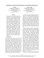
Case presentation
A 13-year-old female referred to the clinic with dermatosis
at the dorsal aspect of the nose. The lesion was 0.3 cm in
diameter, flat, soft, swollen; and light pink in color, with
superficial telangiectasias. Reportedly the lesion had
appeared 6 months ago, and had remained asymptomatic
(figure 1). The patient history was non-contributory,
including the absence of prior trauma. The incisional skin
biopsy showed atrophic epidermis with lax hyperkerato-
sis, sub-papillar moderate lymphocytic infiltrate; dense
dermal infiltrate of fusiform cells arranged in nests with
mitoses and hypotrophic adnexae. The collagen sur-
rounding the neoformation had a normal aspect (figures
2). Mucin stain (Mucicarmin of Mayer) and neurofila-
ment stains (Bielchowsky) were positive [17].
Immunohistochemistry was strongly positive for vimen-
tin, lightly postitive for s-100, negative for EMA and posi-
tive for mucin. Based on biopsy and
immunohistochemistry, the diagnosis of Neurothekeoma
was made.
A 0.3 cm, soft, light pink neoformation in the dorsal aspect of noseFigure 1
A 0.3 cm, soft, light pink neoformation in the dorsal aspect of nose.
Head & Face Medicine 2007, 3:39 />Page 3 of 4
(page number not for citation purposes)
The treatment included complete extirpation of the
lesion, followed by esthetic correction with a Limbert flap
(figure 3).
Defect correction with a Limbert flapFigure 3
Defect correction with a Limbert flap.
H-E 40× – Dense infiltrates of fusiform cells arranged in nests, with some mitoses (pleomorphic cytology)Figure 2
H-E 40× – Dense infiltrates of fusiform cells arranged in nests, with some mitoses (pleomorphic cytology).
Publish with Bio Med Central and every
scientist can read your work free of charge
"BioMed Central will be the most significant development for
disseminating the results of biomedical research in our lifetime."
Sir Paul Nurse, Cancer Research UK
Your research papers will be:
available free of charge to the entire biomedical community
peer reviewed and published immediately upon acceptance
cited in PubMed and archived on PubMed Central
yours — you keep the copyright
Submit your manuscript here:
/>BioMedcentral
Head & Face Medicine 2007, 3:39 />Page 4 of 4
(page number not for citation purposes)
Conclusion
In the present case, clinical diagnosis of neurothekeoma
was difficult to make since the lesion had a scar-like pat-
tern, without previous history of trauma. Histological
examination is definetely essential for the diagnosis, and
must be interpreted by experienced dermatopathologists.
An interesting feature of this case is that it was positive for
both of the neurofilament stains (Bielchowsky) and
mucin stain (Mucicarmin of Mayer) showing its dual dif-
ferentiation: neural and myxoid.
Authors' contributions
All author(s) read and approved the final manuscript.
LDLC, made the clinical diagnosis, follow up of the
patient, surgical treatment and writing the manuscript.
GNF and JNS, made the histological diagnosis.
JEM, planned and assisted the patient's surgical treatment.
Acknowledgements
Dr. Victor Jaimes, Dermatology Department, "Centro Médico Nacional 20
Noviembre, ISSSTE", for helping us obtain immunohistochemistry stains.
José Alberto Castillo Naranjo, Histotechnologist, "Centro Dermatológico
Pascua", for the slides with special stains.
Dr. Maria del Mar Paola Campos Fernández, for her Assistance in writing
the manuscript.
Written consent was obtained from the patient prior to submission of the
manuscript.
References
1. Del Río de la Torre E, Requema V A: Neurotecoma (mixoma
cutáneo de la vaina nerviosa). Estudio histopatológico de 4
casos. Piel 1993, 8:116-121.
2. Gallager RL, Helwig EB: Neurothekeoma – a benign cutaneous
tumor of neural origin. Am J Clin Pathol 1980, 74:759-64.
3. Watanabe K, Kusakabe T, Hoshi N, Suzuki T: Subcutaneous cellu-
lar neurothekeoma: a pseudosarcomatous tumor. Br J Derma-
tol 2001, 144:1273-1274.
4. Requena L, Sangüeza O: Benign neoplasms with neural differen-
tiation: a review. Am J Dermatopathol 1995, 17:75-96.
5. Zelger BG, Steiner H, Kutzner H, Maier H, Zelger B: Cellular neu-
rothekeoma: an epitheloid variant of dermatofibroma? His-
topathology 1998, 32:414-22. summary
6. Calonje E, Wilson-Jones E, Smith NP, Fletcher CDM: Cellular Neu-
rothekeoma: An epithelioid variant of pilar leiomioma?
Analysis of series. J Cutan Pathol 1991, 18:363.
7. Jurecka W: Nerve sheath myxoma: An immunohistochewmi-
cal study of a case. Dermatology 1992, 184:228.
8. Nagai Y, Ohno Y, Ishikawa O, Miyachi Y: Cellular neurothekeoma
on the lower lip, letter. Br J Dermatol 1997, 137:314-315.
9. Pepine M, Flowers F, Ramos-Caro F: Neurothekeoma in a 15 year
old boy: case report. Pediatr Dermatol 1992, 9:272-274.
10. Penarrocha M, Bonet J, Minués JM, Vera F: Nerve sheath myxoma
(neurothekeoma) in the tongue of a newborn. Oral Surg Oral
Med Oral Pathol Oral Radiol Endod 2000, 90:74-7.
11. You TT, Kaiser PK, Netland TP, Jakobiec FA: Neurothekeoma
palpebrae: a rare nerve sheath tumor arising the eyelid. Oph-
thal Plast Reconstr Surg 1999, 15:448-9.
12. Barnhill RL, Dickersin GR, Nickeleit V, Bhan AK, Muhlbauer JE, Philips
ME, Mihm MC Jr: Studies on the cellular origin of neurotheke-
oma: clinical, light microscopic, immunohistochemical and
ultrastructural observations. J Am Acad Dermatol 1991, 25:80-88.
13. Busam KJ, Mentzel T, Colpaert C, Barnhilla RL, Fletcher CD: Atypi-
cal or worrisome features in cellular neurothekeoma: a
study of 10 cases. Am J Surg Pathol 1998, 22:1067-72.
14. Husain S, Silvers D, Halperin A, Mc Nutt NS: Histologic Spectrum
of neurothekeoma and the value of immunoperoxidase
staining for s-100 protein in distinguishing it from
melanoma. Am J Dermatopathol 1994, 16:496-503.
15. Aronson P: Unmasking myelin proteins in neurothekeoma.
Letter 1992, 26:659.
16. Wang AR, May D, Bourne P, Scout G: PGP9.5: a marker for cel-
lular neurothekeoma. Am J Surg Pathol 1999, 23:1401-7.
17. Armed Forces Institute of Pathology: Manual of histologic and
special staining technics. Washington D.C; 1957.