ADVANCED PAEDIATRIC LIFE SUPPORT - part 4 potx
Bạn đang xem bản rút gọn của tài liệu. Xem và tải ngay bản đầy đủ của tài liệu tại đây (370.82 KB, 35 trang )
Bronchiolitis emergency treatment
As there is no specific treatment for bronchiolitis, management is supportive.
Humidified oxygen is delivered into a headbox at a rate that will maintain Sa
O
2
above
92%, and intravenous or nasogastric fluids are commenced if required. Pulse oximetry
is helpful in assessing the severity of hypoxemia. Because of the risk of apnoea, small
infants and those with severe disease should be attached to oxygen saturation and
respiratory monitors. Antibiotics, bronchodilators and steroids are of no value. The
precise role of the nebulised antiviral agent ribavirin is unclear and its use should be
reserved for children with pre-existing lung disease, those with impaired immunity and
infants with congenital heart disease. Mechanical ventilation is required in 2% of infants
admitted to hospital, either because of recurrent apnoea, exhaustion, or hypercapnia
and hypoxaemia secondary to severe small airways obstruction. All intubated infants
must have continuous Sa
O
2
and CO
2
monitoring. Naso-pharyngeal CPAP may be
sufficient ventilatory support for some infants.
Most children recover from the acute infection within two weeks. However, as many
as half will have recurrent episodes of cough and wheeze over the next 3–5 years. Rarely,
there is severe permanent damage to the airways (bronchiolitis obliterans).
Background information on asthma and bronchiolitis
Acute exacerbation of asthma is the commonest reason for a child to be admitted to
hospital in this country. Admissions for acute asthma in children aged 0–4 years
increased seven-fold between 1970 and 1986 and admissions for children in the 5-14
age group tripled. In the early 1990s asthma represented 10–20% of all acute medical
admissions in children but rates have fallen over the last 3–5 years.There were 24 deaths
from asthma in children in England and Wales in 1998 (ONS). Consultations with
General Practitioners for asthma have doubled in the last 15 years. These increases
reflect a real increase in the prevalence of asthma in children.
Except in the young infant, there is rarely any problem in making a diagnosis of acute
asthma. An inhaled foreign body, bronchiolitis, croup and acute epiglottitis should be
considered as alternative diagnoses. The classic features of acute asthma are cough,
wheeze and breathlessness. An increase in these symptoms and difficulty in walking,
talking or sleeping, all indicate worsening asthma. Decreasing relief from increasing
doses of a bronchodilator always indicates worsening asthma.
Upper respiratory tract infections are the commonest precipitant of symptoms of
asthma in the preschool child. Ninety per cent of these infections are caused by
viruses. Exercise-induced symptoms are more frequent in the older child. Heat and
water loss from the respiratory mucosa appears to be the mechanism by which exercise
induces bronchoconstriction. Acute exacerbations may also be precipitated by
emotional upset, laughing or excitement. It is hard to assess the importance of allergen
exposure to the onset of acute symptoms in an individual asthmatic, partly because of
the ubiquitous nature of the common allergens (house dust mite, grass pollens,
moulds) and partly because delay in the allergic response makes a cause and effect
relationship difficult to recognise. A rapid fall in air temperature, exposure to a smoky
atmosphere and other chemical irritants such as paints, and domestic aerosols may
trigger an acute attack.
Bronchiolitis is the most common serious respiratory infection of childhood: it occurs
in 10% of all infants and 2–3% are admitted to hospital with the disease each year.
Ninety per cent of patients are aged 1–9 months: it is rare after one year of age. There
is an annual winter epidemic. Respiratory syncytial virus is the pathogen in 75% cases,
the remainder of cases being caused by other respiratory viruses, such as parainfluenza,
THE CHILD WITH BREATHING DIFFICULTIES
91
BMJ Paediatrics 9/11/0 10:04 pm Page 91
influenza and adenoviruses. Acute bronchiolitis is never a primary bacterial infection,
and it is likely that secondary bacterial involvement is uncommon.
Fever and a clear nasal discharge precede a dry cough and increasing breathlessness.
Wheezing is often, but not always, present. Feeding difficulties associated with increasing
dyspnoea are often the reason for admission to hospital. Recurrent apnoea is a serious
and potentially fatal complication and is seen particularly in infants born prematurely.
Children with pre-existing chronic lung disease (e.g. cystic fibrosis, bronchopulmonary
dysplasia in premature infants), and children with congenital heart disease or immune
deficiency syndromes are at particularly high risk of developing severe respiratory failure
with bronchiolitis.
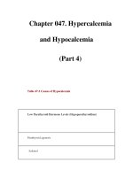
The findings on examination are characteristic.
Table 9.4. Bronchiolitis – characteristic findings on examination
The chest radiograph shows hyperinflation with downward displacement and
flattening of the diaphragm due to small airways obstruction and gas-trapping. In one
third of infants there is also evidence of collapse or consolidation, particularly in the
upper lobes. Respiratory syncytial virus can be cultured or identified with a fluorescent
antibody technique on nasopharyngeal secretions. Blood gas analysis, which is required
in only the most severe cases, shows lowered oxygen and raised carbon dioxide levels.
APPROACH TO THE CHILD WITH FEVER
Although many causes of breathing difficulties are associated with infection, a high
fever is usually associated only with pneumonia, epiglottitis and bacterial tracheitis.
Although many cases of asthma are precipitated by an URTI, the asthmatic child is
rarely febrile and a low grade fever is characteristic of bronchiolitis. Therefore in the
absence of stridor and wheeze, breathing difficulties in association with a significant
fever are likely to be due to pneumonia.
Reassess ABC
Airway and breathing support may be especially needed in children with neurological
THE CHILD WITH BREATHING DIFFICULTIES
92
Risk factors for severity in bronchiolitis
• Age under 6 weeks
• Premature birth
• Chronic lung disease
• Congenital heart disease
• Immunodeficiency
Tachypnoea 50-100 breaths/minute
Recession Subcostal and intercostal
Cough Sharp, dry
Hyperinflation of the chest Sternum prominent, liver depressed
Tachycardia 140-200 beats per minute
Crackles Fine end-inspiratory
Wheezes High-pitched expiratory > inspiratory
Colour Cyanosis or pallor
Breathing pattern Irregular breathing/recurrent apnoea
BMJ Paediatrics 9/11/0 10:04 pm Page 92
handicap who may have poor airway control and weak respiratory muscles even when
well.
Caution should be exercised in fluid administration to children with pneumonia.
Some have inappropriate ADH secretion which can contribute to fluid overload and
worsening breathlessness.
Pneumonia emergency treatment
• As it is not possible to differentiate reliably between bacterial or viral infection on
clinical or radiological grounds, all children diagnosed as having pneumonia should
receive antibiotics. Cefotaxime will be effective against most bacteria but
flucloxacillin should be added if Staphylococcus aureus is suspected and erythromycin
added if Chlamydia or Mycobacteria pneumoniae thought to be responsible.
• Clinical examination and the chest radiograph may reveal a pleural effusion. If this
is large, it should be tapped to relieve breathlessness. Details of the procedure can
be found on page 235.
Background to pneumonia
Pneumonia in childhood is still responsible for over 130 deaths each year in England
and Wales. Infants, and children with congenital abnormalities or chronic illnesses are
at particular risk. In adults, two-thirds of cases of pneumonia are caused by either
Streptococcus pneumoniae or Haemophilus influenzae. A much wider spectrum of
pathogens causes pneumonia in childhood, and different organisms are important in
different age groups.
In the newborn, organisms from the mother’s genital tract, such as Escherichia coli and
other Gram-negative bacilli, group B beta-haemolytic Streptococcus and increasingly,
Chlamydia trachomatis, are the most common pathogens. In infancy respiratory viruses,
particularly respiratory syncytial virus, are the most frequent cause, but Pneumococcus,
Haemophilus and, less commonly, Staphylococcus aureus are also important. In older
children, viruses become less frequent pathogens and bacterial infection is more
important. Mycoplasma pneumonia is a common cause of pneumonia in the school-age
child. Bordatella pertussis can present with pneumonia as well as with classical whooping
cough, even in children who have been fully immunised.
Fever, cough, breathlessness, and lethargy following an upper respiratory infection are
the usual presenting symptoms.The cough is often dry initially but then becomes loose.
Older children may produce purulent sputum but in those below the age of 5 years it is
usually swallowed. Pleuritic chest pain, neck stiffness and abdominal pain may be present
if there is pleural inflammation. Classical signs of consolidation such as impaired
percussion, decreased breath sounds and bronchial breathing are often absent,
particularly in infants, and a chest radiograph is needed. This may show lobar
consolidation, widespread bronchopneumonia or less commonly, cavitation of the lung.
Pleural effusions are quite common, particularly in bacterial pneumonia. An ultrasound
of the chest will delineate a pleural effusion and be helpful in the placing of a chest drain.
Blood cultures, swabs for viral isolation, and a full blood count should also be performed.
As it is not possible to differentiate reliably between bacterial or viral infection on
clinical or radiological grounds, all children diagnosed as having pneumonia should
receive antibiotics. The initial choice of antibiotics depends on the age of the child.
Antibiotics should be given for 7–10 days, except in staphylococcal pneumonia, where
a flucloxacillin course of 4–6 weeks duration is needed. Many older children have no
respiratory difficulty and can be treated at home with penicillin, a cephalosporin or
erythromycin. Infants, and children who look toxic or have definite dyspnoea should be
THE CHILD WITH BREATHING DIFFICULTIES
93
BMJ Paediatrics 9/11/0 10:04 pm Page 93
admitted and usually require intravenous treatment initially. Local antibiotic policies
should be followed. Physiotherapy, an adequate fluid intake and oxygen (in severe
pneumonia), are also required. Mechanical ventilation is rarely required unless there is
serious underlying condition. If a child has recurrent or persistent pneumonia,
investigations to exclude underlying conditions such as cystic fibrosis or
immunodeficiency should be performed.
APPROACH TO THE CHILD IN HEART FAILURE
Infants and children with serious cardiac pathology may present with breathlessness,
cyanosis or cardiogenic shock. The immediate management of the latter is described in
Chapter 10.
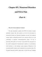
Table 9.5. Causes of heart failure which may present as breathing difficulties
Reassess ABC
HEART FAILURE EMERGENCY TREATMENT
• If there are signs of shock — poor pulse volume or low blood pressure with extreme
pallor and depressed conscious level, treat the child for Cardiogenic Shock (page 109).
• If circulation is adequate and oxygen saturation is normal or improves significantly
with oxygen by face mask but there are signs of heart failure, then the breathing
difficulty is due to pulmonary congestion secondary to a large left to right shunt.
The shunt may be through a VSD, AVSD, PDA or more rarely a truncus arteriosus.
In many cases a heart murmur will be heard. A chest radiograph will also give
confirmatory evidence with a large, usually globular heart and radiological signs of
pulmonary congestion. Give high flow oxygen by face mask with a reservoir and
diuretics such as frusemide (1 mg/kg IV followed by initial maintenance dose of
1–2 mg/kg/day in 1–3 divided doses). If there is no diuresis within 2 hours, the
intravenous bolus can be repeated.
• Babies in the first few days of life who present with breathlessness and increasing
cyanosis largely unresponsive to oxygen supplementation are likely to have a duct-
THE CHILD WITH BREATHING DIFFICULTIES
94
Left ventricular volume overload or excessive pulmonary blood flow
Ventricular septal defect
Atrioventricular septal defect
Persistent arterial duct
Common arterial trunk
Left heart obstruction
Hypertrophic cardiomyopathy
Critical aortic stenosis
Aortic coarctation
Hypoplastic left heart syndrome
Primary “pump” failure
Myocarditis
Cardiomyopathy
BMJ Paediatrics 9/11/0 10:04 pm Page 94
dependent congenital heart disease such as tricuspid or pulmonary atresia. An
infusion of alprostadil at an initial dose of 0·05 micrograms/kg/min will maintain or
increase the patent ductus arteriosus size temporarily until the patient can be
transferred to a neonatal cardiology unit. Patients should be intubated and
ventilated for transfer both because of the seriousness of their condition and also
because the alprostadil may cause apnoea. As oxygen tends to promote ductal
closure, oxygen concentration for ventilation should be individually adjusted using
pulse oximetry to monitor the most effective concentration for each infant.
• Children of all ages who present with breathlessness from heart failure may have
myocarditis.This is characterised by a marked sinus tachycardia and the absence of
signs of structural abnormality. The patients should be treated with oxygen and
diuretics.
Full blood count, serum urea and electrolytes, calcium, glucose and arterial blood
gases should be performed on all patients in heart failure. A routine infection screen
including blood cultures is recommended especially in infants. A full 12-lead
electrocardiogram and chest radiograph are essential. All patients suspected of having
heart disease should be discussed with a paediatric cardiologist, echocardiography will
establish the diagnosis in almost all cases.
Background to heart failure in infancy and childhood
In infancy heart failure is usually secondary to structural heart disease and medical
treatment is directed to improving the clinical condition prior to definitive surgery.With
modern obstetric management many babies are now discharged from the maternity unit
only hours after birth. Therefore babies with serious congenital neonatal heart disease
may present to paediatric or Accident and Emergency departments.
Infants with common congenital heart diseases are usually diagnosed in utero or at
the post-natal examination but a few will present acutely after discharge from medical
care as the lowering pulmonary vascular resistance over the first hours to days of life
allows increasing pulmonary flow in infants with left to right shunts such as VSD,
persistent PDA, truncus arteriosus. The increasing left to right shunt causes increasing
pulmonary congestion and heart failure and the infant presents with poor feeding,
sweating and breathlessness. In addition, some may present at a few months of age when
heart failure is precipitated by a respiratory infection, usually bronchiolitis.
Duct-dependent congenital heart disease
There are also several rarer and more complex congenital heart defects in which the
presence of a patent ductus arteriosus is essential to maintain pulmonary or systemic
flow.The normal patent ductus arteriosus closes functionally in the first 24 hours of life.
This may be delayed in the presence of congenital cardiac anomalies.
The pulmonary obstructive lesions include pulmonary atresia, critical pulmonary
valve stenosis, tricuspid atresia, severe Fallot’s tetralogy and some cases of transposition
of the great vessels. In all of these lesions there is no effective route for blood to take
from the right ventricle into the pulmonary circulation and therefore pulmonary blood
flow and oxygenation of blood are dependent on flow from the aorta via a patent ductus.
Babies with critical pulmonary obstructive lesions present in the first few days of life
with increasing cyanosis, breathlessness or cardiogenic shock. On examination there may
be a characteristic murmur but more frequently there is no murmur audible. An enlarged
liver is a common finding.The clinical situation has arisen from the gradual closure of the
ductus arteriosus. Complete closure will result in the death of the infant from hypoxia.
THE CHILD WITH BREATHING DIFFICULTIES
95
BMJ Paediatrics 9/11/0 10:04 pm Page 95
Additionally, there are some congenital heart malformations where systemic blood
flow is dependent on the ductus arteriosus delivering blood to the aorta from the
pulmonary circulation.This is characteristic of severe coarctation, critical aortic
stenosis and hypoplastic left heart syndrome.
In these congenital heart lesions the baby ceases to be able to feed and becomes
breathless, grey and collapsed with a poor peripheral circulation. On examination the
babies are in heart failure and in more severe cases in cardiogenic shock. In this
situation even in coarctation of the aorta all pulses are difficult to feel.
In the older child myocarditis and cardiomyopathy are the most common causes of
the acute onset of heart failure and remains rare (see Table 9.1.).
How to differentiate the infant with heart failure from
the infant with bronchiolitis
The common features of heart failure in infancy are:
Breathlessness
Feeding difficulty with growth failure
Restlessness
Sweating
Tachycardia
Tachypnoea
Sternal and sub-costal recession
The extremities are cool and pale with cardiomegaly and hepatomegaly
Auscultation reveals a gallop rhythm and occasionally basal crackles
In babies and children peripheral oedema is less commonly seen than in adults. It can
therefore be difficult to differentiate the infant with heart failure from the infant with
bronchiolitis but the cardinal additional features in the infant in heart failure is the greater
degree of hepatomegaly, the enlarged heart with displaced apex beat and the presence of
a gallop rhythm and/or a murmur. A chest radiograph will often be helpful in showing
cardiomegaly and pulmonary congestion rather than the over-inflation of bronchiolitis.
Older children presenting in heart failure will almost certainly have myocarditis or
cardiomyopathy and present with fatigue, effort intolerance, anorexia, abdominal pain
and cough. On examination a marked sinus tachycardia, hepatomegaly and raised JVP
is found.
METABOLIC AND POISONING
Diabetes
As hyperventilation is a feature of the severe acidosis produced by diabetes,
occasionally a child may be presented as a primary breathing difficulty. The correct
diagnosis is usually easy to establish and management is described in Appendix B.
Poisoning
There may be apparent breathing difficulties following the ingestion of a number of
poisons.
The repiratory rate may be increased by poisoning with:
• Salicylates
THE CHILD WITH BREATHING DIFFICULTIES
96
• Ethylene glycol (anti-freeze)
• Methanol
• Cyanide.
But usually only poisoning with salicylates causes any diagnostic dilemma.
Poisoning with drugs that cause a depression of ventilation will present as a
diminished conscious level
The management of the poisoned child is dealt with in Chapter 14.
THE CHILD WITH BREATHING DIFFICULTIES
97
BMJ Paediatrics 9/11/0 10:04 pm Page 97
CHAP TITLE
BMJ Paediatrics 9/11/0 10:04 pm Page 98
CHAPTER
I
10
I
The child in shock
INTRODUCTION
Shock results from an acute failure of circulatory function. Inadequate amounts of
nutrients, especially oxygen, are delivered to body tissues and there is inadequate
removal of tissue waste products. These functions involve several body systems which
means that there are several causes of shock and therefore the clinician must consider
which of several alternative emergency treatments will be effective for an individual
patient. This chapter will provide the student with an approach to the assessment,
resuscitation and emergency management of children in shock.
Maintenance of adequate tissue perfusion depends on a pump (the heart) delivering
the correct type and volume of fluid (blood) through controlled vessels (arteries, veins,
and capillaries) without abnormal obstruction to flow. Inadequate tissue perfusion
resulting in impaired cellular respiration (i.e. shock) may result from defects of the
pump (cardiogenic), loss of fluid (hypovolaemic), abnormalities of vessels (distributive),
flow restriction (obstructive), or inadequate oxygen releasing capacity (dissociative).
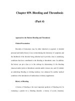
From the box it can be seen that the most common causes of shock in the paediatric
patient are hypovolaemia from any cause, septicaemia, and the effects of trauma.
CHAP TITLE
99
Classification of causes of shock
(common causes are emboldened)
Cardiogenic
Arrhythmias
Cardiomyopathy
Heart failure
Valvular disease
Myocardial contusion
Myocardial infarction
Hypovolaemic
Haemorrhage
Gastroenteritis
Volvulus
BMJ Paediatrics 9/11/0 10:04 pm Page 99
Children in shock are usually presented by parents who are aware that their child is
worryingly ill or seriously injured even though they may not be able to express their
concerns clearly. The child may be presented primarily with a fever, a rash, with pallor,
poor feeding or drowsiness or with a history of trauma or poisoning. The initial
assessment will identify which patients are in shock
APPROACH TO THE CHILD IN SHOCK
PRIMARY ASSESSMENT
Airway
Assess airway patency by the “look, listen, and feel” method.
If the child can speak or cry, this indicates that the airway is patent, that breathing is
occurring and there is adequate circulation.
If there is no evidence of air movement then chin lift or jaw thrust manoeuvres should
be carried out and the airway reassessed. If there continues to be no evidence of air
movement then airway patency can be assessed by performing an opening manoeuvre
and giving rescue breaths (see Basic life support, Chapter 4).
Breathing
Assess the adequacy of breathing
Monitor oxygen saturation with a pulse oximeter.
THE CHILD IN SHOCK
100
Burns
Peritonitis
Distributive
Septicaemia
Anaphylaxis
Vasodilating drugs
Anaesthesia
Spinal cord injury
Obstructive
Tension pneumothorax
Haemopneumothorax
Flail chest
Cardiac tamponade
Pulmonary embolism
Hypertension
Dissociative
Profound anaemia
Carbon monoxide poisoning
Methaemoglobinaemia
• Effort of breathing
Recession
Respiratory rate
Grunting
Accessory muscle use
Flare of the alae nasi
BMJ Paediatrics 9/11/0 10:04 pm Page 100
Circulation
Assess the adequacy of circulation.
Cardiovascular status
Heart rate
A raised heart rate is a common response to many types of stress (fever, anxiety, hypoxia,
hypovolaemia). In shock, tachycardia is caused by catecholamine release, and is an attempt
to maintain cardiac output by increasing heart rate in the face of falling stroke volume.
Bradycardia in a shocked child is caused by hypoxia and acidosis and is a preterminal sign.
Pulse volume
Examination of central and peripheral pulses may reveal a poor pulse volume
peripherally or, more worryingly, centrally. In early septic shock there is sometimes a
high output state which will produce bounding pulses.
Capillary refill
Poor skin perfusion can be a useful early sign of shock. Slow capillary refill (>2 seconds)
after blanching pressure for 5 seconds is evidence of reduced skin perfusion.When testing
for capillary refill press on the skin of the sternum or a digit held at the level of the heart.
Mottling, pallor, and peripheral cyanosis also indicate poor skin perfusion. All these signs
may be difficult to interpret in patients who have just been exposed to cold.
In early shock, there may be a hyperdynamic circulation due to vasodilataion in which
peripheries are warm but the capillary refill is delayed.
Blood pressure
Blood pressure is a difficult measure to obtain and interpret especially in young
infants. A formula for calculating normal systolic blood pressure is:
80 + (2 ҂ Age in years)
Children’s cardiovascular systems compensate well initially in shock. Hypotension is a
late and often sudden sign of decompensation and,if not reversed,will be rapidly followed by death.
Serial measurements of blood pressure should be performed frequently.
Effects of circulatory inadequacy on other organs
Acidotic sighing respirations
The acidosis produced by poor tissue perfusion in shock leads to rapid deep
breathing.
THE CHILD IN SHOCK
101
• Efficacy of breathing
Breath sounds
Chest expansion/abdominal excursion
• Effects of breathing
Heart rate
Skin colour
Mental status
BMJ Paediatrics 9/11/0 10:04 pm Page 101
Pale, cyanosed or cold skin
A core/toe temperature difference of more than 2°C is a sign of poor skin perfusion.
Mental status
Agitation or depressed conscious level. Early signs of brain hypoperfusion are agitation
and confusion, often alternating with drowsiness. Infants may be irritable but drowsy
with a weak cry and hypotonia. They may not focus on the parent’s face. These are
important early cerebral signs of shock. Later the child becomes progressively drowsier
until consciousness is lost.
Urinary output
Urine flow is decreased or absent in shock. Hourly measurement is helpful in
monitoring progress. A minimum flow of 1 ml/kg/h in children and 2 ml/kg/h in infants
indicates adequate renal perfusion.
NOTE: Poor capillary refill, core/toe temperature difference and differential pulse
volumes are neither sensitive nor specific indicators of shock when used in isolation.
There are helpful when used in conjunction with the other signs described.
Look for the presence of signs of heart failure
• Tachycardia
• Raised jugular venous pressure (often not seen in infants in heart failure)
• Lung crepitations on auscultation
• Gallop rhythm
• Enlarged liver
And listen for a heart murmur.
Monitor heart rate/rhythm, blood pressure and core/toe temperature difference. If
heart rate is above 200 in an infant or above 150 in a child or if the rhythm is abnormal
perform a standard ECG.
Disability
Assess neurological function.
• A rapid measure of level of consciousness should be recorded using the AVPU scale.
• A ALERT
• V responds to VOICE
• P responds to PAIN
• U UNRESPONSIVE
• Pupillary size and reaction should be noted.
• Note the child’s posture: children in shock are usually hypotonic.
• The presence of convulsive movements should be noted.
Exposure
• Take the child’s core and toe temperatures.
• Look for a rash: if one is present, ascertain if it is purpuric.
• Look for evidence of poisoning.
THE CHILD IN SHOCK
102
BMJ Paediatrics 9/11/0 10:04 pm Page 102
THE CHILD IN SHOCK
103
RESUSCITATION
Airway
A patent airway is the first requisite. If the airway is not patent an airway opening
manoeuvre should be used. The airway should then be secured with a pharyngeal
airway device or by intubation with experienced senior help.
Breathing
All children in shock should receive high flow oxygen through a face mask with a
reservoir as soon as the airway has been demonstrated to be adequate.
If the child is hypoventilating, respiration should be supported with oxygen via a
bag-valve-mask device and experienced senior help summoned.
Circulation
Gain intravenous or intraosseous access.
Take blood for FBC, U&Es, blood culture, cross-match, glucose stick test and
laboratory test
Give 20 ml/kg rapid bolus of crystalloid to all patients except for those with signs
that heart failure is their primary pathology.
The initial bolus should be colloid and an antibiotic such as cefotaxime 100 mg/kg
should be used for those in whom a diagnosis of septicaemia is made obvious by
the presence of a purpuric rash.
If a tachyarrhythmia is identified as the cause of shock, up to three synchronous
electrical shocks at 0·5, 1·0, 2·0 Joules should be given.
If the arrhythmia is broad complex and the synchronous shocks are not activated by
the defibrillator then attempt an asynchronous shock.
A conscious child should be anaesthetised first if this can be done in a timely
manner.
If the shocked child’s tachyarrhythmia is SVT then he can be treated with
intravenous/intraosseous adenosine if this can be administered more quickly than a
synchronous electrical shock.
Circulatory access
A short, wide-bore peripheral venous or intraosseous cannula should be used. Upper
central venous lines are unsuitable for the resuscitation of hypovolaemic children
because of the risk of iatrogenic pneumothorax, or exacerbation of an unsuspected
neck injury; both these complications can be fatal. Femoral vein access is safer, if
peripheral or intraosseous access is impossible. It is wise to obtain two separate
intravenous and/or intraosseous lines both to give large volumes of fluid quickly and
also in case one line is lost.
Techniques for vascular access are described in Chapter 23.
Antibiotics
In paediatric practice, septicaemia is the commonest cause of a child presenting in
shock. Therefore, unless an alternative diagnosis is very clear (such as trauma,
anaphylaxis or poisoning) an antibiotic, usually a third-generation cephalosporin such as
cefotaxime or ceftriaxone, is given as soon as a blood culture has been taken. An anti-
staphyloccocal antibiotic (flucloxacillin or vancomycin) should be considered in
possible toxic shock syndrome i.e. post burns/cellulitis.
Hypoglycaemia
Hypoglycaemia may give a similar clinical picture to that of compensated shock.This must
always be excluded by urgent glucose stick test and blood glucose estimation. Shock and
hypoglycaemia may coexist as the sick infant or small child has poor glucose-producing reserves.
Key features
While the primary assessment and resuscitation are being carried out a focused
history of the child’s health and activity over the previous 24 hours and any significant
previous illness should be gained.
Certain key features which will be identified clinically in the above assessment, from
the focused history and from the initial blood test results can point the clinician to the
likeliest working diagnosis for emergency treatment.
• A history of vomiting and/or diarrhoea points to fluid loss either externally (e.g.
gastroenteritis) or into the abdomen (e.g. volvulus, intussusception).
• The presence of fever and/or a rash points to septicaemia.
• The presence of urticaria, angio-neurotic oedema and a history of allergen exposure
points to anaphylaxis.
• The presence of cyanosis unresponsive to oxygen or a grey colour with signs of heart
failure in a baby under 4 weeks points to duct-dependent congenital heart disease.
• The presence of heart failure in an older infant or child points to cardiomyopathy.
• A history of sickle cell disease or a recent diarrhoeal illness and a very low
haemoglobin points to acute haemolysis.
• An immediate history of major trauma points to blood loss, and more rarely, tension
pneumothorax, haemothorax, cardiac tamponade or spinal cord transection (see Part IV
The Seriously Injured Child for management).
• The presence of severe tachycardia and an abnormal rhythm on the ECG points to
an arrhythmia (see Chapter 11).
• A history of polyuria and the presence of acidotic breathing and a very high blood
glucose points to diabetes (see Appendix B for management).
• A history of drug ingestion points to poisoning (see Chapter 14 for management).
APPROACH TO THE CHILD WITH FLUID LOSS
Infants are more likely than older children to present with shock due to sudden fluid
loss in gastroenteritis or with concealed fluid loss secondary to a “surgical abdomen”
such as a volvulus.This is due both to the infant’s low physiological reserve and increased
susceptibility to these conditions.
In infants gastroenteritis may occasionally present as a circulatory collapse with little
or no significant preceding history of vomiting or diarrhoea.The infecting organism can
be any of the usual diarrhoeal pathogens, of which the most common is rotavirus. The
mechanism leading to this presentation is that there is a sudden massive loss of fluid
from the bowel wall into the gut lumen, causing depletion of the intravascular volume
and the appearance of shock in the infant.This occurs before the stool is passed so that
the diagnosis may be unsuspected. Usually during resuscitation of these infants, copious
watery diarrhoea is evacuated.
Having completed the primary assessment and resuscitation and identified by means
of the key features that fluid loss is the most likely diagnosis, the child is reassessed to
identify the response to the first fluid bolus.
THE CHILD IN SHOCK
104
BMJ Paediatrics 9/11/0 10:04 pm Page 104
REASSESS ABC
Fluid loss – emergency treatment
If the child still shows clinical signs of shock after the first bolus of fluid, give a second
20 ml/kg bolus of crystalloid. If there is clinical suspicion of a surgical abdominal
problem, such as bile-stained vomiting or abdominal guarding, seek an urgent surgical
opinion. An abdominal radiograph and an ultrasound scan may be helpful in showing
distended bowel, intra-abdominal air or fluid.
In the case of infants with gastroenteritis, two boluses of crystalloid is usually
sufficient to restore the circulating volume. If after this amount of fluid, the child is still
in shock when assessed clinically, give the third bolus as colloid (human albumen is the
most widely used in paediatric practice) and consider whether there is an additional or
alternative diagnosis, such as an intra-abdominal surgical problem (e.g. volvulus,
peritonitis) in the patient originally thought to have gastro-enteritis or co-existent
septicaemia in the patient with the “surgical abdomen”.
Obtain surgical and anaesthetic advice if not already obtained and give antibiotics
intravenously if more than two boluses of fluid have been required
The child should be catheterised in order to assess accurately the urinary output.
Intubation and ventilation should be strongly considered in a patient who has failed
to respond adequately to two boluses of fluid (i.e. half the estimated intravascular
volume). Acid–base status should be checked by means of an arterial blood gas.
In the patient with gastroenteritis who has stabilised after treatment for shock there
will still be a need to treat dehydration and electrolyte imbalance. See Appendix B for
further management
APPROACH TO THE CHILD WITH SEPTICAEMIA
The cardinal sign of meningococcal septicaemia is a purpuric rash in an ill child. At the
onset, however, the rash is not florid and a careful search should be made for purpura in
any unwell child. In about 13% of patients with meningococcal septicaemia, a blanching
erythematous rash replaces a purpuric one, and in 7% of cases no rash occurs. In the
much rarer toxic shock syndrome, the initial clinical picture includes a high fever,
headache, confusion, conjunctival and mucosal hyperaemia, scarlatiniform rash with
secondary desquamation, subcutaneous oedema, vomiting and watery diarrhoea. Early
administration of antibiotics, concurrent with initial resuscitation is vital.
In countries where the vaccine against Meningococcus C has been introduced a fall in
the number of cases of infection is occurring.
Having completed the primary assessment and resuscitation and identified by means
of the key features that septicaemia is the most likely dignosis, the child is reassessed.
REASSESS ABC
Septicaemia emergency management
If the child is still in shock after the first bolus of fluid a second 20 ml/kg fluid bolus
should be given over five to ten minutes. In septicaemia it remains usual practice to give
fluid as 4·5% human albumin. (A discussion of the relative merits of fluids can be found
on page 114)
Children in septic shock often require several boluses of fluid to achieve relative
stability. Once the third bolus of fluid has been commenced, the patient should be
THE CHILD IN SHOCK
105
BMJ Paediatrics 9/11/0 10:04 pm Page 105
intubated by rapid sequence induction of anaesthesia and ventilated. This is done both
to support a seriously ill patient by maximising oxygenation and to anticipate the
development of pulmonary oedema caused by fluid leak in the lungs. All intubated
children must have continuous Sa
O
2
and CO
2
monitoring. The child should be
catheterised in order to assess accurately the urinary output.
In septic shock, myocardial depression is a co-existent feature.Therefore, at the same
time as the third bolus of fluid is commenced an infusion of dobutamine should be
started at an initial rate of 10 micrograms/kg/min. This can be given through a
peripheral vein as it is unlikely that central venous access will yet have been obtained.
The rate of infusion should be adjusted to the patient’s response. Do not hesitate to
increase the infusion rapidly in the face of a poor response. Consider the use of
epinephrine if maximal does of dobutamine and/or dopamine are unsuccessful.
Epinephrine should be preferably given through a central vein but do not delay if this is
not available.
Further investigations
In addition to the blood tests taken during resuscitation, the following blood tests are
needed in the septic child: calcium, magnesium, phosphate, coagulation screen and
arterial blood gas. Electrolyte and acid–base derangements can have a deleterious effect
on myocardial function. They should be sought and corrected.
Table 10.1. Corrective measures for electrolyte and acid–base derangements
It is difficult to manage a patient so seriously ill as to require ventilation and inotropic
support without intensive care facilities and invasive monitoring. If these treatments are
required, a paediatric intensive care unit must be involved early to give advice and to
retrieve the patient
Reassess disability
This is an assessment of the neurological status of the septicaemic child.
• Both hypoxia and shock produce neurological effects on their own account and the
conscious level is part of the assessment of the severity of these conditions. In
addition, in children with meningococcal septicaemia, many have both septicaemia
and meningitis. Of these some, generally in the school age group have clinically
significant raised intracranial pressure (RICP).These children must be identified as
the clinician may need to prevent or treat this problem.
• The level of consciousness should be assessed using the Glasgow Coma Scale.
• Pupillary size and reaction should be noted.
• The presence of abnormal posturing should be noted. This may require a painful
stimulus to demonstrate its presence.
THE CHILD IN SHOCK
106
Result Treat if less than Correct with
Glucose 3 mmol/l 3 ml/kg 10% dextrose
Acid–base 7·15 1 mmol/kg NaHCO
3
: ventilate
Potassium 3·5 mmol/l 0·25 mmol/kg KCl over 30 min: ECG
Calcium 2 mmol/l 0·3 ml/kg 10% Ca gluconate over 30 min
Magnesium 0·75 mmol/l 0·2 ml/kg 50% MgSO
4
over 30 min
Phosphate 0·7 mmol/l 0·2 mmol/kg over 30 min
BMJ Paediatrics 9/11/0 10:04 pm Page 106
THE CHILD IN SHOCK
107
Disability emergency treatment
If, despite effective treatment of shock, the child has a decreasing conscious level
and/or abnormal posturing, possibly also with focal neurological signs, he may
have raised intracranial pressure. He should be intubated using rapid sequence
induction if this has not already been done and capnography used to monitor CO
2
levels which should be kept in the range 4–4·5 kPa. A diuretic such as mannitol
(0·5–1.0 g/kg) or frusemide (1 mg/kg) can be given intravenously. The child should
be catheterised in order to assess accurately the resulting urinary output. This will
temporarily relieve the intracranial pressure. The presence of relative bradycardia
and hypertension is a pre-terminal sign of imminent brain stem coning and death.
This should be treated vigorously with diuretics and hyperventilation.
If the shocked state has been effectively treated, only maintenance fluids should be
continued although close monitoring is required as continued capillary fluid leak
will lead to a return of shock. If the patient is still shocked then treatment of the
shocked state takes priority. An adequate blood pressure is necessary to perfuse a
swollen brain.
Lumbar puncture must be avoided as its performance may cause death through
coning of the brain through the foramen magnum.
Paediatric intensive care skills and monitoring is paramount in these patients. Seek advice
early.
APPROACH TO THE CHILD WITH ANAPHYLAXIS
Anaphylaxis is a potentially life-threatening syndrome which may progress to shock,
although in most cases a rash is the only symptom. It is immunologically mediated.
The most common causes are allergy to penicillin, to radiographic contrast media, and
to certain foods, especially nuts.
Prodromal symptoms of flushing, itching, facial swelling, urticaria, abdominal pain,
diarrhoea, wheeze, and stridor may precede shock or may be the only manifestations of
anaphylaxis. The presence of these additional symptoms confirms anaphylaxis as the
cause of shock in a child. Most patients will have a history of previous attacks and
some may have a “medic-alert” bracelet.
Anaphylaxis can be life-threatening because of the rapid onset of airway compromise
due to laryngeal oedema, breathing difficulties due to sudden severe broncho-
constriction and/or the development of shock due to acute vasodilatation and fluid loss
from the intravascular space caused by increased capillary permeability.
Key points in the history may point to a severe reaction. These are shown in the box.
Symptoms and signs vary according to the body’s response to the allergen. These
are shown in Table 10.2.
Previous severe reaction
History of increasingly severe reaction
History of asthma
Treatment with ß-blockers
Table 10.2. Symptoms and signs in allergic reaction
The management of anaphylactic shock requires good airway management,
administration of epinephrine (adrenaline), and aggressive fluid resuscitation.
Note that the intramuscular route is the preferred route for the delivery of
epinephrine. Intravenous epinephrine should be reserved for children with life-
threatening shock for whom intramuscular injection has been ineffective. The patient
must be carefully monitored.
Having completed the primary assessment and resuscitation and identified by means
of the key features that anaphylaxis is the most likely diagnosis, the child is reassessed
Remove allergen if possible.
Reassess airway
If there is stridor then the child has laryngeal oedema.
Airway emergency management
• If the child has airway obstruction with stridor call for urgent anaesthetic and ENT
help.
• Give epinephrine 10 micrograms/kg IM and also nebulised epinephrine 5 ml 1:1000.
• Consider the need for intubation or a surgical airway.
Reassess breathing
• Assess effort, efficiency and effect
• Check oxygen saturation on the pulse oximeter
• If there is wheeze then the child has bronchoconstriction
Breathing emergency treatment
If the child has bronchoconstriction give nebulised salbutamol 2·5–5 mg. If no
parenteral epinephrine has been given then give epinephrine 10 micrograms/kg IM.
Reassess circulation
• Look for signs of shock.
• Check pulse rate and rhythm on the ECG.
THE CHILD IN SHOCK
108
Symptoms Signs
Mild Burning sensation in mouth Urticarial rash
Itching of lips, mouth, throat Angio-oedema
Feeling of warmth Conjunctivitis
Nausea
Abdominal pain
Moderate (Mild +) Coughing/wheezing Bronchospasm
Loose bowel motions Tachycardia
Sweating Pallor
Irritability
Severe (Moderate +) Difficulty breathing Severe bronchospasm
Collapse Laryngeal oedema
Vomiting Shock
Uncontrolled defaecation Respiratory arrest
Cardiac arrest
BMJ Paediatrics 9/11/0 10:04 pm Page 108
Circulation emergency treatment
If the child is in shock give colloid 20 ml/kg IV/IO. If no parenteral epinephrine has
been given yet then give epinephrine 10 micrograms/kg IM.
Further emergency management
Depending whether upper airway obstruction, bronchoconstriction or shock
predominate in the clinical picture of anaphylaxis, the clinician should
1. secure the airway by intubation
2. follow the protocol for asthma
3. continue to treat for shock with boluses of colloid and ventilatory support
4. give further doses of epinephrine intramuscularly every five minutes if the symptoms
are not reversed.
Additional inotropes will not be needed as the epinephrine used for the treatment of
anaphylaxis is a powerful inotrope. However, in the face of shock resistant to
intramuscular epinephrine and one or two boluses of fluid, an infusion of intravenous
epinephrine may be life-saving.The dose is 0·1–5·0 micrograms/kg/min and the patient
should be closely monitored for pulse and blood pressure.
In addition to the above treatment it is also customary to give patients with
anaphylaxis an antihistamine and steroids. There is no evidence of the part these drugs
play in management and their onset of action is too delayed to be of much benefit in
the first hour.
APPROACH TO THE INFANT WITH A DUCT-DEPENDENT
CONGENITAL HEART DISEASE
Babies with critical pulmonary obstructive lesions present in the first few days of life
with increasing cyanosis, breathlessness or cardiogenic shock. On examination there
may be a characteristic murmur but in fact more frequently there is no murmur audible.
An enlarged liver is a common finding.
Babies with critical systemic obstructive lesions also present in the first few days of
life with inability to feed, breathlessness, a grey appearance and collapse with poor
peripheral circulation. On examination the babies are in heart failure and in more severe
cases in cardiogenic shock. In this situation, even in coarctation of the aorta, all pulses
are difficult to feel.
The clinical situation has arisen from the gradual closure of the ductus arteriosus on
which, in these congenital heart anomalies, a functioning circulation depends.
Complete closure will result in the death of the infant.
THE CHILD IN SHOCK
109
Drug doses in anaphylaxis
Epinephrine 10 micrograms/kg
Chlorpheniramine
>12 years 10–20 milligrams
6–12 years 5–10 milligrams
1–5 years 2·5–5 milligrams
1 month–1 year 250 micrograms/kg
Do not use in neonates
Hydrocortisone 4 milligrams/kg
BMJ Paediatrics 9/11/0 10:04 pm Page 109
Having completed the primary assessment and resuscitation and identified by means
of the key features that duct-dependent congenital heart disease is the most likely
diagnosis, the child is reassessed.
Reassess ABC
DUCT-DEPENDENT CONGENITAL HEART DISEASE
EMERGENCY TREATMENT
Babies in the first few days of life who present with breathlessness and increasing cyanosis
or a grey appearance largely unresponsive to oxygen supplementation are likely to have a
duct-dependent congenital heart disease such as tricuspid or pulmonary atresia, critical
aortic stenosis or hypoplastic left heart syndrome. An infusion of alprostadil at an initial
dose of 0·05 micrograms/kg/min will maintain or increase the patent arteriosus ductus size
temporarily until the patient can be transferred to a neonatal cardiology unit. Patients
should be intubated and ventilated for transfer both because of the seriousness of their
condition and also because the prostaglandin may cause apnoea.
Full blood count, serum urea and electrolytes, calcium, glucose and arterial blood
gases should be performed on all sick infants with congenital heart disease. A routine
infection screen including blood cultures is also recommended. A full 12-lead
electrocardiogram and chest radiograph are essential. All patients suspected of having
heart disease should be discussed with a paediatric cardiologist, echocardiography will
establish the diagnosis in almost all cases.
APPROACH TO THE CHILD WITH CARDIOMYOPATHY
Cardiomyopathy/myocarditis is most uncommon but may rarely be found in an infant
or child presenting in shock and with signs of heart failure but with no history of
congenital heart disease.
If such a patient were in the first few weeks of life, a trial of alprostadil would be
appropriate and harmless.
Having completed the primary assessment and resuscitation and identified by means
of the key features that cardiomyopathy/myocarditis is the most likely diagnosis, the
child is reassessed.
Reassess ABC
Cardiomyopathy emergency treatment
• As the circulation is already overloaded with fluid, a diuretic, such as frusemide,
should be given and the failing heart supported with an infusion of dobutamine
which has some vasodilatory as well as inotropic effects.
• Urgent cardiology advice should be sought. Echocardiography should establish the
diagnosis in almost all cases.
Full blood count, serum urea and electrolytes, calcium, glucose and arterial blood
gases should be performed on all children with heart disease. A routine infection screen
including blood cultures is also recommended. A full 12-lead electrocardiogram and
chest radiograph are essential.
THE CHILD IN SHOCK
110
BMJ Paediatrics 9/11/0 10:04 pm Page 110
APPROACH TO THE CHILD WITH PROFOUND ANAEMIA
The most usual situation in which a child develops sudden severe haemolysis is in the
case of septicaemia associated with sickle cell disease
In this situation, the child should be treated as for sepsis with volume support,
intubation and inotropes. However, the volume infused should be fresh blood as soon
as it can be obtained. These children may have an already damaged myocardium
causing them to be candidates for cardiogenic as well as septic and dissociative shock.
An exchange transfusion may be life-saving in selected cases. These children will all
need early paediatric intensive care advice and transfer.
BACKGROUND TO SHOCK
Shock results from an acute failure of circulatory function. Inadequate amounts of
nutrients, especially oxygen, are delivered to body tissues and there is inadequate removal
of tissue waste products. Shock is a complex clinical syndrome that is the body’s response
to cellular metabolic deficiency.
In hypovolaemic or distributory shock the initial haemodynamic abnormality of fluid
loss or fluid shift is followed by compensatory mechanisms under neuroendocrine
control. Later, shock is worsened by the production of vasoactive mediators and the
products of cellular breakdown.The identity and relative importance of these chemicals
are as yet poorly understood.
Shock is a progressive syndrome but it can be divided into three phases: compensated,
uncompensated, and irreversible. Although artificial, this division is useful because each
phase has characteristic clinicopathological manifestations and outcome.
Phase 1 (compensated) shock
In this phase vital organ function (brain and heart) is conserved by sympathetic
reflexes which increase systemic arterial resistance, divert blood away from non-
essential tissues, constrict the venous reservoir and increase the heart rate to maintain
cardiac output. The systolic blood pressure remains normal whereas the diastolic
pressure may be elevated due to increased systemic arterial resistance. Increased
secretion of angiotensin and vasopressin allows the kidneys to conserve water and salt,
and intestinal fluid is reabsorbed from the digestive tract. Clinical signs at this stage
include mild agitation or confusion, skin pallor, increased heart rate, and cold
peripheral skin with decreased capillary return.
Phase 2 (uncompensated) shock
In uncompensated shock, the compensatory mechanisms start to fail and the
circulatory system is no longer efficient. Areas that have poor perfusion can no longer
metabolise aerobically, and anaerobic metabolism becomes their major source of energy
production. Anaerobic metabolism is comparatively inefficient. Only 2 moles of
adenosine triphosphate (ATP) are produced for each mole of glucose metabolised
compared to 38 moles of ATP per mole of glucose metabolised aerobically.
Anaerobic pathways produce excess lactate leading to systemic acidosis. The acidosis
is compounded by intracellular carbonic acid formed because of the inability of the
circulation to remove CO
2
. Acidosis reduces myocardial contractility and impairs the
response to catecholamines.
A further result of anaerobic metabolism is the failure of the energy dependent
THE CHILD IN SHOCK
111
BMJ Paediatrics 9/11/0 10:04 pm Page 111
sodium–potassium pump, which maintains the normal homoeostatic environment in
which the cell functions.
Lysosomal, mitochondrial, and membrane functions deteriorate without this
homoeostasis. Sluggish flow of blood and chemical changes in small vessels lead to
platelet adhesion, and may produce damaging chain reactions in the kinin and
coagulation systems leading to a bleeding tendency.
Numerous chemical mediators have been identified in shocked patients, but the roles
of each have not been clearly identified. They include histamine, serotonin, cytokines
(especially tumour necrosis factor and interleukin 1), xanthine oxidase (which generates
oxygen radicals), platelet-aggregating factor, and bacterial toxins. They are largely
produced by cells of the immune system, especially monocytic macrophages. It has been
suggested that these mediators, which developed as initial adaptive responses to severe
injury and illness, may have deleterious consequences in the “unnatural” setting of the
resuscitated patient.When the role of these chemical mediators is more fully understood,
blocking agents may be produced which will improve the treatment in phase 2 shock.
The result of these cascading metabolic changes is to reduce tissue perfusion and
oxidation further. Blood pools in some areas because arterioles can no longer control
flow in the capillary system. Furthermore, abnormal capillary permeability allows
further fluid loss from the circulation into the interstitium.
Clinically, the patient in phase 2 shock has a falling blood pressure, very slow capillary
return, tachycardia, cold peripheries, acidotic breathing, depressed cerebral state, and
absent urine output.
Phase 3 (irreversible) shock
The diagnosis of irreversible shock is a retrospective one.The damage to key organs such
as the heart and brain is of such magnitude that death occurs despite adequate restoration
of the circulation. Pathophysiologically, the high energy phosphate reserves in cells
(especially those of the liver and heart) are greatly diminished.The ATP has been degraded
via adenosine to uric acid. New ATP is synthesised at only 2% an hour and the body can
be said to have run out of energy. This underlies the clinical observation that during the
progression of shock a point is reached at which death of the patient is inevitable, despite
therapeutic intervention. Hence early recognition and effective treatment of shock are vital.
A closer study of septic shock illustrates many of these points.
Septic shock
In sepsis the cardiac output may be normal or raised but may still be too low to deliver
sufficient oxygen to the tissues. This is because abnormal distribution of blood in the
microcirculation leads to decreased tissue perfusion.
The release of bacterial toxins triggers complex interacting haemodynamic and
metabolic changes. Mediators and activators are released and react to produce the
“septic syndrome”. These activators may be vasodilators or vasoconstrictors; some
promote and activate the coagulation cascade; others are cardiac depressants.
In septic shock cardiac function may be depressed Oxygen delivery to the heart from the
coronary arteries occurs mainly in diastole, and the tachycardia and increased oxygen
demand of the myocardium in septic shock may jeopardise cardiac oxygenation.
Metabolic acidosis also damages myocardial cells at mitochondrial level.The function of
the left ventricle is affected more than the right ventricle.This may be due to myocardial
oedema, adrenogenic receptor dysfunction, or impaired sarcolemmal calcium influx.The
right ventricle is less important in maintaining cardiac output than the left, but increased
THE CHILD IN SHOCK
112
BMJ Paediatrics 9/11/0 10:04 pm Page 112
pulmonary vascular resistance can limit the hyperdynamic state and oxygen delivery.
In septic shock cells do not use oxygen properly There appears to be a block at the
mitochondrial level in the mechanism of oxygen uptake, and in progressive shock the
difference between arterial and venous saturation levels of oxygen is inappropriately narrow.
This progressive deterioration in cell oxygen consumption heralds multiple organ failure.
Early (compensated) septic shock
This is characterised by a raised cardiac output, decreased systemic resistance, warm
extremities, and a wide pulse pressure.This pattern is seen more typically in adults and
may never be seen in infants in whom cold peripheries are much more common. The
hyperdynamic state is recognised by hyperpyrexia, hyperventilation, tachycardia, and
mental confusion. All of these signs may be minimal: mental confusion in particular
needs to be looked for carefully, if septic shock is not to be overlooked at this stage.
Decreased capillary return is a useful sign in these circumstances.
Late (uncompensated) septic shock
If no effective therapy is given, the cardiovascular performance deteriorates and
cardiac output diminishes. Even with a normal or raised cardiac output, shock develops.
The normal relationship between cardiac output and systemic vascular resistance
breaks down and hypotension may persist as a result of decreased vascular resistance.
The cardiac output may fall gradually over several hours, or precipitously in minutes.
As tissue hypoxia develops, plasma lactic acid levels increase.
Infants, who have little cardiac reserve, often present with hypotension and a
hypodynamic picture. These sick babies are a diagnostic challenge but sepsis must be
assumed and treated as quickly as possible.
Survival in septic shock depends on the maintenance of a hyperdynamic state. Several
factors mitigate against this by encouraging hypovolaemia:
1. Increased microvascular permeability.
2. Arteriolar and venous dilatation with peripheral pooling of blood.
3. Inadequate fluid intake.
4. Fluid loss secondary to fever, diarrhoea, and vomiting.
5. Inappropriate polyuria.
AFTER RESUSCITATION AND EMERGENCY TREATMENT
Following successful restoration of adequate circulation, varying degrees of organ
damage may remain, and should be actively sought and managed after the initial
resuscitation and emergency treatment has stabilised the patient. The problems are
similar but of less degree than those expected following resuscitation from cardiac arrest.
Kidneys
Prerenal failure, acute tubular necrosis, and the more severe cortical necrosis may be
sequelae of phase 2 shock. Once haemodynamic parameters are improving, fluid
administration should be reviewed and serum electrolytes, urea, and creatinine analysed.
Lung
“Shock lung” appears to be a more common sequel in adults than in children.
Patients with this complication develop respiratory failure because of increased lung
THE CHILD IN SHOCK
113
BMJ Paediatrics 9/11/0 10:04 pm Page 113
water. Ventilation with high inspired oxygen is necessary, and positive end-expiratory
pressure (PEEP) may be required.
Heart
Despite adequate volume restoration, and even if shock was not primarily
cardiogenic, poor myocardial perfusion often leads to decreased contractility. Inotropic
agents need to be continued and vasodilators may be required.
Coagulation abnormalities
As described above, sludging of blood and the production of chemical mediators may
initiate microvascular clotting which leads to a consumption coagulopathy. Clotting
times and a platelet count should be estimated and fresh frozen plasma given if clinically
indicated.
Other organs
The liver and bowel may be damaged in shock, leading to gastrointestinal bleeding.
Endocrine organs may be variously affected and patients must be monitored for glucose
and mineral homoeostasis.
FLUID RESUSCITATION
Underlying considerations
Crystalloid or colloid fluids or blood are available for volume replacement.
The distribution of different fluids through the main compartments within the body
(in decreasing volume: intracellular, interstitial and intravascular) is determined by
constituents of the fluid. In general, the large molecules in colloids ensure that a greater
proportion of the volume given as colloid will be retained in the intravascular space, the
compartment where fluid resuscitation is directed. Blood is retained best in the
intravascular space. The ability of the osmotically active particles of colloid to remain
intravascular, and retain intravascular fluid volume, is varied. The complex starches
used in heta- or pentastarch remain in the vascular space for a prolonged period. The
gelatin derivatives of Gelofusine or Haemaccel of other colloids do so for only a few
hours. Albumin will exchange readily with the albumin in the interstitial fluid, but
remains in the intravascular space for more than 24 hours in health. Albumin loss to the
tissue fluid will be enhanced where the endothelial barrier function is degraded by
endothelial inflammation.
Again with crystalloids, distribution is determined by the constituents. The sodium
and chloride of normal saline will ensure that it is localised more to the whole
extracellular compartment (where sodium is the main osmotically active particle), and
so when given intravenously, only a minor part will remain in the intravascular
compartment as the majority of extracellular fluid is tissue fluid. This is in contrast to
the distribution of 5% glucose, which after the metabolism of the glucose is effectively
free water, which then disperses through all the fluid compartments of the body and so
even less is retained intravascularly.
Those who support colloid resuscitation emphasis the importance of oncotic pressure
in maintaining intravascular volume and tissue perfusion. Those who favour crystalloids
THE CHILD IN SHOCK
114
BMJ Paediatrics 9/11/0 10:05 pm Page 114
respond that as the endothelium becomes leakier in ill patients, the colloid will also leak
and serve to retain fluid in the tissues. To equal the increase of intravascular volume
produced by a colloid, approximately three to five times as much crystalloid must be given.
Colloids are in general more expensive than crystalloids, and of the colloids, human
albumin solution (HAS) is the most expensive and most restricted in availability.
Anaphylactic reactions are commoner with the colloids and more so with the gelatine-
based colloids. Transmission of viral infections is a concern with the use of HAS.
Most of the fluids used in resuscitation are (close to) isotonic. Hypertonic solutions,
particularly hypertonic saline has been used to resuscitate patients usually following
blood loss. Experience in paediatrics is not extensive, but certainly some reports are
favourable. An underlying concept is that smaller volumes of hypertonic solutions may
adequately resuscitate the intravascular volume, without excess tissue oedema.
Further details on the composition of fluids can be found in Appendix B.
If blood is needed, it may be given after full cross-match which takes about 1 hour to
perform. In more urgent situations type-specific non-cross-matched blood (which is ABO
rhesus compatible but has a higher incidence of transfusion reactions) should be requested.
It takes about 15 minutes to prepare. In dire emergencies O-negative blood must be given.
Fluids should be warmed if this can be done without delay. Isotonic electrolyte solution
should be kept available in a warmed cabinet. Further details on the management of
shock in trauma, burns and diabetes can be found in Chapters 15, 20 and Appendix B.
Clinical considerations
Many trials contrasting fluid resuscitation regimens have been carried out, though few
have been in paediatrics. None have produced a definitive answer. A recent Cochrane
analysis suggesting that use of albumin increased mortality provoked considerable debate
in the literature but there were few paediatric trials included and many of the studies were
not done in the emergency situation. A further Cochrane review found no evidence that
any colloid solution was more effective than any other, though neither was there a
demonstrable benefit to albumin resuscitation
Furthermore, although all forms of shock are often treated as one, there is no reason
to expect all forms of shock to respond to treatment in the same way, as their underlying
biology differs.
Although the debate is often described as “crystalloid versus colloid”, within each group
there are important differences between individual crystalloids and individual colloids.
Where the electrolytes or tonicity are disturbed, the immediate concern is to reverse
shock or disturbances of perfusion. Chronic disturbances of electrolytes or tonicity
should be corrected more slowly (over 24–48 hours) as compensatory mechanisms will
have developed. Over rapid correction is likely to contribute to morbidity.The fluid used
will depend on the disturbance of electrolytes.
Clinical decisions
The clinical decisions which must be made are essentially:When should we give fluid;
how much fluid should we give, and which fluid should we give?
When should we give fluid?
Fluids should be given where perfusion is compromised. Assessment of perfusion is
difficult, and relies on assessment of organ function – urine output, mentation,
peripheral perfusion. In a retrospective review of children with septic shock, early
administration of large volumes of fluid (>40 ml/kg in the first hour) was associated with
better outcome than smaller volume resuscitation encouraging a vigorous approach in
THE CHILD IN SHOCK
115
BMJ Paediatrics 9/11/0 10:05 pm Page 115