Báo cáo y học: " Integration of bovine herpesvirus 4 genome into cultured persistently infected host cell genome potx
Bạn đang xem bản rút gọn của tài liệu. Xem và tải ngay bản đầy đủ của tài liệu tại đây (291.24 KB, 4 trang )
SHOR T REPOR T Open Access
Integration of bovine herpesvirus 4 genome into
cultured persistently infected host cell genome
Gaetano Donofrio
1*
, Antonio Capocefalo
1
, Valentina Franceschi
1
, Lisa De Lorenzi
2
, Vicky van Santen
3
,
Pietro Parma
2
Abstract
Persistent infection of macrophages with bovine herpesvirus 4 (BoHV-4) has been proposed to play a secondary
causal role, along with bacterial infection, in bovine post-partum metritis. Mechanisms of maintenance of BoHV-4
persistent infection are not understood. We previously generated in vitro models of BoHV-4 persistent infection in
human rhadomyosarcoma and bovine macrophage cell lines by drug selection of cells infected with BoHV-4 carry-
ing a drug-resistance marker, and demonstrated circular episomal BoHV -4 genomes. In the present study, we used
fluorescent in situ hybridization (FISH) to demonstrate BoHV-4 genomes also integrated into the genomes of these
persistently infected cells.
Findings
Bovine herpesvirus 4 (BoHV-4), a member of the
Gammaherpesvirinae subfamily, was first isolated in
Europe from respiratory and ocular diseases by Bartha
and colleagues [1] and later in the United States by
Mohanty and colleagues [2]. BoHV-4 has been isolated
from a variety of samples and cells from healthy cattle
and from cattle with abortion, metritis, pneumonia, diar-
rhea, respiratory infection, and mammary pustular der-
matitis [3]. However, only a few investigators have
successfully produced experimental disease. Although
no clear direct disease associations have been demon-
strated, abundant evidence consistent with a secondary
role for persistent infection by BoHV-4 in bovine post-
partum metriti s has accumulated [4]. Like other herpes-
viruses, BoHV-4 establishes persistent infections in its
natural host [5,6] and in an experimental host, the rab-
bit [7]. Although BoHV-4 has been demonstrated in
many tissues, accumulated evidence suggests that the
main site of persistence in bo th natural and experimen-
tal hosts is cells of the monocyte/macro phage lineage
[8]. Based on this and other evidence, a pathogenetic
model of persistent BoHV- 4 infection along with bacter-
ial co-i nfectio n has been postulated. Bacterially induced
metritis in cattle persistently infected with BoHV-4
could possibly be exacerbated or become chronic follow-
ing the recruitment of macrophages persistently infected
with BoHV-4 from the blo odstream to the site o f
inflammation [9,10]. This model could explain the fact
that BoHV-4 can also be isolated from healthy animals,
where, in the absence of inflammation, the pathogenic
potential of BoHV-4 i s ameliorated. Therefo re, persis-
tent infection represents a prerequisite for BoHV-4
potential pathogenicity. However little information is
available about BoHV-4 persistent infection. We pre-
viously generated in vitro models of BoHV-4 persistent
infection in a human rhabdomy osarcoma cell line, RD-4
[11], and a bovine macrophage cell line, BOMAC [12].
RD-4 cells and BOMAC cells were infected with the
recombinant BoHV-4 26A3neo, which carries the neo-
mycin-resistance gene, and infected cells were selected
with geneticin (G418). In both cases, colonies developed
from cells surviving both the virus infection and the
drug selection. These colonies were cultivated into cell
lines that could be passaged in the presence of the selec-
tive drug. These cells contained the BoHV-4 genome, as
demonstrated both by in situ lysis gel analysis [also
called Gardella gel electrophoresis, a method capable of
distinguishing cellular genomic DNA from covalently
closed circular DNA (episomes) and from linear viral
DNA [13]] a nd by PCR. Therefore, the presence of the
BoHV-4 genome was compatible with both the survival
and replication of RD-4 and BOMAC cells. These cells
also produced low amounts of infectious BoHV-4. The
* Correspondence:
1
Dipartimento di Salute Animale, sezione di Malattie Infettive degli Animali,
Università di Parma, Via del Taglio 10, 43100 Parma, Italy
Full list of author information is available at the end of the article
Donofrio et al. Virology Journal 2010, 7:246
/>© 2010 Donofrio et al; licensee BioMed Central Ltd. This is an Open Access article distributed und er the terms of the Creati ve
Commons Attribution License (http://creativecommons. org/licenses/by/2.0), which permits unrestri cted use, distribution, and
reproduction in any medium, provided the original work is properly cited.
ability to select geneticin-resistant cells 30 passages after
infection with recombinant BoHV-4 was further
evidence that persistent infection could be established.
Further, for BOMAC cells persistent infection could be
established even in the absence of drug selection, as
would be the case following the natural infection of cat-
tle with wild-type virus. Although the persistence of the
viral genome was well documented, its possible integra-
tion in the cellular host genome was not investigated.
The possibility of integration of the BoHV-4 genome
into the genome of cells persistently infected with
BoHV-4 was suggested by the following two observa-
tions: 1) When the physical state of BoHV-4 genome in
the drug-selected BoHV-4 persistently i nfected cells was
analysed by Gardella gel electrophoresis and Southern
hybridization with a BoHV-4 probe, a specific signal
besides episomal circular and linear BoHV-4 DNA, was
detected at the loading well of the Gardella gel, corre-
sponding to the host genomic fraction. That signal
could correspond to integrated BoHV-4 genome. Alter-
natively, the signal associated with the host genomic
fraction might be due to large, complex concat americ
viral genome replication intermediates or to mishybridi-
zation of the probe with host genomic DNA. However,
neither alternative seems to be the case, because such
signal was absent from the genomic fraction of acutely
infected control cells. 2) Using an alternative approach
to determine the status of the BoHV-4 genome in per-
sistently infected cells, based on Southern blot a nalysis
of the complete cellular genome digested with a suitable
restriction enzyme and hybridised with a specific viral
DNA probe, multiple distinct fragments that speci fically
hybridised with probe were generated, instead of a single
fragment of the expected size corresponding to episomal
or linear non-integrated forms (unpublished results).
This was not due to partial digestion, because control
DNA from cells acutely infected with BoHV-4 generated
a single f ragment of the expected size. Therefore, size
differences of th e hybridising fragments could depend
on different distances to restriction sites in linked cellu-
lar flanking sequences resulting from independent inte-
gration events.
Based on this evidence, the possibility of integration of
theBoHV-4genomeintothehostgenomeinpersis-
tently infected cells was further investigated. To provide
independent verification of these observations, two inde-
pen dent BoHV-4 drug selected pers istently infected cell
lines, an RD-4 cell line [11] and a BOMAC cell line
[12], passed for 30 passages and kept under G418 selec-
tion, were analysed by fluorescent in situ hybridization
(FISH) with a probe corresponding to the entire BoHV-
4 genome cloned as a bacterial artificial chromosome
(BAC) [14,15]. This technique can readily demonstrate
integrated BoHV-4 genomes in metaphase cells becau se
of the identical labelling of the sister chromatids in the
same chromosome. In contrast, cells containing epi-
somes, which are always present in multiple copies,
should demonstrate multiple single dots of hybridization
signal, randomly associated with the chromosomes.
Metaphasechromosomepreparationswereprepared
from BoHV-4-persistently-infected RD-4 [11] and
BOMAC [12] cells, after 30 passages in the presence of
selection drug, at log phase of growth. Cells were
blocked in metaphase with 1 μg/ml of colcemid (Sigma,
Milano, Italy) in growth medium (Dulbecco’s Modified
Eagle Medium supplemented with 10% fetal bovine
serum, 2 mM L-glutamine, 100 IU/ml penicillin and 10
μg/ml streptomycin) for 2 hours at 37°C in a humidified
atmosphere of 95% air/5% CO
2
. Harvest of metaphase
cells and chromosome spreads were done according to
standard procedures [16]. Probe containing the full gen-
ome of BoHV-4 cloned as bacterial artificial chromo-
some and purified by standard method [14] was labelled
with Cy3-dUTP (Amersham Bioscience, Milano, Italy)
by nick translation (Roche, Milano, Italy). Species-speci-
fic BAC probes, the human BAC RP11-153M12 and the
bovine BAC INRA-115C10, were labelled with biotin.
Briefly, 500 ng of labeled probe were hybridized to chro-
mosomes at 3 7°C in 2 × SSC, 50% (vol/vol) formamide,
10% (wt/vol) dextran sulfate containing 7 μgBLOCK-
iT™ DNA (Boehringer, Mannheim, Germany) and 3 μg
of sonicated salmon sperm DNA, in a volume of 25 μL.
Posthybridization washing was at 60°C in 0.1× SSC. Cy3
was directly detected as a red signal, whereas biotin-
labeled DNA was detected using FITC-conjugated avidin
(green signal) (Vector laboratories, Burlingame, CA).
The chromosomes were identified by simultaneous 4’,6-
diamidino-2-phenylindolo (DAPI) staining. Digital
images were obtained using a L eica DMR epifluores-
cence microscope (Leica Imaging Systems Ltd., Cam-
bridge, UK) equipped with a CCD camera (Cohu Inc.,
san Diego, CA). Cy3 and DAPI fluorescence signals,
detected using specific filters, were recorded, pseudo-
colored, and merged using QFISH software (Leica Ima-
ging Systems Ltd., Cambridge, UK).
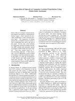
Metaphase c hromosomes from drug-selected BoHV-4
persistently infected RD-4 and BOMAC cell lines,
showed specific fluorescent signals on their chromo-
somes, some of which showed symmetrical double
signals on both chromatids (Fig. 1A and 1B). Observa-
tion of many chromatin spreads revealed integration at
many different, apparently random sites. All of the
metaphase spreads showed labelling and the hybridiza-
tion efficien cy and signal-to-noise ratio of this approach
was very high. In parallel experiments, uninfect ed RD-4
and BOMAC or PHA-stimulated mononuclear cells
from healthy bovines did not shown any BoHV-4 signals
after hybridization (results not shown).
Donofrio et al. Virology Journal 2010, 7:246
/>Page 2 of 4
The molecular analyses de scribed above suggest that,
in the majority of BoHV-4 drug selected persistently
infected RD-4 and BOMAC cells, the BoHV-4 genome
can exist as a chromosomal integrated form. The pre-
vious observations made by Gardella gel electrophoresis
and Southern analysis, suggesting the possible integra-
tion of the BoHV-4 genome, were thus corroborated.
Although signals were present in identical positions on
each sister c hromatid, consistent with the presence of
integrated DNA, other hybridization signals were also
present (Fig. 1A and 1B), suggesting coexistence of inte-
grated and episomal DNA. Further analysis would be
required to unequivocally demonstrate such coexistence.
Although this short report provides the direct visualiza-
tion in vitro of the relationship between persistent
BoHV-4 DNA with the cellular genome at the single
cell level, many questions could be raised. First, does
viral genome integration occur in vivo, or did the virus
integrate in v itro during long-term cultivation? The sec -
ond important question is concerned with the specificity
of the cellular genomic site of BoHV-4 genome integra-
tion. Is integration random, or does it occur at specific
chromosomal loci? Further work in different model sys-
tems, possibly i n vivo u sing macrophage cells coming
from persistently infected animals, will give an answer
to the above questions. However, this is the first demon-
stration of the possible integration of BoHV-4 genome
into the host genome.
Acknowledgements
We would like to thank Italian Ministry of University and Scientific Research
and the Fondazione Cariparma (Cassa di Risparmio di Parma, Italy) for
funding contributions to the project.
Author details
1
Dipartimento di Salute Animale, sezione di Malattie Infettive degli Animali,
Università di Parma, Via del Taglio 10, 43100 Parma, Italy.
2
Dipartimento di
Scienze Animali, Sezione di Zootecnica Agraria, Università di Milano, Via
Caloria, 220133 Milano, Italy.
3
Department of Pathobiology, 264 Greene Hall,
College of Veterinary Medicine, Auburn University, Auburn, Alabama 36849-
5519, USA.
Authors’ contributions
GD: Study design, performed the experiments, interpretation of the data and
wrote the manuscript. AC: Contributed to perform the experiments. VF:
Contributed to perform the experiments. LDL: Contributed to perform the
experiments. VVS: Intellectually contributed. PP: Contributed to study design,
performed the experiments and interpretation of the data. All authors read
and approved the final manuscript.
Competing interests
The authors declare that they have no competing interests.
Received: 18 August 2010 Accepted: 21 September 2010
Published: 21 September 2010
References
1. Bartha A, Juhasz M, Liebermann H: Isolation of a bovine herpesvirus from
calves with respiratory disease and keratoconjunctivitis. A preliminary
report. Acta veterinaria Academiae Scientiarum Hungaricae 1966,
16(3):357-358.
2. Mohanty SB, Rockemann DD, Snyder DB: Serologic cross-reaction between
bovine herpesviruses 1 and 4 by the enzyme-linked immunosorbent
assay. Microbiologica 1984, 7(2):179-186.
3. Thiry E, Bublot M, Dubuisson J, Van Bressem MF, Lequarre AS, Lomonte P,
Vanderplasschen A, Pastoret PP: Molecular biology of bovine herpesvirus
type 4. Veterinary microbiology 1992, 33(14):79-92.
Figure 1 Repr esentative images of drug selected BoHV-4
persistently infected RD-4 (A) and BOMAC (B) cells, analyzed
by FISH. An example of BoHV-4 hybridization signals of
symmetrically labelled sister chromatids in the same spread
metaphase chromosome is indicated by a white square and
enlarged as an insert picture at the corner of each image. Spread
chromosomes were counterstained with DAPI. Red dot signals
asymmetrically labelling the chromosomes correspond to
episomally-maintained BoHV-4 genomes. Green signals show
hybridization of species-specific BACs. The human BAC RP11-
153M12 maps on HSA 20 at 20p13 and the bovine BAC INRA-
115C10 maps on BTA 4 at a subcentromeric position.
Donofrio et al. Virology Journal 2010, 7:246
/>Page 3 of 4
4. Sheldon IM, Cronin J, Goetze L, Donofrio G, Schuberth HJ: Defining
postpartum uterine disease and the mechanisms of infection and
immunity in the female reproductive tract in cattle. Biology of
reproduction 2009, 81(6):1025-1032.
5. Dubuisson J, Thiry E, Bublot M, Thomas I, van Bressem MF, Coignoul F,
Pastoret PP: Experimental infection of bulls with a genital isolate of
bovine herpesvirus-4 and reactivation of latent virus with
dexamethasone. Veterinary microbiology 1989, 21(2):97-114.
6. Osorio FA, Reed DE: Experimental inoculation of cattle with bovine
herpesvirus-4: evidence for a lymphoid-associated persistent infection.
American journal of veterinary research 1983, 44(6):975-980.
7. Osorio FA, Reed DE, Rock DL: Experimental infection of rabbits with
bovine herpesvirus-4: acute and persistent infection. Veterinary
microbiology 1982, 7(6):503-513.
8. Osorio FA, Rock DL, Reed DE: Studies on the pathogenesis of a bovine
cytomegalo-like virus in an experimental host. The Journal of general
virology 1985, 66(Pt 9):1941-1951.
9. Donofrio G, Cavirani S, van Santen V, Flammini CF: Potential secondary
pathogenic role for bovine herpesvirus 4. J Clin Microbiol 2005,
43(7):3421-3426.
10. Donofrio G, Ravanetti L, Cavirani S, Herath S, Capocefalo A, Sheldon IM:
Bacterial infection of endometrial stromal cells influences bovine
herpesvirus 4 immediate early gene activation: a new insight into
bacterial and viral interaction for uterine disease. Reproduction 2008,
136(3):361-366.
11. Donofrio G, Cavirani S, van Santen VL: Establishment of a cell line
persistently infected with bovine herpesvirus-4 by use of a recombinant
virus. The Journal of general virology 2000, 81(Pt 7):1807-1814.
12. Donofrio G, van Santen VL: A bovine macrophage cell line supports
bovine herpesvirus-4 persistent infection. The Journal of general virology
2001, 82(Pt 5):1181-1185.
13. Gardella T, Medveczky P, Sairenji T, Mulder C: Detection of circular and
linear herpesvirus DNA molecules in mammalian cells by gel
electrophoresis. Journal of virology 1984, 50(1):248-254.
14. Donofrio G, Franceschi V, Capocefalo A, Cavirani S, Sheldon IM: Isolation
and characterization of bovine herpesvirus 4 (BoHV-4) from a cow
affected by post partum metritis and cloning of the genome as a
bacterial artificial chromosome. Reprod Biol Endocrinol 2009, 7:83.
15. Donofrio G, Sartori C, Franceschi V, Capocefalo A, Cavirani S, Taddei S,
Flammini CF: Double immunization strategy with a BoHV-4-vectorialized
secreted chimeric peptide BVDV-E2/BoHV-1-gD. Vaccine 2008,
26(48):6031-6042.
16. De Lorenzi L, De Giovanni A, Molteni L, Denis C, Eggen A, Parma P:
Characterization of a balanced reciprocal translocation, rcp(9;11)(q27;
q11) in cattle. Cytogenetic and genome research 2007, 119(3-4):231-234.
doi:10.1186/1743-422X-7-246
Cite this article as: Donofrio et al.: Integration of bovine herpesvirus 4
genome into cultured persistently infected host cell genome. Virology
Journal 2010 7:246.
Submit your next manuscript to BioMed Central
and take full advantage of:
• Convenient online submission
• Thorough peer review
• No space constraints or color figure charges
• Immediate publication on acceptance
• Inclusion in PubMed, CAS, Scopus and Google Scholar
• Research which is freely available for redistribution
Submit your manuscript at
www.biomedcentral.com/submit
Donofrio et al. Virology Journal 2010, 7:246
/>Page 4 of 4