Báo cáo y học: "Evaluation of Fractional Analysis of Bronchoalveolar Lavage Combined with Cellular Morphological Features"
Bạn đang xem bản rút gọn của tài liệu. Xem và tải ngay bản đầy đủ của tài liệu tại đây (1.12 MB, 8 trang )
Int. J. Med. Sci. 2009, 6
1
I
I
n
n
t
t
e
e
r
r
n
n
a
a
t
t
i
i
o
o
n
n
a
a
l
l
J
J
o
o
u
u
r
r
n
n
a
a
l
l
o
o
f
f
M
M
e
e
d
d
i
i
c
c
a
a
l
l
S
S
c
c
i
i
e
e
n
n
c
c
e
e
s
s
2009; 6(1):1-8
© Ivyspring International Publisher. All rights reserved
Research Paper
Evaluation of Fractional Analysis of Bronchoalveolar Lavage Combined
with Cellular Morphological Features
Namiko Taniuchi
1,2
, Mohammad Ghazizadeh
1
, Tatsuji Enomoto
3
, Kiyoshi Matsuda
4
, Masashi Sato
5
, Yuko
Takizawa
1
, Enjing Jin
1
, Seiko Egawa
1
, Arata Azuma
2
, Akihiko Gemma
2
, Shoji Kudoh
2
, Oichi Kawanami
1
1. Department of Molecular Pathology, Institute of Development and Aging Sciences, Nippon Medical School, Graduate
School of Medicine, Kawasaki, Japan.
2. Fourth Department of Internal Medicine, Nippon Medical School, Tokyo, Japan.
3. Department of Respiratory Medicine, Tokyo Metropolitan Hiroo Hospital, Tokyo, Japan.
4. Department of Emergency and Critical Care Medicine, Yamanashi Prefectural Central Hospital, Yamanashi, Japan.
5. Department of Radiology, Nippon Medical School Musashi-kosugi Hospital, Kawasaki, Japan.
Correspondence to: Oichi Kawanami, MD, PhD, Professor and Chairman, Department of Molecular Pathology, Institute
of Development and Aging Sciences, Nippon Medical School, Graduate School of Medicine, 1-396 Kosugi-cho, Nakahara-ku,
Kawasaki, Japan 211-8533. Tel:81-44-733-1823; Fax:81-44-733-1293;
Received: 2008.10.20; Accepted: 2008.11.26; Published: 2008.12.01
Background. The value of bronchoalveolar lavage (BAL) still remains controversial, prompting a need for further
improvement. The purpose of this study was to develop and evaluate a sequential analysis of cell content in fractional
BAL (FBAL) from the airways and alveolar sacs with incorporation of the cellular morphologic features. Methods.
Initially, 30 ml saline was infused into a subsegmental lobe of the lung and the recovered fluid was assigned as FBAL-I
being mainly originated from whole airways. The second and third lavages (FBAL-II and FBAL-III) each were per-
formed using 50 ml saline being from more distal portions of airways and alveolar sacs respectively in the same lobe.
Total cell number/ml and percentages of macrophages, lymphocytes, neutrophils, and eosinophils in each fraction
together with their morphological alterations and mast cells, basophils and Masson bodies were assessed. Results.
In the 12 controls, percentage of neutrophils was high and lymphocytes and macrophages were low in FBAL-I while
in FBAL-III, neutrophils decreased to nearly nil and lymphocytes and macrophages were increased. Analysis of FBAL
from 76 patients with sarcoidosis and 14 with hypersensitivity pneumonitis (HP) revealed that a predominance of
small, round and well-differentiated lymphocytes with relative absence of neutrophils, basophils and Masson bodies
correlated best with sarcoidosis. In contrast, neutrophil predominance and presence of lymphocytes having deep
nuclear indentations and abundant cytoplasm with a process resembling a “hand-mirror” correlated well with HP.
Conclusions. Evaluation of FBAL together with cellular morphological features especially characteristics of lym-
phocytes provides valuable information for establishing the diagnosis in interstitial lung diseases.
Key words: Bronchoalveolar lavage procedure; interstitial lung diseases; sarcoidosis; hypersensitivity pneumonia; lympho-
cyte morphology; neutrophil; Masson bodies.
Introduction
Bronchoalveolar lavage (BAL) has been widely
applied as a routine tool in laboratories of respiratory
disease
1
. The BAL technique is safe and minimally
invasive, and the presence of cell patterns can sup-
port clinical diagnosis in the absence of biopsy or
when the clinical feature is compatible
2, 3
. However,
the application of BAL still remains controversial as
the results obtained so far were based on a variety of
techniques or methods thus introducing considerable
variations in the implications
4
. Although it is well
recognized that correlations between BAL fluid cells
and corresponding tissue cell extractions are better
for lymphocyte infiltration in granulomatous lung
disease
5
, morphological features of lymphocytes
have not been well defined and incorporated in rela-
tion to the pathological conditions.
Int. J. Med. Sci. 2009, 6
2
The primary objective of this study was to de-
velop a sequential analysis of cells in FBAL with spe-
cial references to the distribution of inflammatory
cells in the airways and alveolar sacs combined with
morphological alteration of lymphocytes in relation
to the functional states; migration of mast cells and
basophils. Using this method, lymphocyte morphol-
ogy in hypersensitivity pneumonitis (HP) was
sharply contrasted with the other lymphocyte-rich
diseases such as sarcoidosis.
Materials and Methods
Fractional analysis of cells in BAL fluid. After pre-
medication with atropine and hydroxyzine, a bron-
choscope was inserted under local anesthesia with
lidocaine and wedged at orifice of the objective
sub-segment of a lobe. The first lavage was done with
a 30ml of sterile 0.9% saline (FBAL-I) which was as-
signed as “whole airway” lavage. Subsequently, the
second (FBAL-II) and third (FBAL-III) lavages were
performed each with a 50 ml saline and were consid-
ered to be from “distal airway” and “alveolar sac”
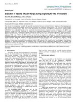
respectively (Figure 1). The percent recovery rate of
lavage fluid and a total cell number were estimated in
each FBAL, and then cytocentrifuging was done for 5
minutes at 1,500 rpm at room temperature (Model
Cytospin 3, Shandon Southern Products Ltd., UK) for
microscopic evaluation. The cells were stained by
May-Grunwald Giemsa and Papanicolaou methods.
The differential cell
counts were performed under a
light microscope by counting more than 300 cells.
Mast cells and basophils in FBAL were counted and
graded as 0, 1+ (1/10
3
cells), 2+ (2 ~5/10
3
cells) and
3+ (> 5/10
3
cells). We further checked non-cellular
components, including tiny organized proteinous
accumulations or Masson bodies, foreign bodies, ag-
gregates.
Figure 1. Fractional analysis of bronchoalveolar lavage (FBAL) procedure. PCR: polymerase chain reaction.
Control individuals and patients with lung diseases.
Control individuals were comprised 8 males (mean
age ±SD: 45.0±13.0), and 4 females (44.8±17.7). We
reviewed clinicopathological records of a total of 2260
patients with various lung diseases between 1984 and
2006 at our Nippon Medical School affiliated hospi-
tals (Table 1). Among them, 76 of sarcoidosis and 14
of HP patients had both a definite diagnosis with
histopathological confirmation and all three FBAL
samples available and therefore selected for detailed
studies. Written informed
consent was obtained from
each patient when they underwent the examinations.
This study was also approved by the Ethical Com-
mittee of the Nippon Medical School.
Clinical features of patients with sarcoidosis. The 76
patients, including 39 men (35.8±14.5) and 37 women
(51.8±13.5), were proved by lung biopsy to have de-
veloped characteristic non-caseating epithelioid
granuloma. Based on chest radiographs, the patients
were divided into four stages according to the criteria
established by the American Thoracic Society, Euro-
pean Respiratory Society and WASOG
6
. Seven pa-
tients (9.2%) met stage 0, 39 patients (51.3%) stage I,
14 patients (18.4%) stage II, and 16 patients (21.1%)
Int. J. Med. Sci. 2009, 6
3
stage III. Thirty-seven patients (48.7%) showed mul-
tiple organ involvements of sarcoidosis including
skin, eyes and/or heart. As to smoking habits, 31
(40.8%) were current smokers, 37 patients (48.7%) had
never smoked or were past smokers.
Table 1. Clinical Diagnoses
(Nippon Medical
School-affiliated hospitals, April 1984 - February 2006).
Clinical diagnoses Patients (%)
Interstitial lung diseases 734 (32.5)
IPF/UIP
101 (4.5)
NSIP
33 (1.5)
COP/BOOP
109 (4.8)
CVD-IP
145 (6.4)
The others with IP
346 (15.3)
Sarcoidosis 265 (11.7)
Radiographic abnormal shadows 136 (6.0)
Eosinophilic pneumonia 119 (5.3)
Leukemia, lymphoma and others 104 (4.6)
Hypersinsitivity pneumonitis 104 (4.6)
Pneumoconiosis (Asbestosis, Coal workers) 95 (4.2)
Malignant tumor (Primary or metastat. Cancer) 81 (3.4)
Tuberculosis 73 (3.2)
ARDS/AIP 64 (2.8)
Fungus, P.Carinii 42 (1.9)
Bloody sputum 30 (1.3)
Langerhans cell histiocytosis 19 (0.8)
Alveolar proteinosis 12 (0.5)
Mycobacterium infection 11 (0.5)
Wegener’s granulomatosis 4 (0.2)
The others 367 (16.2)
Total 2260
IPF: idiopathic pulmonary fibrosis
UIP: usual interstitial pneumonia
NSIP: non-specific interstitial pneumonia
COP: cryptogenic organizing pneumonia
BOOP: bronchiolitis obliterans organizing pneumonia
CVD-IP: collagen vascular disease-related interstitial pneumonia
ARDS: acute respiratory distress syndrome
AIP: acute interstitial pnemonia
Clinical features of hypersensitivity pneumonitis.
The 14 patients, including 6 men (65.3±7.1) and 8
women (59.0±9.4), were proved by lung biopsy to
have developed HP. Six patients were non-smokers,
and 8 current smokers. Four patients were diagnosed
as having summer type hypersensitivity pneumoni-
tis
7
, 4 patients had hypersensitivity to chemical mate-
rial, 1 to feathers of bedding, and 5 to unknown
house dust antigens. In these 5 patients, typical
symptoms were provoked, and besides, laboratory
data were compatible when they returned home or to
work place after cessation of symptoms during brief
hospitalization. Biopsied lung tissues were compati-
ble with HP, including diffuse infiltration of lym-
phocytes, intra-alveolar corporation of collagenous
exudate (Masson body) and tiny non-caseating epi-
thelioid granuloma formation.
Statistical Analysis. Paired-sample t-test was used
to determine whether there was a statistically signifi-
cant difference between each cell differentials of pa-
tients with sarcoidosis or HP (SPSS Software, SPSS
Japan Inc.). We focused on the relationship between
the fractions, including FBAL-I, FBAL-III and
FBAL-total, radiographic staging and smoking his-
tory. A p-value < 0.05 was considered statistically sig-
nificant.
Results
Cell differential rates in control individuals. Among
control group, neutrophil percentage was highest in
FBAL-I and decreased to nearly nil in FBAL-III
(p<0.05). In contrast, lymphocytes and macrophages
were slightly increased in FBAL-III. The differential
cell rates in each FBAL did not show any significant
differences related to the smoking habit (Table 2).
Table 2. Cell differential ratios in control individuals
(n=12) and when subdivided into smokers and
non-smokers.
Control individuals (n=12)
Cell number
(x10
5
/mL)
Mφ(%) Ly(%) Neu(%) Eo(%)
FBAL-I 1.08±0.45 88.5±3.0 5.8±1.2 4.7±1.9∗ 1.0±0.45
FBAL-II 1.83±0.63 90.5±1.1 7.5±0.99 1.5±0.50 0.33±0.21
FBAL-III 2.52±0.76 91.7±1.2 7.3±0.84 0.33±0.21 0.33±0.21
FBAL-total 1.84±0.47 91.0±1.2 7.0±0.73 1.3±0.33∗ 0.50±0.22
Control smokers (n=6)
Cell number
(x10
5
/mL)
Mφ(%) Ly(%) Neu(%) Eo(%)
FBAL-I 1.4±1.07 85.0±4.6 7.7±0.88 6.0±3.5 1.3±0.67
FBAL-II 3.01±1.39 89.0±1.2 8.3±1.9 2.3±0.67 0.33±0.33
FBAL-III 3.08±1.04 90.3±1.2 8.7±0.67 0.67±0.33 0.33±0.33
FBAL-total 2.45±0.78 89.7±1.2 8.0±0.58 1.7±0.33 0.67±0.33
Control nonsmkers (n=6)
Cell number
(x10
5
/mL)
Mφ(%) Ly(%) Neu(%) Eo(%)
FBAL-I 0.87±0.35 92.0±3.2 4.0±1.5 3.3±1.9 0.67±0.67
FBAL-II 1.04±0.33 92.0±1.5 6.7±0.88 0.67±0.33 0.33±0.33
FBAL-III 1.96±1.18 93.0±2.1 6.0±1.2 0.0±0.0 0.33±0.33
FBAL-total 1.22±0.47 92.3±2.0 6.0±1.2 1.0±0.58 0.33±0.33
* p<0.05 as compared with % neutrophils in FBAL-III.
Mφ: macrophages, Ly: lymphocytes, Neu: neutrophils, Eo: eosi-
nophils
Patients’ group showing the highest rate of lympho-
cytes. Of the 2260 patients, 488 (21.6%) showed a
lymphocyte rate of higher than 40%. Clinical diagno-
ses of these patients included sarcoidosis (22.1%), IIP
(14.5%), HP (11.5%), leukemia/lymphoma including
tumor cells (10.9%), COP/BOOP (9.0%) and others
(Table 3). Sarcoidosis and HP are the most distin-
guished group of diseases with lymphocyte-related
Int. J. Med. Sci. 2009, 6
4
etiology and the diagnosis of these granulomatous
diseases were well defined by lung biopsy.
Table 3. Patients’ group showing a highest ratio of lym-
phocyte in FBAL-total
Clinical Diagnosis Patients (%)
Sarcoidosis 108 (22.1)
IIPs 72 (14.5)
Hypersensitivity Pneumonitis 38 (11.5)
Leukemia/Lymphoma 53 (10.9)
COP/BOOP 44 (9.0)
Tuberculosis 18 (3.7)
Lung cancer 10 (2.0)
Others 145 (29.7)
Lymphocytes >40% in FBAL-total 488
IIPs: idiopathic interstitial pneumonias
COP: cryptogenic organizing pneumonia
BOOP: bronchiolitis obliterans organizing pneumoni
Differential cell rates in sarcoidosis. The mean cell
numbers in 76 sarcoidosis patients were 1.64±0.30
x10
5
/mL in FBAL-I, 3.26±0.38 x10
5
/mL in FBAL-III,
and 2.96±0.36 x10
5
/mL in FBAL-total (1.6-fold in-
crease vs. control). The higher percentages of lym-
phocytes in each fraction were 40.8±2.2% (3.9–82.8%)
in FBAL-total, 28.5±2.2% in FBAL-I, and 43.9±2.3% in
FBAL-III respectively (Figure 2). The mean lympho-
cyte percentage of patients who had lung involve-
ment (stage II or III) did not differ in clinical symp-
toms and laboratory data from those of patients with
no lung involvement (stage 0 or I) (40.3±3.7% vs.
41.2±2.7%). There were 40 patients, including 18
smokers, who showed no neutrophil recovery in any
fractions. Among sarcoidosis smoker patients (n=31),
mean neutrophil rate was 4.6±2.1 % in FBAL-I com-
pared to 0.8±0.2 % in FBAL-III (p=0.06). On the other
hand, among sarcoidosis nonsmokers (n=37), mean
neutrophil rate was 9.7±3.4 % in FBAL-I compared to
3.2±1.2 % in FBAL-III (p<0.05).
Differential cell rates in hypersensitivity pneumoni-
tis. The average total cell number was 1.59±0.33
x10
5
/mL in FBAL-I, 6.45±0.92 x10
5
/mL in FBAL-III,
and 5.29±0.70 x10
5
/mL in FBAL-total showing a
2.9-fold increase versus normal control (p<0.05) and
1.8-fold higher than sarcoidosis (p<0.05). A signifi-
cantly higher proportion of lymphocytes was seen
(mean 65.2±4.1% ranging from 35.9 to 84.3%) and the
difference of lymphocyte rates between FBAL-I
(47.5±5.0%) and FBAL-III (68.9±4.5%) was statistically
significant. Neutrophil rate in FBAL-I was 17.4±3.4%
in contrast to 2.9±0.7% in FBAL-III (p<0.05) and
4.4±0.9% in FBAL-total among HP patients, which
was significantly higher than in the normal control
(1.3±0.33%; P<0.05) (Figure 3). Lymphocyte rate in
FBAL-total in HP smoking patients tended to be
lower than non-smoking patients (60.6±4.9% vs.
68.1±9.7%), and neutrophil rate in FBAL-total showed
a higher level in nonsmokers than in smokers
(5.5±1.9% vs. 3.6±1.2%; no statistical significance).
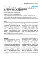
Figure 2. Cell differentials in sarcoidosis (n=76). BAL fluid
cells in sarcoidosis patients were characterized by a higher
percentage of lymphocyte at each fraction, showing
40.8±2.2% (3.9–82.8%) FBAL-total (A), while that in FBAL-I
(B) was 28.5±2.2% compared to 43.9±2.3% in FBAL-III (C)
(p<0.05). Mean neutrophil rate was 3.1±0.9% in FBAL-total
among all patients with sarcoidosis, while mean neutrophil
rate in FBAL-I was 6.9±1.9% compared to 2.0±0.6% in
FBAL-III (p<0.05). Asterisk indicates a significant difference
from FBAL-III (p<0.05).
Int. J. Med. Sci. 2009, 6
5
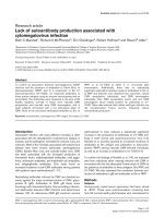
Figure 3. Cell differentials in hypersensitivity pneumonitis
(n=14). A significantly higher proportion of lymphocyte
was seen in HP patients (mean 65.2±4.1% ranging from
35.9 to 84.3%). The difference of lymphocyte rates be-
tween FBAL-I (A; 47.5±5.0%) and FBAL-III (C; 68.9±4.5%)
was statistically significant (p<0.05). Neutrophil rate in
FBAL-I was 17.4±3.4% in contrast to 2.9±0.7% in FBAL-III
(p<0.05) and 4.4±0.9% in FBAL-total (A) among HP pa-
tients, which was significantly higher than in the normal
control (1.3±0.33%) (P<0.05). Asterisk indicates a signifi-
cant difference from FBAL-III (p<0.05).
Comparison of lymphocyte morphology between sar-
coidosis and HP. Sarcoid-lymphocytes were generally
small in size and appeared mature and well differen-
tiated in morphology (Figure 4). They had a round
nucleus and the cytoplasm was fairly thin. In con-
trast, HP lymphocytes had fairly abundant cytoplasm
and occasionally developed an elongated cytoplasmic
process resembling a hand-mirror in shape. A num-
ber of lymphocytes looked like plasma cell, having
large basophilic cytoplasm and nucleus with
wheel-like appearance. In others, the nuclear mem-
brane was highly convoluted like cerebelli-form or
clover-shape. Nucleus often contained a large nu-
cleolus. Sarcoidosis patients consistently showed a
different morphology compared with HP patients.
Other inflammatory cells, particularly eosinophils
and mast cells were also frequently seen in BALF of
HP patients (Figure 5).
Figure 4. Morphology of sarcoid lymphocytes. The sar-
coid lymphocytes displayed mature, small shape dominant,
and also regular size cells. They had round-shape nuclei
and scanty cytoplasm. (Giemsa stain x original magnifica-
tion x 200).
Eosinophils, mast cells and basophils in FBAL.
Higher eosinophil recovery (>5%) in FBAL-total was
found in 120 of 546 (22%) of biopsy proven cases. The
distribution of diseases were as follows; IIP 43
(35.8%), eosinophilic pneumonia 18 (15.0%), sarcoi-
dosis 6 (5.0%), drug-induced pneumonia 3 (2.5%) and
others. On the other hand, eosinophil recovery rate
was highest in the group of eosinophilic pneumonia
showing 28.2% following by NSIP 6.1%, IPF 5.0% and
COP/BOOP 4.6%. With May-Grunwald-Giemsa
staining, mast cells were identifiable by the pur-
ple-red granules in cytoplasm, and were encountered
in half of a total 546 biopsy cases. Highest level of
mast cells (3+) were encountered in 96 patients with a
variety of interstitial lung diseases, including 37 pa-
tients with IIP, 10 sarcoidosis, 7 eosinophilic pneu-
monia, and others. Distribution of mast cells in 7 HP
patients examined in this study were 3+ (n=3), 2+
(n=3) and 1/0 (n=1). Basophilic leukocytes were
never found in the normal control and most patients.
Basophils were occasionally found in 13 patients with
IIP, 3 HP, 5 eosinophilic pneumonia, and others. The
three HP patients who contained basophils were all at