Báo cáo y học: "Use of a highly sensitive strand-specific quantitative PCR to identify abortive replication in the mouse model of respiratory syncytial virus disease" pptx
Bạn đang xem bản rút gọn của tài liệu. Xem và tải ngay bản đầy đủ của tài liệu tại đây (534.9 KB, 11 trang )
RESEARC H Open Access
Use of a highly sensitive strand-specific
quantitative PCR to identify abortive replication
in the mouse model of respiratory syncytial virus
disease
Richard Bannister, Deborah Rodrigues, Edward J Murray, Carl Laxton, Mike Westby, Helen Bright
*
Abstract
Background: The BALB/c mouse is commonly used to study RSV infection and disease. However, despite the
many advantages of this well-characterised model, the inoculum is large, viral replication is restricted and only a
very small amount of virus can be recovered from infected animals. A key question in this model is the fate of the
administered virus. Is replication really being measured or is the model measuring the survival of the virus over
time? To answer these questions we developed a highly sensitive strand-specific quantitative PCR (QPCR) able to
accurately quantify the amount of RSV replication in the BALB/c mouse lung, allowing characterisation of RSV
negative and positive strand RNA dynamics.
Results: In the mouse lung, no increase in RSV genome was seen above the background of the original inoculum
whilst only a limited transient increase (< 1 log) in positive strand, replicative intermediate (RI) RNA occurred. This
RNA did however persist at detectable levels for 59 days post infection. As expected, ribavirin therapy reduced
levels of infectious virus and RI RNA in the mouse lung . However, whilst Palivizumab therapy was also able to
reduce levels of infectious virus, it failed to prevent production of intracellular RI RNA. A comparison of RSV RNA
kinetics in human (A549) and mouse (KLN205) cell lines demonstrated that RSV replication was also severely
delayed and impaired in vitro in the mouse cells.
Conclusions: This is the first time that such a sensitive strand-specific QPCR technique has been to the RSV mouse
system. We have accurately quantified the restricted and abortive nature of RSV replication in the mouse. Further in
vitro studies in human and mouse cells suggest this restricted replication is due at least in part to species-specific
host cell-viral interactions.
Background
Respiratory Syncytial Virus (RSV) is the leading cause of
lower respiratory tract infection (LRTI) i n infants and
children world-wide and is increasingly recognised as a
cause of serious disease in adults and immune compro-
mis ed transplant patients [1,2]. Over half of all childre n
will be infected with RSV by their first birthday and by
the age of 2 n early all c hildren will have been infected
with RSV at least once [3]. LRTI caused by RSV infec-
tion is a major cause of both infant hospitalisation and
infant viral induced death [4]. A number of medical
treatments, including use of bronchodilators, palliative
care (supportive ventilation, nitric oxide) and use of anti
inflammatory agents are availa ble but none of these
treatments relieve the viral burden in RSV-in fected
patients. The only small molecule antiviral therapeutic
agent for treating RSV is Virazole (aerosolised ribavirin),
which has been shown to be of limited use because of
its lengthy administration and questionable efficacy
[5,6]. Palivizumab (Synagis) is a humanised monoclonal
IgG1 antibody specifically directed to the RSV fusion
protein which has been used prophylactically to good
effect in at-risk infants. However, a therapeutic treat-
ment did not result in significant clinical benefit [7].
* Correspondence:
Infectious Diseases Group, Pfizer Global Research and Development,
Sandwich, Kent, CT13 9NJ, UK
Bannister et al. Virology Journal 2010, 7:250
/>© 2010 Banniste r et al; licensee BioMed Central Ltd. This is an Open Access article distributed under the terms of the Creative
Commons Attribution License ( /lic enses/by/2.0), which permits unrestricted use, distribution, and
reproduction in any medium, pr ovided the original work is properly cited.
Thus, there is a clear, unmet medical need to develop
therapies able to ameliorate RSV disease [8-10].
RSV is a negative-stranded RNA virus belonging to
the Paramyxoviridae family. The negative sense single-
strand RSV genome c ompris es a RNA molecule encod-
ing 11 proteins. Upon host cell infection positive-sense
viral mRNAs are synthesise d by the viral RNA polymer-
ase, these mRNAs make use of host-cell machinery to
synthesise viral prote ins. Genome replication occurs via
the production of a positive sense replicative interme di-
ate (RI) RN A strand by the same viral RNA polymerase ;
this RI RNA is used as a template for the synthesis of
more negative sense genome [11-13].
The use of in vivo models with good clinical transla-
tion is vital in the search for new treatments for dis-
ease A number of different animal models have been
used to study RSV infection and replication and to
evaluate potential therapies, including primates,
bovines and rodents [14]. The majority of in vivo stu-
dies have been conducted using either the BALB/c
mouse [15] or the cotton rat (Sigmodon hispidus)[16]
models. The cotton rat is moderately permissive to
human respiratory viral infection and RSV is able to
replicate and produce viral progeny in the lungs [17].
The BALB/c mouse is also susceptible to RSV infection
[18] and, though less permissive than the cotton rat
[19], constitutes a more practical model due to the
availability of a larger number of immunological and
molecular reagents as well as the availability of trans-
genic animals. Like the cotton rat, the mouse requires
inoculation with a high dose (usually 10
6
PFU) to
achieve viral replication. The actual amount o f viral
replication occurring following infection with such a
supra-physiological dose of RSV has never been accu-
rately determined.
We therefore developed a strand-specific real-time
quantitative polymerase chain reaction (QPCR) method
to monitor the kinetics of RSV RNA replication in the
mouse lung. BALB/c mice were infected with RSV A2
and viral RNA in m ouse lungs were monitored ov er an
extended time course. Levels of infectious virus in lungs
were also measured. Taken together, results from these
2 assays showed that RSV RNA synthesis and viral repli-
cation was severely limited in the mouse. Treatment
with a prophylactic antibody (palivizumab) did not affect
viral RNA replication and persistence, but did impair
the production of infectious progeny virus, indicating
that abortive replication [16] o ccurs in the mouse. By
contrast, positive sense viral RNA and infectious virus
production were both disrupted by ribavirin. Further in
vitro studies in human and mouse cells demonstrated
that although both cell types were equally susceptible to
infection; viral RNA synthesis was delayed and impaired
in mouse cells. This finding suggests that a species-
specific host-virus interaction inhibits the capacity for
RSV replication in the mouse.
Methods
Animals
Female BALB/c mice (6-8 weeks o ld), specific pathogen
free, were purchased from Charles River Laboratories
and housed in an animal care facility in ventilated isola-
tion cubicles. Water and chow were provided ad libitum.
Mice were allowed to acclimate to the new environment
for 1-2 weeks and housed in groups according to experi-
mental setup. All experiments with animals were carried
out in compliance with UK legislation and subject to
local ethical review.
Virus, cells and viral assays
RSV-A2 was obtained from Advanced Biotechnologies
Inc. Stocks were produced by infecting Hep-2 cells at a
multiplicity of infection (MOI) of 0.1 focus forming
units (FFU) per cell. Following 4-5 days incubation,
infected cells were harvested and snap frozen in dry ice
and methanol and stored at -80°C. Viral titres were
deter mined by a HEp2 based immunoflu orescence assay
and expressed as FFU/ml [20]. UV-inactivated RSV
(UVRSV) was generated by exposing RSV A2 to UV
radiation at 254 nm for 20 minutes using a Stratalinker
(Stratagene). Loss of infectivity of UVRSV was co n-
firmed by infecting Hep2 cells (MOI ranging from 0.1-1
FFU/cell). For animal studies, viral titres were expressed
as geometric means +/- standard errors of means (SEM)
for all animals in a group.
A549 cells (human lung carcinoma) and KLN205 cells
(DBA/2 mouse lung squamous cell carcinoma) were
purchased from ATCC and maintained in DMEM or
EMEM respectively, each supplemented with 100 IU/ml
of penicillin, 100 μg/ml streptomycin, 2 mM L-gluta-
mine, and 10% foetal calf serum (FCS).
Drugs
Ribavirin was obtained from Sigma-Aldrich and Palivi-
zumab (Synagis, MedImmune) was obtained from
Idis Ltd.
RSV infection in vivo
For preliminary RSV replication and dynamics studies,
mice were inoculated once intranasally (i.n.) with 50 μl
of either RSV A2 (1 × 10
6
FFU per animal) or an
equivalent concentration of UVRSV. One group of con-
trol mice was left untreated. Anima ls were sacrificed at
1, 5, 8, 17, 24, 48 and 72 hours and 7, 10, 37 and 59
days after infection (3 mice per time point). The lungs
were removed from the thorax, dissected into two and
each weighed. One lung was placed into RNAlater
(Ambion) for subsequent RNA extraction and Taqman
Bannister et al. Virology Journal 2010, 7:250
/>Page 2 of 11
analyses. The second lung was processed by hand-held
homogeniser (Omni) in 1 ml MEM (Invitrogen). Homo-
genates were centrifuged, clarified viral supernatant
diluted 1:3 in MEM and 50 μlusedintriplicatein
immunofluorescence assay [20].
For experiments conducted to investigat e inhibition of
RSV replication, one group of animals were adminis-
tered a single intramuscular injection of palivizumab
(5 mg/kg of body weight) 24 hours prior to infection
with RSV. A second group were administered ribavirin
(100 mg/kg of body weight) intraperitoneally one hour
prior to RSV challenge. These groups, plus a further
untreated group, were inoculated intra nasally with
75 μLRSVA2(2.6×10
6
FFU/mouse). Ribavirin treat-
ment was re-administered 5 hours post virus inoculation
and twice daily dosing of this compound continued for a
further day. Ribavirin treatment was not administered
on day 2. Dosing continued on day 3 at 50 mg/kg twice
daily until day 6 post virus infection. Animals were
sacrificed at 1, 8, 24, 48 an d 72 hours and 5, 7 and
10 days post infection (6 animals per group per time
point). Lungs were harvested for viral titrations and
RNA extraction.
In-vitro transcript standard production
A region of the RSV A2 nucleocapsid domain was iso-
lated using a nested primer approach. RSV A2 viral RNA
was prepared from crude preparation using the QIAamp
viral RNA minikit (Qiagen). RNA was reverse transcribed
using the High Capacity cDNA reverse-transcription kit
(Applied Biosystems) with random primers. PCR was
conducted using Pwo Superyield polymerase (Roche) with
external primers (Table 1) at an annealing temperature of
60°C for 35 cycles followed b y nested primer (Table 1)
PCR using cycling conditions as described above.
Gel-purified PCR product was restriction-cloned into
pGEM-4Z vector (Prom ega), grown in Oneshot TOP10
chemically competent E. coli (Invitrogen) and plasmid
purified by QIAprep Spin Miniprep Kit (Qiagen). Clones
were sequence-checked at Lark Technologie s, UK. The
insert plus bacterial promoter vector sequences of verified
clones was isolated by PCR usin g Pwo Superyield poly-
merase with M13 forward (-20) and reverse primers at an
annealing temperature of 55°C for 35 cycles. Positive and
negative sense in vitro transcripts were synthesised by
Sp6 and T7 RNA polymerase (Promega) respectively,
these products were treated with Turbo DNase (Ambion)
and purified by 3 M sodium acetate (pH 5.5) precipita-
tion. Stocks of 10
8
absolute copies per μl were prepared
and stored at -80°C.
Strand-specific real time QPCR
RNA was prepared from mouse lungs using an RNeasy
kit (Qiagen) following manufacturer’s instructions. First
strand cDNA was synthesised from RNA using Reve rse
Transcription Reagents (Applied Biosystems) with gene
specific primers targeted to the positive or negative
sense RSV A2 nucleoca psid region RNA (Table 1).
Primers contain a tag sequence recognised by a tag-spe-
cific primer in QPCR reactions; this reduces the detec-
tion of non-specific, self-primed cDNAs [21]. Reactions
(10 μl) comprised 1 × reaction b uffer, 5.5 mM MgCl
2
,
0.5mMdNTPmix,2.5μM strand-specific primers, 4 U
RNase inhibitor and 12.5 U reverse transcriptase with
4 μl total RNA preparation in water. Reactions were
performed at 50°C for 40 mins followed by 95°C for
5 mins. Positive strand detection by QPCR was per-
formed using TaqMan® Universal PCR mastermix
(Applied Biosystems) with positive sense RNA specific
primer, 800 nM tag-specific primer and 100 nM probe
(Table 1). Reactions were performed using an Applied
Biosystems 7900 HT. Samples were held at 50°C for
Table 1 Primer sequences used for RNA standard generation, cDNA synthesis and QPCR.
Primer Sequence
In vitro standard external positive sense TCCAGCAAATACACCATCCA
In vitro standard external negative sense CTGCTTCACCACCCAATTTT
In vitro standard nested positive sense ATA
GAATTCGGTATGTTATATGCGATGTCTAGGT
1
In vitro standard nested positive sense ATAGGATCCTGCTAAGACTCCCCACCGTAA
2
Positive sense RNA-specific cDNA synthesis CGGTCATGGTGGCGAATAATCCTGCAAAAATCCCTTCAACT
3
Negative sense RNA-specific cDNA synthesis CGGTCATGGTGGCGAATAAACTTTATAGATGTTTTTGTTCA
3
Positive sense-specific QPCR primer CCCCACTTTATAGATGTTTTTGTTCA
Negative sense-specific QPCR primer TCCTGCAAAAATCCCTTCAACT
QPCR tag primer CGGTCATGGTGGCGAATAA
Probe FAM-TTGGTATAGCACAATCTTCTACCAGAGGTGGC-TAMRA
1
Sequence contains an EcoRI restriction site (bold, underlined)
2
Sequence contains a BamHI restriction site (bold, underlined)
3
Tag sequence is indicted (bold, underlined)
Bannister et al. Virology Journal 2010, 7:250
/>Page 3 of 11
2 mins followed by 95°C for 10 mins and then 40 cycles
of 95°C for 15 secs and 60°C for 1 min. Negative sense
strand detection was performed as described for the
positive sense RNA reaction but substituting the positive
senseRNAspecificprimerforanegativesenseRNA
primer. Positive and negative sense RNA transcript stan-
dard ranges (10-10
7
absolute copies/μl) were processed
alongside samples. The limits of detection for this assay
were defined as values measured outside the range of
the standard curves. RSV copy number per μloftotal
mouse lung RNA were normalised to beta-actin
detected using commercially available TaqMan® VIC/
MGB primer-limited endogenous control (Applied Bio-
systems) with random-primed 1
st
strand cDNA synthe-
sised using the High Capacity cDNA reverse-
transcription kit (Applied Biosystems). Absolute valu es
of normalised RSV copy number were subsequently
dividedbytheweightofthelungtissuefromwhich
RNA was extracted and expressed as norma lised copy
number/g lung wt.
RSV infection in vitro
To investigate whether RSV RNA synthesis occurs effec-
tively in a mouse cell line compared to a human cell
line in vitro, human lung carcinoma cells (A549) and
mouse lung epithelial s quamous cells (KLN205) were
plated at a density of 1 × 10
4
cells per well in 96 well
plates and infected with R SV A2 to yield various multi-
plicities of infection (MOIs) ranging from 1 × 10
-3
to 1.
Media containing 10% FCS was replaced with fresh
media containing 2% FCS after 24 hours. Cells were
lysed with RLT buffer (Qiagen) at 1, 8, 24, 48 and 72
hour s and after 5 (A549) or 6 (KLN205), 7 and 10 days.
Total RNA was prepared using the RNeasy 96 kit (Qia-
gen). RSV strand-specific QPCR was performed as
described above. RSV copy number per μloftotalRNA
were not normalised to beta-actin but rather analysed
separately due to variable rates of cell death observed
throughout the experiment and expressed as RSV copy
number.
Statistics
For QPCR analyses the ratio of positive to negative
copy number is analysed on the logarithmic scale.
Treatments are compared t o untreated RSV infected
controls at each time point by two-sample t-test incor-
porating Satterthwaite’s adjustment to the degrees to
freedom. To allow for testing of multiple time points
within a treatment a Bonferroni adjustment was made
to achieve an approximate 5% significanc e level within
that treatment. Infectious virus assay data were ana-
lysedby1wayanalysisofvariance(ANOVA)forsig-
nificant differences (p = < 0.05) between treated
groups and untreated RSV in fected controls at each
time point.
Results
Development of an RSV strand-specific real time
quantitative PCR method
A strand-speci fic QPCR method was developed to study
RSV intracellular RNA dynamics. This method distin-
guishes between negative sense (genomic) RNA and
positive sense RNAs (nucleocapsi d mRNA and RI RNA)
by discrimination at the 1
st
strand cDNA synthesis
stage. The strand-specific RSV primers used in the
reverse transcription stage contain tag sequences that
are incorporated into specifically primed cDNA and this
sequence can be specifically targeted by a tag-specific
primer during QPCR cycling (Table 1). The use of this
tag is designed to reduce detection of cDNAs synthe-
sised due to RNA self-priming in the reverse
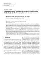
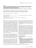
Figure 1 An RSV strand-specific QPCR method. Strand-specific
priming was performed during cDNA synthesis and QPCR was
performed using a primer/probe set designed to amplify part of the
nucleocapsid region. A) Negative sense RNA standard curve. B) Positive
sense strand RNA standard curve. Duplicate measurements are plotted.
Bannister et al. Virology Journal 2010, 7:250
/>Page 4 of 11
transcription reaction. Standard curves generated using
in vitro transcribed RNA standards to monitor negative
(Figure 1A) and positive (Figure 1B) strand specific
QPCR revealed that both assays were ≥95% efficient
with R
2
values above 0.99 (Table 2). The specificity of
the reaction s was assessed by spiking positive and nega-
tive sense RSV RNA st andards into naïve mouse lung
RNA and using both positive and negative strand-speci-
ficreagentstomeasureRSVRNA.Thedetectionof
non-specific RNA strand in mouse lung RNA back-
ground was <0.001% of specific strand detection in bot h
positive and n egative sense-specific reactions (Table 2).
No signal was detected when naïve mouse lung alone
was assayed, ruling out any non-specific effect from self-
priming RNA species.
RSV replication dynamics in BALB/c mouse lungs
RSV RNAs were analysed in the lungs of female BALB/c
mice dosed i.n. with 10
6
FFU per animal over a 59 day
period. Another group were infected with UV-inacti-
vated RSV as a control.
Mean normalised copy number/g lung wt. of both
positive and negative RSV strands from infected mouse
lung RNA preparations remained above 1 × 10
6
for 24
hours following infection (Figure 2A). From 24 hour s
onwards the amount of RSV negative strand reduced
and t his trend continued until day 10 when mean mea-
sured RNA reached a basal level of approximately 10
2
normalised copies/g lung wt. that persisted to 59 days
post-infection. By contrast, the mean positive sense
strand RNA remained above 10
6
normalised copies/g
lung wt. until 72 hours post-infection . The mean nega-
tive and positive strand UVRSV RNA both declined in a
time dependent manner from >10
6
normalised copies/g
lung wt. 1 hour post infection and could not be detected
after day 7 p.i. (Figure 2A). FFU assay performed on
lung homogenates revealed a high mean FFU/g lung wt.
of >10
4
at 1 hour post-dosing that was markedly
reduced to <10
3
FFU/g lung wt. by 5 hours (Figure 2B).
Infectious virus remained at this low level until 72
hours post infection when an increase to 10
4
FFU/g
lung wt. was observed. Infectious virus reduced again on
day 7. No infectious virus was detected from lungs
excised from UVRSV dosed mice. Note that no RSV
RNA or infectious virus could be detected in the lungs
of control, untreated mice. These FFU data agree well
with previously published results describing detection of
infectious virus from RSV-infec ted BALB/c mouse lungs
over a time-course [22].
Effect of ribavirin and palivizumab on RSV replication in
BALB/c mouse lungs
Hav ing conducted a time-course overview of intracellu-
lar RSV RNA in BALB/c mouse lungs, we investigated
the effects that palivizumab and ribavirin treatments
have on RNAs in RSV-infected BALB/c mice and how
these correlated with their effectsoninfectiousvirus
production. A group of mice infected with 2.6 × 10
6
FFU RSV were treated prophylactic ally with palivizumab
(5 mg/kg of body weight) 24 hours prior to infection
with RSV. A second group were administered ribavirin
(100 mg/kg of body weight) intraperitoneally one hour
prior to RSV challenge and re-administered throughout
the experiment as described in Materials and Methods.
Untreated RSV infected mice were also monitored in
this experiment.
The use of either ribavirin or palivizumab had no effect
on the quantities of intracellular negative sense genomic
RNA measured throughout the experimen t when com-
pared to untreated RSV dosed mice (Figure 3A). How-
ever, ribavirin treatment did correlate with an alteration
in the time c ourse profile of positiv e sense RI RNA in
mouse lungs compared to untreated RSV dosed mice
(Figure 3B). There was a ≥1 log reduction in mean posi-
tive strand RNA relative to untreated RSV infected mice
on days 3 and 5. There was no drop in positive strand
copy numbers between days 5 and 7 in ribavirin treated
mice, however positive strand copy numbers decreased to
between 10
3
-10
4
normalised copies/g lung wt. on day 10,
as was also measured in untreated mice. In palivizu mab
treated mice the positive sense RNA profile tracked
closely that observed in untreated RSV infected mice.
MeasuredRNAquantitieswereexpressedasratiosof
positive to negative strand RNA for each treatment
(Figure 3C). Statistical analyses reveal that the positive/
negative RNA ratio in ribavirin treated mouse lungs is
significantly lower than that of untreated mice at days 1,
2, 3 and 5, and significantly higher at day 7. There is no
sig nificant difference to untreated RSV infected mice at
day 10. It should be noted that dosing of ribavirin to
mice was stopped at day 6, which coincides with the time
at which the ratio of positive to negative strand RNA in
Table 2 Standard curve fits, R
2
and specificity of the QPCR.
RNA strand Slope Reaction Efficiency (%) R
2
Specificity (% non-specific strand detected)
Negative (genomic) Y = -3.44x+41.11 95 0.997 < 0.001
Positive (mRNA and RI) Y = -3.37x+41.06 98 0.997 < 0.001
Specificity was assessed by spiking non strand-specific RNA standards into naïve mouse lung RNA. Detection of spiked non strand-specific RNA is expressed as a
percentage of measured specific sense standards spiked to mouse lung. Reaction Efficiency defined as 10
(-1/slope)
-1.
Bannister et al. Virology Journal 2010, 7:250
/>Page 5 of 11
ribavirin treated mouse lungs switched from being signifi-
cantly lower than untreated RSV dosed mice at day 5 to
significantly greater at day 7. The RSV RNA strand ratio
from palivizumab-treated mouse lungs is not significantly
different to that of untreated RSV infected mice at any
time point (Figure 3C).
Infectious virus was quantified from lungs 1 hour
post-infection by FFU assay(Figure3D).Meanvalues
from the 3 RSV infected groups were all approximately
10
5
FFU/g lung weight. In untre ated RSV infected mice,
levels of quantified infectious virus increased by 1-2 logs
from approximately 1 0
3
FFU/g lung wt. at 24 hours to
almost 10
5
FFU/g lung wt. at 3 days post infection.
Measured infectious virus remained above 10
4
FFU/g
lung weight to day 5 but became undetectable at 7 days.
In ribavirin treated mice infectious virus in lung homo-
genates was significantly lower than in untreated RSV
infected mice at 24 hours and was undetectable at 48
hours. Infectious virus was again detectable in this
group at day 5 and increased at day 7. This increase
coincides with a persistence of positive strand RNAs
above 10
5
copies/g lung wt. at a time when positive
sense RNA in untreated mice fell below 10
5
copies/g
lung wt. (Figure 3B).
Infectious virus d etected in lun gs from mice treated
with palivizumab was significantly lower than untreated
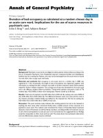
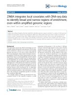
Figure 2 RSV infection and replication in BALB/c mouse lungs. Mice were dosed with either 1 × 10
6
FFU RSV A2 or an equivalent concentration
of UV-inactivated RSV. Three mice per treatment were sampled at 1, 5, 8, 24, 48 and 72 hours and after 7, 10, 37 and 59 days post-infection. One lung
per animal was processed for QPCR analyses, the other for infectivity assay. A) Levels of positive and negative sense RSV RNA in mouse lungs were
monitored using strand-specific QPCR. Normalised RSV copy number was determined from strand-specific RNA standard curves corrected by beta
actin arbitrary copy number. Means ± SEM for 3 animals per time point are plotted. Lower limit of detection = 80 actin normalised copies per gram
lung wt. B) Infectious live virus in mouse lungs was monitored by FFU assay (lower limit of detection = 10
2
FFU/g lung wt.) up to day 7 post infection.
Individual measurements are plotted and bars indicate mean values.
Bannister et al. Virology Journal 2010, 7:250
/>Page 6 of 11
RSV infected mice at 24 and 48 hours post infection.
Infectious virus was undetectable from palivizumab trea-
ted mouse lungs at all time points past 48 hours.
RSV RNA replication is severely impaired in mouse
cells in vitro
In human A549 cells infected with a low MOI of 1 ×
10
-3
or 1 × 10
-2
, viral RNAs increased from below the
limit of detection at 1 hour post infection to maximum
levels (negative sense >10
6
copies; positive strand >10
7
copies)atday5whichweresustaineduptotheendof
the experiment at day 10. When the cells were infected
with higher MOIs of 1 × 10
-1
or greater, the positive
and negative RSV RNA attained similar maximum levels
to those observed in the lower MOI infections (negative
sense >10
6
copies; positive strand >10
7
copies) (Figure
4C and 4D). Viral RNA r eaches maximum expression
values earlier in cells infected with higher MOI. A
decrease in measured negative and positive RNA was
observed in MOI 1 × 10
-1
and 1 infections after day 5
(Figure 4C and 4D) whic h correlated with a progressive
decrease in beta actin gene expression, indicative of cell
death (Figure 4E).
In mouse KLN205 cells infected with RSV at a low
MOI of 1 × 10
-3
no viral RNA could be detected (Figure
4A).AtanMOIof1×10
-2
very low levels of positive
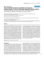
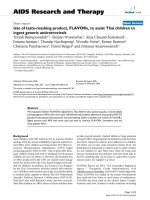
Figure 3 Palivizumab and ribavirin reduce infect ious virus in mouse lungs, but only ribavirin affects intracellular v iral replication.
Naïve Balb/c mice and mice treated prophylactically with 5 mg/kg palivizumab were infected intra nasally with 2.3 × 10
6
FFU RSV A2. A third
group were treated with ribavirin prior to RSV administration and throughout the study period as described in materials and methods. A)
Negative and B) positive sense strand RSV RNAs were quantified by strand-specific QPCR. Means ± SEM for 6 animals per time point are plotted.
Normalised RSV copy number was determined from strand-specific RNA standard curves corrected by beta actin arbitrary copy number. C) A
ratio index of positive to negative strand RNA was constructed and time course profiles for ribavirin and palivizumab treatments are plotted
against untreated RSV infected values. Means ± 95% confidence intervals (n = 6) are shown. D) Infectious live virus in mouse lungs was
monitored by FFU assay throughout the time-course. Means ± SEM (n = 6) are plotted. Asterisks indicate significant difference (p ≤ 0.05) to
untreated RSV-infected values at each sampling time. No data was collected post day 7 and is depicted as ND on the graph.
Bannister et al. Virology Journal 2010, 7:250
/>Page 7 of 11
MOI = 1
0 2 4 6 8 10
10
0
10
1
10
2
10
3
10
4
10
5
10
6
10
7
10
8
Time (days)
RSV copy number
MOI = 0.1
0 2 4 6 8 10
10
0
10
1
10
2
10
3
10
4
10
5
10
6
10
7
10
8
Time (days)
RSV copy number
MOI = 0.01
0 2 4 6 8 10
10
0
10
1
10
2
10
3
10
4
10
5
10
6
10
7
10
8
Time (days)
RSV copy number
MOI = 0.001
0 2 4 6 8 10
10
0
10
1
10
2
10
3
10
4
10
5
10
6
10
7
10
8
Time (days)
RSV copy number
beta actin - A459 cells
0 2 4 6 8 10
10
0
10
1
10
2
10
3
10
4
10
5
10
6
10
7
10
8
Time (days)
arbitrary copy number
beta actin - A459 cells
0 2 4 6 8 10
10
0
10
1
10
2
10
3
10
4
10
5
10
6
10
7
10
8
Time (days)
arbitrary copy number
A
F
E
D
C
B
beta actin - KLN205 cells
0 2 4 6 8 10
10
0
10
1
10
2
10
3
10
4
10
5
10
6
10
7
10
8
Time (days)
arbitrary copy number
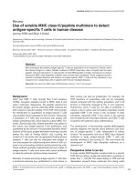
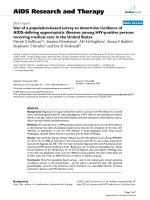
Figure 4 RSV RNA synthesis in human and mouse cell lines. A-D) Positive and negative sense viral RNA were monitored by strand-specific
QPCR from A549 and KLN205 cells treated with RSV A2 at MOIs of A) 1 × 10
-3
,B)1×10
-2
C) 1 × 10
-1
or D) 1. Copy numbers were determined
from strand-specific RNA standard curves. RSV RNA was not detected in KLN205 cells treated with RSV at MOI = 1 × 10
-3
. E-F) Beta actin,
expressed as arbitrary copy number, was measured by QPCR from E) A549 or F) KLN205 cells treated with RSV A2 at the MOIs shown. Means ±
SEM (n = 2) are plotted.
Bannister et al. Virology Journal 2010, 7:250
/>Page 8 of 11
and negative sense RSV RNA could be detected (Figure
4B), but at day 10 the mean amounts of neither strand
were higher than those measured 1 hour post-infection
(10
2
copies).
In cells infected at a MOI of 1 × 10
-1
, mean positive
sense RSV RNA increased by 2 logs from 10
4
copies on
day 3 to 10
6
copies by day 7 (Figure 4C). However, no
increase in negative sense RNA copy number was
observed over the 10 day culture period. A limited
increase in positive sense RNA was also observed in cul-
tures infected with an MOI of 1, rising from day 2 (10
5
copies) to day 7 (10
6
copies) and was maintained until
the end of the study at day 10. Similar t o cells infected
with the MOI of 1 × 10
-1
, no increase in negative strand
RNA was observed (Figure 4D). Beta actin levels in the
mouse KLN205 cells fell less than 0.5 logs between days
2 and 10 indicating t hat no appreciable cell death had
occurred throughout the study (Figure 4F).
Discussion
We have developed a stra nd-specific QPCR method to
measure RSV in vitro and in vivo.Thismethoddistin-
guishes between negative sense viral RNA (genome) and
positive sense RNA (replicative intermediate and nucleo-
capsid mRNA). Using this method, we provide a detailed
insight into RS V RNA production in infected BALB/c
mouse lung. To our knowledge, this is the first time
that a strand specific method has been applied to profile
RSV RNA dynamics in the BALB/c mouse over such a
detailed time course.
Early viral RNA synthesis in mouse lungs is charac-
terised by absolute m easures of positive and negative
sense RNA being equivalent at infection, followed by a
1-2 logs relative increase in positive stra nd RNA by day
3 post infection. This disparity between RNA strands
decreas es again from day 7. It should be noted that this
window of maximum disparity between the positive and
negative strand copy numbers at day 3 coincides with
the highest level of infectious progeny virus detected
from mouse lungs following infection. It is known that
paramyxovirus replicative intermediate RNA represent
10-40% of the genome [16], therefore the majority of
positive strand RNA synthesis seen here is accounted
for by nucleocapsid mRNA production.
That RSV genome and positive strand RNA can be
detected in mouse lungs up to at least 59 days post-
infection has been reported both here and elsewhere
[15,23]. It therefore appears that mice are unable to
fully clear the virus following infection. The fact that
UV killed RSV was not detected by QPCR past day 7
supports this view of viral persistence. RSV persistence
in the lungs has been reported from humans with
chronic obstructive pulmonary disease (COPD) [24],
although in another study, RSV infections in COPD
were attributed to acute infec tion rather than low-level
persistence [25]. The significance of persistent low levels
of RSV in this and other conditions is unclear at present
and further studies are required to elucidate the scope
and impact of this phenomenon [26]. However, it is pos-
sible that low levels of persistent virus exist between
RSV seasons and it is apparent that RSV persistence and
strategies for complete viral clearance may be studied in
rodent models.
Viral RNA replication has been studied by strand-dis-
criminate QPCR previously in the cotton rat [16]. Viral
genome levels increased by approximately 2 logs from
6 hours post infection to a peak measured on day
4 whereas our studies indicate that in the mouse lung
total g enomic RNA did not increase in t his time frame.
Indeed, in the mouse model we observed that viral gen-
ome load either decreased after 24 hours or (if a higher
inoculum was applied), was maintained for a period of
time before decreasing after d ay 5. These data suggest
that RSV has a greater replicative capacity in the cotton
rat model compared to the mouse. H owever until a
direct head to head comparison is made between the
two species, this cannot be concluded.
Ribavirin has been used extensively as an antiviral
therapeutic. Its exact mode of action is poorly defined
although several mechanisms have been proposed [27].
Here, as expected, ribavirin treatment had a marked
effect on RSV intracellular RNA dynamics as evidenced
by the reduction in positive sense RNA in mouse lungs.
However, there was lit tle difference seen in t he time-
course profiles of total genomic RNA in ribavirin treated
and untreated RSV infected mice. This sugg ests that the
amount of new genome synthesised following infection
is only a small fraction of that dosed initially and that
measuring positive sense RNA specifically is vital to the
study of the intracellular viral processes in mouse lung
following supra-physiologic dosing.
Prophylactic treatment of RSV-infected mice with the
neutralising antibody palivizumab resulted in a reduc-
tion in infectious progeny virus detected in the l ung,
although a reduction in positive sense strand RNA was
not observed. These findings agree with thos e previously
observed in the cotton rat, where a lack of detectable
progeny virus occurred despite intracellular replication
taking place. This phenomenon was termed abortive
replication [16]. The authors speculated that ab ortive
replication could occur due to the blocking of produc-
tion and release of large amounts of progeny virus
despite infection occurring in the presence of high titres
of neutralising antibody. Our data s upport this hypo th-
esis. We conclude that the evaluation of antibody-
mediated viral therapies in the mouse model may be
confounded by the high viral titres required for effective
infection.
Bannister et al. Virology Journal 2010, 7:250
/>Page 9 of 11
To investigate whether the restricted replication pat-
tern seen in the mouse is purely an in vivo phenomenon,
we infected lung epithelial carcinoma cells from human
(A549) and mouse (KLN205) with RSV and studied viral
replication by strand-specific QPCR. One hour post
infection, the input viral RNA levels were very similar in
bot h hum an and mouse cells, irrespective of MOI or cell
type, indicating that the mouse and human cells had
bee n exposed to equivalent amounts of viral RNA. How-
ever, a clear increase in either viral RNA s trand only
occurred in mouse cells when they were infected with a
high MOI of 0.1 or 1. This situation mirrors that which
occurs in the mouse in vivo modelinthatanextremely
high viral ti tre is required for repl ication [14]. Moreover,
the increase in positive strand viral RNA was consider-
ably delayed, occurring after a lag time of 3 days in cul-
ture suggesting that the virus has undergone a p eriod of
adaptation. Overall, RSV RNA synthesis in human A549
cells was at least 3 orders of magnitude more efficient
than that observed in mouse cells, illustrating that RSV
cannot replicate efficiently in mouse KLN205 cells. This
data suggests that some host-specif ic block to viral repli -
cation exists, tho ugh a wider range of human and mouse
cell lines require testing to confirm this.
It is unclear why the murine cells did not facilitate
RSV RNA synthesis to the same extent as seen in
human cells. It may be that RNA replication in KLN205
cells is inhibited either by the presence or absence of
one or more host factors required for the viral life cycle.
For example, it is known that RSV can modulate host
cell anti-viral responses, such as the degradation of
STAT2 by NS1 [28], which inhibits the interferon
response. Poor replication of RSV in mouse embryo
cells has been descr ibed previously [29]. This was attrib-
uted to the mouse interferon response as treatment of
infected cells with ant i-mouse interferon improved virus
yields. Perhaps RSV is not able to modulate the mouse
interferon response to the same extent as human inter-
feron. Alternatively, it is also known that RSV requires
host proteins to replicate efficiently. Phosphorylation o f
the RSV P protein by casein 2 is required for transcrip-
tion elongation activity of the viral polymerase in-vitro
[30]. It is plausible that species-specific differences in
host factors may impair the ability of RSV to replicate
efficiently in mouse cells, as is exemplified with HIV
and APOBEC3G [31].
In conclusion, we have demonstrated and quantified
the abortive and restricted nature of RSV RNA synthesis
and replication in mouse using a highly sensitive and
specificQPCRmethod.Wehavegoneontoprovide
evidence that the impaired replication may be due to a
murine host-virus interaction. We suggest a number of
candidates and work is ongoing to identify these
interactions.
Acknowledgements
The authors thank Julien Browne, Frances Burden, Bhavika Desai, Tansi
Khodai, Susanne Lang, Hannah Perkins and Joanne Strawbridge for practical
support. We would also like to thank Chloe Brown, Lisa-Marie Burrows and
Lindsey Cousens in Pfizer CM. The statistical support of Katrina Gore and
Richard Lyons is also gratefully acknowledged.
Authors’ contributions
RB carried out the molecular and cellular studies and drafted the
manuscript. DR carried out the in vivo and cellular assays and analysis and
interpretation of data, EJM, MW and CL participated in the design of the
study and analysis and interpretation of data. HB conceived of the study,
participated in its design and coordination and helped to draft the
manuscript. All authors read and approved the final manuscript.
Competing interests
All authors are or were employed in a full-time capacity by Pfizer Research
and Development.
Received: 15 June 2010 Accepted: 22 September 2010
Published: 22 September 2010
References
1. Han LL, Alexander JP, Anderson LJ: Respiratory syncytial virus pneumonia
among the elderly: an assessment of disease burden. J Infect Dis 1999,
179:25-30.
2. McCarthy AJ, Kingman HM, Kelly C, Taylor GS, Caul EO, Grier D, Moppett J,
Foot AB, Cornish JM, Oakhill A, et al: The outcome of 26 patients with
respiratory syncytial virus infection following allogeneic stem cell
transplantation. Bone Marrow Transplant 1999, 24:1315-1322.
3. Glezen WP, Greenberg SB, Atmar RL, Piedra PA, Couch RB: Impact of
respiratory virus infections on persons with chronic underlying
conditions. Jama 2000, 283:499-505.
4. Leader S, Kohlhase K: Recent trends in severe respiratory syncytial virus
(RSV) among US infants, 1997 to 2000. J Pediatr 2003, 143:S127-132.
5. DeVincenzo JP: Therapy of respiratory syncytial virus infection. Pediatr
Infect Dis J 2000, 19:786-790, discussion 802-784, 811-783.
6. Greenough A, Thomas M: Respiratory syncytial virus prevention: past and
present strategies. Expert Opin Pharmacother 2000, 1:1195-1201.
7. Saez-Llorens X, Moreno MT, Ramilo O, Sanchez PJ, Top FH Jr, Connor EM:
Safety and pharmacokinetics of palivizumab therapy in children
hospitalized with respiratory syncytial virus infection. Pediatr Infect Dis J
2004, 23:707-712.
8. Andries K, Moeremans M, Gevers T, Willebrords R, Sommen C, Lacrampe J,
Janssens F, Wyde PR: Substituted benzimidazoles with nanomolar activity
against respiratory syncytial virus. Antiviral Res 2003, 60:209-219.
9. Douglas JL, Panis ML, Ho E, Lin KY, Krawczyk SH, Grant DM, Cai R,
Swaminathan S, Cihlar T: Inhibition of respiratory syncytial virus fusion by
the small molecule VP-14637 via specific interactions with F protein.
J Virol 2003, 77:5054-5064.
10. Wyde PR, Chetty SN, Timmerman P, Gilbert BE, Andries K: Short duration
aerosols of JNJ 2408068 (R170591) administered prophylactically or
therapeutically protect cotton rats from experimental respiratory
syncytial virus infection. Antiviral Res 2003, 60:221-231.
11. Cowton VM, McGivern DR, Fearns R: Unravelling the complexities of
respiratory syncytial virus RNA synthesis. J Gen Virol 2006, 87:1805-1821.
12. Hacking D, Hull J: Respiratory syncytial virus–viral biology and the host
response. J Infect 2002, 45:18-24.
13. Sugrue RJ: Interactions between respiratory syncytial virus and the host
cell: opportunities for antivirus strategies? Expert Rev Mol Med 2006,
8:1-17.
14. Domachowske JB, Bonville CA, Rosenberg HF: Animal models for studying
respiratory syncytial virus infection and its long term effects on lung
function. Pediatr Infect Dis J 2004, 23:S228-234.
15. Estripeaut D, Torres JP, Somers CS, Tagliabue C, Khokhar S, Bhoj VG,
Grube SM, Wozniakowski A, Gomez AM, Ramilo O, et al: Respiratory
syncytial virus persistence in the lungs correlates with airway
hyperreactivity in the mouse model. J Infect Dis 2008, 198:1435-1443.
16. Boukhvalova MS, Prince GA, Blanco JC: Respiratory syncytial virus infects
and abortively replicates in the lungs in spite of preexisting immunity.
J Virol 2007, 81:9443-9450.
Bannister et al. Virology Journal 2010, 7:250
/>Page 10 of 11
17. Boukhvalova MS, Prince GA, Blanco JC: The cotton rat model of
respiratory viral infections. Biologicals 2009, 37:152-159.
18. Taylor G, Stott EJ, Hughes M, Collins AP: Respiratory syncytial virus
infection in mice. Infect Immun 1984, 43:649-655.
19. Vaux-Peretz F, Meignier B: Comparison of lung histopathology and
bronchoalveolar lavage cytology in mice and cotton rats infected with
respiratory syncytial virus. Vaccine 1990, 8:543-548.
20. Malhotra R, Ward M, Bright H, Priest R, Foster MR, Hurle M, Blair E, Bird M:
Isolation and characterisation of potential respiratory syncytial virus
receptor(s) on epithelial cells. Microbes Infect 2003, 5:123-133.
21. Bessaud M, Autret A, Jegouic S, Balanant J, Joffret ML, Delpeyroux F:
Development of a Taqman RT-PCR assay for the detection and
quantification of negatively stranded RNA of human enteroviruses:
evidence for false-priming and improvement by tagged RT-PCR. J Virol
Methods 2008, 153:182-189.
22. Chavez-Bueno S, Mejias A, Gomez AM, Olsen KD, Rios AM, Fonseca-Aten M,
Ramilo O, Jafri HS: Respiratory syncytial virus-induced acute and chronic
airway disease is independent of genetic background: an experimental
murine model. Virol J 2005, 2:46.
23. Schwarze J, O’Donnell DR, Rohwedder A, Openshaw PJ: Latency and
persistence of respiratory syncytial virus despite T cell immunity. Am J
Respir Crit Care Med 2004, 169:801-805.
24. Wilkinson TM, Donaldson GC, Johnston SL, Openshaw PJ, Wedzicha JA:
Respiratory syncytial virus, airway inflammation, and FEV1 decline in
patients with chronic obstructive pulmonary disease. Am J Respir Crit
Care Med 2006, 173:871-876.
25. Falsey AR, Formica MA, Hennessey PA, Criddle MM, Sullender WM,
Walsh EE: Detection of respiratory syncytial virus in adults with chronic
obstructive pulmonary disease. Am J Respir Crit Care Med 2006,
173:639-643.
26. Sikkel MB, Quint JK, Mallia P, Wedzicha JA, Johnston SL: Respiratory
syncytial virus persistence in chronic obstructive pulmonary disease.
Pediatr Infect Dis J 2008, 27:S63-70.
27. Sidwell RW, Barnard DL: Respiratory syncytial virus infections: recent
prospects for control. Antiviral Res 2006, 71:379-390.
28. Elliott J, Lynch OT, Suessmuth Y, Qian P, Boyd CR, Burrows JF, Buick R,
Stevenson NJ, Touzelet O, Gadina M, et al: Respiratory syncytial virus NS1
protein degrades STAT2 by using the Elongin-Cullin E3 ligase. J Virol
2007, 81:3428-3436.
29. Hanada N, Morishima T, Nishikawa K, Isomura S, Nagai Y: Interferon-
mediated self-limiting growth of respiratory syncytial virus in mouse
embryo cells. J Med Virol 1986, 20:363-370.
30. Dupuy LC, Dobson S, Bitko V, Barik S: Casein kinase 2-mediated
phosphorylation of respiratory syncytial virus phosphoprotein P is
essential for the transcription elongation activity of the viral polymerase;
phosphorylation by casein kinase 1 occurs mainly at Ser(215) and is
without effect. J Virol 1999, 73:8384-8392.
31. Browne EP, Littman DR: Species-specific restriction of apobec3-mediated
hypermutation. J Virol 2008, 82:1305-1313.
doi:10.1186/1743-422X-7-250
Cite this article as: Bannister et al.: Use of a highly sensitive strand-
specific quantitative PCR to identify abortive replication in the mouse
model of respiratory syncytial virus disease. Virology Journal 2010 7:250.
Submit your next manuscript to BioMed Central
and take full advantage of:
• Convenient online submission
• Thorough peer review
• No space constraints or color figure charges
• Immediate publication on acceptance
• Inclusion in PubMed, CAS, Scopus and Google Scholar
• Research which is freely available for redistribution
Submit your manuscript at
www.biomedcentral.com/submit
Bannister et al. Virology Journal 2010, 7:250
/>Page 11 of 11