Báo cáo y học: " Molecular characterization, structural analysis and determination of host range of a novel bacteriophage LSB-1" docx
Bạn đang xem bản rút gọn của tài liệu. Xem và tải ngay bản đầy đủ của tài liệu tại đây (991.45 KB, 7 trang )
RESEARC H Open Access
Molecular characterization, structural analysis and
determination of host range of a novel
bacteriophage LSB-1
Yaming Chai, Hongyan Xiong
*
, Xiangyu Ma, Liqing Cheng, Guorong Huang, Zhonglin Rao Lin Zhang
Abstract
Background: Bacteriophages (phages) are widespread in the environment and play a crucial role in the evolution
of their bacterial hosts and the emerg ence of new pathogens.
Results: LSB-1, a reference coliphage strain, was classified as a member of the Podoviridae family with a cystic
form (50 ± 5 nm diameter) and short tail (60 ± 5 nm long). The double stranded DNA was about 30 kilobase pa irs
in length. We identified its host range and determined the gp17 sequences and protein structure using shotgun
analysis and bioinformatics technology.
Conclusions: Coliphage LSB-1 possesses a tailspike protein with endosialidase activity which is probably
responsible for its specific enteroinvasive E.coli host range within the laboratory.
Background
Bacteriophages (phages) are widespread in the environ-
ment and play a crucial role in the evolution of their
bacterial hosts and the emergence of new pathogens.
They have enormous potential for the development of
new drugs, therapies and environmental control technol-
ogies, such as natural, non-toxic alternatives for control-
ling bacterial pathogens. Recent interest in phages has
been stimulated by studies that demonstrate the efficacy
of phages in preventing and treating infections [1-3].
Phages are readily isolated from water samples in the
environment. Some of the i solated phages have shown
broad-host range interaction with the bacterial isolates
and others have shown either species or strain level spe-
cificity. Both polyvalent phages and non-polyvalent
phages are morphologically and genetically diverse [4,5].
They are efficient at host recognition but there is no sin-
gle method of adsorptio n, and different phages employ
different strategies [6,7]. Identification of the mechanisms
of adsorption and the host ranges of different phages may
allow genetic manipulation to alter the phage host bind-
ing profile artificially. To gain a better understanding of
the biological properties of phages, we have sequenced
thegenomeofthegp17fromphageLSB-1,whichwas
isolated from sewage samples. We deter mined its host
range and analyzed its 3-dimensional structure to identify
possible functional domains.
Results
Coliphage morphology
The LSB-1 phage has a cystic form of 50 ± 5 nm in dia-
meter, with a short noncontractile tail 60 ± 5 nm long
(Fig. 1). It is classified as a member of the Podoviridae
family [8-10].
Nucleic acid characterization
LSB-1 coliphage nucleic acid was sensitive to Dnase I,
but resistant to Rnase A and S1 nuclease (Fig. 2). It was
concluded that all the extracts contained double-
stranded linear DNA.
Preliminary overview of the coliphages genome
The shotgun sequencing and a primer-walking method
was used to assemble the whole linear LSB-1 genome of
approximately 30 kb. Fifteen major potential ORFs were
identified, all of which could be assigned functions
based on homology with corresponding genes in the
K1F coliphage in a BLAST search (Fig. 3 and 4).
* Correspondence:
Department of Epidemiology, Faculty of Preventive Medicine, Third Military
Medical University, Chongqing 400038, China
Chai et al. Virology Journal 2010, 7:255
/>© 2010 Chai et al; licensee BioMed Central Ltd. This is an O pen Access article distributed under the terms of the Creative Commons
Attribution License (http://creativec ommons.org/licenses/by/2.0), which permits unrestricted use, distribution, and reproduction in
any medium , provided the original work is properly cited.
Genome relationship of LSB-1 and other phages
We chose four important proteins, capsid protein
(gp10), tail tubular protein (gp12), internal virion pro-
tein D (gp16), and endosialidase (gp17), which are
present in phage T7 supergroup members, to build
phylogenetic trees. As shown in Fig. 5, 6, 7 and 8,
trees built using different proteins are broadly similar.
Coliphages LSB-1, EcoDs1, and K1F closely clustered
towards the end of the group and phages T7, T3,
phiYeO3-12 and K11 cluster within another branch of
the same group. These data suggest that LSB-1 is
most closely related to the K1F-like phages, and
should be classified as a new member of the K1F
supergroup.
Gp17 DNA sequencing and function analysis
As shown in Fig. 9 and 10, the secondary structure of gp17
protein was composed of 33.10% Alpha helix (Hh), 25.25%
Extended strand (Ee) and 30.29% Random coil (Cc).
The PHYRE program generated several possible tem-
plates classified as best fit when the gp17 sequence was
submitted. All of these templates included the sequences
of hydrolase as one of its domains. It also provided the
SCOP codes, E-values and estimated precision values for
the predicted models. Table 1 shows these parameter s
provided by different templates.
The endosialidase 3-dimensional structure was chosen
to generate coordinates for the Gp17 of LSB-1 based on
an E-value of 1.1 and an estimated precision of 70%. Using
the PHYRE program, it was difficult to find another closer
coordinate template for Gp17 of LSB-1 even though an
E-value of less than 2e-6 was considered [11-13]. Gp17 of
LSB-1 included endosialidase amino acid residues from
Thr290 to Glu713, with the with the exception of forty-
one residues (Ser 465 to Trp475, Tyr497 to Val512, and
Lys543 to Leu562). These forty-one residues d id not
match corresponding sequences within the prediction ser-
ver. The Gp17 3-dimensional structure model predicted
two domains: Domain A with hydrolase activity connected
by an intervening unstructured sequence to Domain B.
The later domain may have a host-anchoring function.
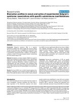
A worm algorithm representation of th e model i s shown
in Fig. 11. Domain A, the larger of the tw o domains,
includes residu es Thr290 to Phe528 a nd corresponds to
the N-terminal domain of endosialidase. It comprises 4 a
Figure 1 Electron micrograph of phage LSB-1.Electron
micrographs of the phages with a short, stout tail. The polyhedral
nature of the viral head is shown.
Figure 2 Agarose gel electrophoresis of DNA of phage LSB-1.
Lane 1: whole genome DNA digested by Nhe I. Lane 2: whole
genome DNA without digestion. Molecular size markers (kb) are
HindIII-digested lambda DNA.
Figure 3 Comparative genomic arrangements of coliphage
LSB-1 with K1F. Genome of coliphage LSB-1 is aligned. Arrows
indicate functions of the ORFs.
Figure 4 Comparative genomic arrangements of coliphage LSB-1 with K1F. Genome of coliphage K1F is aligned. Arrows indicate functions
of the ORFs.
Chai et al. Virology Journal 2010, 7:255
/>Page 2 of 7
helices and 9 b strands with intermittent unstructured
intervening regions. The arrangement is characteristic of
crystallized hydrolase. Domain B is composed of 1 a helix
and 9 b strands with intermittent unstructured intervening
regions. It includes residues Gln542 to Glu713 and corre-
sponds to the C-terminal domain of endosialidase. The re
were no predicted structural constraints in the connecting
region between domains A and B which included the resi-
dues of Tyr529 to Asp541.
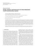
The molecular surface structure of the Gp17 is pre-
sented in Fig. 12. It shows the catalytic pocket in Domain
A comprising the catalytic triad, Glut405, Arg415, and
Arg487, identical to that found in the endosialidase
family of enzymes. Domain A is shown topographically
in front of Domain B. The connecting region was difficult
to see in this representation because of its opaqueness.
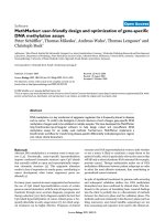
Fig. 13 and Fig. 14 show the worm algorithm structures
of Gp17and the KIF endosialidase respectively.
Discussion
Previous studies have suggested that gene 17 from the
T7-like and K1F-like phage may be playing an important
role in host range recognition processes [14,1 5], but lit-
tle work has been done on comparing their molecular
characterization. K1F-like phages are known to possess
tailspike proteins with endosialidase activity that
degrades polySia with high substrate specificity. These
tailspike-associated enzymatic activities enable the
phages to penetrate the capsular layer and are important
determinants of t he bacteriophage host range [16]. The
T7-like phage encodes a tail fiber protein that specifi-
cally recognizes and binds to lipopolysaccharide [17]
and recognizes E. coli B and many E. coli K-12 strains.
It was expected that molecular characterization would
provideevidenceforahostadsorbingmechanism
of coliphage LSB-1. We found that the LSB-1 has some
common features of the phage K1F supergroup.
A tailspike protein with endosialidase activity is implicated
in allowing a specific enteroinvasive E.coli host range.
Insertionofsuchanendosialidasegeneintoanon-
polyvalent virulent phage may artificially increase its host
range to enteroinvasive E.coli. Using manipulation of the
phage g enome to kill pathogenic bacteria has broad
Figure 5 Phylogenetic analysis of the capsid protein (gp10)
from seven phages in the T7 supergroup. The alignment of
whole sequence was used to construct the neighbor-joining tree.
The scale bar represents 0.05 fixed mutations per amino acid
position. Bootstrap values based on 1000 computer-generated tree
are indicated at the nodes.
Figure 6 Phylogenetic analysis of the tail t ubular protein
(gp12) from seven phages in the T7 supergroup. The alignment
of whole sequence was used to construct the neighbor-joining tree.
The scale bar represents 0.05 fixed mutations per amino acid
position. Bootstrap values based on 1000 computer-generated tree
are indicated at the nodes.
Figure 7 Phylogenetic analysis of the internal virion protein
(gp16) from seven phages in the T7 supergroup. The alignment
of whole sequence was used to construct the neighbor-joining tree.
The scale bar represents 0.05 fixed mutations per amino acid
position. Bootstrap values based on 1000 computer-generated tree
are indicated at the nodes.
Figure 8 Phylogenetic analysis of the endosialidase (gp17)
from seven phages in the T7 supergroup. The alignment of
whole sequence was used to construct the neighbor-joining tree.
The scale bar represents 0.2 fixed mutations per amino acid
position. Bootstrap values based on 1000 computer-generated tree
are indicated at the nodes.
Chai et al. Virology Journal 2010, 7:255
/>Page 3 of 7
implications for the welfare of both man and animals. Our
results may inform new ways of genetic manipulation of
phages to alter their host binding profile.
Materials and methods
Purification of phage particles
Bacteriophage LSB-1 was propagated in liquid culture.
The EIEC strain 8401 was infected with phages at a multi-
plicity of infection of 0.1. Fol lowing complete lysis of the
host cells, cell debris was removed by centrifugation at
Figure 9 Secondary structure prediction of gp17 protein.
Figure 10 Supplements and reports of Fig. 9, the secondary
structure of gp17 protein comprised 33.1% Alpha helix(Hh),
25.25% Extended strand(Ee) and 30.29% Random coil(Cc).
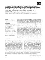
Table 1 Parameters provided by different templates
No SCOP Code E-value Estimated Precision Classification
1 c2c1lA_ 1.1 70% Hydrolase
2 d7a3ha_ 4.2 45% glycosidases
3 c2cksA_ 4.6 40% Hydrolase
4 c1wkyA_ 16 10% Hydrolase
Chai et al. Virology Journal 2010, 7:255
/>Page 4 of 7
4000 × g for 10 min, and phage particles in the superna-
tant were concentrated by adding polyethylene glycol 6000
and NaCl to achieve final concentrations of 10% and 1.0
M, respectively. Phage particles were collected by centrifu-
gation at 8000 × g for 10 min. Pellets were re- suspended
in 0.01× the original volume of sterile SM buffer (5.8 g
sodium chloride, 2 g magnesium sulphate, 100 mg gelatin,
50 mL 1 mol·L-1 Tris (pH 7.5) and 945 mL distilled
water). For isopycnic centrifugation, the phage suspension
was placed on a cesium chloride gradient stepwise using
three solutions whose densities were 1.45, 1.50, and 1.70,
respectively. After centrifugation for 60 min at 150,000 ×
g, the phage band was withdrawn and dialyzed against 10
mM Tris-HCl (pH 7.5) containing 10 mM MgSO. The
purified phage (approximately 10
11
PFU/ml) was stored at
4°C until use.
Host range determination
Seventeen bacterial strains, listed in Table 2, were tested
for sensitivity against the isolated phages. The Spot Test
Figure 11 Computer-generated 3-dimensional modeling of
gp17 protein. Worm algorithm representation of the 3-dimensional
model showing the two main domains. The larger Domain A is
homologous to hydrolase sequences. The smaller Domain B is
homologous to the sequence of the anchoring domain.
Figure 12 Computer-generated 3-dimensional modeling of gp17
protein. Molecular surface presentation of the predicted catalytic
pocket of endosialidase. The active site triad (yellow), Glut405, Arg415
and Arg487, is the amidase reaction site of endosialidase.
Figure 13 Worm algorithm representation of the model of
endosialidase of K1F.
Figure 14 Molecular surface presentation of K1F endosialidase.
The active site triad (yellow) is Glut581, Arg596 and Arg647.
Chai et al. Virology Journal 2010, 7:255
/>Page 5 of 7
[18] was used to determine the host range of the phage.
Lytic activity was examined foll owing overnight incuba-
tion at 37°C and recorded on a scale as follows: (N) no
plaques, (T) turbid plaques, (C) clear plaques. To obtain
an accurate estimate of relative phage lytic activity, all
host range determinations were carried out simulta-
neously using a single high-titer stock of purified bacter-
iophage and all host cells were incubated at 37°C in LB
medium.
Electron microscopy
To examine phage LSB-1 morphology, 50 μl of purified
viral stock solution was fixed by addition of 50 μlof
0.5% glutaraldehyde in 4% paraformaldehy de and a drop
of this solution was placed on a carbon coated copper
grid After waiting 30 min for settlement, excess liquid
was removed and the grid was allowed to dry. A drop of
2% phosphotungstic acid was added for 2 min before
excess was removed with filter paper before drying and
then examination by TEM (Hitachi, model S-800) at an
acceleration voltage of 45 kV.
Nucleic acid characterization
Themethodusedwasbaseduponthatdescribedby
Sambrook and Russell [19]. Bacteriophage from the con-
centrated solutions were lysed with the addition of
EDTA (final concentration 20 mmo l·L-1), proteinase K
(final concentration 50 μg·mL-1) and SDS (final concen-
tration 0.5%) and incubation at 56°C for 1 h. The
nucleic acid was purified using phenol, phenol/chloro-
form and chloroform extraction. The final aqueous
phase was dialy zed overnight against Tris EDTA buffer
(TE). In this method, nucleic acid yield was estimated
by agarose gel electrophoresis and compar ison of ethi-
dium bromide stain intensity with known DNA stan-
dards (Hind III cut lambda phage DNA, New England
Biolabs Inc, USA).
The nucleic acid extracts were diluted to a standard
concentration of ~20 ng·μL-1. Approximately 250ng of
each extract was subjected to digestion with DNase I
(Sigma Al drich), RNase A (Sigma Aldrich) a nd S1
nuclease (Promega). All reactions wer e terminated with
the addition of EDTA (10 mmol·L-1 final concentration)
and analyzed using 0.8% agarose gel electrophoresis at
5 V cm-1.
Gp17 DNA sequencing and analysis
The coliphage genome was sequenced by the shotgun
method. Genomic DNA was sheared by sonication, cloned
into pUC18 and sequenced with an ABI 3700 automated
DNA sequencer, to giv e 13-fold coverage of the genome.
Sequences were assembled into contigs, and gaps linked
using a primer-walking technique (Kaczorowski and
Szybalski, 1998) [20]. Potential ope n reading frames
(ORFs) were predicted using ORF Finder i.
nlm.nih.gov/projects/gorf/ and manual correction. Trans-
lated ORFs were used in a BLAST search against the
Swiss-Prot and NCBI pro-
tein databases />CMD=Web&PAGE_TYPE=BlastHome. Protein secondary
structures were predicted with SOPMA http://npsa-pbil.
ibcp.fr/cgi-bin/npsa_automat.pl?page=npsa_sopma.html.
Phylogenetic trees were constructed using Me ga 4
software following published protocols [21,22].
The amino acid sequence of endosialidase was determined
and compared with the protein structure family databases
PDB [23], SCOP [24,25], and PFAM [26] to identify the most
suitable template structure. The e ventual template structure
was taken from PDB. Three-dimensional models were created
using PHYRE by map-
ping the coordinates of the template structure wit h
aligned residues of the endosialidase. Computer-generated
three-dimensional models were viewed and analyzed using
CN3 D 4.1 application software programs obtained from
/>Conclusions
In summary, a typical Podoviridae morphology and the
double-stranded nature of its DNA give the coliphage
LSB-1 some common features with the phage K1F
supergroup. It possesses a tailspike protein with
Table 2 Phage and bacterial strains used in the study
and host range spectrum of the bacteriophages
No Species Strain Plaque formation
1 Escherichia 285
a
N
2 Escherichia ATCC 25922
b
N
3 Escherichia ATCC 8099
b
N
4 Escherichia ATCC 10536
b
N
5 Escherichia JM109
c
N
6 Escherichia BL21
c
N
7 Escherichia HDSa
c
N
8 Escherichia O157H7 44752
d
N
9 Escherichia EIEC 8401
d
C
10 Escherichia EIEC ATCC 43893
d
C
11 Staphylococcus aureus ATCC 6538
b
N
12 Staphylococcus aureus ATCC 26001
b
N
13 Salmonella typhi ATCC 50071
b
N
14 Pseudomonas aeruginosa ATCC 10145
b
N
15 Shigella dysenteriae ATCC 13313
b
N
16 Α-hemolytic streptococcus ATCC 32213
b
N
17 Β-hemolytic streptococcus ATCC 32210
b
N
Obtained from
a
Zhenghui L, Institute of Medical Equipment, Academy of
Military Medical Science, China;
b
Bacterial culture center, Institute of
Microbiology and Epidemiology, Academy of Military Medical Science, China;
c
Jing a. Department of Microbiology, Third Military Medical University, China;
d
Bacterial culture center, Institute of Epidemiology and Microbiology, Chinese
Academy of Preventive Medicine.
C = clear plaque; T = turbid plaque; N = no plaque.
Chai et al. Virology Journal 2010, 7:255
/>Page 6 of 7
endosialidase activity which is probably responsible for
its specific enteroinvasive E.coli host range within the
laboratory.
Acknowledgements
The authors would like to thank Jinxin Ke and Wenqi Huang for their help in
electron microscopy. The project was supported by National Natural Science
Foundation of China (Grant No.30571637), Scientific Research Project of the
Chongqing China (Grant No. CSTC, 2008AC5005) and National Key
Technology R&D Program of China (Grant No. 2008BAD96B06-05).
Authors’ contributions
YC and HX conceived of the study, carried out the experimental work,
analysis and drafted the manuscript. XM and LC participated in its design
and coordination and helped to draft the manuscript. GH and ZR
participated in its design and experimental work. LZ participated in
coordination of the study. All authors read and approved the final
manuscript.
Competing interests
The authors declare that they have no competing interests.
Received: 12 July 2010 Accepted: 28 September 2010
Published: 28 September 2010
References
1. Hermoso JL, García : Taking aim on bacterial pathogens: from phage
therapy to enzybiotics. Curr Opin Microbiol 2007, 10:461-472.
2. Mann : The potential of phages to prevent MRSA infections. Res Microbiol
2008, 159:400-405.
3. Sulakvelidze : Phage therapy: an attractive option for dealing with
antibiotic-resistant bacterial infections. Drug Discov Today 2005,
10:807-809.
4. Hyman ST: Bacteriophage Host Range and Bacterial Resistance. Adv Appl
Microbiol 2010, 70:217-248.
5. Song HY, Xu XM, Zhang : Isolation of a novel polyvalent virulent
bacteriophage of E. coli. J Med Coll PLA 2007, 22:261-267.
6. Konopa KT: Isolation of coliphage lambda ghosts able to adsorb onto
bacterial cells. Biochim Biophys Acta 1975, 399:460-467.
7. Paranchych PM, Bradley : Stages in phage R17 infection. Virology 1970,
41:465-473.
8. Duda : Icosahedral Tailed dsDNA Bacterial Viruses. Encyclopedia of Virology
2008, 30-37.
9. Molineux : T7-Like Phages (Podoviridae). Encyclopedia of Virology 2004,
1722-1729.
10. Rothman-Denes : Enterobacteria Phage N4 (Podoviridae). Encyclopedia of
Virology 2004, 450-454.
11. Bujnicki AE, Fischer LR: LiveBench-2: large-scale automated evaluation of
protein structure prediction servers. Proteins 2001, , Suppl 5: 184-191.
12. Bujnicki AE, Fischer LR: Structure prediction meta server. Bioinformatics
2001, 17:750-751.
13. Cristobal AZ, Fischer , et al: A study of quality measures for protein
threading models. BMC Bioinformatics 2001, 2:5.
14. Machida KM, Hattori SY, Kawase SI: Structure and function of a novel
coliphage-associated sialidase. FEMS Microbiol Lett 2000, 182:333-337.
15. Schulz DS, Frank KS, Mühlenhoff AD, Gerardy-Schahn RF: Structural Basis
for the Recognition and Cleavage of Polysialic Acid by the
Bacteriophage K1F Tailspike Protein EndoNF. J Mol Biol 2010, 397:341-351.
16. Bull ER, Molineux : A tale of tails: Sialidase is key to success in a model of
phage therapy against K1-capsulated Escherichia coli. Virology
2010,
398:79-86.
17. Steven BL, Maizel MU, Parry JS, Hainfeld FW: Molecular substructure of a
viral receptor-recognition protein: The gp17 tail-fiber of bacteriophage
T7. J Mol Biol 1988, 200:351-365.
18. Champagne NG: The spot test method for the in-plant enumeration of
bacteriophages with paired cultures of Lactobacillus delbrueckii subsp.
bulgaricus and Streptococcus salivarius subsp. thermophilus. Int Dairy J
1995, 5:417-425.
19. Sambrook DW: Molecular cloning. A laboratory manual CSHL Press. New
York 2001.
20. Kaczorowski WS: Genomic DNA sequencing by SPEL-6 primer walking
using hexamer ligation. Gene 1998, 223:83-91.
21. Heres DV: Phylogenetic analysis of the pathogenic bacteria Spiroplasma
penaei based on multilocus sequence analysis. J Invertebr Pathol 2010,
103:30-35.
22. El Ossmani BB, Aboukhalid MB, Gazzaz DZ, Chafik JT: Assessment of
phylogenetic structure of Berber-speaking population of Azrou using 15
STRs of Identifiler kit. Leg Med 2010, 12:52-56, 22. Ossmani, B.B., boukhalid,
M.B., Gazzaz, D.Z. & Chafik, J.T. (2010). Assessment of phylogenetic structure
of Berber-speaking population of Azrou using 15 STRs of Identifiler kit. Leg
Med. 12, 52-56
23. Berman KH, Nakamura : Announcing the worldwide Protein Data Bank.
Nat Struct Biol 2003, 10:980.
24. Andreeva DH, Brenner : SCOP database in 2004: refinements integrate
structure and sequence family data. Nucleic Acids Res 2004, 32:D226-D229.
25. Brenner CC, Hubbard AG: Understanding protein structure: using scop for
fold interpretation. Methods Enzymol 1996, 266:635-643.
26. Bateman EB, Durbin : Pfam 3.1: 1313 multiple alignments and profile
HMMs match the majority of proteins. Nucleic Acids Res 1999, 27:260-262.
doi:10.1186/1743-422X-7-255
Cite this article as: Chai et al.: Molecular characterization, structural
analysis and determination of host range of a novel bacteriophage LSB-
1. Virology Journal 2010 7:255.
Submit your next manuscript to BioMed Central
and take full advantage of:
• Convenient online submission
• Thorough peer review
• No space constraints or color figure charges
• Immediate publication on acceptance
• Inclusion in PubMed, CAS, Scopus and Google Scholar
• Research which is freely available for redistribution
Submit your manuscript at
www.biomedcentral.com/submit
Chai et al. Virology Journal 2010, 7:255
/>Page 7 of 7