báo cáo khoa học: " Differential patterns of reactive oxygen species and antioxidative mechanisms during atrazine injury and sucrose-induced tolerance in Arabidopsis thaliana plantlets" ppsx
Bạn đang xem bản rút gọn của tài liệu. Xem và tải ngay bản đầy đủ của tài liệu tại đây (1.15 MB, 18 trang )
BMC Plant Biology
BioMed Central
Open Access
Research article
Differential patterns of reactive oxygen species and antioxidative
mechanisms during atrazine injury and sucrose-induced tolerance
in Arabidopsis thaliana plantlets
Fanny Ramel1, Cécile Sulmon1, Matthieu Bogard1,2, Ivan Couée1 and
Gwenola Gouesbet*1
Address: 1Centre National de la Recherche Scientifique, Université de Rennes I, UMR 6553 ECOBIO, Campus de Beaulieu, bâtiment 14A, F-35042
Rennes Cedex, France and 2INRA, UMR 1095 Génétique, Diversité et Ecophysiologie des Céréales, 234-avenue du Brezet, F-63100 ClermontFerrand, France
Email: Fanny Ramel - ; Cécile Sulmon - ;
Matthieu Bogard - ; Ivan Couée - ; Gwenola Gouesbet* -
* Corresponding author
Published: 13 March 2009
BMC Plant Biology 2009, 9:28
doi:10.1186/1471-2229-9-28
Received: 4 December 2008
Accepted: 13 March 2009
This article is available from: />© 2009 Ramel et al; licensee BioMed Central Ltd.
This is an Open Access article distributed under the terms of the Creative Commons Attribution License ( />which permits unrestricted use, distribution, and reproduction in any medium, provided the original work is properly cited.
Abstract
Background: Besides being essential for plant structure and metabolism, soluble carbohydrates
play important roles in stress responses. Sucrose has been shown to confer to Arabidopsis
seedlings a high level of tolerance to the herbicide atrazine, which causes reactive oxygen species
(ROS) production and oxidative stress. The effects of atrazine and of exogenous sucrose on ROS
patterns and ROS-scavenging systems were studied. Simultaneous analysis of ROS contents,
expression of ROS-related genes and activities of ROS-scavenging enzymes gave an integrative view
of physiological state and detoxifying potential under conditions of sensitivity or tolerance.
Results: Toxicity of atrazine could be related to inefficient activation of singlet oxygen (1O2)
quenching pathways leading to 1O2 accumulation. Atrazine treatment also increased hydrogen
peroxide (H2O2) content, while reducing gene expressions and enzymatic activities related to two
major H2O2-detoxification pathways. Conversely, sucrose-protected plantlets in the presence of
atrazine exhibited efficient 1O2 quenching, low 1O2 accumulation and active H2O2-detoxifying
systems.
Conclusion: In conclusion, sucrose protection was in part due to activation of specific ROS
scavenging systems with consequent reduction of oxidative damages. Importance of ROS
combination and potential interferences of sucrose, xenobiotic and ROS signalling pathways are
discussed.
Background
Although molecular oxygen (O2) is used as stable terminal electron acceptor in many essential metabolic processes, its partially reduced or activated forms, singlet
oxygen (1O2), superoxide radical (O2.-), hydrogen peroxide (H2O2) and hydroxyl radical (HO.), are highly reactive
[1]. Overproduction of these reactive oxygen species
(ROS) can initiate a variety of autooxidative chain reac-
Page 1 of 18
(page number not for citation purposes)
BMC Plant Biology 2009, 9:28
tions on membrane unsaturated fatty acids, thus yielding
lipid hydroperoxides and cascades of events ultimately
leading to destruction of organelles and macromolecules
[2].
In plants, ROS are continuously produced as byproducts
of various metabolic pathways, principally through electron transport chains in chloroplasts and mitochondria,
photorespiration in peroxisomes, oxidases and peroxidases [3]. ROS, which also act as signalling molecules,
have been shown to affect the expression of multiple
genes [2,4], and to be involved in activation and control
of various genetic stress-response programs [5].
However, numerous environmental factors such as UVradiation, high light, drought, low or high temperature,
mechanical stress and some xenobiotics disturb the
prooxidant-antioxidant balance and lead to irreparable
metabolic dysfunctions and cell death [6]. Different
classes of herbicides are direct or indirect sources of oxidative damages in plants. The herbicide atrazine, of the triazine family, binds to the D1 protein, which results in
inhibition of photosystem II (PSII) by blocking electron
transfer to the plastoquinone pool [7], thus leading to
production of triplet chlorophyll and 1O2 [8,9].
Because of widespread use, atrazine is a common contaminant in soils, streams, rivers and lakes [10,11]. The length
of water residence time associated with high loading rates
results in prolonged exposure of phytoplankton communities to atrazine. Numerous studies have been carried out
on the sensitivity of aquatic photosynthetic communities
towards atrazine and on effects of this herbicide on reduction of photosynthesis, chlorophyll synthesis, cell growth
and nitrogen fixation [12,13]. In the case of wild terrestrial plants, most studies deal with mutations of D1 protein in atrazine-resistant weeds [14], rather than with
atrazine-related toxic effects.
Exogenous supply of soluble sugars, particularly sucrose,
has been shown to confer to Arabidopsis plantlets a high
level of atrazine tolerance [15-17]. Transcriptome profiling revealed that atrazine sensitivity and sucrose-induced
atrazine tolerance were associated with important modifications of gene expression related to ROS defence mechanisms, repair mechanisms, signal transduction and
cellular communication [18]. Thus, sucrose-induced atrazine tolerance was shown to depend on modifications of
gene expression, which to a large extent resulted from
combined effects of sucrose and atrazine. This strongly
suggested important interactions of sucrose and xenobiotic signalling or of sucrose and ROS signalling, thus
resulting in induction of specific transcription factors and
in an integrated response to changing environmental conditions [18].
/>
Complex arrays of detoxification mechanisms have been
selected in plants against ROS accumulation and toxicity.
Biochemical antioxidants, such as ascorbate, glutathione,
tocopherol, flavonoids, anthocyanins and carotenoids
[19,20], and ROS-scavenging enzymes, such as superoxide dismutase (SOD), ascorbate peroxidase (APX), glutathione peroxidase (GPX) and catalase (CAT) [21-23],
are involved in maintaining the redox balance of cells. For
example, transgenic plants with enhanced SOD activity
exhibit increased tolerance to oxidative stress [22,24,25].
Moreover, Ramel et al. [18] have shown that, during
sugar-induced protection against atrazine, expression of
several ROS defence-related genes was enhanced.
The present work analyses the relationships between ROS
patterns, expression of genes involved in synthesis of antioxidant molecules or antioxidative processes and respective enzyme activities in order to characterize atrazine
sensitivity and sucrose-induced tolerance against atrazinedependent oxidative stress. Atrazine-treated plantlets were
found to exhibit an original pattern of ROS with increased
levels of 1O2 and H2O2 associated with a decrease of O2.content, whereas the protective sucrose-atrazine combination favored accumulation of O2.- and H2O2. These ROS
patterns were associated with differences of antioxidant
gene expression and enzyme activities, thus suggesting
that atrazine injuries might be due to deficient ROSdetoxification mechanisms. The possible interferences of
sucrose, xenobiotic and ROS signalling are discussed.
Results
Patterns of accumulation of singlet oxygen, superoxide
radical and hydrogen peroxide
The transfer of plantlets after 3 weeks of growth to control
and treatment media, as described in Methods, was
designed to compare plantlets at the same developmental
and physiological stages. As previously described in
numerous studies of sugar effects in plants, mannitol
treatment was used as osmotic control. Moreover, we previously showed that the deleterious effects of atrazine on
Arabidopsis plantlets followed the same dose-response
curve and the same time dependence in the absence or
presence of 80 mM mannitol [16,17]. It was also verified
that, in accordance with previous studies [26], exogenous
sugar treatment resulted in increased levels of endogenous
soluble sugars in Arabidopsis plantlets (data not shown).
At the end of treatments, plantlets were specifically
stained for singlet oxygen, superoxide radical, and hydrogen peroxide. Hideg et al. [27] described some limitations
in the use of vacuum infiltration of ROS probes and reagents with excised leaves or leaf segments from pea, spinach or tobacco. However, vacuum infiltration has been
successfully used on whole Arabidopsis thaliana plantlets
under various experimental conditions [28-30]. Moreo-
Page 2 of 18
(page number not for citation purposes)
BMC Plant Biology 2009, 9:28
/>
ver, under the conditions of the present work, whatever
the dye used and therefore the ROS detected, the nonstressed plantlets, transferred to 80 mM mannitol or 80
mM sucrose media, presented expected responses related
to ROS production (Fig. 1, 2 and 3; Additional files 1, 2
and 3). Plantlets that were transferred for 12 h on mannitol medium presented the same ROS levels as three-week-
old plantlets prior to transfer (Fig. 1, 2 and 3; Additional
files 1, 2 and 3).
Detection and quantification of singlet oxygen (1O2) were
performed with the specific Singlet Oxygen Sensor Green®
reagent [31]. For atrazine-containing treatments (MA and
SA), green fluorescence indicating primary events of 1O2
06
0
6
0$
6$
KRXUV
KRXUV
KRXUV
KRXUV
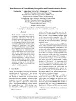
Figure 1
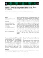
Visualization of singlet oxygen detected with the SOSG fluorescent probe
Visualization of singlet oxygen detected with the SOSG fluorescent probe. Detections have been done on 3-weekold MS-grown Arabidopsis thaliana plantlets subjected to subsequent treatment (12, 24, 48 or 72 hours) with 80 mM mannitol
(M), 80 mM sucrose (S), 80 mM mannitol plus 10 M atrazine (MA) or 80 mM sucrose plus 10 M atrazine (SA). The fluorescence of SOSG corresponds to the green coloration, while the red color corresponds to chlorophyll autofluorescence. Green
fluorescence of roots corresponds to flavonoid and porphyrin autofluorescence. Individual plantlets under the microscope are
shown. Quantification of singlet oxygen is presented in Additional file 1.
Page 3 of 18
(page number not for citation purposes)
BMC Plant Biology 2009, 9:28
/>
06
0
6
0$
6$
KRXUV
KRXUV
KRXUV
KRXUV
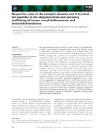
Figure 2
Visualization of superoxide radical detected by NBT staining
Visualization of superoxide radical detected by NBT staining. Detections have been done on 3-week-old MS-grown
Arabidopsis thaliana plantlets subjected to subsequent treatment (12, 24, 48 or 72 hours) with 80 mM mannitol (M), 80 mM
sucrose (S), 80 mM mannitol plus 10 M atrazine (MA) or 80 mM sucrose plus 10 M atrazine (SA). Groups of 15 plantlets are
shown. Quantification of superoxide radical is presented in Additional file 2.
Page 4 of 18
(page number not for citation purposes)
BMC Plant Biology 2009, 9:28
/>
06
0
6
0$
6$
KRXUV
KRXUV
KRXUV
KRXUV
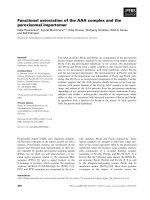
Figure 3
Visualization of hydrogen peroxide detected by DAB staining
Visualization of hydrogen peroxide detected by DAB staining. Detections have been done on 3-week-old MS-grown
Arabidopsis thaliana plantlets subjected to subsequent treatment (12, 24, 48 or 72 hours) with 80 mM mannitol (M), 80 mM
sucrose (S), 80 mM mannitol plus 10 M atrazine (MA) or 80 mM sucrose plus 10 M atrazine (SA). Groups of 15 plantlets are
shown. Quantification of hydrogen peroxide is presented in Additional file 3.
Page 5 of 18
(page number not for citation purposes)
BMC Plant Biology 2009, 9:28
accumulation was detected in cotyledons as soon as after
12 hours of treatment (Fig. 1 and Additional file 1). Tolerance treatment (SA) maintained a low level of 1O2 in
cotyledons throughout the treatment, while atrazine treatment (MA) strongly increased 1O2 production in cotyledons and leaves from 12 to 72 hours of treatment. The
presence of sucrose in herbicide-containing medium thus
appeared to prevent accumulation of 1O2 generated by
atrazine.
Superoxide radical (O2.-) detection and quantification
were performed using the nitroblue tetrazolium (NBT)
staining method. The levels of superoxide radical staining
after 12 hours of transfer (Fig. 2 and Additional file 2)
were quite similar in the absence (M or S) or presence (MA
or SA) of 10 M atrazine. However, the time-course
revealed constant levels of O2.- in control plantlets (M),
while a strong blue coloration appeared in plantlets
treated with sucrose (S). This increase was more visible in
young leaves. Superoxide radical levels in atrazine-treated
plantlets (MA) decreased from 24 hours of treatment. The
combination of sucrose plus atrazine (SA) led to an intermediate state with slight coloration maintained in young
leaves throughout the treatment. Low levels of O2.-, relatively to the mannitol control, were also observed when a
drop of 10 M atrazine solution was directly applied to
leaf tissue (data not shown).
H2O2 detection and quantification were performed using
the 3,3'-diaminobenzidine (DAB) staining method [32].
Polymerization of DAB, visible as a brown precipitate in
the presence of H2O2, was detected under all conditions.
No coloration was observed when infiltration was carried
out in the presence of ascorbic acid, thus confirming the
H2O2 specificity of DAB staining, in accordance with previous reports [33-36]. Figure 3 and Additional file 3 summarize the time-course of H2O2 accumulation. From 24
hours of transfer, control (M) and sucrose-treated (S)
plantlets exhibited a much weaker level of H2O2 than
plantlets of atrazine-containing treatments (MA and SA).
No variation of H2O2 accumulation was detected in the
presence of mannitol, whereas H2O2 content decreased in
sucrose-treated plantlets. In contrast, atrazine in the
absence or presence of sucrose tended to increase progressively H2O2 levels until 72 hours of treatment. This
increase could be detected as early as the fourth hour of
atrazine treatment (data not shown). Likewise, an immediate increase of H2O2 levels was also observed when a
drop of 10 M atrazine solution was directly applied to
leaf tissue (data not shown).
Patterns of singlet oxygen quenching mechanisms
Transcriptomic analysis showed that genes linked to the
synthesis of 1O2-quenchers presented contrasted patterns
of expression in relation to atrazine sensitivity and toler-
/>
ance (Table 1). Some genes exhibited higher transcript
levels under tolerance condition (SA) and repression
under atrazine injury condition (MA), thus suggesting the
possibility of more efficient quenching mechanisms in the
presence of sucrose. Thus, seven genes encoding thioredoxin family proteins (At2g32920, At2g35010,
At2g47470, At3g06730, At4g27080, At5g42980 and
At5g60640) were characterized by significant atrazine
repression of expression, which was lifted by sucrose-atrazine tolerance treatment (Table 1). Only two genes encoding thioredoxin family proteins exhibited higher
expression under atrazine treatment (At5g06690 and
At1g08570) than under sucrose plus atrazine treatment
(Table 1). In contrast, two thioredoxin genes (At1g69880
and At1g45145) and one thioredoxin reductase gene
(At2g17420) were significantly induced under tolerance
conditions (SA) compared to atrazine treatment (MA)
(Table 1). Thioredoxins have been shown to be involved
in supplying reducing power to reductases detoxifying
lipid hydroperoxides or repairing oxidized proteins [37].
Thioredoxins could also act as regulators of scavenging
mechanisms [38-40] and as components of signalling
pathways of plant antioxidant network. Finally, Das and
Das [41] presented evidence that human thioredoxin was
a powerful 1O2 quencher, which could protect cells and
tissues against oxidative stress.
Another group of genes exhibited induction of expression
under atrazine conditions, whereas they were less induced
or not differentially expressed under sucrose-atrazine conditions. Activation of these genes might reflect stress signalling due to high 1O2 content in atrazine treated-cells, as
revealed by ROS detection ((Fig. 1 and Additional file 1).
Some of these genes belonged to carotenoid biosynthesis
pathways, such as Zeta-carotene desaturase ZDS1
(At3g04870), beta-carotene hydroxylase (At4g25700) or
4-hydroxyphenylpyruvate dioxygenase HPD (At1g06570)
(Table 1). Carotenoids, which are known to act in chloroplasts as accessory pigments in light harvesting, can
detoxify 1O2 and triplet chlorophyll and dissipate excess
excitation energy [9].
Transcriptome profiling was carried out after 24 hours of
treatment [18]. Measurements of carotenoid levels at different times of treatment showed that modifications were
most contrasted after 48 hours of treatment [18]. Thus,
given the potential delay between transcription and metabolic fluxes, modifications of carotenoid levels after 48
hours of treatment were compared with transcript-level
modifications after 24 hours of treatment. Carotenoid
(xanthophylls and carotenes) levels in Arabidopsis thaliana
plantlets after 48 hours of treatment are presented in
Table 2. Atrazine treatment tended to reduce carotenoid
contents, while addition of sucrose in presence of atrazine
maintained carotenoid levels near control levels. How-
Page 6 of 18
(page number not for citation purposes)
BMC Plant Biology 2009, 9:28
/>
Table 1: Expression of genes involved in singlet oxygen quenching after 24 hours of treatment.
Log2(ratio)
Accession number Gene description
At1g08570
At1g45145
At1g69880
At2g17420
Localisation
Treatment comparison
S/M MA/M SA/M
At2g32920
At2g35010
At2g47470
At3g06730
At4g27080
At5g06690
At5g42980
At5g60640
Thioredoxin family protein
Thioredoxin H-type 5 (TRX-H-5) (TOUL)
Thioredoxin, putative
Thioredoxin reductase 2/NADPH-dependent thioredoxin reductase 2
(NTR2)
Thioredoxin family protein
Thioredoxin family protein
Thioredoxin family protein
Thioredoxin family protein
Thioredoxin family protein
Thioredoxin family protein
Thioredoxin H-type 3 (TRX-H-3) (GIF1)
Thioredoxin family protein
No classification
Cytosol
No classification
Cytoplasm
nde
nde
2.05
1.22
1.04
nde
nde
nde
nde
0.75
2.42
1.51
Endomembrane system
Mitochondrion
Endomembrane system
Chloroplast
Endoplasmic reticulum
Chloroplast
Cytosol
Endomembrane system
nde
nde
nde
nde
nde
-1.15
nde
nde
-1.54
-1.00
-1.74
-0.74
-0.96
1.14
-0.94
-1.19
nde
nde
nde
nde
nde
nde
nde
nde
At1g06570
At3g04870
At4g25700
4-hydroxyphenylpyruvate dioxygenase (HPD)
Zeta-carotene desaturase (ZDS1)/carotene 7.8-desaturase
Beta-carotene hydroxylase
Chloroplast
Chloroplast
Chloroplast
-0.75
nde
nde
3.18
0.94
1.07
2.11
nde
nde
At1g08550
Violaxanthin de-epoxidase precursor. putative (AVDE1)
Photosystem II
-1.26
0.91
nde
At3g26900
At4g36810
Shikimate kinase family protein
Geranylgeranyl pyrophosphate synthase (GGPS1)/GGPP synthetase/
farnesyltranstransferase
Chloroplast
Chloroplast
nde
nde
1.69
0.88
nde
nde
At3g55610
Delta 1-pyrroline-5-carboxylate synthetase B/P5CS B (P5CS2)
Cytoplasm
0.82
3.63
2.24
Relative expressions of gene are given with their log2(ratio) for sucrose versus mannitol (S/M), mannitol plus atrazine versus mannitol (MA/M) and
sucrose plus atrazine versus mannitol (SA/M) comparison. nde: not differentially expressed. Genes with a Bonferroni P-value higher than 5% were
considered as being not differentially expressed as described by Lurin et al. [85].
ever, carotenoid/chlorophyll ratios were not significantly
different, thus indicating that the photoprotection role of
carotenoids was maintained in the presence of atrazine.
Higher induction by atrazine treatment was also found for
the violaxanthin de-epoxidase precursor (At1g08550)
gene, which is involved in the xanthophyll cycle (Table 1).
Together with carotenoids, zeaxanthin, synthesized from
violaxanthin via the xanthophyll cycle, protects chloroplasts by accepting excitation energy from singlet chlorophyll [42]. Two genes involved in the shikimate
(shikimate kinase, At3g26900) and terpenoid pathways
(geranylgeranyl pyrophosphate synthase, At4g36810),
which are essential for tocopherol synthesis [43], were
also induced by the herbicide and not differentially
expressed by the tolerance treatment (SA) (Table 1). The
antioxidant tocopherol is known to scavenge oxygen free
radicals, lipid peroxy radicals and 1O2 [44]. Finally, the
presence of atrazine alone was found to induce the
At3g55610 gene, which is involved in proline synthesis,
with a higher intensity than under conditions of combina-
Table 2: Carotenoid content and carotenoid/chlorophyll ratios in leaves of Arabidopsis thaliana plantlets after 48 hours of treatment.
Treatment
Mannitol (M)
Sucrose (S)
Mannitol atrazine (MA)
Sucrose atrazine (SA)
Carotenoid content
(Mean ± SE)
g g-1 FW
Carotenoid/Chlorophyll ratios
78.6 ± 0.3
78.8 ± 0.6
61.2 ± 0.6
72.1± 0.8
0.172 ± 0.008
0.168 ± 0.009
0.176 ± 0.012
0.186 ± 0.010
Page 7 of 18
(page number not for citation purposes)
BMC Plant Biology 2009, 9:28
/>
tion with sucrose (Table 1). Proline is also known to be an
1O quencher [45].
2
Patterns of superoxide radical scavenging mechanisms
Excess of superoxide radical caused by numerous environmental stresses is detoxified by superoxide dismutase
(SOD) enzymes and converted into H2O2. Seven isoenzymes have been identified, differing by their metal cofactor (Fe, Mn, or Cu and Zn), in Arabidopsis thaliana [46].
Transcriptome profiling was carried out after 24 hours of
treatment [18]. Measurements of enzyme activities at different times of treatment showed that modifications were
most contrasted after 48 hours of treatment (data not
shown). Thus, given the potential delay between transcription and protein synthesis, modifications of global
SOD activities after 48 hours of treatment were compared
with modifications of SOD-encoding transcript levels
after 24 hours of treatment.
SOD activity (Fig. 4) was decreased by atrazine treatment
(MA) in comparison to the mannitol control (M). In contrast, addition of sucrose in the presence of atrazine (SA)
maintained a functional level of SOD activity equivalent
to that of the mannitol control. Since sucrose alone was
found to increase SOD activity, it thus seemed that
sucrose might balance the negative effect of atrazine in the
situation of SA treatment.
62' DFWLYLW\ XQLW J ):
D
E
E
F
0
6
0$
6$
Figure 4
Effects of atrazine and sucrose on SOD enzyme activity
Effects of atrazine and sucrose on SOD enzyme
activity. SOD activity was measured in protein extracts
from 3-week-old MS-grown Arabidopsis thaliana plantlets subjected to subsequent treatment (48 hours) with 80 mM mannitol (M), 80 mM sucrose (S), 80 mM mannitol plus 10 M
atrazine (MA) or 80 mM sucrose plus 10 M atrazine (SA).
SOD activity is expressed in unit/g FW as defined in Methods. Statistical analysis was carried out as described in Methods.
Among the six isoenzyme-encoding genes represented in
this microarray analysis (Table 3), three exhibited significant variations of transcript levels in comparison with
control conditions, thus suggesting their potential
involvement in O2.--detoxifying processes in relation to
atrazine sensitivity and tolerance. Three genes, encoding
CSD1, MSD1, FSD3, were characterized by significant
repression under conditions of atrazine treatment compared to control, in accordance with the measurement of
global SOD activity (Fig. 4). The CSD1 gene (At1g08830),
encoding cytosolic Cu-Zn superoxide dismutase, exhibited an induction under tolerance conditions (SA). In contrast, MSD1 (At3g10920) and FSD3 (At5g23310) genes,
which, respectively, encode mitochondrial and chloroplastic superoxide dismutases, were not differentially
expressed in the presence of sucrose. Exogenous sucrose,
whether combined or not with atrazine, therefore reestablished the basal level of transcripts (Table 3) and of
global activity (Fig. 4), thus avoiding the repressive effects
of the herbicide.
Potential origin of hydrogen peroxide accumulation in the
presence of atrazine
H2O2 contents in atrazine-treated plantlets in the presence
or absence of sucrose seemed to be independent from O2.dismutation. Indeed, O2.- level was low in sucrose plus
atrazine-treated plantlets and null in atrazine-treated
plantlets. Thus, atrazine, in the absence or presence of
sucrose, may promote H2O2-producing pathways independently from O2.- and 1O2 accumulation. Transcriptomic analysis revealed induction of two genes encoding
H2O2-producing enzymes in atrazine-treated plantlets in
the presence or absence of sucrose (SA and MA) (Table 4):
amine oxidase (At1g57770) and proline oxidase
(At3g30775). Moreover, other potentially H2O2-producing genes were upregulated either under MA condition: a
glycolate oxidase putative gene (At3g14420) and a glyoxal
oxidase-related gene (At3g53950); or under SA condition:
two genes encoding acyl-CoA oxidases (At4g16760,
At5g65110) (Table 4).
Patterns of hydrogen peroxide scavenging mechanisms
In order to investigate the efficiency of hydrogen peroxide
scavenging mechanisms, global H2O2-scavenging enzyme
activities and transcript levels of related genes were analysed. As explained above, modifications of enzyme activities after 48 hours of treatment were compared with
modifications of transcript levels after 24 hours of treatment.
H2O2 can be principally scavenged by two different ways:
ascorbate-glutathione cycles and catalases, which play
important roles in plant defence and senescence. Ascorbate-glutathione cycles are catalysed by a set of four
enzymes: ascorbate peroxidase (APX), monodehy-
Page 8 of 18
(page number not for citation purposes)
BMC Plant Biology 2009, 9:28
/>
Table 3: Expression of genes encoding enzymes involved in O2.- scavenging after 24 hours of treatment.
Log2(ratio)
Accession number Gene description
At1g08830
At2g28190
At3g10920
At4g25100
At5g18100
At5g23310
Localisation
Superoxide dismutase (Cu-Zn) (SODCC)/copper/zinc superoxide dismutase
(CSD1)
Superoxide dismutase (Cu-Zn). chloroplast (SODCP)/copper/zinc superoxide
dismutase (CSD2)
Superoxide dismutase (Mn). mitochondrial (SODA)/manganese superoxide
dismutase (MSD1)
Superoxide dismutase (Fe). chloroplast (SODB)/iron superoxide dismutase (FSD1)
Superoxide dismutase (Cu-Zn)/copper/zinc superoxide dismutase (CSD3)
Superoxide dismutase (Fe)/iron superoxide dismutase 3 (FSD3)
Treatment comparison
S/M MA/M SA/M
Cytoplasm
0.80
-0.70
1.22
Chloroplast
-0.73
nde
-0.76
Mitochondrion
nde
-1.23
nde
Chloroplast
Peroxisome
Chloroplast
nde
nde
nde
nde
nde
-1.34
nde
nde
nde
Relative expressions of gene are given with their log2(ratio) for sucrose versus mannitol (S/M), mannitol plus atrazine versus mannitol (MA/M) and
sucrose plus atrazine versus mannitol (SA/M) comparison. nde: not differentially expressed. Genes with a Bonferroni P-value higher than 5% were
considered as being not differentially expressed as described by Lurin et al. [85].
droascorbate reductase (MDAR), glutathione-dependent
dehydroascorbate reductase (DHAR), and glutathione
reductase (GR) [47].
The five enzymes belonging to H2O2-scavenging mechanisms presented two different profiles of global activity
according to the different treatments. The majority of
enzymes involved in ascorbate-glutathione cycles (APX,
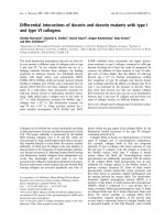
DHAR and MDAR) were differentially affected by the different treatments. Activity of these three enzymes was significantly reduced by addition of atrazine, while sucrose
treatment had an opposite effect and significantly
increased these activities (Fig. 5a, b, c). The tolerance condition (SA) succeeded to limit repressive effects of the herbicide and maintained enzyme activities at the control
level. The fourth enzyme of the ascorbate-glutathione
cycles, GR, did not present any significant variation of
activity between the different treatments (Fig. 5d). Finally,
catalase exhibited slightly lower activity under conditions
of sucrose plus atrazine, when compared to control and
atrazine-containing medium (Fig. 5e).
The repressive effect of atrazine in the absence of sucrose
(MA treatment) on APX global activity was correlated with
a general repression of APX genes (Fig. 5a, Table 5).
Among the six APX genes present in the microarray, the
cytosolic APX1 (At1g07890), the stromal sAPX
(At4g08390) and the chloroplastic APX4 (At4g09010)
genes exhibited important decrease of transcript levels
under conditions of atrazine treatment (MA) compared to
mannitol control, while the other APX genes were not differentially expressed in the presence of atrazine. Whereas
APX4 expression remained downregulated in the presence
of sucrose plus atrazine, this tolerant condition balanced
the repressive effects of atrazine for APX1 and sAPX genes,
which recovered a level of transcript similar to the control.
Finally, and in contrast with global APX activity, the thylakoid-bound tAPX (At1g77490) gene was not affected by
Table 4: Expression of genes potentially encoding H2O2-producing enzymes after 24 hours of treatment.
Log2(ratio)
Accession number
At1g57770
At3g14420
At3g30775
At3g53950
At4g16760
At5g65110
Gene description
Localisation
Treatment comparison
S/M
MA/M
SA/M
Amine oxidase family
(S)-2-hydroxy-acid oxidase, peroxisomal, putative/glycolate
oxidase, putative/short chain alpha-hydroxy acid oxidase, putative
Proline oxidase, mitochondrial/osmotic stressresponsive proline dehydrogenase (POX) (PRO1) (ERD5)
Glyoxal oxidase-related
Acyl-CoA oxidase (ACX1)
Acyl-CoA oxidase (ACX2)
Chloroplast
Peroxisome
nde
-1.08
1.59
1.33
0.80
nde
Mitochondrion
Endomembrane system
Peroxisome
Peroxisome
nde
nde
0,87
0.91
2.51
1.00
nde
nde
1.22
nde
1.48
1.83
Relative expressions of gene are given with their log2(ratio) for sucrose versus mannitol (S/M), mannitol plus atrazine versus mannitol (MA/M) and
sucrose plus atrazine versus mannitol (SA/M) comparison. nde: not differentially expressed. Genes with a Bonferroni P-value higher than 5% were
considered as being not differentially expressed as described by Lurin et al. [85].
Page 9 of 18
(page number not for citation purposes)
BMC Plant Biology 2009, 9:28
/>
Figure 5
Effects of atrazine and sucrose on antioxidative enzyme activities
Effects of atrazine and sucrose on antioxidative enzyme activities. Activities of ascorbate peroxidase (APX) (A),
dehydroascorbate reductase (DHAR) (B), monodehydroascorbate reductase (MDAR) (C), glutathione reductase (GR) (D) and
catalase (CAT) (E) were measured in protein extracts from 3-week-old MS-grown Arabidopsis thaliana plantlets subjected to
subsequent treatment (48 hours) with 80 mM mannitol (M), 80 mM sucrose (S), 80 mM mannitol plus 10 M atrazine (MA) or
80 mM sucrose plus 10 M atrazine (SA). Enzymatic activities are expressed in nkatal/g FW, nkatal corresponds to the amount
of enzymatic activity that catalyzes the transformation of one nmole of substrate per second. Statistical analysis was carried out
as described in Methods.
Page 10 of 18
(page number not for citation purposes)
BMC Plant Biology 2009, 9:28
/>
Table 5: Expression of genes encoding enzymes involved in ascorbate-glutathione cycles after 24 hours of treatment.
Log2(ratio)
Accession number
Gene description
Localisation
Treatment Comparison
S/M
MA/M
SA/M
At1g07890
At1g77490
At3g09640
At4g08390
At4g09010
At4g35000
L-ascorbate peroxidase 1. cytosolic (APX1)
L-ascorbate peroxidase. thylakoid-bound (tAPX)
L-ascorbate peroxidase 2 (APX2)
L-ascorbate peroxidase. stromal (sAPX)
L-ascorbate peroxidase 4 (APX4)
L-ascorbate peroxidase 3 (APX3)
Cytosol
Chloroplast
Cytoplasm
Chloroplast
Chloroplast
Peroxisome
nde
-1.03
nde
1.46
-0.79
nde
-1.92
nde
nde
-1.18
-1.12
nde
nde
-1.08
nde
nde
-1.40
nde
At1g75270
At5g16710
At5g36270
Dehydroascorbate reductase (DHAR2)
Dehydroascorbate reductase (DHAR3)
Dehydroascorbate reductase. putative
Cytoplasm
Chloroplast
Cytoplasm
1.92
nde
0.80
-0.91
nde
nde
2.70
nde
1.20
At1g63940
At3g09940
At3g27820
At3g52880
At5g03630
Monodehydroascorbate reductase (MDAR5)
Monodehydroascorbate reductase (MDAR3)
Monodehydroascorbate reductase (MDAR4)
Monodehydroascorbate reductase (MDAR1)
Monodehydroascorbate reductase (MDAR2)
Chloroplast
Cytoplasm
Cytoplasm
Cytoplasm
Cytoplasm
nde
nde
nde
nde
nde
nde
nde
nde
nde
-1.35
nde
nde
nde
nde
1.11
At3g24170
At3g54660
Glutathione reductase. putative (GR1)
Gluthatione reductase. chloroplast (GR2)
Cytoplasm
Chloroplast
1.15
nde
nde
nde
0.92
nde
Relative expressions of gene are given with their log2(ratio) for sucrose versus mannitol (S/M), mannitol plus atrazine versus mannitol (MA/M) and
sucrose plus atrazine versus mannitol (SA/M) comparison. nde: not differentially expressed. Genes with a Bonferroni P-value higher than 5% were
considered as being not differentially expressed as described by Lurin et al. [85].
atrazine, while sucrose repressed its expression under S
and SA conditions.
Dehydroascorbate reductase (DHAR) is a key component
of the ascorbate recycling system. DHAR recycles dehydroascorbate into ascorbate by using reduced glutathione
as a reductant. Two functional DHAR genes, among three
that are encoded in the Arabidopsis thaliana genome, plus
a putative gene, were represented in the microarray (Table
5). The cytosolic DHAR2 (At1g75270) and putative
DHAR (At5g36270) genes exhibited high induction in the
combined presence of sucrose and atrazine, and, respectively, a slight repression or no variation in the presence of
atrazine in comparison to control condition. In contrast
to the repressive effects of atrazine, which were associated
with a decrease of DHAR activity, the increase of DHAR
transcript levels in the combined presence of sucrose and
atrazine was not associated with an increase of global
DHAR enzyme activity (Fig. 5b).
Reduction of monodehydroascorbate by monodehydroascorbate reductase (MDAR) is also an important step
in ascorbate recycling. Among the five MDAR genes
present in the microarray, only cytosolic MDAR2
(At5g03630) exhibited differential expression patterns
according to the treatment applied. While atrazine
repressed its expression, the protective combination of
sucrose and atrazine upregulated it (Table 5). Atrazine
was also found to decrease global MDAR activity, while
sucrose plus atrazine treatment resulted in maintenance
of MDAR activity relatively to the mannitol control (Fig.
5c).
Glutathione serves as a reductant in oxidation-reduction
processes, such as recycling of oxidised ascorbate by dehydroascorbate reductase [48]. Reduction of oxidised glutathione is catalysed by glutathione reductase (GR), which
requires NADPH. Among the two isoenzymes present in
the microarray, only the cytosolic glutathione reductase
GR1 (At3g24170) was found to be induced by sucroseatrazine and sucrose treatments, while no variation of
expression was detected in the presence of atrazine (Table
5). These variations of expression were not associated
with changes of global GR activity, since no significant difference of activity was observed between treatments (Fig.
5d).
The second way to reduce H2O2 content in cells is activation of catalases (CAT), which catalyse dismutation of
H2O2 into water and oxygen [49]. Little variation of transcript levels was detected for the three catalase isoenzymes
(Table 6). CAT2 (At4g35099) exhibited upregulation by
atrazine stress, while CAT3 was slightly downregulated by
the protective sucrose plus atrazine treatment. In relation
Page 11 of 18
(page number not for citation purposes)
BMC Plant Biology 2009, 9:28
/>
Table 6: Expression of genes encoding enzymes involved in H2O2 scavenging after 24 hours of treatment.
Log2(ratio)
Accession number
Gene description
Localisation
S/M
At1g20620
At1g20630
At4g35090
Catalase 3
Catalase 1
Catalase 2
Peroxisome
Peroxisome
Peroxisome
nde
nde
nde
Treatment comparison
MA/M
SA/M
nde
nde
0.96
-0.74
nde
nde
Relative expressions of gene are given with their log2(ratio) for sucrose versus mannitol (S/M), mannitol plus atrazine versus mannitol (MA/M) and
sucrose plus atrazine versus mannitol (SA/M) comparison. nde: not differentially expressed. Genes with a Bonferroni P-value higher than 5% were
considered as being not differentially expressed as described by Lurin et al. [85].
with these slight changes of transcript levels (Table 6), global catalase activities were found to show little variation
(Fig. 5e).
Discussion
Characterisation of the impact of atrazine on ROS
patterns
ROS patterns appear to depend strongly on the nature and
intensity of stress conditions applied to plants [50]. It is
therefore of great importance to characterise ROS accumulation kinetics associated with a particular stress, and not
to rely on expected effects. Thus, while, as expected, atrazine inhibition of photosystem II was associated with 1O2
accumulation [7] (Fig. 1 and Additional file 1), decrease
of superoxide radical levels and increase of H2O2 levels
were also observed (Figs. 2, 3 and Additional files 2, 3).
This disagreed with the proposed, but experimentally
unproven, accumulation of superoxide radical by triazine
treatment in Arabidopsis leaves [51]. It was however
coherent with inhibition of photosynthetic activity and of
the Mehler reaction, whereby superoxide radical is formed
by reduction of oxygen at the PSI site [52]. Atrazine binding to D1 protein of PSII and inhibition of electron feeding to PSI were indeed likely to decrease superoxide
radical production by blocking the Mehler reaction.
The induction of H2O2 accumulation by atrazine was all
the more surprising as it occurred rapidly after transfer to
atrazine (Fig. 3 and Additional file 3) and in the absence
or in the presence of sucrose, which by itself had a negative effect on H2O2 accumulation. This is, to our knowledge, the first demonstration of rapid in vivo H2O2
accumulation under conditions of atrazine treatment. The
negative effect of sucrose on H2O2 accumulation was consistent with the previously-described repression of protein
and lipid catabolism, including a number of oxidasebased processes, by soluble sugars [18,53]. In contrast,
atrazine by itself was found to induce a number of genes
encoding oxidases, the most highly induced being a gene
encoding a proline oxidase (Table 4). Since this induction
occurred prior to significant impairment of photosystems
and phototrophic growth [18], it could not be ascribed to
a situation of metabolic starvation. Activation of protein
and lipid catabolism and of oxidase-based processes has
been reported to occur under conditions of carbohydrate
limitation or starvation [54,55]. In this context, it was
extremely interesting that, in the presence of exogenous
sucrose, i.e. in a situation of carbohydrate optimum, atrazine was able to induce a number of oxidase-encoding
genes and other genes typical of carbohydrate-limitation
response, such as the gene encoding isovaleryl-CoA dehydrogenase [18,56].
Numerous abiotic stressors, including xenobiotics, are
known to produce oxidative stress in photosynthetic
organisms. This is the case for benzoxazolinone [57], metronidazole, dinoterb [58], acetochlor [59], copper [60],
wounding [61] and high light [6]. Studies on these
stresses mainly focus on the effects of a single ROS and
rarely consider the effects of ROS combination. However,
ROS are chemically distinct and selectively perceived for
the fine control of adjusting antioxidants and photosynthesis to different environmental stress conditions [62].
Indeed, cross-talk between 1O2 and H2O2 has been clearly
demonstrated by Laloi et al. [50], who suggested antagonistic interactions between 1O2 and H2O2 with a reduction
of 1O2-mediated cell death and stress signalling response
by H2O2 content. In contrast, the present condition of
atrazine treatment, which eventually leads to plantlet
death, was characterised by high 1O2, high H2O2 and low
superoxide radical levels. Laloi et al. [63], who described
antagonistic effects between 1O2, and H2O2, modulated
H2O2 levels in Arabidopsis transgenic plants at the plastid
level. It was thus possible that non-antagonistic effects of
H2O2 and 1O2 under conditions of atrazine treatment
were due to differences of ROS localisation.
Finally, among the set of 29 induced transcription factors
that have been characterized as 1O2-specific by Gadjev et
al. [64], only one was slightly induced (data not shown)
during the course of atrazine treatment despite the high
accumulation of singlet oxygen (Fig. 1 and Additional file
1). The analysis of 1O2 responses by Gadjev et al. [64] was
based on studies of the Arabidopsis conditional flu
Page 12 of 18
(page number not for citation purposes)
BMC Plant Biology 2009, 9:28
mutant [65]. It was thus clear that other signals than 1O2
were perceived by atrazine-treated plantlets or that atrazine-induced 1O2 accumulation involved other processes
and responses than flu-mutant-dependent 1O2 accumulation [50,64,65]. However, full characterisation of the signalling events associated with xenobiotic exposure in
plants remains to be carried out.
Impairment of antioxidant defences in the presence of
atrazine
Atrazine-treated plantlets were characterised by low O2.levels and high H2O2 levels, in contrast with sucrosetreated atrazine-tolerant plants, which showed high O2.and high H2O2 levels. These differences of ROS patterns
were associated with striking differences of gene expressions and enzyme activities involved in ROS-scavenging
pathways.
Thus, atrazine sensitivity was associated with down-regulation of key players of H2O2 scavenging. Among the four
enzymes involved in ascorbate-glutathione cycles, which
are essential to remove large amounts of H2O2 generated
by stress [48,66], three enzymes (APX, MDAR and DHAR)
exhibited a significant decrease of global activities in atrazine-treated plantlets. Moreover, this repression was correlated with a global down-regulation of typical
corresponding transcripts (APX1, sAPX, DHAR2, and
MDAR2), which, conversely, have already been shown to
undergo important induction during responses to several
environmental abiotic stresses. APX1, a cytosolic enzyme,
has previously been described as a central component of
the reactive oxygen gene network of Arabidopsis [67].
Involvement of sAPX in response to oxidative stress has
also been reported by transcriptional induction in the
presence of H2O2, methylviologen, FeCl3 or UV treatments in soybean seedlings [68]. Finally, Yoshida et al.
[69] reported the importance of DHAR2 under conditions
of ozone treatment, with higher sensitivity to ozone in a
DHAR2-deficient mutant, probably due to insufficient
recycling of ascorbate.
Consequently, repression of these transcripts and decrease
of the corresponding enzyme activities in the presence of
atrazine might accentuate the effects of H2O2 accumulation by reduction of ascorbate recycling, thus leading to
disruption of antioxidant mechanisms and propagation
of atrazine injuries. It was thus clear that the effects of atrazine at transcript level [18] had actual negative consequences on biochemical defences and could be involved
in xenobiotic sensitivity. This is strong evidence that xenobiotic sensitivity may be linked to gene regulation effects
in plants. Correlatively, the situation of sucrose-induced
tolerance was characterised by the lifting of atrazine
repression, in the case of APX1 and sAPX, or by the induction by sucrose-atrazine combination, in the case of
/>
DHAR2 and MDAR2. These positive effects on transcript
levels were associated with maintenance of the corresponding enzyme activities at control levels. Although
ROS can mediate induction of protective proteins
involved in the stability of specific mRNAs [70], they can
also cause RNA oxidative damages and induce protein
inactivation and degradation [71]. Increase of transcript
levels was therefore likely to be an adaptive response to
ensure protein synthesis under stress conditions resulting
in higher protein turnover.
The decline of O2.- levels in atrazine-treated plantlets,
which, as explained above, could be ascribed to inhibition
of electron transfer through PSI, was associated with a
general repression of transcripts encoding the different
isoenzymes of SOD and with a decrease of the global
activity of this O2.--scavenging enzyme family, thus indicating that atrazine-treated cells responded to the low
superoxide radical situation. Association of low O2.- and
high H2O2 may be a cause for the ill-adapted response of
anti-oxidant defences in atrazine-treated plantlets, thus
suggesting that further work should be carried out on the
adaptation of organisms to fluctuations of ROS combinations.
Mechanisms of sucrose-induced tolerance to singlet
oxygen
In contrast with non-induction of H2O2-scavenging systems, atrazine-treated plantlets seemed to be able to sense
the increase of 1O2 levels and induce some genes potentially involved in 1O2 quenching (Table 1). Thus, atrazinetreated plantlets, in the absence or presence of sucrose
showed increased expression of 4-Hydroxyphenylpyruvate dioxygenase (HPD) gene (At1g06570), which could
be involved in the maintenance of the photoprotective
role of carotenoids. The At5g06690 gene, encoding a chloroplastic thioredoxin, which is a potential 1O2-quencher
[41], was also induced in atrazine-treated plantlet. However, generally, the thioredoxin gene family was negatively
affected by atrazine treatment, with 7 genes among 12 significantly repressed by atrazine. Correlatively, ten of these
twelve genes showed lifting of repression or significant
induction in the combined presence of sucrose and atrazine. Nevertheless, most of these genes encoded extraplastidial thioredoxins or thioredoxins of unknown
localisation (Table 1). The link of thioredoxin gene family
differential expression with efficient 1O2 quenching in the
presence of atrazine plus sucrose (Fig. 1 and Additional
file 1) was thus difficult to ascertain. On one hand, several
studies have shown the efficiency of thioredoxins in maintenance of cellular reductant environment and in cytoprotective mechanisms [37-40]. On the other hand, efficient
1O quenching in the case of PSII inhibition by atrazine
2
would require the involvement of chloroplastic TRXs. Two
TRX genes, At3g06730 and At5g06690, have been
Page 13 of 18
(page number not for citation purposes)
BMC Plant Biology 2009, 9:28
described as encoding chloroplastic TRXs (Table 1).
Expression of these two genes showed contrasted patterns
in the presence of atrazine or in the presence of sucrose
plus atrazine, with At3g06730 being repressed by atrazine, and At5g06690 being induced by atrazine, whereas
the presence of sucrose and atrazine resulted in a return to
baseline levels. Thus, further work would be required to
analyse the physiological significance of this different pattern, and whether the At3g06730 gene product may play
an important role in atrazine responses. Further work
would also be required to characterise the potential
importance of At1g69880 and At2g17420 TRX genes,
which are induced by sucrose and by sucrose plus atrazine, in sucrose-induced tolerance.
Conclusion
Parallel and integrative analysis therefore revealed correlated modifications of ROS patterns, antioxidant biochemical defences, and corresponding transcript markers,
under conditions of atrazine sensitivity and of sucroseinduced tolerance. Atrazine injury was shown to be
related with increased levels of singlet oxygen and hydrogen peroxide in leaves. Sucrose-treated plantlets were able
to sense changing ROS levels and activate efficient
quenching and antioxidant systems, whereas, in the
absence of sucrose protection, atrazine-treated plantlets
failed to develop fully these defence mechanisms. It thus
seemed that atrazine may generate signals that activate
some H2O2-producing pathways, and that impair the
induction and activation of antioxidant defence mechanisms. Further work is needed to characterise completely
the complex signalling events associated with xenobiotic
exposure in plants.
Methods
Plant material and growth conditions
Seeds of Arabidopsis thaliana (ecotype Colombia, Col0)
were surfaced-sterilized in bayrochlore/ethanol (1/1, v/v),
rinsed in absolute ethanol and dried overnight. Germination and growth were carried out under axenic conditions
in square Petri dishes. After seeds were sowed, Petri dishes
were placed at 4°C for 48 h in order to break dormancy
and homogenize germination and transferred to a control
growth chamber at 22°C under a 16 h light period regime
at 85 mol m-2 s-1 for 3 weeks. Growth medium consisted
of 0.8% (w/v) agar in 1× Murashige and Skoog (MS) basal
salt mix (M5519, Sigma-Aldrich) adjusted to pH 5.7.
Plantlets were then transferred to fresh MS agar medium
containing 80 mM mannitol (M, control), 80 mM mannitol and 10 M atrazine (MA, lethal treatment), 80 mM
sucrose (S, sugar treatment) and 80 mM sucrose and 10
M atrazine (SA, tolerance treatment).
/>
Chlorophyll and carotenoid extraction and quantification
Pigments were extracted by pounding aerial parts of seedlings in 80% acetone, and absorbance of the resulting
extracts was measured at 663 nm, 646 nm and 470 nm.
Levels of chlorophyll and total carotenoids (xanthophylls
and carotenes) were determined from the equations given
by Lichtenthaler and Wellburn [72]. Measurements were
done on 3 replicas of 5–10 pooled seedlings each.
Singlet oxygen staining
Three week-old plantlets were transferred for 12, 24, 48 or
72 hours to the different control and treatment media
described above (M, S, MA and SA). Plantlets, prior to the
transfer and at the end of the treatment, were immersed
and infiltrated in the dark under vacuum with a solution
of 100 M Singlet Oxygen Sensor Green® reagent (SOSG)
(S36002, Invitrogen) [31] in 50 mM phosphate potassium buffer (pH 7.5). Infiltrated plantlets were then
placed again on control and treatment media during 30
minutes in the light before being photographed under the
microscope. Following excitation at 480 nm, the fluorescence emission at 530 nm was then detected by an Olympus BX41 spectrofluorometer coupled with a camera. The
presence of red chlorophyll autofluorescence from chloroplasts did not alter the green fluorescence of SOSG. The
infiltration method was chosen in order to measure singlet oxygen levels after the different times of treatment.
Image analysis and quantification of level fluorescence
were performed using the ImageJ software [73]. Experiments were repeated four times on at least 15 plantlets.
Superoxide radical staining
The nitroblue tetrazolium (NBT) (N6876, Sigma-Aldrich)
staining method of Rao and Davis [74] was modified as
follows for in situ detection of superoxide radical. Three
week-old plantlets were transferred for 12, 24, 48 or 72
hours to the different control and treatment media
described above (M, S, MA and SA). Plantlets, prior to the
transfer and at the end of the treatment, were immersed
and infiltrated under vacuum with 3.5 mg ml-1 NBT staining solution in potassium phosphate buffer (10 mM) containing 10 mM NaN3. After infiltration, stained plantlets
were bleached in acetic acid-glycerol-ethanol (1/1/3) (v/
v/v) solution at 100°C during 5 min. Plantlets were then
stored in a glycerol-ethanol (1/4) (v/v) solution until
photographs were taken. O2.- was visualized as a blue
color produced by NBT precipitation. A modified version
of previously described assays for superoxide quantification was used [75,76]. Briefly, NBT-stained plantlets were
ground in liquid nitrogen, the formazan content of the
obtained powder was solubilized in 2 M KOH-DMSO (1/
1.16) (v/v), and then centrifuged for 10 min at 12,000 g.
The A630 was immediately measured, and compared with
a standard curve obtained from known amounts of NBT
Page 14 of 18
(page number not for citation purposes)
BMC Plant Biology 2009, 9:28
in the KOH-DMSO mix. Experiments were repeated four
times on at least 15 plantlets.
Hydrogen peroxide staining
The H2O2 staining agent, 3,3'diaminobenzidine (DAB)
(D5637, Sigma-Aldrich), was dissolved in H2O and
adjusted to pH 3.8 with KOH. The DAB solution was
freshly prepared in order to avoid auto-oxidation [32].
Three week-old plantlets were transferred for 12, 24, 48 or
72 hours to the different control and treatment media
described above (M, S, MA and SA). Plantlets, prior to the
transfer and at the end of the treatment, were immersed
and infiltrated under vacuum with 1.25 mg ml-1 DAB
staining solution. Stained plantlets were then bleached in
acetic acid-glycerol-ethanol (1/1/3) (v/v/v) solution at
100°C during 5 min, and then stored in glycerol-ethanol
(1/4) (v/v) solution until photographs were taken. H2O2
was visualized as a brown color due to DAB polymerization. Quantification of H2O2 contents was determined
using the method of Kotchoni et al. (2006) [77]. The
DAB-stained plantlets were ground in liquid nitrogen. The
resulting powder was homogenized in 0.2 M HClO4, and
then centrifuged for 10 min at 12,000 g. The A450 was
immediately measured and compared with a standard
curve containing known amounts of H2O2 in 0.2 M
HClO4-DAB. Experiments were repeated four times on at
least 15 plantlets. The specificity of DAB staining towards
H2O2 was assessed in control infiltrations in the presence
of 10 mM ascorbic acid.
Enzyme activities
Three week-old plantlets were transferred for 48 hours to
the different control and treatment media described
above (M, S, MA and SA). Whole plantlets (100 mg FW)
were ground in liquid nitrogen to extract total proteins.
The powder obtained was suspended in 500 l of extraction buffer containing 50 mM phosphate buffer (pH 7.5),
1% (w/v) polyvinylpyrrolidone (PVP), 0.5% (v/v) Triton
X-100, 1 mM EDTA and a cocktail of protease inhibitors
(P9599, Sigma-Aldrich). In the specific case of APX activity measurement, the plant powder was suspended in 50
mM Hepes (pH 7) buffer containing 0.5 mM ascorbate,
0.5% (v/v) Triton X-100 and 1% (w/v) PVP. After centrifugation (15 min, 10,000 g), the supernatant was recovered and a second extraction of the pellet was identically
realized. The two supernatants were pooled and constituted the total protein extract that was immediately used
for enzyme activity measurement.
Superoxide dismutase (SOD) activity (EC 1.15.1.1) was
determined using the method of Beauchamp and Fridovich [78] that spectrophotometrically measures inhibition
of the photochemical reduction of nitroblue tetrazolium
(NBT) at 560 nm. One unit of SOD activity was defined as
the amount of enzyme required to inhibit the reduction
/>
rate of NBT by 50%. The reaction mixture contained 50
mM potassium phosphate buffer (pH 7.5), 10 mM
methionine, 2 M riboflavin, 0.1 mM EDTA, 70 M NBT
and enzyme sample. Reactions were carried out at 25°C
under a light intensity of about 120 mol m-2 s-1 for 10
min.
Ascorbate peroxidase (APX) activity (EC 1.11.1.11) was
measured according to Nakano and Asada [79] by monitoring the rate of hydrogen peroxide-dependent oxidation
of ascorbate at 290 nm (E = 2.8 mM-1 cm-1). The reaction
mixture contained 50 mM potassium phosphate buffer
(pH 7), 0.5 mM ascorbic acid, 0.1 mM H2O2, 1 mM EDTA
and enzyme sample.
Dehydroascorbate reductase (DHAR) activity (EC 1.8.5.1)
was measured as described by Hossain and Asada [80].
DHAR was assayed spectrophotometrically by monitoring
the increase in absorbance at 265 nm due to ascorbate formation (E = 14 mM-1 cm-1). The reaction mixture, freshly
prepared in N2-saturated buffer, consisted of 50 mM
potassium phosphate buffer (pH 7), 0.5 mM dehydroascorbate, 5 mM reduced glutathione, 1 mM EDTA
and enzyme sample. Correction was made for non-enzymatic reduction rate of DHA in absence of protein extract.
Monodehydroascorbate reductase (MDAR) activity (EC
1.6.5.4) was measured as described by Hossain et al. [81].
MDAR was assayed spectrophotometrically by following
the decrease in absorbance at 340 nm due to NADH oxidation (E = 6.2 mM-1 cm-1). The reaction mixture consisted of 50 mM buffer TES (pH 7.5), 0.1 mM NADH, 2.5
mM ascorbate, ascorbate oxidase (1 U ml-1) (Curcubita
enzyme (EC 1.10.3.3), A0157, Sigma-Aldrich) and
enzyme sample.
Glutathione reductase (GR) activity (EC 1.6.4.2) was
measured as described by Smith et al. [82] following spectrophotometrically the disappearance of NADPH at 340
nm (E = 6.2 mM-1 cm-1). The reaction mixture contained
50 mM Hepes-NaOH buffer (pH 7.5), 0.5 mM oxidized
glutathione, 0.25 mM NADPH, 0.5 mM EDTA and
enzyme sample.
Catalase (CAT) activity (EC 1.11.1.6) was measured spectrophotometrically at 250 nm by following the disappearance of H2O2 (E = 39.4 mM-1 cm-1) in a reaction mixture
containing 50 mM potassium phosphate buffer (pH 7)
and protein extract. The reaction of dismutation was initiated by the addition of H2O2 (10 mM) as described by
Aebi [83].
Transcriptome profiling
Gene expression data were extracted from the transcriptomic profiling experiment registered as E-MEXP-411 in
Page 15 of 18
(page number not for citation purposes)
BMC Plant Biology 2009, 9:28
/>
ArrayExpress [18,84]. Genes with a Bonferroni P-value
higher than 5% were considered as being not differentially expressed as described by Lurin et al. [85]. Differentially expressed genes are those genes showing at least one
P-value 0.05 after Bonferroni correction, in one of the
MA/M, SA/M or S/M comparisons [18]. This P-value corresponds to genes whose Log2(ratio) was greater than 0.73
or lower than -0.73 (corresponding to 1.6586-fold
changes). This transcriptomic experiment compared the
RNA profiles of three-week-old MS-grown plantlets transferred for 24 hours to the different control and treatment
media described above (M, S, MA and SA).
Patterns of accumulation of superoxide radical.
Detections and quantification have been done on 3-week-old MSgrown Arabidopsis thaliana plantlets subjected to subsequent treatment (12, 24, 48 or 72 hours) with 80 mM mannitol (M), 80 mM
sucrose (S), 80 mM mannitol plus 10 M atrazine (MA) or 80 mM
sucrose plus 10 M atrazine (SA). Superoxide radical content was
expressed as nmoles of reduced NBT per g DW.
Click here for file
[ />
Statistical analysis
Statistical analysis was carried out with the Minitab®
15.1.1.0 software (Minitab SARL, Paris, France). The nonparametrical Mann-Whitney test was used for the different
comparisons of means. Means that were not significantly
different (P > 0.05) show the same letter in graph representations.
Patterns of accumulation of hydrogen peroxide.
Detections and quantification have been done on 3-week-old MSgrown Arabidopsis thaliana plantlets subjected to subsequent treatment (12, 24, 48 or 72 hours) with 80 mM mannitol (M), 80 mM
sucrose (S), 80 mM mannitol plus 10 M atrazine (MA) or 80 mM
sucrose plus 10 M atrazine (SA). Hydrogen peroxide content was
expressed as moles of H2O2 per g DW.
Click here for file
[ />
Additional file 2
Additional file 3
Abbreviations
APX: ascorbate peroxidase; CAT: catalase; DAB: diaminobenzidine; DHAR: dehydroascorbate reductase; GR:
glutathione reductase; H2O2: hydrogen peroxide; HO.:
hydroxyl radical; MDAR: monodehydroascorbate reductase; MS: Murashige and Skoog; NBT: nitroblue tetrazolium; O2: molecular oxygen; 1O2: singlet oxygen; O2.-:
superoxide radical; PSII: photosystem II; ROS: reactive
oxygen species; SOD: superoxide dismutase; SOSG: Singlet Oxygen Sensor Green®; DW: dry weight.
Authors' contributions
FR, CS, MB, IC and GG conceived the study and designed
experiments. FR, MB and GG performed the experiments.
FR, CS, MB, IC and GG carried out analysis and interpretation of experimental data including statistical analyses.
FR, CS, IC and GG wrote the manuscript. All authors read
and approved the final manuscript.
Additional material
Additional file 1
Patterns of accumulation of singlet oxygen.
Singlet oxygen detections using the SOSG probe have been done on 3week-old MS-grown Arabidopsis thaliana plantlets subjected to subsequent treatment (12, 24, 48 or 72 hours) with 80 mM mannitol
(M), 80 mM sucrose (S), 80 mM mannitol plus 10 M atrazine (MA)
or 80 mM sucrose plus 10 M atrazine (SA). Image analysis and
quantification of fluorescence was performed using ImageJ software.
Changes in average intensities are shown as percentage of mean fluorescence intensity of MS-grown plantlets as control.
Click here for file
[ />
Acknowledgements
This work was supported in part by the interdisciplinary program
"Ingénierie écologique" (CNRS, France), by Rennes Métropole (France)
local council and by a fellowship (to F.R.) from the Ministère de l'Enseignement Supérieur et de la Recherche (France).
References
1.
2.
3.
4.
5.
6.
7.
8.
9.
10.
11.
12.
Salin ML: Chloroplast and mitochondrial mechanisms for protection against oxygen toxicity. Free Radic Res Commun 1991,
12–13(Pt 2):851-858.
Mittler R, Vanderauwera S, Gollery M, Van Breusegem F: Reactive
oxygen gene network of plants. Trends in Plant Science 2004,
9(10):490-498.
Dat J, Vandenabeele S, Vranova E, Van Montagu M, Inze D, Van
Breusegem F: Dual action of the active oxygen species during
plant stress responses. Cell Mol Life Sci 2000, 57(5):779-795.
Foyer CH, Noctor G: Leaves in the dark see the light. Science
1999, 284(5414):599-601.
Dalton TD, Shertzer HG, Puga A: Regulation of gene expression
by reactive oxygen. Annu Rev Pharmacol Toxicol 1999, 39:67-101.
Scandalios JG: Oxidative stress responses – what have
genome-scale studies taught us?
Genome Biol 2002,
3(7):REVIEWS1019.
Rutherford AW, Krieger-Liszkay A: Herbicide-induced oxidative
stress in photosystem II.
Trends in Biochem Sci 2001,
26(11):648-653.
Macpherson AN, Telfer A, Barber J, Truscott TG: Direct-detection
of singlet oxygen from isolated photosystem-II reaction
centers. Biochim Biophys Acta 1993, 1143(3):301-309.
Telfer A, Dhami S, Bishop SM, Phillips D, Barber J: -carotene
quenches singlet oxygen formed by isolated photosystem-II
reaction centers. Biochemistry 1994, 33(48):14469-14474.
Solomon KR, Baker DB, Richards RP, Dixon DR, Klaine SJ, LaPoint
TW, Kendall RJ, Weisskopf CP, Giddings JM, Giesy JP, et al.: Ecological risk assessment of atrazine in North American surface
waters. Environ Toxicol Chem 1996, 15(1):31-74.
Clark GM, Goolsby DA, Battaglin WA: Seasonal and annual load
of herbicides from the Mississippi River basin to the Gulf of
Mexico. Environ Sci Technol 1999, 33(7):981-986.
Millie DF, Hersh CM: Statistical characterizations of the atrazine-induced photosynthetic inhibition of Cyclotella
Page 16 of 18
(page number not for citation purposes)
BMC Plant Biology 2009, 9:28
13.
14.
15.
16.
17.
18.
19.
20.
21.
22.
23.
24.
25.
26.
27.
28.
29.
30.
31.
32.
33.
meneghiniana (Bacillariophyta).
Aquat Toxicol 1987,
10(4):239-249.
Hersh CM, Crumpton WG: Atrazine tolerance of algae isolated
from 2 agricultural streams. Environ Toxicol Chem 1989,
8(4):327-332.
Sibony M, Rubin B: Molecular basis for multiple resistance to
acetolactate synthase-inhibiting herbicides and atrazine in
Amaranthus blitoides (prostrate pigweed).
Planta 2003,
216(6):1022-1027.
Sulmon C, Gouesbet G, Binet F, Martin-Laurent F, El Amrani A,
Couee I: Sucrose amendment enhances phytoaccumulation
of the herbicide atrazine in Arabidopsis thaliana. Environ Pollut
2007, 145(2):507-515.
Sulmon C, Gouesbet G, El Amrani A, Couee I: Sugar-induced tolerance to the herbicide atrazine in Arabidopsis seedlings
involves activation of oxidative and xenobiotic stress
responses. Plant Cell Rep 2006, 25(5):489-498.
Sulmon C, Gouesbet G, Couee I, El Amrani A: Sugar-induced tolerance to atrazine in Arabidopsis seedlings: interacting
effects of atrazine and soluble sugars on psbA mRNA and D1
protein levels. Plant Sci 2004, 167(4):913-923.
Ramel F, Sulmon C, Cabello-Hurtado F, Taconnat L, Martin-Magniette
ML, Renou JP, Elamrani A, Couee I, Gouesbet G: Genome-wide
interacting effects of sucrose and herbicide-mediated stress
in Arabidopsis thaliana : novel insights into atrazine toxicity
and sucrose-induced tolerance. BMC Genomics 2007, 8(1):450.
Foyer CH: Prospects for enhancement of the soluble antioxidants, ascorbate and glutathione. Biofactors 2001, 15(2–
4):75-78.
DellaPenna D, Pogson BJ: Vitamin synthesis in plants: Tocopherols and carotenoids. Annu Rev Plant Biol 2006, 57:711-738.
Asada K: Production and scavenging of reactive oxygen species in chloroplasts and their functions. Plant Physiol 2006,
141(2):391-396.
Bowler C, Slooten L, Vandenbranden S, De Rycke R, Botterman J,
Sybesma C, Van Montagu M, Inze D: Manganese superoxide dismutase can reduce cellular damage mediated by oxygen radicals in transgenic plants. EMBO J 1991, 10(7):1723-1732.
Bowler C, Alliotte T, De Loose M, Van Montagu M, Inze D: The
induction of manganese superoxide dismutase in response
to stress in Nicotiana plumbaginifolia. EMBO J 1989, 8(1):31-38.
Perl A, Perltreves R, Galili S, Aviv D, Shalgi E, Malkin S, Galun E:
Enhanced oxidative-stress defense in transgenic potato
expressing tomato Cu, Zn superoxide dismutases. Theor Appl
Genet 1993, 85(5):568-576.
Gupta AS, Heinen JL, Holaday AS, Burke JJ, Allen RD: Increased
resistance to oxidative stress in transgenic plants that overexpress chloroplastic Cu/Zn superoxide-dismutase. Proc Natl
Acad Sci USA 1993, 90(4):1629-1633.
Martin T, Oswald O, Graham IA: Arabidopsis seedling growth,
storage lipid mobilization, and photosynthetic gene expression are regulated by carbon: nitrogen availability. Plant Physiol 2002, 128(2):472-481.
Hideg E, Barta C, Kalai T, Vass I, Hideg K, Asada K: Detection of singlet oxygen and superoxide with fluorescent sensors in
leaves under stress by photoinhibition or UV radiation. Plant
Cell Physiol 2002, 43(10):1154-1164.
Hoffmann A, Hammes E, Plieth C, Desel C, Sattelmacher B, Hansen
UP: Effect of CO2 supply on formation of reactive oxygen species in Arabidopsis thaliana. Protoplasma 2005, 227(1):3-9.
Nakagami H, Soukupova H, Schikora A, Zarsky V, Hirt H: A
mitogen-activated protein kinase kinase kinase mediates
reactive oxygen species homeostasis in Arabidopsis. J Biol
Chem 2006, 281(50):38697-38704.
Kalbina I, Strid A: Supplementary ultraviolet-B irradiation
reveals differences in stress responses between Arabidopsis
thaliana ecotypes. Plant Cell Environ 2006, 29(5):754-763.
Flors C, Fryer MJ, Waring J, Reeder B, Bechtold U, Mullineaux PM,
Nonell S, Wilson MT, Baker NR: Imaging the production of singlet oxygen in vivo using a new fluorescent sensor, Singlet
Oxygen Sensor Green®. J Exp Bot 2006, 57(8):1725-1734.
Fryer MJ, Oxborough K, Mullineaux PM, Baker NR: Imaging of
photo-oxidative stress responses in leaves. J Exp Bot 2002,
53(372):1249-1254.
Thordal-Christensen H, Yangdou Wei ZZ, Collinge DB: Subcellular
localization of H2O2 in plants. H2O2 accumulation in papillae
/>
34.
35.
36.
37.
38.
39.
40.
41.
42.
43.
44.
45.
46.
47.
48.
49.
50.
51.
52.
53.
54.
55.
and hypersensitive response during the barley-powdery mildew interaction. Plant J 1997, 11(6):1187-1194.
Huckelhoven R, Fodor J, Trujillo M, Kogel KH: Barley Mla and Rar
mutants compromised in the hypersensitive cell death
response against Blumeria graminis f.sp hordei are modified in
their ability to accumulate reactive oxygen intermediates at
sites of fungal invasion. Planta 2000, 212(1):16-24.
Lee BH, Lee H, Xiong L, Zhu JK: A mitochondrial complex I
defect impairs cold-regulated nuclear gene expression. Plant
Cell 2002, 14(6):1235-1251.
Dutilleul C, Garmier M, Noctor G, Mathieu C, Chetrit P, Foyer CH,
de Paepe R: Leaf mitochondria modulate whole cell redox
homeostasis, set antioxidant capacity, and determine stress
resistance through altered signaling and diurnal regulation.
Plant Cell 2003, 15(5):1212-1226.
Laloi C, Mestres-Ortega D, Marco Y, Meyer Y, Reichheld JP: The
Arabidopsis cytosolic thioredoxin h5 gene induction by oxidative stress and its W-box-mediated response to pathogen
elicitor. Plant Physiol 2004, 134(3):1006-1016.
Marchand C, Le Marechal P, Meyer Y, Miginiac-Maslow M, IssakidisBourguet E, Decottignies P: New targets of Arabidopsis thioredoxins revealed by proteomic analysis. Proteomics 2004,
4(9):2696-2706.
Wong JH, Balmer Y, Cai N, Tanaka CK, Vensel WH, Hurkman WJ,
Buchanan BB: Unraveling thioredoxin-linked metabolic processes of cereal starchy endosperm using proteomics. FEBS
Letters 2003, 547(1–3):151-156.
Yamazaki D, Motohashi K, Kasama T, Hara Y, Hisabori T: Target
proteins of the cytosolic thioredoxins in Arabidopsis thaliana.
Plant Cell Physiol 2004, 45(1):18-27.
Das KC, Das CK: Thioredoxin, a singlet oxygen quencher and
hydroxyl radical scavenger: Redox independent functions.
Biochem Biophys Res Commun 2000, 277(2):443-447.
Havaux M, Dall'Osto L, Bassi R: Zeaxanthin has enhanced antioxidant capacity with respect to all other xanthophylls in
Arabidopsis leaves and functions independent of binding to
PSII antennae. Plant Physiol 2007, 145(4):1506-1520.
Fryer MJ: The antioxidant effects of thylakoid vitamin-E
(alpha-tocopherol). Plant Cell Environ 1992, 15(4):381-392.
Havaux M, Eymery F, Porfirova S, Rey P, Dormann P: Vitamin E protects against photoinhibition and photooxidative stress in
Arabidopsis thaliana. Plant Cell 2005, 17(12):3451-3469.
Matysik J, Alia , Bhalu B, Mohanty P: Molecular mechanisms of
quenching of reactive oxygen species by proline under stress
in plants. Curr Sci 2002, 82(5):525-532.
Kliebenstein DJ, Monde RA, Last RL: Superoxide dismutase in
Arabidopsis: An eclectic enzyme family with disparate regulation and protein localization.
Plant Physiol 1998,
118(2):637-650.
Apel K, Hirt H: Reactive oxygen species: Metabolism, oxidative stress, and signal transduction. Annu Rev Plant Biol 2004,
55:373-399.
Noctor G, Foyer CH: Ascorbate and glutathione: Keeping
active oxygen under control. Annu Rev Plant Physiol Plant Mol Biol
1998, 49:249-279.
Willekens H, Chamnongpol S, Davey M, Schraudner M, Langebartels
C, VanMontagu M, Inze D, VanCamp W: Catalase is a sink for
H2O2 and is indispensable for stress defence in C-3 plants.
EMBO J 1997, 16(16):4806-4816.
Laloi C, Przybyla D, Apel K: A genetic approach towards elucidating the biological activity of different reactive oxygen species in Arabidopsis thaliana. J Exp Bot 2006, 57(8):1719-1724.
Scott I, Logan DC: Mitochondrial morphology transition is an
early indicator of subsequent cell death in Arabidopsis. New
Phytol 2008, 177(1):90-101.
Asada K, Kiso K, Yoshikawa K: Univalent reduction of molecular
oxygen by spinach chloroplasts on illumination. J Biol Chem
1974, 249(7):2175-2181.
Couée I, Sulmon C, Gouesbet G, El Amrani A: Involvement of soluble sugars in reactive oxygen species balance and responses
to oxidative stress in plants. J Exp Bot 2006, 57(3):449-459.
Brouquisse R, James F, Raymond P, Pradet A: Study of glucose starvation in excised maize root-tips.
Plant Physiol 1991,
96(2):619-626.
Hooks MA, Bode K, Couee I: Regulation of acyl-coa oxidases in
maize seedlings. Phytochemistry 1995, 40(3):657-660.
Page 17 of 18
(page number not for citation purposes)
BMC Plant Biology 2009, 9:28
56.
57.
58.
59.
60.
61.
62.
63.
64.
65.
66.
67.
68.
69.
70.
71.
72.
73.
74.
75.
76.
Däschner K, Couée I, Binder S: The mitochondrial isovalerylcoenzyme A dehydrogenase of Arabidopsis oxidizes intermediates of leucine and valine catabolism. Plant Physiol 2001,
126(2):601-612.
Batish DR, Singh HP, Setia N, Kaur S, Kohli RK: 2-Benzoxazolinone
(BOA) induced oxidative stress, lipid peroxidation and
changes in some antioxidant enzyme activities in mung bean
(Phaseolus aureus). Plant Physiol Biochem 2006, 44(11–12):819-827.
Shao N, Krieger-Liszkay A, Schroda M, Beck CF: A reporter system
for the individual detection of hydrogen peroxide and singlet
oxygen: its use for the assay of reactive oxygen species produced in vivo. Plant J 2007, 50(3):475-487.
Chao L, Zhou QX, Chen S, Cui S, Wang ME: Single and joint stress
of acetochlor and Pb on three agricultural crops in northeast
China. J Environ Sci (China) 2007, 19(6):719-724.
Tewari R, Hahn E-J, Paek K-Y: Modulation of copper toxicityinduced oxidative damage by nitric oxide supply in the
adventitious roots of Panax ginseng. Plant Cell Reports 2008,
27(1):171-181.
Orozco-Cardenas M, Ryan CA: Hydrogen peroxide is generated
systemically in plant leaves by wounding and systemin via the
octadecanoid pathway.
Proc Natl Acad Sci USA 1999,
96(11):6553-6557.
Laloi C, Apel K, Danon A: Reactive oxygen signalling: the latest
news. Curr Opin Plant Biol 2004, 7(3):323-328.
Laloi C, Stachowiak M, Pers-Kamczyc E, Warzych E, Murgia I, Apel K:
Cross-talk between singlet oxygen- and hydrogen peroxidedependent signaling of stress responses in Arabidopsis thaliana. Proc Natl Acad Sci USA 2007, 104(2):672-677.
Gadjev I, Vanderauwera S, Gechev TS, Laloi C, Minkov IN, Shulaev V,
Apel K, Inze D, Mittler R, Van Breusegem F: Transcriptomic footprints disclose specificity of reactive oxygen species signaling
in Arabidopsis. Plant Physiol 2006, 141(2):436-445.
op den Camp RGL, Przybyla D, Ochsenbein C, Laloi C, Kim CH,
Danon A, Wagner D, Hideg E, Gobel C, Feussner I, et al.: Rapid
induction of distinct stress responses after the release of singlet oxygen in Arabidopsis. Plant Cell 2003, 15(10):2320-2332.
Jimenez A, Hernandez JA, delRio LA, Sevilla F: Evidence for the
presence of the ascorbate-glutathione cycle in mitochondria
and peroxisomes of pea leaves.
Plant Physiol 1997,
114(1):275-284.
Davletova S, Rizhsky L, Liang HJ, Zhong SQ, Oliver DJ, Coutu J, Shulaev V, Schlauch K, Mittler R: Cytosolic ascorbate peroxidase 1 is
a central component of the reactive oxygen gene network of
Arabidopsis. Plant Cell 2005, 17(1):268-281.
Moon H, Baek D, Lee B, Prasad DT, Lee SY, Cho MJ, Lim CO, Choi
MS, Bahk J, Kim MO, et al.: Soybean ascorbate peroxidase suppresses Bax-induced apoptosis in yeast by inhibiting oxygen
radical generation.
Biochem Biophys Res Commun 2002,
290(1):457-462.
Yoshida S, Tamaoki M, Shikano T, Nakajima N, Ogawa D, Ioki M,
Aono M, Kubo A, Kamada H, Inoue Y, et al.: Cytosolic dehydroascorbate reductase is important for ozone tolerance in
Arabidopsis thaliana. Plant Cell Physiol 2006, 47(2):304-308.
Chung J-S, Zhu J-K, Bressan RA, Hasegawa PM, Shi H: Reactive oxygen species mediate Na+-induced SOS1 mRNA stability in
Arabidopsis. Plant J 2008, 53(3):554-565.
Xiong Y, Contento AL, Bassham DC: Disruption of autophagy
results in constitutive oxidative stress in Arabidopsis.
Autophagy 2007, 3(3):257-258.
Lichtenthaler HK, Wellburn AR: Determinations of total carotenoids and chlorophylls a and b of leaf extracts in different solvents. Biochem Soc Trans 1983, 11:591-592.
[ />Rao MV, Davis KR: Ozone-induced cell death occurs via two
distinct mechanisms in Arabidopsis: the role of salicylic acid.
Plant J 1999, 17(6):603-614.
Rook GA, Steele J, Umar S, Dockrell HM: A simple method for the
solubilisation of reduced NBT, and its use as a colorimetric
assay for activation of human macrophages by gamma-interferon. J Immunol Methods 1985, 82(1):161-167.
Mookerjee A, Basu JM, Majumder S, Chatterjee S, Panda GS, Dutta P,
Pal S, Mukherjee P, Efferth T, Roy S, et al.: A novel copper complex
induces ROS generation in doxorubicin resistant Ehrlich ascitis carcinoma cells and increases activity of antioxidant
enzymes in vital organs in vivo. BMC Cancer 2006, 6:267.
/>
77.
78.
79.
80.
81.
82.
83.
84.
85.
Kotchoni SO, Kuhns C, Ditzer A, Kirch HH, Bartels D: Overexpression of different aldehyde dehydrogenase genes in
Arabidopsis thaliana confers tolerance to abiotic stress and
protects plants against lipid peroxidation and oxidative
stress. Plant Cell Environ 2006, 29(6):1033-1048.
Beauchamp C, Fridovich I: Superoxide dismutase: improved
assays and an assay applicable to acrylamide gels. Anal Biochem 1971, 44(1):276-287.
Nakano Y, Asada K: Hydrogen-peroxide is scavenged by ascorbate-specific peroxidase in spinach-chloroplasts. Plant Cell
Physiol 1981, 22(5):867-880.
Hossain MA, Asada K: Purification of dehydroascorbate reductase from spinach and its characterization as a thiol enzyme.
Plant Cell Physiol 1984, 25(1):85-92.
Hossain MA, Nakano Y, Asada K: Monodehydroascorbate
reductase in spinach-chloroplasts and its participation in
regeneration of ascorbate for scavenging hydrogen-peroxide. Plant Cell Physiol 1984, 25(3):385-395.
Smith IK, Vierheller TL, Thorne CA: Assay of glutathione-reductase in crude tissue-homogenates using 5,5'-dithiobis(2nitrobenzoic acid). Anal Biochem 1988, 175(2):408-413.
Aebi H: Catalase in vitro. Methods Enzymol 1984, 105:121-126.
[ />Lurin C, Andres C, Aubourg S, Bellaoui M, Bitton F, Bruyere C, Caboche M, Debast C, Gualberto J, Hoffmann B, et al.: Genome-wide
analysis of Arabidopsis pentatricopeptide repeat proteins
reveals their essential role in organelle biogenesis. Plant Cell
2004, 16(8):2089-2103.
Publish with Bio Med Central and every
scientist can read your work free of charge
"BioMed Central will be the most significant development for
disseminating the results of biomedical researc h in our lifetime."
Sir Paul Nurse, Cancer Research UK
Your research papers will be:
available free of charge to the entire biomedical community
peer reviewed and published immediately upon acceptance
cited in PubMed and archived on PubMed Central
yours — you keep the copyright
BioMedcentral
Submit your manuscript here:
/>
Page 18 of 18
(page number not for citation purposes)