báo cáo khoa học: " Removing celiac disease-related gluten proteins from bread wheat while retaining technological properties: a study with Chinese Spring deletion lines" ppt
Bạn đang xem bản rút gọn của tài liệu. Xem và tải ngay bản đầy đủ của tài liệu tại đây (611.74 KB, 12 trang )
BioMed Central
Page 1 of 12
(page number not for citation purposes)
BMC Plant Biology
Open Access
Research article
Removing celiac disease-related gluten proteins from bread wheat
while retaining technological properties: a study with Chinese
Spring deletion lines
Hetty C van den Broeck*
†1
, Teun WJM van Herpen
†1,2,3
, Cees Schuit
1
,
Elma MJ Salentijn
1
, Liesbeth Dekking
4,5
, Dirk Bosch
1
, Rob J Hamer
2
,
Marinus JM Smulders
1,3
, Ludovicus JWJ Gilissen
1,3
and Ingrid M van der
Meer
1,3
Address:
1
Plant Research International, Wageningen UR, PO Box 16, NL-6700 AA Wageningen, The Netherlands,
2
Laboratory of Food Chemistry,
Wageningen UR, PO Box 8129, NL-6700 EV Wageningen, The Netherlands,
3
Allergy Consortium Wageningen, PO Box 16, NL-6700 AA
Wageningen, The Netherlands,
4
Leiden University Medical Center, PO Box 9600, NL-2300 RC Leiden, The Netherlands and
5
Dynomics BV,
Erasmus Medical Centre, Department of Immunology, PO Box 82, NL-1400 AB Bussum, The Netherlands
Email: Hetty C van den Broeck* - ; Teun WJM van Herpen - ; Cees Schuit - ;
Elma MJ Salentijn - ; Liesbeth Dekking - ; Dirk Bosch - ;
Rob J Hamer - ; Marinus JM Smulders - ; Ludovicus JWJ Gilissen - ; Ingrid M van der
Meer -
* Corresponding author †Equal contributors
Abstract
Background: Gluten proteins can induce celiac disease (CD) in genetically susceptible individuals.
In CD patients gluten-derived peptides are presented to the immune system, which leads to a
CD4
+
T-cell mediated immune response and inflammation of the small intestine. However, not all
gluten proteins contain T-cell stimulatory epitopes. Gluten proteins are encoded by multigene loci
present on chromosomes 1 and 6 of the three different genomes of hexaploid bread wheat
(Triticum aestivum) (AABBDD).
Results: The effects of deleting individual gluten loci on both the level of T-cell stimulatory
epitopes in the gluten proteome and the technological properties of the flour were analyzed using
a set of deletion lines of Triticum aestivum cv. Chinese Spring. The reduction of T-cell stimulatory
epitopes was analyzed using monoclonal antibodies that recognize T-cell epitopes present in gluten
proteins. The deletion lines were technologically tested with respect to dough mixing properties
and dough rheology. The results show that removing the α-gliadin locus from the short arm of
chromosome 6 of the D-genome (6DS) resulted in a significant decrease in the presence of T-cell
stimulatory epitopes but also in a significant loss of technological properties. However, removing
the ω-gliadin, γ-gliadin, and LMW-GS loci from the short arm of chromosome 1 of the D-genome
(1DS) removed T-cell stimulatory epitopes from the proteome while maintaining technological
properties.
Conclusion: The consequences of these data are discussed with regard to reducing the load of T-
cell stimulatory epitopes in wheat, and to contributing to the design of CD-safe wheat varieties.
Published: 7 April 2009
BMC Plant Biology 2009, 9:41 doi:10.1186/1471-2229-9-41
Received: 5 November 2008
Accepted: 7 April 2009
This article is available from: />© 2009 Broeck et al; licensee BioMed Central Ltd.
This is an Open Access article distributed under the terms of the Creative Commons Attribution License ( />),
which permits unrestricted use, distribution, and reproduction in any medium, provided the original work is properly cited.
BMC Plant Biology 2009, 9:41 />Page 2 of 12
(page number not for citation purposes)
Background
Celiac disease (CD) is a disorder that is characterized by a
permanent intolerance to gluten proteins from wheat, rye,
and barley. Over 0.5% of the Western population suffers
from CD, which presents itself by chronic diarrhea,
fatigue, osteoporosis, lymphoma, and several other clini-
cal symptoms after prolonged gluten consumption. Until
now, the only treatment is a complete and life long elim-
ination of gluten from the daily diet [1]. In the small intes-
tine, several native gluten peptides can bind directly to
specific human leukocyte antigen (HLA)-DQ2 or DQ8
receptors on antigen presenting cells (APCs). However,
after deamidation by tissue transglutaminase (tTG), the
affinity of the peptides for these HLA-receptors is strongly
increased. The gluten peptides can be presented by APCs
to gluten-sensitive T-cell lymphocytes leading to the
release of cytokines, which will cause inflammation reac-
tions and result in damaged intestinal villi [2].
Gluten are major storage proteins and have many interest-
ing characteristics for food industrial applications, e.g. in
baking bread. Gluten proteins can be divided into three
main groups: high molecular weight glutenin subunits
(HMW-GS), low molecular weight glutenin subunits
(LMW-GS), and gliadins. The HMW-GS are divided in x-
type and y-type subunits [3]. The LMW-GS are divided
into B-, C-, and D-type subunits [4]. Gliadins are divided
into α/β-, γ-, and ω-gliadins [5]. Multiple T-cell activating
gluten peptides were mainly found in α-gliadins, but also
in γ-gliadins and both LMW-GS and HMW-GS [1,2,6,7].
Especially peptides derived from α-gliadins are recognized
by T-cells from most CD patients, while T-cell responses
to γ-gliadins and glutenins are less frequently found [2,7-
10].
Wheat varieties with very low amounts of T-cell stimula-
tory epitopes may be tolerated by many CD-patients
[9,11], while a diet based on wheat varieties reduced in T-
cell stimulatory epitopes may help in the prevention of
CD, as it has been observed that the amount and duration
to gluten exposure is associated with the initiation of CD
[12-14]. Breeding for bread wheat (Triticum aestivum) with
less T-cell stimulatory gluten may result, however, in vari-
eties with unwanted loss of technological properties,
because glutenins and gliadins together contribute largely
to dough quality. A correct mixture of both glutenins and
gliadins is essential to obtain optimal viscoelastic dough
[15], and the quantity and the size distribution of the glu-
ten proteins are important factors for polymerization
[16,17].
Gluten-encoding genes are located on the three homoeol-
ogous genomes of bread wheat: A, B, and D. A few (for
HMW-GS) to a hundred (for α-gliadins) gene copies are
present in hexaploid wheat. Sequences of individual gene
copies within the same gluten family, such as the α-glia-
dins, are very similar and may contain multiple and differ-
ent T-cell stimulatory epitopes [18]. Gluten proteins are
encoded by 15 major loci. The HMW-GS are encoded by
loci on the long arm of group 1 chromosomes (Glu-A1, -
B1, and -D1) [19]. The LMW-GS are mainly encoded by
the Glu-3 loci on the short arms of group 1 chromosomes
(Glu-A3, -B3, and -D3) [20] and are tightly linked to the
loci encoding the γ-gliadins and ω-gliadins (Gli-A1,-B1,
and -D1 and Gli-A3, -B3, and -D3). Most α/β-gliadins are
encoded by loci on the short arms of group 6 chromo-
somes (Gli-A2, B2, and D2) [21].
In this study, deletion lines of Triticum aestivum cv. Chi-
nese Spring (CS) were selected [22-24]. These deletion
lines are generally lacking one locus containing gluten
genes from one of the three homoeologous chromo-
somes. Here, we explore the feasibility to reduce T-cell
stimulatory epitopes in hexaploid bread wheat by screen-
ing with epitope-specific monoclonal antibodies [25-27],
while maintaining the technological properties.
Results
Protein database search
The NCBI protein database search was performed to ana-
lyze the number of proteins that contain the different
sequences recognized by mAb and T-cells. This search pro-
vided insight in how many proteins were expected to con-
tain the different sequences and which different
sequences were present within the proteins. The numbers
of protein sequences that contain the various sequences
involved in the onset of CD that are recognized by T-cells
and mAbs are shown in Table 1. It was observed that the
mAb and T-cell minimal sequences were specific for the
epitopes in each of the expected protein group, with the
exception of the mAb recognizing Glia-α9, whose mini-
mally recognized sequence was also present in a number
of γ- and ω-gliadin proteins. The sequence recognized by
the T-cells was not present within any other protein group
except for the α/β-gliadins. The minimal sequences recog-
nized by mAbs LMW-1 and LMW-2 were more frequently
found in the LMW-GS group than the sequence recog-
nized by the corresponding T-cells. The sequences recog-
nized by mAb and T-cells for HMW-glt was present in
nearly all HMW-GS protein sequences.
SDS-PAGE
To obtain the gluten protein patterns from the CS deletion
lines, gluten proteins were extracted and analyzed by SDS-
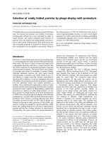
PAGE followed by silver staining. Major differences com-
pared to CS wild type are indicated by boxes in Figure 1.
Differences in gluten protein content compared to CS
wild type were mostly observed in the B-, C- type LMW-GS
and α/β-, γ-gliadin region. Lines with deletions of the
short arms of chromosomes 1D were missing several glu-
ten protein bands in the ω-gliadin/D-type LMW-GS
region. The double deletion line, 1BS-19/6DS-4 (Figure
BMC Plant Biology 2009, 9:41 />Page 3 of 12
(page number not for citation purposes)
1), was missing the largest number of gluten protein
bands because of two deletions in gluten encoding
regions. Unexpected results were obtained for deletion
line 1BS-18, which is the line with the smallest deletion of
chromosome arm 1BS. This line is missing an extra band
compared to the other 1BS deletion lines having larger
deletions. This does not fit with reported results on dele-
tion lines [22,23]. Deletion line 6BS-4 (Figure 1) missed a
gluten protein band that is present in the other deletion
lines of chromosome 6B, even though deletion line 6BS-1
has been reported (WGGRC; Figure 2C) to contain a larger
deletion than 6BS-4. Deletion line 6BS-4 also contains the
5BS-2 deletion, but, to our knowledge, no gluten protein
locus has ever been identified onto the short arm of chro-
mosome 5B. We do not have any explanation for these
discrepancies
Gli-1 deletions
CS deletion lines were analyzed for their contribution to
T-cell stimulatory epitopes by using various mAbs recog-
nizing different T-cell epitopes. In Figure 2A, immunoblot
results are presented using mAbs Glia-α9 and Glia-α20 for
deletion lines of the short arm of chromosomes 1 (Gli-1)
and 6 (Gli-2). Major differences, compared to CS wild
type, are indicated with arrowheads. Deletion lines 1AS-3
and 1AS-1 were missing one gluten protein band by using
mAb Glia-α9 and no gluten protein bands by using mAb
Glia-α20 (Figures 2A and 2B). This suggests that this miss-
ing gluten protein only contains the epitope sequence rec-
ognized by mAb Glia-α9 and the loci encoding these
gluten protein map to bin 1AS3-0.86–1.00 (the terminal
14% of chromosome arm 1AS) (Figure 2C). All five dele-
tion lines of the short arm of chromosome 1B (Figure 2A)
lacked one gluten protein band by using mAb Glia-α9 and
no gluten protein band by using mAb Glia-α20. The dou-
ble deletion line 1BS-19/6DS-4 (Figure 2A) was missing
two extra bands using mAb Glia-α9 and four by using
mAb Glia-α20, which is caused by the 6DS-4 deletion.
Two gluten protein bands were recognized by both mAbs
Glia-α9 and Glia-α20. All 1BS deletion lines (Figure 2A)
lacked the same gluten protein band recognized by mAb
Glia-α9 and because of that the loci encoding correspond-
ing gluten protein map to bin 1BSsat18-0.50–1.00 (Figure
2C). All three deletion lines of the short arm of chromo-
some 1D (Figure 2A) lacked four gluten protein bands by
using mAb Glia-α9 and two gluten protein bands by using
mAb Glia-α20. These missing protein bands correspond
to the boxed (missing) proteins in Figure 1. One gluten
protein band did not completely disappear by using mAb
Glia-α9. This is probably because of the presence of gluten
Table 1: Results of database search for sequences recognized by mAbs and T-cells.
Protein groups
Epitope α/β-gliadins γ-gliadins ω-gliadins/D-type LMW-GS LMW-GS HMW-GS
mAb Glia-α9 (QPFPQPQ) 68 67 3 - -
T-cell Glia-α9 (PFPQPQLPY) 44 - - - -
mAb Glia-α20 (RPQQPYP) 48 - - - -
T-cell Glia-α20 (FRPQQPYPQ) 48 - - - -
mAb LMW-1 (PPFSQQ) - - - 233 -
mAb LMW-2 (QSPF) - - - 163 -
T-cell LMW-glt (PFSQQQQSPF) - - - 21 -
mAb HMW-glt (QGQQGYYP) - - - - 67
T-cell HMW-glt (QGYYPTSPQ) - - - - 65
Number of sequences retrieved 84 93 6 263 67
The number in each cell represents the presence of the recognized sequence by mAb or T-cell within a protein group. '-' in a cell means that the
sequence was not present.
SDS-PAGE analysis of prolamin extracts from Chinese Spring deletion linesFigure 1
SDS-PAGE analysis of prolamin extracts from Chi-
nese Spring deletion lines. CS: Chinese Spring wild type.
Boxes indicate differences in protein bands.
ω-gliadins
D-type LMW-GS
B-, C-LMW-GS
α/β-, γ-gliadins
HMW-GS
kDa
200.0
116.3
97.4
66.2
45.0
31.0
CS
1AS-3
1AS-1
1BS-18
1BS-19/6DS-4
1BS-2
1BS-9
1BS-10
1DS-5
1DS-1
1DS-3
CS
6AS-1
6BS-5
6BS-4
6BS-1
6DS-6
6DS-2
BMC Plant Biology 2009, 9:41 />Page 4 of 12
(page number not for citation purposes)
proteins from different loci but having the same molecu-
lar weights, therefore becoming visible only as one gluten
protein band. The loci encoding the recognized gluten
proteins map to bin 1DS5-0.7–1.0 (the terminal 30% of
1DS) (Figure 2C). The two gluten protein bands recog-
nized by mAb Glia-α20 were the same as recognized by
mAb Glia-α9.
Gli-2 deletions
When analyzing CS deletion lines that are lacking parts of
the short arm of chromosome 6, deletion line 6AS-1 (Fig-
ure 2A) lacked one gluten protein band in immunoblot-
ting using mAb Glia-α9 and two bands by using mAb
Glia-α20. Deletion line 6BS-4 (Figure 2A) lacked one glu-
ten protein band by using mAb Glia-α9, but this was not
the case for the other two 6BS deletion lines, 6BS-1 and
6BS-5 (Figure 2A), which is not consistent with the
reported sizes of the deletions. In the 6BS deletion lines,
no changes were observed in gluten protein bands com-
pared with CS wild type by using mAb Glia-α20 (Figure
2B). These results suggest that the short arm of chromo-
some 6B encodes no gluten proteins containing T-cell
stimulatory epitopes recognized by both mAbs Glia-α9
and Glia-α20, at least not mapping to bin 6BS-0.25–1.00
(terminal 75% of 6BS) (Figure 2C). Deletion line 6DS-2,
the line with the largest deletion (Figures 2A and 2B)
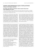
Immunoblot analysis of Chinese Spring deletion lines of the short arm of chromosome 1 and 6Figure 2
Immunoblot analysis of Chinese Spring deletion lines of the short arm of chromosome 1 and 6. (A) Using mAb
Glia-α9. (B) Using mAb Glia-α20. CS: Chinese Spring wild type. Arrowheads indicate absent protein bands. (C) Physical maps
of the short (S) arms of wheat chromosomes 1A, 1B, 1D, 6A, 6B, and 6D from centromer to telomeric ends (Wheat Genetic
and Genomic Resources Centre, Kansas State University, USA). Arrows on the right of each chromosome indicate the dele-
tion lines with their breakpoint (indicated as fraction length from the centromer). The banding patterns within the chromo-
somes are according to Gill et al. [24]
CS
1AS-3
1AS-1
1BS-18
1BS-19/6DS-4
1BS-2
1BS-9
1BS-10
1DS-5
1DS-1
1DS-3
CS
6AS-1
6BS-5
6BS-4
6BS-1
6DS-6
6DS-2
60
50
40
30
A
B
kDa
60
50
40
30
C
centromer
centromer
centromer
centromer
centromer centromer
satellite
satellite
BMC Plant Biology 2009, 9:41 />Page 5 of 12
(page number not for citation purposes)
lacked two gluten protein bands recognized by mAb Glia-
α9 and four bands by mAb Glia-α20. One gluten protein
band has not completely disappeared probably because of
the presence of different gluten proteins having the same
molecular weight within one gluten protein band. The
same gluten protein bands are also absent in the double
deletion line 1BS-19/6DS-4 (Figure 2A). These missing
protein bands correspond to the boxed (missing) proteins
in Figure 1. Hence, the loci encoding these gluten proteins
map to bin 6DS4-0.79–0.99 (Figure 2C).
Glu-3 deletions
The immunoblot results using mAb LMW-2 for the dele-
tion lines of the short arm of chromosome 1 are shown in
Figure 3. One band was observed in all the deletion lines
and in CS wild type without significant differences.
Immunoblot results using mAb LMW-1 showed similar
patterns (results not shown).
Glu-1 deletions
Within the protein database, nearly all HMW-GS had
epitope sequences recognized by mAb HMW-glt. The
immunoblot results for the deletion lines of the long arm
of chromosome 1 using the mAb recognizing HMW-glt
are shown in Figure 4A. In CS wild type, all four HMW
glutenin subunits were detected. No contribution to
HMW-GS was observed for the long arm of chromosome
1A, as expected for a transcriptional silent locus. Two
HMW-GS, 1Bx7 and 1By8, were absent in deletion lines
1BL-1 and 1BL-6. This suggests that the locus encoding
HMW-GS 1Bx7 and 1By8 map to bin 1BL1-0.47–0.69
(Figure 4B). The two HMW-GS, 1Dx2 and 1Dy12, were
absent in deletion line 1DL-4. This suggests that the loci
encoding HMW-GS 1Dx2 and 1Dy12 map to bin 1DL4-
0.18–0.41 (Figure 4B).
Rheological parameters of Chinese Spring deletion lines
The lines with the largest deletions from chromosomes 1
and 6, according to our results, were used for technologi-
cal testing. Parameters among flours of different deletion
lines are presented in Figure 5 and in the Additional file 1:
Rheological parameters.
Total protein content in flour (% w/w) of all deletion lines
was higher compared to CS wild type flour. Especially pro-
tein content in flour of line 6AS-1 was high (20.5%), fol-
lowed by protein content in flour of deletion line 1BS-19/
6DS-4 (18.6%).
The glutenin macro polymer (GMP) content expressed as
volume per mg protein was decreased in deletion line
1BL-1 and was nil in deletion line 1DL-4 (Figure 5 and
Additional file 1: Rheological parameters). GMP repre-
sents the highly aggregated glutenin protein network that
is the prime determinant of dough elastic properties. A
decrease in GMP is therefore expected to lead to a decrease
in dough strength [28-30]. Because of the low amount of
GMP present in flour of the deletion lines 1BL-1 and 1DL-
4, it was impossible to estimate glutenin particle sizes for
these lines. Flours of the two deletion lines, 1BS-10 and
6DS-2, showed a small decrease in GMP volume. For all
other deletion lines, the GMP volume was increased.
Glutenin particle size is a predictor of dough mixing prop-
erties [31]. Average glutenin particle size was increased in
flours of deletion line 1AL-1 and 6AS-1. In deletion lines
Immunoblot analysis of Chinese Spring deletion lines of the short arm of chromosome 1, using mAb LMW-2Figure 3
Immunoblot analysis of Chinese Spring deletion lines
of the short arm of chromosome 1, using mAb LMW-
2. CS: Chinese Spring wild type.
kDa
60
50
40
30
CS
1AS-3
1AS-1
1BS-18
1BS-19/6DS-4
1BS-2
1BS-9
1BS-10
1DS-5
1DS-1
1DS-3
Immunoblot analysis of Chinese Spring deletion lines of the long arm of chromosome 1Figure 4
Immunoblot analysis of Chinese Spring deletion lines
of the long arm of chromosome 1. (A) Using mAb
HMW-glt. CS: Chinese Spring wild type. (B) Physical maps of
the long (L) arms of wheat chromosomes 1A, 1B, and 1D
from centromer to telomeric ends (Wheat Genetic and
Genomic Resources Centre, Kansas State University, US).
Arrows on the right of each chromosome indicate the dele-
tion lines with their breakpoint (indicated as fraction length
from the centromer). The banding patterns within the chro-
mosomes are according to Gill et al [24].
B
centromer
centromer
centromer
A
kDa
120
84
CS
1AL-3
1AL-5
1AL-1
1BL-3
1BL-2
1BL-1
1BL-6
1DL-2
1DL-4
BMC Plant Biology 2009, 9:41 />Page 6 of 12
(page number not for citation purposes)
6DS-2, 6BS-1, 1BS-10, 1BS-19/6DS-4 and 1AS-1 the aver-
age particle size was decreased compared to CS wild type.
Dough made from flours of the two deletion lines 1BL-1
and 1DL-4, lacking HMW-GS, showed a significant
decrease in dough development time (DDT) (Figure 5 and
Additional file 1: Rheological parameters). Dough made
from all other deletion lines showed increase in DDT,
especially the lines with deletions of the short arm of
chromosome 6 (6AS-1, 1BS-19/6DS-4, 6DS-2, and 6BS-1)
and 1AS-1. Deletions of the Gli-2 loci seem to have a sub-
stantial effect on increasing DDT.
Bandwidth at peak resistance (BWPR) is a measure of
dough stability. The BWPR was slightly decreased for dele-
tion line 1DL-4 and was increased for all other deletion
lines compared to CS wild type dough (Figure 5 and Addi-
tional file 1: Rheological parameters). The BWPR was
especially high for deletion lines 6AS-1 and 1BS-19/6DS-
4. It is relevant to note that these are the same deletion
lines having the highest protein content in flour.
Dough elasticity, indicated by relaxation half time (T
1/2
),
was decreased in flours of deletion lines 1BL-1 and 1DL-
4, which lack HMW-GS, and in deletion lines 6BS-1 and
6DS-2 (Figure 5 and Additional file 1: Rheological param-
eters). In contrast, deletion lines 1BS-19/6DS-4 and 1AS-
1, showed an increase in T
1/2
, indicating more elastic
dough [32,33].
To summarize, in Figure 6 immunoblots are shown for
Chinese Spring wild type and the gliadin proteins reacting
with mAbs Glia-α9 and Glia-α20 are numbered. In Table
2 the relation is shown of these proteins together with
their bin-location on the chromosomes and the rheologi-
cal parameters if these proteins are missing in the deletion
lines. Deletions of the long arms of chromosome 1A, 1B,
and 1D are not included because the HMW-GS encoded
by the loci on these arms (1BL and 1DL) seem to be
required for good technological properties.
Discussion
In this study, we examined the possibilities to develop a
bread wheat variety with both reduced levels of T-cell
stimulatory epitopes and good technological properties.
We used a set of Chinese Spring deletion lines that lack
different gluten protein-encoding loci from the group 1
and 6 chromosomes to determine whether reduction in T-
cell stimulatory epitopes can be achieved by removal of
certain gluten protein encoding genes with minimal effect
on the technological properties of bread wheat. Many
cytogenetic resources have been developed in T. aestivum
cv Chinese Spring, which is considered as a model variety
for hexaploid wheat. However, differences among varie-
ties may exist.
CD immunogenic epitopes
On the short arm of the group 6 chromosomes, the gluten
loci that encode α-gliadins are located. The α-gliadins are
considered the most immunogenic concerning both the
adaptive immune response and the innate immune
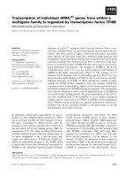
Rheological parameters tested for Chinese Spring deletion linesFigure 5
Rheological parameters tested for Chinese Spring
deletion lines. All technological measurements were per-
formed in duplicate, except the relaxation test (T
1/2
) for dele-
tion lines 1DL-4, 6AS-1, 6DS-2, and 6DS-4/1BS-19. Error
bars represent the standard error. 'NA' means not analyzed
for particle surface area (D
3,2
) because the amount of GMP
was too low.
DDT (min)
0
1
2
3
4
5
BWPR (%)
0
5
10
15
20
25
30
35
40
45
T
½
(sec)
0
10
20
30
40
50
60
70
total protein content in flour (%)
0
4
8
12
16
20
24
GMP volume (μl/mg)
0
2
4
6
8
10
12
14
Particle surface area D3.2 (μm)
0
2
4
6
8
10
12
14
16
18
20
WT 1AL-1 1BL-1 1DL-4 1AS-1 1BS-10 1DS-1 6AS-1 6BS-1 6DS-2 1BS-19/
6DS-4
NA
NA
BMC Plant Biology 2009, 9:41 />Page 7 of 12
(page number not for citation purposes)
response [2,8,10,11]. We observed that the locus on the
short arm of chromosome 6D, mapped to bin 6DS-0.45–
0.99, is responsible for most of the T-cell stimulatory α-
gliadin proteins. These results are in agreement with the
results obtained by Molberg et al. [34] who showed no
decrease in response of DQ2-α-II T-cells for deletion line
6DS-6 and a significant decrease in T-cell response for
deletion lines 6DS-4 and 6DS-2. In addition, results are in
agreement with results of Van Herpen et al. [18], based on
relative presence of CD-epitopes in α-gliadin ESTs from
the three homoeologous loci, and with results of Salentijn
et al. [35] on the presence in cDNAs from two hexaploid
and two tetraploid cultivars. When using mAb Glia-α20 in
immunoblotting also two gluten protein bands were
stained that were encoded by the short arm of chromo-
some 1D. We tentatively assign these as ω-gliadins/D-type
LMW-GS containing the mAb Glia-α20 sequence. Only a
few ω-gliadin proteins have been sequenced so far
because they are difficult to clone due to the presence of
large repetitive domains [36]. It has been shown that ω-
gliadins may have epitopes that are involved in gluten-
sensitive response of CD patients [37,38]. The α-gliadins
encoded by chromosome 6 seem to be related to gliadins
encoded by chromosome 1 from which they might have
originated through gene duplication and/or translocation
[39,40]. Analysis of the minimal sequence recognized by
mAb Glia-α9 indicated that this sequence also occurs in
some γ- and ω-gliadins. Indeed, mAb Glia-α9 recognized
gluten protein bands that disappeared in deletion lines of
the short arm of chromosome 1A, 1B, and 1D (where γ-
and ω-gliadin encoding genes are located). We observed
that genes mapped to bin 1DS-0.48–1.00 had the highest
contribution to the number of T-cell stimulatory epitopes.
Technological properties
Studies have shown that the technological parameters of
wheat flours are influenced by alleles encoding different
HMW-GS [41-44], LMW-GS [45,46], and gliadins [47].
Deleting parts of the short arm of chromosome 1A
resulted in an increased dough development time (DDT)
and volume of glutenin macro polymer (GMP). A
decrease in LMW-GS or gliadins results in a relative
increase of ratios for HMW-GS/LMW-GS or glutenins/
gliadins. Such a change was suggested to increase dough
strength [15,16]. Indeed, we found that removal of the
locus from the short arm of chromosome 1A resulted in
increased dough elasticity. In the deletion lines 1AS-1 and
1DS-1, higher GMP volumes were observed, while in dele-
tion line 1BS-10 a decreased GMP volume was found
together with decreased DDT. On chromosome 1B, also a
Glu-B2 locus is located encoding a B-type LMW-GS
[48,49] and a Glu-B3 locus is located encoding two tightly
linked genes for an ω-gliadin and a B-type LMW-GS [50].
This suggests that LMW-GS encoded by these loci are
important for the formation of the GMP [51,52]. Removal
of the loci could affect the ratios for HMW-GS/LMW-GS or
glutenins/gliadins. Chromosome 1D encodes a D-type
LMW-GS containing a single cysteine residue and there-
fore may act as a chain terminator [53,54]. The absence of
the protein could increase the GMP volume in deletion
line 1DS-1. It would be expected that the GMP volume
would decrease in deletion line 1AS-1 because of removal
of the locus encoding major LMW-GS. We observed, how-
ever, that no T-cell stimulatory epitopes present in LMW-
GS disappeared from the immunoblot using mAbs LMW-
1 and LMW-2, which is possible if expression from the
deleted locus is compensated for by the other two loci
present on the homoeologous chromosomes, for example
by a higher expression of Glu-B3. Compensation behavior
of storage protein synthesis in wheat was observed by
Wieser et al. [55] after inhibition of the expression of α-
gliadins by RNA interference (RNAi). Also Gil-Humanes
et al. [56] recently observed while RNAi reduced the pro-
portion of γ-gliadins by 55–80% and α-gliadins by 63%,
this did not lead to similar reduction in proteins detected
by the sandwich ELISA using the R5 monoclonal anti-
body. The R5 assay was, however, developed for the detec-
tion of gluten proteins from different sources and not
optimized to detect T-cell stimulatory gluten proteins
[57]. Hence, although the R5 assay is currently considered
the standard test for identification of gluten contami-
Numbering of protein bands reacting with mAbs Glia-α9 and Glia-α20 in Chinese Spring wild typeFigure 6
Numbering of protein bands reacting with mAbs
Glia-α9 and Glia-α20 in Chinese Spring wild type.
Immunoblots of Chinese Spring wild type using mAbs Glia-α9
(left) and Glia-α20 (right).
Glia-α9 Glia-α20
Chinese Spring
1
2
3
4
5
6
7
9
8
10
11
12
BMC Plant Biology 2009, 9:41 />Page 8 of 12
(page number not for citation purposes)
nants, we regarded this test unsuitable in the context of
this study.
With respect to technological properties, deletion line
6AS-1 showed an increase in GMP volume and a strong
increase in glutenin particle size. In contrast, deletion
lines 6BS-1 and 6DS-2 showed a decrease in glutenin par-
ticle size and a decrease in GMP volume for deletion line
6DS-2. Gliadins of the α- and γ-type have been identified
to contain an extra cysteine residue that makes them act as
chain terminators. We suggest that the short arm of chro-
mosome 6A in CS is encoding a chain terminating α-glia-
din. The lower content of chain terminators could account
for a larger size of glutenin particles as observed in dele-
tion line 6AS-1. Because of compensation, deletions of the
short arm of chromosome 6B and 6D could lead to an
increased expression of chain terminating α-gliadins
encoded by the short arm of chromosome 6A and result
in observed smaller glutenin particle sizes. The deletions
of the short arm of chromosome 6B and 6D resulted in
stronger dough as shown by increased DDT. This effect on
dough strength is expected because a decrease in α-glia-
dins results in a relative increase of the glutenin/gliadin
ratio. The GMP volume of flour from deletion line 6DS-2
was decreased, which indicates weaker dough, whereas
the DDT was increased, which indicates stronger dough.
Because of this effect, the decrease in GMP volume in dele-
tion line 6DS-2 resulted in decreased elasticity rather than
decreased dough strength.
We observed that technological properties of flour from
deletion lines were strongly affected by the removal of the
different HMW-GS with the strongest effect in deletion
line 1DL-4. Dough strength (as expressed as DDT and
GMP volume) and dough elasticity (T
1/2
) were both
strongly decreased, which is in agreement with published
results [15,58,59]. Deletion of the locus on the long arm
of chromosome 1A resulted in some increase in dough
Table 2: Bin location of gliadin proteins and effect on rheological parameters if absent in deletion lines.
mAb Rheological parameters
Gliadin protein
bands in CS
Glia-α9 Glia-α20 Bin location Total protein
in flour (%)
DDT
a
(min) BWPR
b
(%) T
1/2
c
(sec) GMP
d
volume
(μl/mg)
Particle surface
area (D
3,2
) (μm)
1 yes yes 1DS5-0.70–
1.00
00+0+ 0
2 yes yes 1DS5-0.70–
1.00
00+0+ 0
3 yes no 1DS5-0.70–
1.00
00+0+ 0
4 yes no 1DS5-0.70–
1.00
00+0+ 0
5 yes no 1BSsat0.50–
1.00
00+0- -
6 yes no 1AS3-0.86–
1.00
+++++ 0
7noyes6DS4-0.79–
0.99
+++ -
8 yes yes 6DS4-0.79–
0.99
+++ -
9 yes yes 6DS4-0.79–
0.99
+++ -
10 yes no 6DS4-0.79–
0.99
+++ -
11 yes yes 6AS-0.35–
1.00
+0++ + ++
12 no yes 6AS-0.35–
1.00
+0++ + ++
Chinese Spring wild type 0 0 0 0 0 0
Protein bands are numbered as shown in Figure 6 and their bin location is determined. Bin location is linked to rheological parameters. Results are
compared to Chinese Spring, which is set at "0". "+" in cell means value is up to 50% higher. "++" in cell means value is between 50 to100% higher.
"-" in cell means values is up to 50% lower. " " in cell means value is between 50 to100% lower. "Yes" or "no" in cell means the reaction with the
mAb.
a
Dough development time
b
Band Width at Peak Resistance
c
Flow-relaxation half time
d
Glutenin Macro Polymer
BMC Plant Biology 2009, 9:41 />Page 9 of 12
(page number not for citation purposes)
strength (DDT and GMP volume) and elasticity (T
1/2
). In
addition, glutenin particle sizes were significantly
increased. Both the x-type and y-type encoding genes of
CS at Glu-A1 are silent [19]. In most studies, the silent
locus at Glu-A1 was not found to be important to deter-
mine dough strength compared to non-silent loci [45,46],
so the effect of deletion of the long arm of chromosome
1AL might be because of the absence of other gene prod-
ucts. Based on these results, the Glu-1 loci of CS are con-
sidered inappropriate as a focus to breed for wheat with
less T-cell stimulatory epitopes if technological properties
are to be preserved.
Conclusion
A strategy to breed for bread wheat with less T-cell stimu-
latory gluten epitopes while retaining technological prop-
erties is feasible by focusing on eliminating genes present
on the short arms of chromosome 1D and 6D. This will
result in a wheat variety with highly decreased T-cell stim-
ulatory epitopes. However, eliminating genes might
decrease dough elasticity because of a changed ratio in
glutenin and gliadin proteins. This ratio could be com-
pensated for by the addition of monomeric proteins with
no T-cell stimulatory to the flour, for example from safe
sources like oats, or by the introduction through breeding
or genetic modification of CD-safe gliadin genes. In addi-
tion, wheat varieties with limited but not complete
reduced levels of T-cell stimulatory epitopes may still con-
tribute to lower the gluten load for the entire population
and it may reduce the development of CD in a number of
potential patients.
Methods
Wheat materials
From the Wheat Genetic & Genomic Resources Center
(WGGRC) Kansas State University, USA http://www.k-
state.edu/wgrc/Germplasm/Deletions/del_index.html,
twenty-six T. aestivum Chinese Spring deletion lines were
selected as described [22-24]. The deletion lines had par-
tial deletions of the long and short arms of chromosomes
1 and 6, which was characterized by cytogenetics (Figures
2C and 4B). One line contained deletions of both the
short arm of chromosome 1 and chromosome 6 (1BS-19/
6DS-4, Figure 2C). All deletion lines were grown in con-
tainment glasshouses. No morphological differences were
observed. Seeds were harvested from mature wheat plants.
Database search for the specificity of the sequences
recognized by mAbs compared to T-cell epitopes
The frequency of occurrence of known T-cell epitopes
involved in the onset of CD was analyzed by searching
within the National Center for Biotechnology Informa-
tion (NCBI) database. From the NCBI protein database
/> five different groups of
gluten protein sequences were extracted and subsequently
converted into FASTA formats, using the following search
queries: 'alpha gliadin', 'gamma gliadin', 'omega gliadin'
'D-type LMW-GS', 'LMW glutenin', and 'HMW glutenin'.
All non-Triticum, non-Aegilops entries, and sequences con-
taining less than 100 amino acids were removed. For the
'HMW glutenin' group only full size sequences were ana-
lyzed. The obtained protein sequences were aligned using
ClustalW to validate if the correct groups were assigned to
the sequences. Within the 'gamma gliadin' group, four
sequences (AAA34286, P04729, P04730, and AAA34285)
were more similar to LMW glutenins and were transferred
to the 'LMW glutenin' group. In the 'omega gliadin/D-type
LMW-GS' group, one sequence (ABI20696) was specific
for the 'alpha gliadin' group and was transferred to the
'alpha gliadin' group. The sequences in the five estab-
lished groups were analyzed for the different minimal rec-
ognition sequences of mAbs and T-cells [25]. No
mismatches were allowed. Scores were expressed as the
number of sequences and as the percentage of the
sequences in the established group that contained one or
more recognition sequences. The T-cell minimal recogni-
tion sequences used in the analyses were: Glia-α9
(PFPQPQLPY), Glia-α20 (FRPQQPYPQ), LMW-glt
(PFSQQQQSPF), HMW-glt (QGYYPTSPQ) and mAb
minimal recognition sequences used were: Glia-α9 (QPF-
PQPQ), Glia-α20 (RPQQPYP), LMW-1 (PPFSQQ), LMW-
2 (QSPF), HMW-glt (QGQQGYYP) [25-27,60].
Extraction of gluten proteins
Gluten proteins were extracted from wheat grains accord-
ing to Van den Broeck et al. [61]. Grains were ground in
an analytical mill (A 11 Basic, IKA-Werke) and sieved
through mesh (0.5 mm). Gluten proteins were extracted
from 50 mg wheat flour by addition of 0.5 ml of 50% (v/
v) aqueous iso-propanol with continuous mixing (MS1
Minishaker, IKA Works, Inc.) at 1000 rpm for 30 min at
room temperature, followed by centrifugation at 10,000
rpm for 10 min at room temperature. The residue was re-
extracted twice with 50% (v/v) aqueous iso-propanol,
50mM Tris-HCl, pH 7.5 containing 1% (w/v) DTT, for 30
min at 60°C with mixing every 5 to 10 min followed by
centrifugation at 10,000 rpm for 10 min at room temper-
ature. After addition of each next extraction solution, the
residue was resuspended by shaking in a Fastprep
®
FP220A Instrument for 10 sec at 6.5 m/sec followed by
sonication for 10 min in an ultrasonic bath (Branson
3510, Branson Ultrasonics Corporation). The three
obtained supernatants were combined and considered the
gluten protein extract. The protein content was quantified
using the Biorad Protein Assay (Bio-Rad Laboratories),
based on the Bradford dye-binding procedure, according
to manufacturer's instruction with BSA as a standard.
SDS-PAGE
Gluten proteins were separated on SDS-PAGE gels (10%)
using a SE260 mighty small II system (GE Healthcare,
UK). SDS-PAGE was followed by immunoblotting or by
BMC Plant Biology 2009, 9:41 />Page 10 of 12
(page number not for citation purposes)
silver staining [62] with some modifications. Gels were
fixed in 50% (v/v) ethanol/10% (v/v) acetic acid in water
for 30 min. Then, gels were washed in 5% (v/v) ethanol/
1% (v/v) acetic acid in water for 10 min, followed by three
times washing for 5 min in MilliQ water. Gels were sensi-
tized in 0.02% (w/v) sodium thiosulfate for 1 min and
again washed three times for 30 sec in MilliQ water. Gels
were incubated in 0.1% (w/v) silver nitrate for at least 20
min. After this incubation, gels were rinsed 2 times for 5
sec in MilliQ water and developed in 6% (w/v) sodium
carbonate containing 0.05% (v/v) formaldehyde (37%)/
0.4‰ (w/v) sodium thiosulfate. Development of staining
was stopped by addition of 5% HAc/water.
Immunoblotting
Proteins were blotted onto nitrocellulose (0.2 μm, Bio-
Rad Laboratories), in buffer omitting methanol, using a
Mini Trans-Blot Cell (Bio-Rad Laboratories) at 100 V for 1
hour. Blots were incubated and visualized as described
[63] using mAbs specific for T-cell stimulatory epitopes
against Glia-α9 [26,60], Glia-α20 [25,60], GLT-156
(LMW-1 and LMW-2) [27,60], HMW-glt [26,60]. Mono-
clonal Ab binding was visualized by staining for alkaline
phosphatase, using Nitro Blue tetrazolium (NBT) and 5-
Bromo-4-chloro-3-indolyl phosphate (BCIP) (Sigma).
Quadrumat milling
To obtain white wheat flour, wheat kernels (total weight
ranging 7.6–36 g) were milled using a Quadrumat JR
(Brabender, Germany). Kernel moisture was adjusted to
16.5%. Bran was separated from endosperm flour by siev-
ing through mesh (150 μm). After sieving the average
yield was 50% (w/w), noting that samples 6AS-1 and
6DS-2 had a typically higher flour yield of 64% and 60%,
the other samples ranged from 43% to 51%.
Total protein content in flour
Flour protein content was estimated by the Dumas method
[64] using an NA2100 Nitrogen and Protein Analyzer (Ther-
moQuest-CE Instruments, Rodeno, Italy). The Dumas
method is based on the measurement of total nitrogen in the
sample (N × 5.7). Methionine was used as a standard.
Isolation of glutenin macro polymer from flour and
glutenin particle size analysis
Dough strength is correlated to the amount of the glute-
nin macro polymer (GMP) and to the size of glutenin par-
ticles. Glutenin macro polymer was isolated by dispersing
wheat flour in 1.5% (w/v) SDS followed by ultracentrifu-
gation as described [29]. Fresh GMP from flour was dis-
persed in 1.5% (w/v) SDS (10 ml) by rotating overnight at
room temperature. Particle size distributions were meas-
ured using a Mastersizer 2000 (Malvern Instruments, UK).
The laser diffraction pattern obtained with the instrument
was correlated to the particle size distribution based on
Fraunhofer theory, assuming a spherical particle shape.
The range of the instrument was 0.02–2000 μm. Disper-
sions of GMP were transferred to the water filled sample
vessel at an obscuration of approximately 8%. The surface
area mean (D
3,2
) was used from the particle size distribu-
tion data for comparisons. Further details of this method
are described by Don et al. and Wang et al. [31,65].
Mixing experiments
Dough strength was determined using a micro-Mix-
ograph. A 2 g Mixograph (National Manufacturing Co.,
USA) pin-mixer was used to analyze the mixing properties
of the different flour samples. Mixing was performed at
20°C. Water was added according to the Plastograph
method (ICC 115/1 (ICC, 1992) [66]. Dough contained
2% (w/w) sodium chloride (Merck, Germany). Band-
width at peak resistance (BWPR) in percentages and
dough development time (DDT) in minutes were used
from the midline analysis for comparison.
Flow-relaxation measurements
Relaxation tests were performed to study dough elasticity.
Longer relaxation half times indicate more elastic dough
behavior [32,33]. Dough was mixed to peak in the 2 g
Mixograph pin-mixer, carefully removed from the mixer
and transferred to the Bohlin VOR rheometer (Bohlin
Instruments, Sweden). Flow-relaxation measurements
were performed using an aluminum grooved plate geom-
etry with a cross-section of 30 mm and a gap of 1 mm
[33]. Moisture loss from the dough piece was prevented
using paraffin oil. The actual measurement was performed
after an equilibration time of 30 min to allow appropriate
release of dough stress. The measuring temperature was
20°C. During measurement, the sample was deformed to
a strain of 100% at a shear rate of 0.0208 s
-1
. The strain
was kept constant and the subsequent decrease of stress of
the dough was recorded as a function of time. The time
necessary for the dough to relax to a stress of 50% of the
initial stress, recorded directly after stopping deformation,
was used as the flow-relaxation half time (T
1/2
).
Authors' contributions
IMM and MJMS initiated this study. MJMS and CS selected
Chinese Spring deletion lines. CS and HCB extracted glu-
ten proteins from deletion lines. EMJS and TWJMH per-
formed data base search. LD raised monoclonal
antibodies. HCB performed SDS-PAGE and immunoblot-
ting. TWJMH performed technological tests. IMM super-
vised HCB and CS. RJH, DB, MJMS, and LJWJG supervised
TWJMH. HCB, TJWMH, RJH, MJMS, LJWJG, and IMM
contributed to writing the manuscript. EMJS, LD, and DB
gave editorial comments. All authors read and approved
the final manuscript.
BMC Plant Biology 2009, 9:41 />Page 11 of 12
(page number not for citation purposes)
Additional material
Acknowledgements
We are grateful to W.J. Lichtendonk for discussions on the technological
part of this work and for his help during the technological experiments. This
research was funded by the Celiac Disease Consortium, an Innovative Clus-
ter approved by the Netherlands Genomics Initiative and partially funded
by the Dutch Government (BSIK03009), and by the Dutch Ministry of Agri-
culture, Nature, and Food Safety (project KB-05-001-019-PRI). We thank
the WGGRC Kansas State University, USA, for providing us with Chinese
Spring deletion lines.
References
1. Koning F: The molecular basis of celiac disease. J Mol Recognit
2003, 16:333-336.
2. Vader W, Kooy Y, van Veelen P, de Ru A, Harris D, Benckhuijsen W,
Peña S, Mearin L, Drijfhout JW, Koning F: The gluten response in
children with celiac disease is directed toward multiple glia-
din and glutenin peptides. Gastroenterology 2002, 122:1729-1737.
3. Payne P, Holt L, Law C: Structural and genetical studies on the
high-molecular-weight subunits of wheat glutenin. Theor Appl
Genet 1981, 60:229-236.
4. Jackson EA, Holt LM, Payne PI: Characterization of high molec-
ular-weight gliadin and low-molecular-weight glutenin subu-
nits of wheat endosperm by two-dimensional
electrophoresis and the chromosomal localization of their
controlling genes. Theor Appl Genet 1983, 66:29-37.
5. Woychik JH, Boundy JA, Dimler RJ: Starch gel electrophoresis of
wheat gluten proteins with concentrated urea. Arch Biochem
Biophys 1961, 94:477-482.
6. Sollid LM: Coeliac disease: Dissecting a complex inflamma-
tory disorder. Nat Rev Immunol 2002, 2:647-655.
7. Arentz-Hansen H, Korner R, Molberg Ø, Quarsten H, Vader W,
Kooy Y, Lundin KEA, Koning F, Roepstorff P, Sollid LM, McAdam SN:
The intestinal T-cell response to α-gliadin in adult celiac dis-
ease is focused on a single deamidated glutamine targeted
by tissue transglutaminase. J Exp Med 2000, 191:603-612.
8. Arentz-Hansen H, McAdam SN, Molberg Ø, Fleckenstein B, Lundin
KEA, Jorgensen TJD, Jung G, Roepstorff P, Sollid LM: Celiac lesion
T cells recognize epitopes that cluster in regions of gliadins
rich in proline residues. Gastroenterology 2002, 123:803-809.
9. Vader LW, Stepniak DT, Bunnik EM, Kooy YMC, de Haan W, Drijf-
hout JW, van Veelen PA, Koning F: Characterization of cereal
toxicity for celiac disease patients based on protein homol-
ogy in grains. Gastroenterology 2003, 125:1105-1113.
10. Molberg Ø, Flæte NES, Jensen T, Lundin KEA, Arentz-Hansen H,
Anderson OD, Uhlen AK, Sollid LM: Intestinal T-cell responses to
high-molecular-weight glutenins in celiac disease. Gastroenter-
ology 2003, 125:337-344.
11. Janatuinen EK, Kemppainen TA, Julkunen RJK, Kosma VM, Maki M,
Heikkinen M, Uusitupa MIJ: No harm from five year ingestion of
oats in coeliac disease. Gut 2002, 50:332-335.
12. Fasano A: Systemic autoimmune disorders in celiac disease.
Curr Opin Gastroenterol 2006, 22(6):674-679.
13. Ivarsson A, Persson LA, Nystrom L, Ascher H, Cavell B, Danielsson
L, Dannaeus A, Lindberg T, Lindquist B, Stenhammar L, Hernell O:
Epidemic of coeliac disease in Swedish children. Acta Paediatr
2000, 89:165-171.
14. Ventura A, Magazzù G, Greco L: Duration of exposure to gluten
and risk for autoimmune disorders in patients with celiac dis-
ease. Gastroenterology 1999, 117:297-303.
15. Wieser H: Chemistry of gluten proteins. Food Microbiol 2007,
24:115-119.
16. Gupta RB, Khan K, Macritchie F: Biochemical basis of flour prop-
erties in bread wheats. I. Effects of variation in the quantity
and size distribution of polymeric protein. J Cereal Sci 1993,
18:23-41.
17. Gupta RB, Popineau Y, Lefebvre J, Cornec M, Lawrence GJ,
Macritchie F: Biochemical basis of flour properties in bread
wheats .II. Changes in polymeric protein-formation and
dough/gluten properties associated with the loss of low M
R
or high M
R
glutenin subunits. J Cereal Sci 1995, 21:103-116.
18. van Herpen TWJM, Goryunova SV, Schoot J van der, Mitreva M, Sal-
entijn EMJ, Vorst O, Schenk MF, van Veelen PA, Koning F, van Soest
LJM, Vosman B, Bosch D, Hamer RJ, Gilissen LJWJ, Smulders MJM:
Alpha-gliadin genes from the A, B, and D genomes of wheat
contain different sets of celiac disease epitopes. BMC Genomics
2006, 7:1.
19. Harberd NP, Bartels D, Thompson RD: DNA restriction-frag-
ment variation in the gene family encoding high molecular
weight (HMW) glutenin subunits of wheat. Biochem Genet
1986, 24:579-596.
20. Singh NK, Shepherd KW: Linkage mapping of genes-controlling
endosperm storage proteins in wheat .1. Genes on the short
arms of group-1 chromosomes. Theor Appl Genet 1988,
75:628-641.
21. Marino CL, Tuleen NA, Hart GE, Nelson JC, Sorrells ME, Lu YH,
Leroy P, Lopes CR: Molecular genetic maps of the group 6
chromosomes of hexaploid wheat (Triticum aestivum L. em.
Thell.). Genome 1996, 39:359-366.
22. Endo TR, Gill BS: The deletion stocks of common wheat. J
Hered 1996, 87:295-307.
23. Qi L, Echalier B, Friebe B, Gill B: Molecular characterization of a
set of wheat deletion stocks for use in chromosome bin map-
ping of ESTs. Funct Integr Genomics 2003, 3:39-55.
24. Gill BS, Friebe B, Endo TR: Standard karyotype and nomencla-
ture system for description of chromosome bands and struc-
tural aberrations in wheat (Triticum aestivum). Genome 1991,
34:830-839.
25. Mitea C, Havenaar R, Drijfhout JW, Edens L, Dekking L, Koning F:
Efficient degradation of gluten by a prolyl endoprotease in a
gastrointestinal model: implications for coeliac disease. Gut
2008, 57:25-32.
26. Spaenij-Dekking EHA, Kooy-Winkelaar EMC, Nieuwenhuizen WF,
Drijfhout JW, Koning F: A novel and sensitive method for the
detection of T cell stimulatory epitopes of α/β- and γ-gliadin.
Gut 2004,
53:1267-1273.
27. Spaenij-Dekking L, Kooy-Winkelaar Y, van Veelen P, Drijfhout JW,
Jonker H, van Soest L, Smulders MJM, Bosch D, Gilissen LJWJ, Koning
F: Natural Variation in Toxicity of Wheat: Potential for
Selection of Nontoxic Varieties for Celiac Disease Patients.
Gastroenterology 2005, 129:797-806.
28. Graveland A, Bosveld P, Lichtendonk WJ, Marseille JP, Moonen HHE,
Scheepstra A: A model for the molecular structure of the glu-
tenins from wheat flour. J Cereal Sci 1985, 21:117-126.
29. Graveland A, Bosveld P, Lichtendonk WJ, Moonen HHE, Scheepstra
A: Extraction and fractionation of wheat flour proteins. J Sci
Food Agric 1982, 33:1117-1128.
30. Weegels PL, Pijpekamp AM van de, Graveland A, Hamer RJ, Schofield
JD: Depolymerisation and re-polymerisation of wheat glute-
nin during dough processing. I. Relationships between glute-
nin macropolymer content and quality parameters. J Cereal
Sci 1996, 23:103-111.
31. Don C, Lookhart G, Naeem H, MacRitchie F, Hamer RJ: Heat stress
and genotype affect the glutenin particles of the glutenin
macropolymer-gel fraction. J Cereal Sci 2005, 42:69-80.
32. Lásztity R: The Chemistry of cereal proteins Boca Raton: Taylor & Fran-
cis Ltd; 1995.
33. Lichtendonk WJ, Kelfkens M, Orsel R, Bekkers ACAPA, Plijter JJ: The
impact of water-soluble pentosans on dough properties. In
Wheat Gluten. Proceedings of the Seventh International Gluten Workshop:
2–6 April; Bristol, UK Edited by: Shewry PR, Tatham AS. Cambridge:
Royal Society of Chemistry; 2000:512-518.
34. Molberg Ø, Uhlen AK, Jensen T, Flæte NES, Fleckenstein B, Arentz-
Hansen H, Raki M, Lundin KEA, Sollid LM: Mapping of gluten T-cell
epitopes in the bread wheat ancestors: Implications for
celiac disease. Gastroenterology 2005, 128:393-401.
Additional file 1
Rheological parameters. Rheological parameters of Chinese Spring wild
type and deletion lines.
Click here for file
[ />2229-9-41-S1.doc]
Publish with BioMed Central and every
scientist can read your work free of charge
"BioMed Central will be the most significant development for
disseminating the results of biomedical research in our lifetime."
Sir Paul Nurse, Cancer Research UK
Your research papers will be:
available free of charge to the entire biomedical community
peer reviewed and published immediately upon acceptance
cited in PubMed and archived on PubMed Central
yours — you keep the copyright
Submit your manuscript here:
/>BioMedcentral
BMC Plant Biology 2009, 9:41 />Page 12 of 12
(page number not for citation purposes)
35. Salentijn EMJ, Goryunova SV, Bas N, Meer IM van der, Broeck HC van
den, Bastien T, Gilissen LJWJ, Smulders MJM: Tetraploid and hex-
aploid wheat varieties reveal large differences in expression
of alpha-gliadins from homoeologous Gli-2 loci. BMC Genomics
2009, 10:48.
36. Hassani ME, Shariflou MR, Gianibelli MC, Sharp PJ: Characterisa-
tion of a ω-gliadin gene in Triticum tauschii. J Cereal Sci 2008,
47:59-67.
37. Denery-Papini S, Nicolas Y, Popineau Y: Efficiency and limitations
of immunochemical assays for the testing of gluten-free
foods. J Cereal Sci 1999, 30:121-131.
38. Ensari A, Marsh MN, Moriarty KJ, Moore CM, Fido RJ, Tatham AS:
Studies in vivo of omega-gliadins in gluten sensitivity (coeliac
sprue disease). Clin Sci 1998, 95:419-424.
39. Gu YQ, Crossman C, Kong X, Luo M, You FM, Coleman-Derr D,
Dubcovsky J, Anderson OD: Genomic organization of the com-
plex α-gliadin gene loci in wheat. Theor Appl Genet 2004,
109:648-657.
40. Shewry PR, Tatham AS: The prolamin storage proteins of cereal
seeds – Structure and evolution. Biochem J 1990, 267:1-12.
41. Don C, Mann G, Bekes F, Hamer R: Linking glutenin particles to
HMW subunit composition. In The Gluten Proteins. Proceedings of
the 8th International Gluten Workshop: 8–10 September; Viterbo, Italy
Edited by: Lafiandra D, Masci S, D'Ovidio R. Cambridge: Royal Society
of Chemistry; 2003:188-192.
42. Payne PI, Nightingale MA, Krattiger AF, Holt LM: The relationship
between HMW glutenin subunit composition and the bread-
making quality of British-grown wheat varieties. J Sci Food
Agric 1987, 40:51-65.
43. Lafiandra D, D'Ovidio R, Porceddu E, Margiotta B, Colaprico G: New
data supporting high M
r
glutenin subunit 5 as the determi-
nant of quality differences among the pairs 5 + 10 vs. 2 + 12.
J Cereal Sci 1993, 18:197-205.
44. Popineau Y, Cornec M, Lefebvre J, Marchylo B: Influence of high M
r
glutenin subunits on glutenin polymers and rheological prop-
erties of glutens and gluten subfractions of near-isogenic
lines of wheat Sicco. J Cereal Sci 1994, 19:231-241.
45. Branlard G, Dardevet M, Saccomano R, Lagoutte F, Gourdon J:
Genetic diversity of wheat storage proteins and bread wheat
quality. Euphytica 2001, 119:59-67.
46. Eagles HA, Eastwood RF, Hollamby GJ, Martin EM, Cornish GB: Revi-
sion of the estimates of glutenin gene effects at the Glu-B1
locus from southern Australian wheat breeding programs.
Aust J Agric Res 2004, 55:1093-1096.
47. Van lonkhuijsen HJ, Hamer RJ, Schreuder C: Influence of specific
gliadins on the breadmaking quality of wheat. Cereal Chem
1992, 69:174-177.
48. Liu CY, Shepherd KW: Inheritance of B subunits of glutenin and
ω- and γ-gliadins in tetraploid wheats. Theor Appl Genet 1995,
90:1149-1157.
49. Metakovsky EV, Branlard G, Chernakov VM, Upelniek VP, Redaelli R,
Pogna NE: Recombination mapping of some chromosome 1A-
, 1B-, 1D- and 6B-controlled gliadins and low-molecular-
weight glutenin subunits in common wheat. Theor Appl Genet
1997, 94:788-795.
50. Ruiz M, Carrillo JM: Linkage relationships between prolamin
genes on chromosomes 1A and 1B of durum wheat. Theor
Appl Genet 1993, 87:353-360.
51. Masci S, Lew EJL, Lafiandra D, Porceddu E, Kasarda DD: Character-
ization of low-molecular-weight glutenin subunits in durum-
wheat by reversed-phase high-performance liquid-chroma-
tography and N-terminal sequencing. Cereal Chem 1995,
72:100-104.
52. Masci S, D'Ovidio R, Lafiandra D, Kasarda DD: Characterization of
a low-molecular-weight glutenin subunit gene from bread
wheat and the corresponding protein that represents a
major subunit of the glutenin polymer. Plant Physiol 1998,
118:1147-1158.
53. Masci S, Egorov TA, Ronchi C, Kuzmicky DD, Kasarda DD, Lafiandra
D: Evidence for the presence of only one cysteine residue in
the D-type low molecular weight subunits of wheat glutenin.
J Cereal Sci 1999,
29:17-25.
54. Masci S, Lafiandra D, Porceddu E, Lew EJL, Tao HP, Kasarda DD: D-
glutenin subunits – N-terminal squences and evidence for
the presence of cysteine. Cereal Chem 1993, 70:581-585.
55. Wieser H, Koehler P, Folck A, Becker D: Characterization of
wheat with strongly reduced α-gliadin content. In Proceedings
of the 9th International Gluten Workshop, Gluten Proteins: 14–16 Septem-
ber; San Francisco, USA Edited by: Lookhart GL, Ng PKW. AACC
International; 2007:13-16.
56. Gil-Humanes J, Pistón F, Hernando A, Alvarez JB, Shewry PR, Barro
F: Silencing of γ-gliadins by RNA interference (RNAi) in
bread wheat. J Cereal Sci 2008, 48:565-568.
57. Valdés I, García E, Llorente M, Méndez E: Innovative approach to
low-level gluten determination in foods using a novel sand-
wich enzyme-linked immunosorbent assay protocol. Eur J
Gastroen Hepat 2003, 15:465-474.
58. Wieser H, Kieffer R: Correlations of the amount of gluten pro-
tein types to the technological properties of wheat flours
determined on a micro-scale. J Cereal Sci 2001, 34:19-27.
59. Don C, Mann G, Bekes F, Hamer RJ: HMW-GS affect the proper-
ties of glutenin particles in GMP and thus flour quality. J
Cereal Sci 2006, 44:127-136.
60. Mitea C, Kooy-Winkelaar Y, van Veelen P, de Ru A, Drijfhout JW,
Koning F, Dekking L: Fine specificity of monoclonal antibodies
against celiac disease-inducing peptides in the gluteome. Am
J Clin Nutr 2008, 88:1057-1066.
61. Broeck HC Van den, America AHP, Smulders MJM, Bosch D, Hamer
RJ, Gilissen LJWJ, Meer IM van der: A modified extraction proto-
col enables detection and quantification of celiac disease-
related gluten proteins from wheat. J Chrom B 2009,
877:975-982.
62. Rabilloud T, Carpentier G, Tarroux P: Improvement and simplifi-
cation of low-background silver staining of proteins by using
sodium dithionite. Electrophoresis 1988, 9:288-291.
63. Cordewener JHG, Hause G, Görgen E, Busink R, Hause B, Dons HJM,
Van Lammeren AAM, van Lookeren Campagne MM, Pechan P:
Changes in synthesis and localization of members of the 70-
kDa class of heat-shock proteins accompany the induction of
embryogenesis in Brassica napus L. microspores. Planta 1995,
196:747-755.
64. Sebecic B, Balenovic J: Rapid ecologically acceptable method for
wheat protein content determination – Comparison of
methods. Deut Lebensm-Rundsch 2001, 97:221-225.
65. Wang MW, van Vliet T, Hamer RJ: Interaction of water unex-
tractable solids and xylanase with gluten protein: effect of
wheat cultivar. J Cereal Sci 2005, 41:251-258.
66. Shewry PR, Lookhart GL: Wheat gluten protein analysis St. Paul, MN,
USA: AACC International; 2003.