Tài liệu Báo cáo khoa học: Leptin protects H9c2 rat cardiomyocytes from H2O2-induced apoptosis docx
Bạn đang xem bản rút gọn của tài liệu. Xem và tải ngay bản đầy đủ của tài liệu tại đây (706.3 KB, 9 trang )
Leptin protects H9c2 rat cardiomyocytes from
H
2
O
2
-induced apoptosis
Megumi Eguchi*, Yuantao Liu*, Eyun-Jung Shin and Gary Sweeney
Department of Biology, York University, Toronto, Canada
The rapid increase in the prevalence of obesity to epi-
demic proportions is a serious concern, as obesity is
associated with the development of many complications
including type 2 diabetes, hypertension and heart failure
[1]. Heart failure is a leading cause of mortality in indus-
trialized countries, and is accompanied by progressive
left ventricular remodeling characterized by hypertro-
phy of the myocytes, impaired vascularization in the
heart, abnormal extracellular matrix composition (fibro-
sis) and elevated cardiomyocyte cell death [2]. However,
in addition to increasing the risk for initial myocardial
infarction, obesity may confer protective effects that
limit cardiac remodeling post-infarction, the so-called
obesity paradox [3]. Necrosis was initially viewed as the
major pathway by which cardiomyocytes are lost during
remodeling; however, research in the past 10–15 years
has indicated that apoptosis has important pathophysio-
logical consequences in the development and progres-
sion of heart failure [4,5]. Indeed, the apoptotic rate is
significantly increased (from 0.001% to 0.08%) in the
failing heart [2].
Adipokines, collectively referring to factors derived
from adipose tissue, have attracted tremendous
research interest in recent years as an important mech-
anistic link between obesity and various associated
complications [6]. The circulating adipokine profile is
altered in obese individuals, and it is now clear that
development of heart failure can be directly influenced
by adipokines [1]. Leptin is the product of the obese
(ob) gene, and its main function is to control appetite
and energy expenditure by acting on the hypothalamus
[7]. There is a positive correlation between circulating
leptin concentration and the body mass of an indiv-
idual. This suggests the existence of leptin resistance in
Keywords
apoptosis; caspase; heart failure; leptin;
mitochondria
Correspondence
G. Sweeney, Department of Biology, York
University, Toronto, M3J 1P3 Canada
Fax: +1 416 736 5698
Tel: +1 416 736 2100 ext. 66635
E-mail:
*These authors contributed equally to this
work
(Received 23 January 2008, revised 25
March 2008, accepted 14 April 2008)
doi:10.1111/j.1742-4658.2008.06465.x
Obesity is a known risk factor for induction of myocardial infarction, but,
paradoxically, may also confer a protective effect against subsequent
remodeling leading to heart failure. In this study, we investigated the effect
of leptin, the product of the obese (ob) gene, on cardiomyocyte apoptosis,
a well-characterized component of cardiac remodeling after myocardial
infarction. Exposing H9c2 cells to H
2
O
2
decreased cell viability, and this
was attenuated by pretreating cells with leptin for 1 h, but not 24 h. Leptin
also attenuated the ability of H
2
O
2
to increase phosphatidylserine exposure
and annexin V binding. Further investigation of underlying mechanisms of
leptin’s protective effect demonstrated that the H
2
O
2
-induced decrease in
mitochondrial membrane potential (Y) leading to cytochrome c release was
attenuated by leptin pretreatment, and this was associated with reduced
translocation of the pro-apoptotic Bax protein to the mitochondrial mem-
brane. Finally, leptin prevented H
2
O
2
-induced increases in caspase-3 cleav-
age and activity, although again 24 h leptin pretreatment did not confer
significant protection. In summary, we have demonstrated that acute leptin
pretreatment mediates anti-apoptotic effects in H9c2 rat cardiomyocytes,
which may be of significance in clarifying the direct impact of leptin on the
heart.
Abbreviations
JC-1, 5,5¢,6,6¢-tetrachloro-1,1¢,3,3¢-tetraethylbenzimidazoyl carbocyanide iodide; MTT, 3-(4,5-dimethylthiazol-2-yl)-2,5-diphenyl-tetrazolium
bromide; PS, phosphatidylserine.
3136 FEBS Journal 275 (2008) 3136–3144 ª 2008 The Authors Journal compilation ª 2008 FEBS
the hypothalamus of such individuals; however,
whether the heart is leptin-resistant is still controversial
[8,9]. Leptin’s action is mediated by six isoforms of
leptin receptors [10]. These receptors can be classified
as secreted (Ob-Re), short (Ob-Ra, c, d and f) and
long (Ob-Rb) forms. The adult heart has been shown
to express both long and short ObR isoforms, but pre-
dominantly short forms of the receptor [11], and it has
also been shown that the heart is a site of leptin
production [12]. The local expression of leptin and
its receptors in the heart further suggests that leptin
can potentially affect cardiac function by directly
acting on the heart, and this has been confirmed by
several recent studies [13–15].
Exposure of cardiomyocytes to H
2
O
2
and other reac-
tive oxygen species is increased in the heart, especially
after short ischemia ⁄ reperfusion, and excessive oxida-
tive stress contributes to the pathogenesis of heart fail-
ure [16]. A high circulating leptin concentration is seen
in the majority of obese individuals. In this study, we
have investigated the effects of leptin on H
2
O
2
-induced
cell death in H9c2 cells, the most appropriate in vitro
model of cardiomyocytes currently available. This
was accomplished by analyses of apoptotis [3-(4,5-
dimethylthiazol-2-yl)-2,5-diphenyl-tetrazolium bromide
(MTT) assay and annexin V binding], together with
investigation of the mechanistic role played by the
intrinsic pathway of apoptosis (change in mitochon-
drial membrane potential (Y), cytochrome c release
and caspase-3 activity).
Results
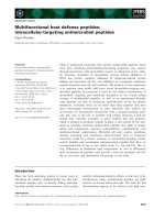
Leptin treatment for 1 h but not 24 h protects
H9c2 cells from H
2
O
2
-induced decreases in cell
viability
The effect of H
2
O
2
treatment on the cell viability of
H9c2 cells was measured by the uptake and reduction
of MTT to an insoluble formazan dye. H
2
O
2
treatment
for 5 h significantly reduced the cell viability as
expected, and this effect was attenuated upon 1 h pre-
incubation with 6 nm leptin but not 24 h pre-incuba-
tion (Fig. 1). The dose of leptin was chosen based on
preliminary experiments, previous work by ourselves
and others in vitro [17,18], and because it is relevant to
the circulating levels observed in obesity [19].
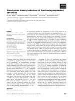
H
2
O
2
-induced phosphatidylserine exposure is
decreased by leptin treatment
Appearance of phosphatidylserine (PS) in the outer
leaflet of the phospholipid bilayer without disrupted
integrity of the membrane is one of the earliest char-
acteristics of apoptotic cells. In order to study the
effect of leptin on H
2
O
2
-induced PS exposure, cells
were incubated with appropriate treatments as indi-
cated, and analyzed for the degree of annexin V
binding to the surface of intact cells (Fig. 2A,B).
Cells were counterstained with propidium iodide to
allow distinction between apoptosis and necrosis.
Leptin treatment alone did not affect PS exposure,
but an increase in annexin V binding was observed
after as little as 2 h H
2
O
2
treatment. No increase in
propidium iodide staining was apparent under these
conditions, but was seen in positive control experi-
ments (data not shown), indicating that the cell
death was predominantly due to apoptosis. Quanti-
tative assessment of fluorescence (Fig. 2C) showed
that 1 h leptin pretreatment significantly attenuated
the level of annexin V binding detected in response
to H
2
O
2
. Although apparently decreasing the effects
of H
2
O
2
, 24 h leptin pretreatment did not have a
significant effect.
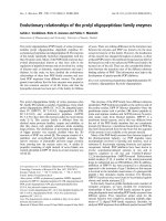
Leptin pretreatment attenuates H
2
O
2
-induced
loss of mitochondrial membrane potential
The mitochondrial membrane potential (Y) is a critical
factor in maintaining the integrity of mitochondria
and subsequent regulation of apoptosis. Loss of mito-
chondrial membrane potential will lead to release of
the cytochrome c from mitochondria, which in turn
0 1 24 0 1 24
Leptin (h)
H
2
O
2
–+++––
Cell viability
(fold over control)
0
0.2
0.4
0.6
0.8
1
1.2
*
Fig. 1. Leptin pretreatment for 1 h but not 24 h attenuates the abil-
ity of H
2
O
2
to decrease cell viability. H9c2 cells were treated with
or without 6 n
M leptin for 1 h or 24 h prior to exposure to H
2
O
2
(400 lM) for 5 h, and cell viability was measured using the MTT
assay. Data represent mean ± SEM (n = 4). The asterisk indicates
a statistically significant difference from H
2
O
2
treatment alone
(P < 0.05).
M. Eguchi et al. Regulation of cardiomyocyte apoptosis by leptin
FEBS Journal 275 (2008) 3136–3144 ª 2008 The Authors Journal compilation ª 2008 FEBS 3137
activates downstream caspases to cause apoptosis
[20,21]. 5,5¢,6,6¢-tetrachloro-1,1¢,3,3¢-tetraethylbenzimi-
dazoyl carbocyanide iodide (JC-1) accumulates as
aggregates in the normal hyperpolarized mitochondria,
resulting in red fluorescence, but JC-1 exists in the
monomeric form in apoptotic cells and stains cells
green. Here we observed that untreated control cells
exhibit numerous brightly stained mitochondria that
emit red fluorescence (Fig. 3). Cells treated with H
2
O
2
exhibited fewer red JC-1 aggregates, and more green
fluorescence of monomers appeared in the cytoplasm,
indicating dissipation of the mitochondrial membrane
potential. Leptin pretreatment attenuated these H
2
O
2
-
induced changes (Fig. 3).
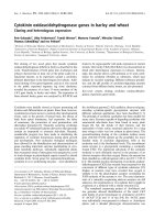
Leptin pretreatment reduces cytochrome
c release from mitochondria
Release of cytochrome c from mitochondria is a criti-
cal step in progression of the intrinsic apoptotic path-
way [20,21]. H
2
O
2
treatment for 2 h increased the
release of cytochrome c from mitochondria, as can be
seen by the loss of co-localization of cytochrome c and
mitochondria (Fig. 4). The effect of H
2
O
2
was again
attenuated by preincubation with leptin for 1 h
(Fig. 4). Co-localization was unaffected in cells treated
with leptin alone.
The mechanism whereby leptin attenuates the
intrinsic pathway of apoptosis involves reduced
Bax integration in the mitochondrial membrane
To assess translocation of the pro-apoptotic Bax pro-
tein to the mitochondrial membrane, we utilized an
approach exploiting the observation that the N-termi-
nal domain is only exposed and recognized by a spe-
cific antibody when this protein translocates and
integrates into the membrane [22]. In viable control
cells, or those treated with leptin, little or no Bax
immunofluorescence was observed (Fig. 5). However,
when cells were exposed to H
2
O
2
, we observed pro-
nounced staining for Bax, with a maximal effect after
4 h, and this was clearly attenuated in cells pretreated
with leptin for 1 h (Fig. 5).
A
B
C
Fig. 2. H
2
O
2
-induced annexin V binding to
the cell surface decreases with leptin
pretreatment. Phosphatidylserine externali-
zation was assessed via annexin V binding
in the absence (A) or presence (B) of 2 h
H
2
O
2
(400 lM) treatment with or without
leptin pretreatment (6 n
M, 1 h or 24 h). Cells
were treated to allow detection of both ann-
exin V (green) and propidium iodide (red),
and images representative of those obtained
for at least eight independent experiments
are shown for each condition. The results
from all experiments (n > 3) were quanti-
fied, and (C) shows the mean fluorescence
(±SEM). The asterisk indicates a significant
difference compared with H
2
O
2
alone
(P < 0.05).
Regulation of cardiomyocyte apoptosis by leptin M. Eguchi et al.
3138 FEBS Journal 275 (2008) 3136–3144 ª 2008 The Authors Journal compilation ª 2008 FEBS
H
2
O
2
-induced increases in caspase-3 cleavage
and activity are attenuated by leptin
pretreatment
Caspase-3 is an executioner of apoptosis, and is
involved in many important events that lead to the
completion of apoptosis [23]. Cleavage of caspase-3 is
indicative of activation, and in cells treated with leptin
alone there was no change in the cleavage of caspase-3
compared to control. H
2
O
2
treatment increased gener-
ation of the cleaved form of caspase-3, and this was
attenuated by 1 h leptin pretreatment (Fig. 6A). The
levels of cleaved caspase-3 correlated well with enzy-
matic activity, which was increased 1.8-fold compared
to control upon H
2
O
2
treatment. This effect of H
2
O
2
was again significantly reduced by leptin 1 h pretreat-
ment, but not significantly by 24 h pretreatment
(Fig. 6B). In order to determine the functional conse-
quences of the above findings, we examined whether
the protective effect of leptin on cell viability was
observed after a prolonged time period subsequent to
H
2
O
2
exposure. When the number of living cells, as
determined by trypan blue exclusion, was counted
three days after exposure to H
2
O
2
, over 2.2-fold more
cells were viable when pretreated with leptin for 1 h as
opposed to exposure to H
2
O
2
alone (data not shown).
Discussion
There has been great interest in the relationship
between circulating leptin levels and the development
of cardiovascular diseases, but the precise role of leptin
is still controversial [24]. Hyperleptinemia, which is
commonly seen in obese individuals, has been pro-
posed to play a role in the development of various car-
diovascular diseases [25,26]. Heart failure is a common
end-stage event resulting from various cardiovascular
diseases, and it is now well established that cardiomyo-
cyte apoptosis is an important component of cardiac
remodeling, ultimately leading to heart failure. An
excellent recent study suggested that leptin can prevent
the increased levels of apoptosis observed upon ageing
in ob ⁄ ob mice [13]. However, the direct effect of leptin
on cardiomyocyte apoptosis and the intracellular
mechanisms involved are still unclear.
H9c2 cells, together with use of H
2
O
2
to induce apop-
tosis, have been used on many occasions as a model
system to study regulation of cardiomyocyte cell death
[27–29]. Here we used this model system to show the
effects of short-term (1 h) and long-term (24 h) exposure
of H9c2 cells to leptin on H
2
O
2
-induced cell death. Our
results indicate that 1 h pretreatment with leptin is able
to significantly decrease the apoptotic effects of H
2
O
2
on H9c2 cells and thus protect them from death. How-
ever, when 24 h preincubation was used, a protective
effect was not observed. This is not entirely without
precedent, as we have previously shown that acute and
chronic leptin treatments have distinct effects on insulin
signaling and subsequent regulation of glucose uptake
in skeletal muscle cells [30,31]. These results suggest that
transient intracellular effects stimulated by acute leptin
treatment play an important role in the cardioprotective
role of leptin, and that the enhanced lipid accumulation
found after 24 h treatment with leptin [18] may convey
deleterious effects [32,33]. The effects observed after a
short period of leptin exposure may be of physiological
relevance given the fact that circulating leptin levels
fluctuate with diurnal rhythm and are not consistently
high for 24 h [34].
We have shown here that leptin’s cardioprotective
effect against H
2
O
2
-induced apoptosis occurs through
the prevention of activation of apoptotic markers at
an early stage, including PS exposure to the outer
membrane – one of the first detectable signs of apopto-
sis [35]. The lack of significant propidium iodide stain-
ing in our annexin V binding studies suggests that 2 h
treatment with H
2
O
2
does not induce significant necro-
sis in these cells. Furthermore, upon investigation of
the mechanisms underlying H
2
O
2
-induced apoptosis
and their regulation by leptin, we observed changes in
Fig. 3. Leptin attenuates H
2
O
2
-induced mitochondrial membrane
potential loss in H9c2 cells. Quiescent H9c2 cells with or without
1 h leptin (6 n
M) pretreatment were exposed to 0.4 lM H
2
O
2
for
30 min. JC-1 fluorescence was measured by confocal microscopy,
assessing the emission shift from green (530 nm) to red (590 nm)
using 488 nm excitation. Composite red and green fluorescence is
shown. Results are representative of those from three separate
experiments.
M. Eguchi et al. Regulation of cardiomyocyte apoptosis by leptin
FEBS Journal 275 (2008) 3136–3144 ª 2008 The Authors Journal compilation ª 2008 FEBS 3139
major components of the intrinsic pathway of apopto-
sis. Notably, the mechanism whereby leptin prevents
activation of the intrinsic pathway of apoptosis
appears to involve prevention of the H
2
O
2
-induced
change in the cellular localization and activity level of
the pro-apoptotic Bax protein [36] detected by immu-
nofluorescence microscopy using a conformation-sensi-
tive antibody [22]. Accordingly, attenuation of a
decrease in mitochondrial membrane potential, and of
the subsequently increased cytochrome c release and
caspase-3 activation was also observed in cells pre-
treated with leptin.
The theory of selective leptin resistance occurring in
obese individuals has been suggested based upon
observations that, while the effects of leptin on satiety
and energy metabolism were blunted, the sympatho-
excitatory effects were maintained in obese individuals
[9,37]. Whether enhanced or suppressed myocardial
leptin action, either direct or centrally mediated,
exists pre- and post-myocardial infarction in obese
MergedCytochrome c Mito Tracker
A
B
C
D
Fig. 4. Leptin decreases H
2
O
2
-induced cyto-
chrome c release from mitochondria. Confo-
cal analysis of H9c2 cells treated with or
without leptin (6 n
M, 1 h) prior to exposure
to H
2
O
2
(400 lM) for 2 h shows immuno-
staining of cytochrome c (green), Mitotrac-
ker staining of mitochondria (red), and
merged images of the two showing co-local-
ization in yellow. Upon release of cyto-
chrome c from mitochondria, green
fluorescence can be seen independently. (A)
Control, (B) leptin treatment for 1 h, (C)
H
2
O
2
treatment, (D) H
2
O
2
treatment with
1 h leptin pretreatment. Images shown
are representative of four independent
experiments.
Fig. 5. Leptin attenuates H
2
O
2
-induced exposure of the Bax N-ter-
minus. Immunofluorescence staining (green) of Bax using Bax
N-terminal (N20) antibody, which only detects Bax localized in mito-
chondrial membrane. The results are for cells after 4 h exposure to
H
2
O
2
(400 lM) with or without leptin pretreatment (6 nM, 1 h).
Images are representative of three independent experiments.
Regulation of cardiomyocyte apoptosis by leptin M. Eguchi et al.
3140 FEBS Journal 275 (2008) 3136–3144 ª 2008 The Authors Journal compilation ª 2008 FEBS
individuals is still a matter of some debate. Our study
clearly indicates a direct, as opposed to systemic or
centrally mediated, role for leptin in mediating cardio-
myocyte apoptosis, and reinforces data from in vivo
studies suggesting a cardioprotective role for leptin via
mediation of anti-apoptotic effects. As mentioned
above, leptin- or leptin receptor-deficient rodents dis-
play an increased rate of cardiac apoptosis. The
increase in apoptotic rate and mortality was abolished
upon leptin injection in ob ⁄ ob mice but not db ⁄ db
mice, indicating that leptin plays an important role in
cardioprotection [13]. Furthermore, it has recently
been shown that perfusion of the heart with leptin dur-
ing a short reperfusion period (35 min) significantly
decreased mitochondrial membrane pore opening and
the infarct size induced by ischemia ⁄ reperfusion [38].
In summary, our current in vitro study suggests that
leptin exerts a protective effect against H
2
O
2
-induced
apoptosis in H9c2 rat cardiomyocytes by preventing
activation of components of the mitochondrial-depen-
dent intrinsic pathway of apoptosis. This is in keeping
with other recent data [13], butt the effect mediated by
leptin in vivo may depend on the development of leptin
resistance, the stage in progression of heart failure or
other variables. Overall, the direct influence of leptin
on cardiac structure and function is still uncertain, but
appears to be of growing importance.
Experimental procedures
Culture of H9c2 rat cardiomyocytes
The rat embryonic ventricular myocardial cell line H9c2
was maintained as described previously [39] in DMEM with
4.5 gÆL
)1
glucose supplemented with 10% (v ⁄ v) fetal bovine
serum and 1% penicillin ⁄ streptomycin (v ⁄ v). Cells were
routinely grown to 80% confluence in 75 cm
2
flasks at
37 °C with an atmosphere of 5% CO
2
prior to passage and
seeding for experiments. All cell-culture materials were pur-
chased from Wisent (Quebec, Canada). For the induction
of cell death, cells were exposed to H
2
O
2
(400 lm, Sigma-
Aldrich, St Louis, MO, USA) for various time periods as
indicated following treatment with leptin (6 nm). We
analyzed ObR expression in these cells by PCR, and found
expression of both long (ObRb) and short (ObRa) receptor
isoforms (data not shown).
Determination of cell viability
The MTT assay was performed as described previously [40]
as a measure of cell viability. In addition, trypan blue
exclusion was used in some experiments, and the number of
trypan blue-negative cells was counted using a hemocytom-
eter 3 days after the end of H
2
O
2
treatment.
Annexin V binding assay
Annexin V Alexa Fluor 488 (Molecular Probes, Eugene,
OR, USA) was used to detect PS exposure to the outer sur-
face of the cell membrane according to the manufacturer’s
protocol. Briefly, cells were grown in a 12-well plate with
cover slips in each well. They were treated with H
2
O
2
follow-
ing incubation with leptin. Then the cells were washed with
cold NaCl ⁄ P
i
and 1· binding buffer (10 mm Hepes pH 7.4,
140 mm NaCl, 2.5 mm CaCl
2
). Cells were then incubated
with annexin V Alexa Fluor 488 (1 : 20 dilution) and
1 lgÆmL
)1
propidium iodide diluted in 1· binding buffer for
15 min. After incubation, cells were washed twice in 1· bind-
ing buffer before mounting the cover slips on glass slides
using DAKO fluorescent mounting medium (DakoCytoma-
tion, Missisauga, Canada). Annexin V Alexa Fluor 488 was
H
2
O
2
Leptin (24h)
+
+
+
+
–
–
– –
Cleaved
caspase 3
To ta l
caspase 3
Leptin (1h)
H
2
O
2
+
+
+
+
–
–
– –
Cleaved
caspase 3
To ta l
caspase 3
0
0.5
1
1.5
2
2.5
H
2
O
2
Leptin (h)
+ + +
–
0
– –
0
1
24 24 1
Caspase 3 activity
(fold above control)
*
A
B
Fig. 6. H
2
O
2
-induced cleavage and activa-
tion of caspase-3 are reduced in leptin pre-
treated cells. (A) Representative western
blots of cell lysates prepared after H
2
O
2
treatment (400 lM, 4 h) with or without lep-
tin pretreatment (6 n
M, 1 h or 24 h). Levels
of the cleaved form of caspase-3
(17 ⁄ 19 kDa) as well as changes in total cas-
pase-3 levels (35 kDa) were analysed by
western blotting. (B) Quantitative analysis of
the activity of caspase-3 measured using a
specific caspase-3 activity assay kit (mean-
s ± SEM, n = 3). The asterisk indicates a
statistically significant difference from H
2
O
2
treatment alone (P < 0.05).
M. Eguchi et al. Regulation of cardiomyocyte apoptosis by leptin
FEBS Journal 275 (2008) 3136–3144 ª 2008 The Authors Journal compilation ª 2008 FEBS 3141
excited at 495 nm, and the fluorescence of cells was deter-
mined using a confocal microscope (Olympus Fluoview
Center Valley, PA, USA). Quantification was performed by
analyzing the fluorescence intensity per cell, and the data
shown are means ± SEM of all experiments, in which two
cover slips were used per condition and nine fields of view
from each cover slip were quantified.
Immunofluorescent detection of conformational
changes in Bax (N-terminal exposure) by confocal
microscopy
For analysis of Bax immunofluorescence, cells grown on
cover slips were washed twice with NaCl ⁄ P
i
, fixed with 4%
paraformaldehyde in NaCl ⁄ P
i
for 15 min, permeabilized
with 0.2% Triton X-100 for 5 min and blocked using 3%
BSA in NaCl ⁄ P
i
for 1 h at room temperature. Cells were
then incubated for 1 h at 37 °C with anti-Bax N-terminal
IgG (Santa Cruz Biotechnology, Santa Cruz, CA, USA;
1 : 150) in blocking buffer. The unique feature of this assay
is that the N-terminal epitope is not detected when Bax is
retained in the cytosol, but is exposed and detected upon
Bax insertion into the mitochondrial membrane [22]. The
cells were washed three times in NaCl ⁄ P
i
, and incubated
for 1 h at room temperature in anti-rabbit IgG Alexa
Fluor 488 serum (Molecular Probes; 1 : 2000). After wash-
ing, cells were mounted using DAKO mounting medium
and confocal images were analysed as above.
Measurement of mitochondrial membrane
potential (Y) using JC-1
H9c2 cells were grown on cover-slips, treated as indicated
in Fig. 3, and then washed twice with NaCl ⁄ P
i
. The cells
were incubated with 5 lm JC-1 dye (Molecular Probes) in
serum-free medium for 15 min at 37 °C. The medium was
then removed, and the cells were washed three times with
NaCl ⁄ P
i
. The cells were examined immediately under a con-
focal microscope. JC-1 fluorescence was measured to assess
the emission shift from green (530 nm) to red (590 nm) in
polarized mitochondria at 488 nm excitation.
Immunofluorescent detection of intracellular
cytochrome c localization by confocal microscopy
To detect cytochrome c release from the mitochondria, cells
grown on cover slips were first treated to stain mitochondria
by incubation for 10 min at room temperature with 10 nm
MitoTracker CMTMRos dye (Molecular Probes) in
NaCl ⁄ P
i
. Cells were fixed with 4% paraformaldehyde for
15 min, permeabilized with 0.2% Triton X-100 for 5 min,
and blocked using 3% serum dissolved in NaCl ⁄ P
i
for
30 min at room temperature. Cells were then probed
with monoclonal anti-cytochrome c IgG (BD Biosciences
Pharmingen, Oakville, Canada; 1 : 250 dilution in blocking
solution) for 1 h at room temperature, followed by staining
with goat anti-mouse Alexa Fluor 488 (Molecular Probes;
1 : 1000) for 1 h at room temperature. After washing, cells
were mounted using DAKO mounting medium, and ana-
lyzed by confocal microscopy.
Caspase-3 activity assay
Caspase-3 activity was measured using an Apo-ONE homo-
geneous caspase-3 assay kit (Promega, Madison, WI, USA)
according to the manufacturer’s protocol. Briefly, cells
grown on 96-well plates were treated with H
2
O
2
with or
without leptin pretreatment. After exposure to H
2
O
2
,
Apo-ONE caspase-3 ⁄ 7 reagent was added, and the mixture
incubated at room temperature for up to 18 h. The level of
fluorescence was measured using a Wallac 1420 Victor 3
apparatus (Perkin Elmer, Waltham, MA, USA) with excita-
tion ⁄ emission at 499 ⁄ 521 nm.
Immunoblotting for total and cleaved forms of
caspase-3
After appropriate treatment of cells, they were washed in
NaCl ⁄ P
i
and lysed using lysis buffer (0.5 m Tris ⁄ HCl pH 6.8,
2% v ⁄ v SDS, 15% v ⁄ v glycerol 10% v ⁄ v 2-mercaptoethanol,
0.2 mm phenylmethanesulfonyl fluoride, 10 lgÆmL
)1
leupep-
tin, 1 mm pepstatin A, 0.5 mm Na
3
VO
4
, 0.2 mm E64, 2 mm
okadoic acid, a few grains of bromophenol blue). Centri-
fugation at 1500 g was used to precipitate floating cells,
which were collected and lysed with the cells growing in
culture dish. Each lysate was collected and transferred to
Eppendorf tubes, which were heated to 65 °C for 15 min,
and the cells were further lysed by passing five times through
a 25-gauge needle ⁄ syringe. After centrifuging each sample at
12 000 g for 2 min at 4 °C, 35 lL aliquots were loaded onto
a 10% SDS–PAGE gel. After protein transfer to poly(vinyli-
dene difluoride) membrane, the membrane was incubated
with the primary caspase-3 antibody solution (1 : 1000, Cell
Signaling Technology, Beverly, MA, USA) at 4 °C overnight.
The antibody detects both total (35 kDa) and cleaved
(17 ⁄ 19 kDa) forms of caspase-3. Then the membrane was
incubated in horseradish peroxidase-linked secondary anti-
body solution (1 : 10 000) for 1 h and analyzed by enhanced
chemilunenescence. The b-actin content was routinely
checked to confirm the accuracy of protein loading on gels
(data not shown). Quantification of band intensity upon wes-
tern blotting was conducted using nih image software
(National Institutes of Health, Bethesda, MD, USA).
Statistical analysis
All data presented are expressed as means ± SEM. Sta-
tistical analysis was undertaken using Student’s t-test.
Regulation of cardiomyocyte apoptosis by leptin M. Eguchi et al.
3142 FEBS Journal 275 (2008) 3136–3144 ª 2008 The Authors Journal compilation ª 2008 FEBS
Differences between groups were considered significant at
P < 0.05.
Acknowledgements
Funding for this work was provided by the Canadian
Institutes of Health Research (CIHR) via an operating
grant and a New Investigator award to GS.
References
1 Abel ED, Litwin SE & Sweeney G (2008) Cardiac
remodeling in obesity. Physiol Rev 88, 389–419.
2 Hilfiker-Kleiner D, Landmesser U & Drexler H (2006)
Molecular mechanisms in heart failure focus on cardiac
hypertrophy, inflammation, angiogenesis, and apoptosis.
J Am Coll Cardiol 48, A56–A66.
3 Hall JA, French TK, Rasmusson KD, Vesty JC,
Roberts CA, Rimmasch HL, Kfoury AG & Renlund
DG (2005) The paradox of obesity in patients
with heart failure. J Am Acad Nurse Pract 17, 542–546.
4 Kunapuli S, Rosanio S & Schwarz ER (2006) ‘How do
cardiomyocytes die?’ Apoptosis and autophagic cell
death in cardiac myocytes. J Card Fail 12, 381–391.
5 Reeve JL, Duffy AM, O’Brien T & Samali A (2005)
Don’t lose heart – therapeutic value of apoptosis
prevention in the treatment of cardiovascular disease.
J Cell Mol Med 9, 609–622.
6 Kobayashi K (2005) Adipokines: therapeutic targets for
metabolic syndrome. Curr Drug Targets 6, 525–529.
7 Ahima RS & Flier JS (2000) Leptin. Annu Rev Physiol
62, 413–437.
8 Rahmouni K, Morgan DA, Morgan GM, Mark AL &
Haynes WG (2005) Role of selective leptin resistance in
diet-induced obesity hypertension. Diabetes 54, 2012–
2018.
9 Mark AL, Correia ML, Rahmouni K & Haynes WG
(2002) Selective leptin resistance: a new concept in
leptin physiology with cardiovascular implications.
J Hypertens 20, 1245–1250.
10 Sweeney G (2002) Leptin signaling. Cell Signal 14, 655–
663.
11 Matsui H, Motooka M, Koike H, Inoue M, Iwasaki
T, Suzuki T, Kurabayashi M & Yokoyama T (2007)
Ischemia ⁄ reperfusion in rat heart induces leptin
and leptin receptor gene expression. Life Sci 80, 672–
680.
12 Purdham DM, Zou MX, Rajapurohitam V &
Karmazyn M (2004) Rat heart is a site of leptin
production and action. Am J Physiol Heart Circ Physiol
287, H2877–H2884.
13 Barouch LA, Gao D, Chen L, Miller KL, Xu W,
Phan AC, Kittleson MM, Minhas KM, Berkowitz
DE, Wei C et al. (2006) Cardiac myocyte apoptosis is
associated with increased DNA damage and decreased
survival in murine models of obesity. Circ Res 98,
119–124.
14 Madani S, De Girolamo S, Munoz DM, Li RK &
Sweeney G (2006) Direct effects of leptin on size and
extracellular matrix components of human pediatric
ventricular myocytes. Cardiovasc Res 69, 716–725.
15 Ren J & Relling DP (2006) Leptin-induced suppression
of cardiomyocyte contraction is amplified by ceramide.
Peptides 27, 1415–1419.
16 Sawyer DB, Siwik DA, Xiao L, Pimentel DR, Singh K
& Colucci WS (2002) Role of oxidative stress in myo-
cardial hypertrophy and failure. J Mol Cell Cardiol 34,
379–388.
17 Tajmir P, Ceddia RB, Li RK, Coe IR & Sweeney G
(2004) Leptin increases cardiomyocyte hyperplasia via
extracellular signal-regulated kinase- and phosphatidyl-
inositol 3-kinase-dependent signaling pathways. Endocri-
nology 145, 1550–1555.
18 Palanivel R, Eguchi M, Shuralyova I, Coe I & Sweeney
G (2006) Distinct effects of short- and long-term leptin
treatment on glucose and fatty acid uptake and metabo-
lism in HL-1 cardiomyocytes. Metabolism 55, 1067–
1075.
19 Caro JF, Kolaczynski JW, Nyce MR, Ohannesian JP,
Opentanova I, Goldman WH, Lynn RB, Zhang PL,
Sinha MK & Considine RV (1996) Decreased cerebro-
spinal-fluid ⁄ serum leptin ratio in obesity: a possible
mechanism for leptin resistance. Lancet 348, 159–161.
20 Suleiman MS, Halestrap AP & Griffiths EJ (2001)
Mitochondria: a target for myocardial protection. Phar-
macol Ther 89, 29–46.
21 Crompton M (1999) The mitochondrial permeability
transition pore and its role in cell death. Biochem J 341,
233–249.
22 Desbiens KM, Deschesnes RG, Labrie MM, Desfosses
Y, Lambert H, Landry J & Bellmann K (2003) c-Myc
potentiates the mitochondrial pathway of apoptosis by
acting upstream of apoptosis signal-regulating kinase 1
(Ask1) in the p38 signalling cascade. Biochem J 372,
631–641.
23 Clerk A, Cole SM, Cullingford TE, Harrison JG, Jor-
makka M & Valks DM (2003) Regulation of cardiac
myocyte cell death. Pharmacol Ther 97, 223–261.
24 Ren J (2004) Leptin and hyperleptinemia – from friend to
foe for cardiovascular function. J Endocrinol 181, 1–10.
25 Schulze PC & Kratzsch J (2005) Leptin as a new diag-
nostic tool in chronic heart failure. Clin Chim Acta 362,
1–11.
26 Schulze PC, Kratzsch J, Linke A, Schoene N, Adams
V, Gielen S, Erbs S, Moebius-Winkler S & Schuler G
(2003) Elevated serum levels of leptin and soluble leptin
receptor in patients with advanced chronic heart failure.
Eur J Heart Fail 5, 33–40.
M. Eguchi et al. Regulation of cardiomyocyte apoptosis by leptin
FEBS Journal 275 (2008) 3136–3144 ª 2008 The Authors Journal compilation ª 2008 FEBS 3143
27 Yasuoka C, Ihara Y, Ikeda S, Miyahara Y, Kondo T &
Kohno S (2004) Antiapoptotic activity of Akt is down-
regulated by Ca
2+
in myocardiac H9c2 cells. Evidence
of Ca(2+)-dependent regulation of protein phosphatase
2Ac. J Biol Chem 279, 51182–51192.
28 Murata H, Ihara Y, Nakamura H, Yodoi J, Sumikawa
K & Kondo T (2003) Glutaredoxin exerts an antiapop-
totic effect by regulating the redox state of Akt. J Biol
Chem 278, 50226–50233.
29 Han H, Long H, Wang H, Wang J, Zhang Y &
Wang Z (2004) Progressive apoptotic cell death trig-
gered by transient oxidative insult in H9c2 rat ven-
tricular cells: a novel pattern of apoptosis and the
mechanisms. Am J Physiol Heart Circ Physiol 286,
H2169–H2182.
30 Tajmir P, Kwan JJ, Kessas M, Mozammel S & Sweeney
G (2003) Acute and chronic leptin treatment mediate
contrasting effects on signaling, glucose uptake, and
GLUT4 translocation in L6–GLUT4myc myotubes.
J Cell Physiol 197, 122–130.
31 Sweeney G, Keen J, Somwar R, Konrad D, Garg R &
Klip A (2001) High leptin levels acutely inhibit insulin-
stimulated glucose uptake without affecting glucose
transporter 4 translocation in L6 rat skeletal muscle
cells. Endocrinology 142, 4806–4812.
32 McGavock JM, Victor RG, Unger RH & Szczepaniak
LS (2006) Adiposity of the heart, revisited. Ann Intern
Med 144, 517–524.
33 Ghosh S & Rodrigues B (2006) Cardiac cell death in
early diabetes and its modulation by dietary fatty acids.
Biochim Biophys Acta 1761, 1148–1162.
34 Wagner R, Oberste-Berghaus C, Herpertz S, Blum WF,
Pelz B, Hebebrand J, Senf W, Mann K & Albers N
(2000) Time relationship between circadian variation of
serum levels of leptin, insulin and cortisol in healthy
subjects. Horm Res 54, 174–180.
35 Laimer M, Ebenbichler CF, Kaser S, Sandhofer A, Weiss
H, Nehoda H, Aigner F & Patsch JR (2002) Weight loss
increases soluble leptin receptor levels and the soluble
receptor bound fraction of leptin. Obes Res 10, 597–601.
36 Cook SA, Sugden PH & Clerk A (1999) Regulation of
bcl-2 family proteins during development and in
response to oxidative stress in cardiac myocytes: associ-
ation with changes in mitochondrial membrane poten-
tial. Circ Res 85, 940–949.
37 Correia ML, Haynes WG, Rahmouni K, Morgan DA,
Sivitz WI & Mark AL (2002) The concept of selective
leptin resistance: evidence from agouti yellow obese
mice. Diabetes 51, 439–442.
38 Smith CC, Mocanu MM, Davidson SM, Wynne AM,
Simpkin JC & Yellon DM (2006) Leptin, the obesity-
associated hormone, exhibits direct cardioprotective
effects. Br J Pharmacol 149, 5–13.
39 Wang L, Ma W, Markovich R, Lee WL & Wang PH
(1998) Insulin-like growth factor I modulates induction
of apoptotic signaling in H9C2 cardiac muscle cells.
Endocrinology 139, 1354–1360.
40 Eguchi M, Gillis LC, Liu Y, Lyakhovsky N, Du M,
McDermott JC & Sweeney G (2007) Regulation of
SOCS-3 expression by leptin and its co-localization with
insulin receptor in rat skeletal muscle cells. Mol Cell
Endocrinol 267, 38–45.
Regulation of cardiomyocyte apoptosis by leptin M. Eguchi et al.
3144 FEBS Journal 275 (2008) 3136–3144 ª 2008 The Authors Journal compilation ª 2008 FEBS