báo cáo khoa học: " In vivo imaging of the tonoplast intrinsic protein family in Arabidopsis roots" ppsx
Bạn đang xem bản rút gọn của tài liệu. Xem và tải ngay bản đầy đủ của tài liệu tại đây (2.17 MB, 9 trang )
BioMed Central
Open Access
Page 1 of 9
(page number not for citation purposes)
BMC Plant Biology
Research article
In vivo imaging of the tonoplast intrinsic protein family in
Arabidopsis roots
Stefano Gattolin, Mathias Sorieul, Paul R Hunter, Roman H Khonsari and
Lorenzo Frigerio*
Address: Department of Biological Sciences, University of Warwick, Coventry CV4 7AL, UK
Email: Stefano Gattolin - ; Mathias Sorieul - ; Paul R Hunter - ;
Roman H Khonsari - ; Lorenzo Frigerio* -
* Corresponding author
Abstract
Background: Tonoplast intrinsic proteins (TIPs) are widely used as markers for vacuolar
compartments in higher plants. Ten TIP isoforms are encoded by the Arabidopsis genome. For
several isoforms, the tissue and cell specific pattern of expression are not known.
Results: We generated fluorescent protein fusions to the genomic sequences of all members of
the Arabidopsis TIP family whose expression is predicted to occur in root tissues (TIP1;1 and 1;2;
TIP2;1, 2;2 and 2;3; TIP4;1) and expressed these fusions, both individually and in selected pairwise
combinations, in transgenic Arabidopsis. Analysis by confocal microscopy revealed that TIP
distribution varied between different cell layers within the root axis, with extensive co-expression
of some TIPs and more restricted expression patterns for other isoforms. TIP isoforms whose
expression overlapped appeared to localise to the tonoplast of the central vacuole, vacuolar bulbs
and smaller, uncharacterised structures.
Conclusion: We have produced a comprehensive atlas of TIP expression in Arabidopsis roots,
which reveals novel expression patterns for not previously studied TIPs.
Background
Tonoplast intrinsic proteins (TIPs) are a subfamily of
aquaporins, small integral membrane proteins belonging
to the major intrinsic protein (MIPs) family. Aquaporins
form channels that facilitate the movement of water,
small uncharged solutes (glycerol, urea, boric acid, silicic
acid, hydrogen peroxide) and gases (ammonia, carbon
dioxide) across biological membranes. (For recent reviews
see [1,2]). TIPs have been either detected, or predicted to
localise, to the tonoplast [3].
The Arabidopsis genome encodes 10 TIP isoforms [4], fur-
ther classified into five subgroups: three γ -TIP (TIP1),
three δ-TIP (TIP2), the seed-specific α- and β-TIP (TIP3;1
and TIP3;2), one ε-TIP (TIP4;1) and one ζ-TIP (TIP5;1).
Several TIP isoforms have been studied in detail as regards
their expression [3,5,6] and function [7,8]. TIPs have also
been widely employed as intracellular markers for vacu-
olar biogenesis and identity. Immunofluorescence experi-
ments in root tips and mature embryos of different plant
species led to the identification of separate vacuolar com-
partments within the same cell [9-13]. These experiments
indicated an association of γ -TIP (TIP1;1) with vegetative,
lytic-type vacuoles and of α-TIP (TIP3;1) and δ-TIP
(TIP2;1) with protein storage vacuoles. The detection of
Published: 18 November 2009
BMC Plant Biology 2009, 9:133 doi:10.1186/1471-2229-9-133
Received: 29 June 2009
Accepted: 18 November 2009
This article is available from: />© 2009 Gattolin et al; licensee BioMed Central Ltd.
This is an Open Access article distributed under the terms of the Creative Commons Attribution License ( />),
which permits unrestricted use, distribution, and reproduction in any medium, provided the original work is properly cited.
BMC Plant Biology 2009, 9:133 />Page 2 of 9
(page number not for citation purposes)
different TIP isoforms on separate tonoplasts provided
evidence for multiple, functionally different vacuolar
compartments within plant cells (reviewed in Frigerio et
al, 2008). Recently we compared expression of TIP3;1 and
TIP1;1 in Arabidopsis and found minimal overlap in the
timing of their expression, with TIP3;1 being abundant in
embryos of mature seeds and sharply declining during
seed germination, to be replaced by TIP1;1 [14]. The latter
was not present in root tips, thus raising some doubt as to
the applicability of these particular isoforms as vacuolar
markers in Arabidopsis [5,14]. As the investigation was
limited to the three TIP isoforms against which peptide
antibodies were raised for the immunofluorescence stud-
ies [10], the possibility remained that other TIP family
members with similar immunoreactivity may be present
in different vacuoles within Arabidopsis root tissues.
Indeed, the tissue-specificity of expression of some TIP
family members has not yet been investigated in detail.
In this report we have mapped the expression of every Ara-
bidopsis TIP isoform that is predicted to be present in root
tissues by transcriptomic analysis [15]. This excludes
TIP3;1 and TIP3;2 (α and β-TIP), which have seed-specific
expression patterns [14,16]; Gattolin and Frigerio, unpub-
lished), and both TIP1;3 (γ -TIP3) and TIP5;1 (ζ-TIP),
which are predicted by bioinformatic analysis to be
expressed solely in floral organs and pollen [15-17].
Our results indicate that expression of some TIP isoforms
under their native promoters is remarkably tissue and cell-
specific. In general, when multiple isoforms are co-
expressed in the same cell, they appear to localise mainly
to the tonoplast of the central vacuole. Our identification
of the sites of expression of every TIP isoform paves the
way to understanding TIP specialisation and function in
Arabidopsis root tissues.
Results
In addition to the fluorescent TIP reporters we generated
previously for TIP1;1 (γ -TIP1; At2g36830) and TIP2;1 (δ-
TIP1; At3g16240) [14], we cloned the genomic sequences
of not previously studied isoforms: TIP1:2 (γ -TIP2;
At3g26520), TIP2;2, TIP2;3 (δ-TIP2 and δ-TIP3;
At4g17340 and At5g47450) and TIP4;1 (ε-TIP;
At2g25810). We produced chimeric constructs in which
either YFP or monomeric RFP were fused in frame to the
C-terminus of each TIP genomic sequence (including their
promoter regions, 5' UTR and introns), and generated
transgenic plants which were analysed for TIP-XFP expres-
sion patterns by confocal laser scanning microscopy
(CLSM). We first observed TIP-YFP expression at low mag-
nification. 8-day old roots from seedlings expressing indi-
vidual YFP-tagged TIPs were stained with propidium
iodide and analysed by CLSM. With the possible excep-
tion of TIP1;2, no YFP-tagged TIP isoforms yielded a
detectable signal in the root cap or meristem (Fig. 1, pan-
els A to F). In general, TIP-YFP expression initiates at the
elongation zone. While TIP1;1-YFP and TIP4;1-YFP are
detectable from the base of the elongation zone (panels A
and F), TIP2;2-YFP and TIP2;3-YFP expression is first
observed at the zone of transition with the differentiation
zone (panels D and E). The onset of fluorescence occurs in
different cell types depending on the isoforms. TIP1;1-YFP
is initially visible in endodermal cells, before extending to
every cell type in the differentiation zone (Fig. 1, compare
panels A and G). This pattern is faithfully replicated in lat-
eral roots (Fig. 1M). TIP1;1 expression is strongest at the
differentiation zone (Fig. 1G). TIP2;2-YFP becomes first
detectable in the cortex and epidermis, but its expression
extends to the pericycle as the root matures (Fig. 1, com-
pare panels D and J). TIP2;3-YFP has a similarly wide-
spread distribution in more mature root axes but its
expression initiates in the pericycle, then extends to cortex
and epidermal cells (Fig. 1, panels E and K). Again, the ini-
tial expression patterns of TIP2;2 and TIP2;3 are mirrored
in the lateral roots (Fig 1, panels P and Q). In contrast to
the previous isoforms, TIP4;1-YFP is only expressed in epi-
dermal and (less strongly) in cortical cells of the differen-
tiation zone (Fig. 1, panels F and L), with fluorescence
decreasing in more mature parts of the root where lateral
roots emerge (Fig. 1R).
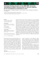
Expression patterns of TIP isoforms in Arabidopsis rootsFigure 1
Expression patterns of TIP isoforms in Arabidopsis
roots. 8-day old roots from the indicated transgenic lines
were excised, stained with propidium iodide for 2 min and
visualised by CLSM. The images show representative results
for each construct. The signals from YFP (green) and propid-
ium iodide fluorescence (red) are merged. Top panels: single
optical sections of the root tips, Middle panels: single optical
sections of root differentiation zones. Bottom panels: maxi-
mal projection of 16 optical z sections (4 μm step-size)
through mature root axes and young lateral roots. Scale bars:
100 μm.
d/Wϭ͖ϭͲz&W d/Wϭ͖ϮͲz&W d/WϮ͖ϭͲz&W d/WϮ͖ϮͲz&W d/WϮ͖ϯͲz&W d/Wϰ͖ϭͲz&W
&
',/ : < >
DKWYZE
BMC Plant Biology 2009, 9:133 />Page 3 of 9
(page number not for citation purposes)
In the case of TIP1;2, expression seems to be exclusively
limited to the root cap and the columella (Fig. 1B). A very
limited YFP signal can also be detected in the same region
of the young lateral root (Fig 1N, arrowhead) and in older
lateral roots (Additional file 1A).
Perhaps the most remarkable expression pattern observed
is that of TIP2;1, which in 8-day old roots is only detecta-
ble in a small region at the base of the lateral roots (Fig.
1O, arrowhead).
Having identified the general patterns of expression of the
different isoforms at low magnification, we then studied
the cell-specificity of TIP-YFP expression in more detail.
We analysed propidium iodide -stained roots by CLSM by
performing optical z-sections through differentiation
zones at 63× magnification (Fig. 2).
TIP1;1-YFP is clearly expressed in epidermis and cortex,
but its expression is particularly strong in the endodermis
and pericycle (Fig. 2A). Here TIP1;1-YFP highlights
numerous bright circular structures in the lumen of the
central vacuole. We hypothesise these are vacuolar 'bulbs',
which have previously been described as tonoplast invagi-
nations, which occur independently of the ectopic expres-
sion of XFP-tagged membrane proteins, and where
TIP1;1-GFP is concentrated [18,19]. It is however difficult
in some cases to observe a continuity between these struc-
tures and the central vacuole tonoplast.
At higher magnification, the overlapping patterns of
expression of TIP2;2-YFP and TIP2;3-YFP are confirmed.
Both are present in pericycle cells, particularly in the rows
of pericycle cells that form the xylem poles [20]. Both TIP-
YFPs tend to be absent from the endodermis (Fig. 2, pan-
els B and D), although we could detect discontinuous
endodermal expression at various positions along most
root axes (Fig. 2, panels C and E; and highlighted in blue
in panels G and H).
In contrast to the previous isoforms labelling inner root
cell layers, TIP4;1 expression is clearly restricted to the
root epidermis and cortex, with no signal detectable in the
inner layers (Fig. 2F).
Localisation of TIP1;1 and TIP1;2
Having analysed the TIPs with the broadest expression
patterns, we focussed on the two TIPs which seem to have
a more limited expression range in roots. TIP1;2-YFP pre-
sented a patchy distribution in cells of the root cap (Fig
1B). To ascertain that this was not an artefact due to
expression of our chimeric gene, we also generated a con-
struct (YFP-TIP1;2) where YFP was fused downstream of
the promoter and 5'UTR and in frame with the 5' of the
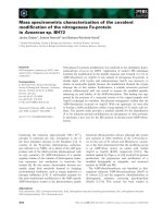
Cell-type specificity of TIP-YFP expression in the root axisFigure 2
Cell-type specificity of TIP-YFP expression in the
root axis. 8-day old roots from the indicated transgenic
lines were excised, stained with propidium iodide for 2 min
and visualised by CLSM. Stacks of 80 optical z sections (1 μm
step-size) were collected from root axes at the differentia-
tion zone. The images show representative results for each
construct. A to F: for each panel, the top section shows a sin-
gle xy optical section, and the bottom section shows the xz
projection of the whole image stack, revealing the cross sec-
tion of the root axis. The signals from YFP fluorescence
(green) and propidium iodide fluorescence (red) are merged.
G and H: the YFP fluorescence trace from representative
image stacks for the indicated transgenic lines was recon-
structed, segmented and rendered in 3D with Mimics 12.1.
The different tissues are colour-coded as follows: brown,
epidermis; red, cortex; blue, endodermis; green, pericycle.
Ep, epidermis; c, cortex; e, endodermis; p, pericycle; x,
xylem. Scale bar: 20 μm.
d/Wϭ͖ϭͲz&W d/WϮ͖ϮͲz&W
d/Wϰ͖ϭͲz&W
d/WϮ͖ϯͲz&W
&
d/WϮ͖ϮͲz&W
d/WϮ͖ϯͲz&W
',
dž
Đ
Ğ
ĞƉ
Ɖ
BMC Plant Biology 2009, 9:133 />Page 4 of 9
(page number not for citation purposes)
TIP coding sequence. In transgenic plants, YFP-TIP1;2
presents a similar expression pattern to TIP1;2-YFP, thus
ruling out YFP fusion artefacts (compare Fig. 3A with Fig.
1B). Expression is confined to the columella and the lat-
eral root cap [21], with the labelled cells disappearing at
the boundary with the elongation zone (Fig. 3A). Some of
the labelled cells are in the process of detaching from the
root (Fig. 3, panels C and F, arrowheads), suggesting that
they may correspond to 'border-like' cells [22]. The distri-
bution of YFP-TIP1;2 is therefore radically different to that
observed for its paralogue, TIP1;1-YFP, which has the wid-
est pattern of expression but is excluded from the root tip,
including the root cap (Fig. 1, panels A to M).
At the subcellular level, YFP-TIP1;2 localises to the endo-
plasmic reticulum (ER) of young root cap cells (Fig 3e:
note the characteristic reticular pattern and the nuclear
envelope; see also Additional file 1B). The chimeric pro-
tein is mostly found on the tonoplast of elongated lateral
root cap cells (Fig. 3D). This is likely to reflect different
stages of TIP1;2 trafficking in cells of different ages, rather
than impaired capacity to reach the tonoplast. This is fur-
ther demonstrated by the fact that in the epidermis of cot-
yledonary cells, where TIP1;2 is uniformly expressed, the
fusion protein appears to localise to the tonoplast (Addi-
tional file 1C-D).
TIP2;1 is localised in lateral root primordia
We have previously shown that TIP2;1 expression
becomes detectable in old root regions nearing the
hypocotyl, and is then widespread in hypocotyl and coty-
ledonary leaves [14]. We did not initially notice expres-
sion in young roots, but closer analysis revealed that in 8-
day old roots TIP2;1-YFP has a very specialised expression
pattern (Fig. 4). The YFP signal is detected in a ring-like
cluster at the base of emerging lateral roots (Fig. 4, panels
A-D). In very early lateral root primordia (LRP), TIP2;1
expression is detectable in 2-4 cells at the LRP. As the LRP
grows further, the number of cells expressing TIP2;1-YFP
increases but remains confined to a cluster underlying the
base the lateral root (Fig. 4, panels F-I). In rare cases, when
the lateral root is fully emerged, the expression of TIP2;1-
YFP can extend to some cells within the lateral root axis
(Fig 4I). Co-labelling with propidium iodide shows that
the TIP2;1 expressing cells are located in close proximity
to the xylem (fig. 4E), suggesting a pericycle localisation.
Co-expression with TIP2;3-RFP, which we found to be
enriched in the pericycle (Fig. 2, panels D, E, H) confirms
that TIP2;1-YFP expression originates from pericycle cells
(Fig. 4, panels F-I). This indicates that the initial expres-
sion of TIP2;1-YFP is likely to occur in the LRP founder
cells. Remarkably, the expression of TIP2;1-YFP and
TIP2;3-RFP appears to be mutually exclusive, with a clear
boundary between cells expressing one or the other iso-
form (Fig. 4H, inset; see Additional file 2 for individual
channels).
Overlapping TIP isoforms are mostly detectable at the
central vacuole tonoplast
We have shown that the various TIP isoforms under study
present diverse tissue specificity within roots. Several iso-
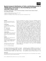
YFP-TIP1;2 is expressed in the root capFigure 3
YFP-TIP1;2 is expressed in the root cap. 8-day old
roots from the indicated transgenic lines were excised,
stained with propidium iodide for 2 min and visualised by
CLSM. Stacks of 80 optical z sections (1 μm step-size) were
collected from root tips. The images show a representative
result for this construct. The signals from YFP fluorescence
(green) and propidium iodide fluorescence (red) are merged.
A: maximal 3D projection of the root tip at the base of the
elongation zone. The image shows two adjacent z-stacks of
the same root, separated by a black line. B and C: xz projec-
tions of the image stack in panel a, revealing two cross-sec-
tions of the root axis, taken in the regions of the root
indicated by the arrowheads in A. D and E: the regions indi-
cated by dotted boxes in A were observed at high magnifica-
tion. Single optical sections are shown. Note YFP-TIP1;2 in
the ER of young root cap cells and in the tonoplast of root
cap cells closer to the elongation zone. F: The fluorescent
traces from YFP (green) and propidium iodide (red) from the
image stack in panels A were reconstructed, segmented and
rendered in 3D with Mimics 12.1. Scale bars: (a), 20 μm; (d)
and (e), 10 μm.
&
BMC Plant Biology 2009, 9:133 />Page 5 of 9
(page number not for citation purposes)
forms, however, are co-expressed in certain tissues,
namely TIP1;1, TIP2;2, TIP2;3, and TIP4;1. In order to
ascertain whether these isoforms were specific to distinct
vacuolar compartments, we focused on the subcellular
localisation of selected pairs of the above isoforms, tagged
with different spectral variants of fluorescent proteins and
co-expressed in transgenic Arabidopsis (Fig. 5).
The individual TIP expression patterns in double trans-
genic lines mirrored those observed in the lines expressing
individual isoforms (Additional file 3). The widespread
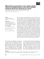
TIP 2;1-YFP expression in lateral root primordiaFigure 4
TIP 2;1-YFP expression in lateral root primordia. A-E:
8-day old roots from TIP2;1-YFP transgenic seedlings were
excised, stained with propidium iodide and visualised by
CLSM. Stacks of 80 optical z sections (1 μm step-size) were
collected from mature root axes. The images show repre-
sentative results for this construct. Maximal projections of
the z-stacks are shown, with the individual signals for YFP
(A), propidium iodide (B) or the merged signals (C and D). E:
The fluorescent traces from YFP (green) and propidium
iodide (red) from the image stack in panels (A-C) was recon-
structed, segmented and rendered in 3D with Mimics 12.1.
Note that the TIP2;1-YFP-expressing cells are in close prox-
imity to the xylem (labelled with x). F-I: Roots from 8-day old
transgenic seedlings expressing TIP2;1-YFP (green) and
TIP2;3-RFP (red) were imaged. Sequential stages of lateral
root development are shown. Inset in H: note the boundary
between pericycle cells expressing TIP2;3-RFP (top) and
TIP2;1-YFP (bottom). Scale bars: 20 μm.
,
/
&'
dž
dž
Overlapping TIP isoforms are mainly detected at the tono-plast of the central vacuoleFigure 5
Overlapping TIP isoforms are mainly detected at the
tonoplast of the central vacuole. Transgenic seedlings
co-expressing the indicated TIP-YFP and TIP-RFP constructs
were grown for 8 days on MS medium agar plates. Roots
were excised and visualised by CLSM. (A, D, G, J): YFP fluo-
rescence (green); (B, E, H, K): RFP fluorescence (red); (C, F,
I, L): merged images. Arrowheads in panel I indicate struc-
tures labelled by TIP2;3-RFP but not TIP2;2-YFP. Scale
bars:10 μm.
d/Wϰ͖ϭͲz&W
d/WϮ͖ϯͲZ&Wd/WϮ͖ϮͲz&W ŵĞƌŐĞ
d/WϮ͖ϯͲZ&W ŵĞƌŐĞ
d/WϮ͖ϯͲZ&Wd/Wϭ͖ϭͲz&W ŵĞƌŐĞ
d/Wϭ͖ϭͲZ&Wd/WϮ͖ϮͲz&W ŵĞƌŐĞ
&
',/
:<>
BMC Plant Biology 2009, 9:133 />Page 6 of 9
(page number not for citation purposes)
TIP1;1-YFP and TIP2;3-RFP are co-expressed in epidermis,
cortex and pericycle cells (Fig. 2). In these tissues, both
proteins are detected on the tonoplast of the central vacu-
ole (Fig. 5A-C). Both the tonoplast and the smaller, bulb-
like vacuolar structures [18] are labelled. Likewise, TIP2;2-
YFP and TIP1;1-RFP mostly label the same tonoplast in
the cell layers where they are co-expressed (Fig. 5D-F).
TIP2;2-YFP and TIP2;3-RFP, which almost overlap in root
tissues (Fig. 2), are also co-localised on tonoplast and
'bulbs' (Fig. 5G-I). Occasionally, TIP2;3-RFP highlighted
smaller vesicular structures that did not appear to contain
TIP2;2-YFP (Fig 5I, arrowheads). The nature of these struc-
tures was not investigated further. Finally, TIP4;1-YFP,
which is restricted to epidermis and cortex (Fig. 2F), co-
localises with TIP2;3 in those tissues (Fig. 5J-L). Note that
the relative abundance of these two isoforms mirrors the
pattern observed in single isoform localisation, with
TIP4;1 expression being strongest in the epidermis and
weaker in the cortex (Fig. 2F), and TIP2;3 expression being
stronger in cortex but weaker in epidermis (Fig. 2, panels
D-E).
Taken together, these co-expression results indicate that
each TIP isoform-fluorescent protein fusion we analysed
is predominantly found at the central vacuole tonoplast in
Arabidopsis root tissues.
Discussion
We have produced a complete expression map for all
members of the TIP family that are present in Arabidopsis
root tissues, including isoforms not previously studied.
The use of XFP fusions to TIP genomic sequences allowed
us to investigate both the tissue specificity and the subcel-
lular localisation of these proteins.
In general, our fluorescent reporter - TIP localisation data
correlate well with the relative TIP transcript levels, as
observed by microarray analysis [15,23,24], with the
exception of TIP1;2. TIP1;2 is indeed the isoform with the
highest level of mRNA expression in the root cap [23],
which matches our observations (Fig. 3). However, tran-
script levels for TIP1;2 have also been shown to be almost
as high as TIP1;1 throughout the root axis [15,23,24]. We
can only speculate at this stage that post-transcriptional
control processes prevent TIP1;2-YFP protein from being
detectable in these tissues.
TIP1;1 is the most widely expressed isoform along the
root axis. TIP2;2 and 2;3 have very similar expression pat-
terns, with their expression being low in the endodermis,
but high in the xylem pole pericycle. It appears that TIP2;1
becomes strongly expressed in the pericycle when this
undergoes differentiation to form the lateral root primor-
dium (Fig. 4). This narrow range of localisation of TIP2;1
is intriguing. Transcripts of the maize aquaporin ZmTIP1
were also localised in the lateral root by in situ hybridisa-
tion, but every cell in the LRP seemed to contain the tran-
script [25]. TIP2;1 can therefore be considered an
additional marker for the Arabidopsis LRP margins,
alongside the auxin efflux carriers Pin4 and Pin6 [26] and
the transcription factor CUC3 [27], which have a similar
localisation. It will be interesting to study the specific role
of TIP2;1 in these cells and determine why this stage of lat-
eral root development demands such a precise TIP iso-
form activation.
As a general observation, we could not detect expression
of any of the TIP-XFP fusions under study in the root tip
meristem. This lack of expression was previously reported
for TIP1;1 both by histochemical detection of GUS
fusions [5] and YFP tagging [14,19]. It is of course possi-
ble that expression levels of our fusions are too low in this
region to be detected by confocal microscopy. However,
the fact that the more sensitive histochemical GUS stain-
ing also fails to detect expression of TIP1;1, which micro-
array data indicate is the most abundantly transcribed
isoform in roots [16], strongly suggest that the protein is
not expressed in the root meristem. This is in contrast with
data from other species such as pea and barley, where TIPs
have been located in isolated root tip cells [10,12] and in
root tip sections by immunohistochemical methods [28].
Analysis in the Olbrich et al. study was performed on 3-
day old seedlings. At the same age in Arabidopsis seed-
lings we could already detect all the TIP isoforms
described in this study, with the exception of TIP2;1.
However their expression pattern was already the same as
observed at 8 days (data not shown). We therefore
resolved to present results at 8 days, when the complete
set of root TIPs is detectable.
This lack of observable expression in root tips makes it dif-
ficult to perform meaningful comparisons between the
vacuolar complement of Arabidopsis root tip cells and
that of other plant species.
TIP-YFP expression was also not detected in the root vas-
culature, regardless of the developmental stage. This mir-
rors observations in barley and pea root sections, where
the stele was not labelled by TIP antisera [28]. While it is
easy to rationalise the absence of a vacuole in the xylem
cells, which underwent autolysis, and in mature sieve ele-
ments, which lack true vacuoles (reviewed in [29]) it was
somewhat surprising not to find TIPs in the companion
and parenchima cells. We think it unlikely that this lack of
detection is caused by a loss of sensitivity by the confocal
microscope detectors in the inner layers of the roots,
because both propidium iodide staining and YFP signal
are easily detected in the xylem and xylem pole pericycle,
BMC Plant Biology 2009, 9:133 />Page 7 of 9
(page number not for citation purposes)
respectively (Fig. 2 and Additional file 3). In addition, we
could easily detect 35S::TIP2;1-YFP in the vascular tissue
using the same settings (Additional file 4). Accordingly,
Boursiac et al [24] recently showed that constitutively
expressed TIP1;1-GFP and TIP1;2-GFP clearly label the
vascular tissue [24].
Recently it has been shown that Arabidopsis knockout
mutants lacking TIP1;1, TIP1;2, or both isoforms, do not
have any major defects [7,19]. This is in contrast with
drastic defects observed in Arabidopsis upon downregula-
tion of TIP1;1 by RNAi [30]. A possible explanation for
the latter result is off-target silencing in the RNAi lines [7].
Our data provide a rationale for the lack of a macroscopic
phenotype in the double TIP1 knockouts observed by
Schussler et al. We have shown that, in roots, expression
of TIP1;1 and TIP1;2 does not appear to overlap, with
TIP1;1 being expressed in epidermis, cortex, endodermis
and perycycle starting from the elongation zone, and
TIP1;2 being restricted to the root cap. As TIP1;1 and 1;2
show different tissue specificities, it seems unlikely that
they are reciprocally redundant. In addition, we have
shown that other TIP isoforms, namely TIP2;2, TIP2;3 and
TIP4;1 would still be present in the tissues lacking TIP1;1
(Fig. 2). It is therefore possible that these remaining iso-
forms compensate for the lack of TIP1;1 in the knockout.
On the other hand, the effect of the absence of TIP1;2
from the root cap may be subtle and may have gone unde-
tected under the experimental conditions adopted for the
whole-plant analysis of the double mutants. A lack of phe-
notype in the aerial parts of the single knockout plants
may be explained by the fact that both TIP1;1 and TIP1;2
are expressed in leaves [14] and Additional file 2) and
may well be acting redundantly there. As for the double
knockout, redundancy may be afforded by TIP2;1 [14]
and TIP2;2 [16], which are also expressed in leaves.
Conclusion
We have identified novel patterns of expression of TIP iso-
forms in Arabidopsis roots. This information may provide
a useful starting point for a more targeted approach to dis-
sect the function of individual TIP isoforms in root tissues.
It also provides the foundation for further analysis of the
intracellular targeting of different TIPs.
Methods
Recombinant DNA and generation of transgenic plants
The constructs encoding native TIP1;1-YFP and native
TIP2;1YFP have been described previously [14].
A full list of primers designed to amplify the genomic
sequences of the root-expressed TIPs is shown in Addi-
tional file 5. Each TIP genomic sequence, including either
the complete promoter region (up to the UTR of the gene
immediately upstream in the chromosome) or 1.5 Kb of
the promoter (if longer than 1.5 Kb), plus 5' UTR and
introns, was amplified from total genomic DNA from Ara-
bidopsis thaliana Columbia ecotype. Primers included
restriction sites KpnI at the 5' and XhoI at the 3' of the tar-
get sequences. Amplified fragments were cloned into the
KpnI and XhoI sites of pGREEN0029, upstream of a XhoI/
SacI fragment containing the YFP coding sequence and
the OCS 3' terminator fragment. A similar strategy was
adopted to fuse TIP sequences to RFP, but in this case the
forward primers included both KpnI and SacI restriction
sites, generating a TIP-RFP cassette that could be mobi-
lized with SacI. To obtain pairwise TIP-YFP/TIP-RFP com-
binations, selected TIP-RFP cassettes were excised with
SacI and ligated into TIP-YFP vectors linearised with SacI,
giving rise to constructs harbouring both reporter genes in
a tandem. All the chimeric constructs were introduced
into strain C58 of Agrobacterium tumefaciens harbouring
the pSoup vector [31]. Arabidopsis plants were then trans-
formed using the floral dip method [32].
Confocal analysis and image processing
About 30 seeds from at least 4 independent transgenic
lines per construct were germinated onto agar plates con-
taining half-strength Murashige and Skoog (MS) Basal
Medium (Sigma-Aldrich) and grown for 8 days at 22°C,
in a 16:8 light:dark regime. Roots were excised, mounted
in half-strength liquid MS medium and immediately
observed with a Leica TSC SP5 confocal laser scanning
microscope, using either a 10× (NA 0.3) air or a 63× (NA
1.4) oil immersion objective. In some cases roots were
preincubated for 2 min in 10 μg/ml propidium iodide,
diluted in half-strength MS medium. YFP was excited at
514 nm and detected in the 525 to 550 nm range. RFP was
excited at 561 nm and detected in the 553 to 638 nm
range. Propidium iodide was excited at 561 nm and
detected in the 650 to 720 nm range. Simultaneous detec-
tion of YFP and RFP or YFP and propidium iodide was
performed by combining the settings indicated above in
the sequential scanning facility of the microscope, as
instructed by the manufacturer.
3D reconstruction of z-stacks of optical sections was per-
formed with the Leica LAS-AF Lite free software (Leica
Microsystems, Germany). Segmentation analysis and 3D
rendering were performed with Mimics 12.1 (Materialise
N.V., Leuwen, Belgium).
Abbreviations
CLSM: confocal laser scanning microscopy; ER: endoplas-
mic reticulum; PI: propidium iodide; TIP: tonoplast
intrinsic protein.
Authors' contributions
SG generated the majority of the constructs and transgenic
plants and performed the bulk of the confocal analysis.
BMC Plant Biology 2009, 9:133 />Page 8 of 9
(page number not for citation purposes)
MS produced the TIP1;2-RFP and 35S:TIP2;1 constructs
and transgenic lines and performed confocal analysis. PH
produced the native TIP1;1-YFP and TIP2;1 YFP constructs
and transgenic plants and performed confocal analysis.
RK performed the 3D image analysis in MIMICS. LF
designed the experimental programme, gave technical
and intellectual guidance and wrote the manuscript. All
authors read and approved the final manuscript.
Additional material
Acknowledgements
We are grateful to Robert Spooner and Alessandro Vitale for critical read-
ing of the manuscript. This work was orted in part by the European Union
(LSH-2002-1.2.5-2 "Recombinant Pharmaceuticals from Plant for Human
Health -Pharma-Planta") and by the Leverhulme Trust (grant F/00215/AP).
A grant from the Fondation 'Les Gueules Cassées' funded the acquisition of
the Mimics software.
References
1. Kaldenhoff R, Fischer M: Functional aquaporin diversity in
plants. Biochim Biophys Acta 2006, 1758:1134-1141.
2. Maurel C: Plant aquaporins: Novel functions and regulation
properties. FEBS Letters 2007, 581:2227-2236.
3. Hofte H, Hubbard L, Reizer J, Ludevid D, Herman EM, Chrispeels MJ:
Vegetative and Seed-Specific Forms of Tonoplast Intrinsic
Protein in the Vacuolar Membrane of Arabidopsis thaliana.
Plant Physiol 1992, 99:561-570.
4. Johanson U, Karlsson M, Johansson I, Gustavsson S, Sjovall S, Fraysse
L, Weig AR, Kjellbom P: The complete set of genes encoding
major intrinsic proteins in Arabidopsis provides a frame-
work for a new nomenclature for major intrinsic proteins in
plants. Plant Physiol 2001, 126:1358-1369.
5. Ludevid D, Hofte H, Himelblau E, Chrispeels MJ: The Expression
Pattern of the Tonoplast Intrinsic Protein gamma-TIP in
Arabidopsis thaliana Is Correlated with Cell Enlargement.
Plant Physiol 1992, 100:1633-1639.
6. Daniels MJ, Chaumont F, Mirkov TE, Chrispeels MJ: Characteriza-
tion of a new vacuolar membrane aquaporin sensitive to
mercury at a unique site. Plant Cell 1996, 8:587-599.
7. Schussler MD, Alexandersson E, Bienert GP, Kichey T, Laursen KH,
Johanson U, Kjellbom P, Schjoerring JK, Jahn TP: The effects of the
loss of TIP1;1 and TIP1;2 aquaporins in Arabidopsis thaliana.
Plant J 2008, 56:756-767.
8. Loque D, Ludewig U, Yuan L, von Wiren N: Tonoplast Intrinsic
Proteins AtTIP2;1 and AtTIP2;3 Facilitate NH3 Transport
into the Vacuole. Plant Physiol 2005, 137:671-680.
9. Jauh GY, Fischer AM, Grimes HD, Ryan CA Jr, Rogers JC: delta-
Tonoplast intrinsic protein defines unique plant vacuole
functions. Proc Natl Acad Sci USA 1998, 95:12995-12999.
10. Jauh G-Y, Phillips TE, Rogers JC: Tonoplast intrinsic protein iso-
forms as markers for vacuolar functions. Plant Cell 1999,
11:1867-1882.
11. Gillespie J, Rogers SW, Deery M, Dupree P, Rogers JC:
A unique
family of proteins associated with internalized membranes
in protein storage vacuoles of the Brassicaceae. Plant J 2005,
41:429-441.
12. Paris N, Stanley CM, Jones RL, Rogers JC: Plant cells contain two
functionally distinct vacuolar compartments. Cell 1996,
85:563-572.
13. Poxleitner M, Rogers SW, Lacey Samuels A, Browse J, Rogers JC: A
role for caleosin in degradation of oil-body storage lipid dur-
ing seed germination. Plant J 2006, 47:917-933.
14. Hunter PR, Craddock CP, Di Benedetto S, Roberts LM, Frigerio L:
Fluorescent Reporter Proteins for the Tonoplast and the
Vacuolar Lumen Identify a Single Vacuolar Compartment in
Arabidopsis Cells. Plant Physiol 2007, 145:1371-1382.
15. Schmid M, Davison TS, Henz SR, Pape UJ, Demar M, Vingron M,
Scholkopf B, Weigel D, Lohmann JU: A gene expression map of
Additional file 1
Expression and subcellular localisation of TIP1;2. 8-day old seedlings
expressing YFP-TIP1;2 were visualised by CLSM. A: 10× magnification
of a lateral root. The signals from YFP fluorescence (green) and propidium
iodide fluorescence (red) are merged. B: single root cap cell with TIP1;2-
YFP showing typical ER labelling. C-D: epidermal cells in cotyledons
where TIP1;2-YFP shows typical tonoplast labelling (green). Red: chloro-
phyll autofluorescence (excitation 514 nm, detection 600-650 nm). Scale
bars: A, 100
μ
m; B and D, 5
μ
m; C, 20
μ
m.
Click here for file
[ />2229-9-133-S1.PDF]
Additional file 2
Mutually exclusive expression of TIP2;1 and TIP2;3 in lateral root pri-
mordia. Roots from 8-day old transgenic seedlings expressing TIP2;1-YFP
(green) and TIP2;3-RFP (red) were visualised by CLSM. Scale bar, 20
μ
m.
Click here for file
[ />2229-9-133-S2.PDF]
Additional file 3
Co-expression of selected TIP-XFP pairs. Transgenic seedlings co-
expressing the indicated TIP-YFP and TIP-RFP constructs were grown for
8 days on MS medium-agar plates. Roots were excised and visualised by
CLSM. Stacks of 80 optical z sections (1
μ
m step-size) were collected from
root axes at the differentiation zone. The images show representative
results for each construct. Each panel shows the xz projection of the whole
image stack, revealing the cross section of the root axis.
Click here for file
[ />2229-9-133-S3.PDF]
Additional file 4
Constitutively expressed TIP2;1-YFP is detectable in every root tissue.
Roots from 8-day old transgenic seedlings expressing 35S::TIP2;1-YFP
(green) were excised, stained with propidium iodide (red) for 2 min and
visualised by CLSM. A: stacks of 80 optical z sections (1
μ
m step-size)
were collected from root axes at the differentiation zone. The images show
representative results for this construct. The signals from YFP fluorescence
(green) and propidium iodide fluorescence (red) are merged. B-D: single
optical section through the vascular tissue, indicating that constitutive
expression of TIP2;1 is easily detectable in these cell types. B: YFP, C, pro-
pidium iodide, D, merged images. Scale bar, 10
μ
m.
Click here for file
[ />2229-9-133-S4.PDF]
Additional file 5
Primers used in this study. The diagram indicates the target sequences
for the indicated primers in the final constructs. Restriction sites are
shown in bold.
Click here for file
[ />2229-9-133-S5.PDF]
Publish with Bio Med Central and every
scientist can read your work free of charge
"BioMed Central will be the most significant development for
disseminating the results of biomedical research in our lifetime."
Sir Paul Nurse, Cancer Research UK
Your research papers will be:
available free of charge to the entire biomedical community
peer reviewed and published immediately upon acceptance
cited in PubMed and archived on PubMed Central
yours — you keep the copyright
Submit your manuscript here:
/>BioMedcentral
BMC Plant Biology 2009, 9:133 />Page 9 of 9
(page number not for citation purposes)
Arabidopsis thaliana development. Nat Genet 2005,
37:501-506.
16. Frigerio L, Hinz G, Robinson DG: Multiple vacuoles in plant cells:
rule or exception? Traffic 2008, 9:1564-1570.
17. Soto G, Alleva K, Mazzella MA, Amodeo G, Muschietti JP: AtTIP1;3
and AtTIP5;1, the only highly expressed Arabidopsis pollen-
specific aquaporins, transport water and urea. FEBS Lett 2008,
582:4077-4082.
18. Saito C, Ueda T, Abe H, Wada Y, Kuroiwa T, Hisada A, Furuya M,
Nakano A: A complex and mobile structure forms a distinct
subregion within the continuous vacuolar membrane in
young cotyledons of Arabidopsis. Plant J 2002, 29:245-255.
19. Beebo A, Thomas D, Der C, Sanchez L, Leborgne-Castel N, Marty F,
Schoefs B, Bouhidel K: Life with and without AtTIP1;1, an Ara-
bidopsis aquaporin preferentially localized in the apposing
tonoplasts of adjacent vacuoles. Plant Mol Biol 2009, 70:193-209.
20. Parizot B, Laplaze L, Ricaud L, Boucheron-Dubuisson E, Bayle V,
Bonke M, De Smet I, Poethig SR, Helariutta Y, Haseloff J, et al.:
Diarch Symmetry of the Vascular Bundle in Arabidopsis
Root Encompasses the Pericycle and Is Reflected in Distich
Lateral Root Initiation. Plant Physiol 2008, 146:140-148.
21. Dolan L, Janmaat K, Willemsen V, Linstead P, Poethig S, Roberts K,
Scheres B: Cellular organisation of the Arabidopsis thaliana
root. Development 1993, 119:71-84.
22. Vicre M, Santaella C, Blanchet S, Gateau A, Driouich A: Root bor-
der-like cells of Arabidopsis. Microscopical characterization
and role in the interaction with rhizobacteria. Plant Physiol
2005, 138:998-1008.
23. Birnbaum K, Shasha DE, Wang JY, Jung JW, Lambert GM, Galbraith
DW, Benfey PN: A gene expression map of the Arabidopsis
root. Science 2003, 302:1956-1960.
24. Boursiac Y, Chen S, Luu DT, Sorieul M, Dries N van den, Maurel C:
Early effects of salinity on water transport in Arabidopsis
roots. Molecular and cellular features of aquaporin expres-
sion. Plant Physiol
2005, 139:790-805.
25. Chaumont F, Barrieu F, Herman EM, Chrispeels MJ: Characteriza-
tion of a maize tonoplast aquaporin expressed in zones of
cell division and elongation. Plant Physiol 1998, 117:1143-1152.
26. Benkova E, Michniewicz M, Sauer M, Teichmann T, Seifertova D, Jur-
gens G, Friml J: Local, efflux-dependent auxin gradients as a
common module for plant organ formation. Cell 2003,
115:591-602.
27. Vroemen CW, Mordhorst AP, Albrecht C, Kwaaitaal MA, de Vries
SC: The CUP-SHAPED COTYLEDON3 gene is required for
boundary and shoot meristem formation in Arabidopsis.
Plant Cell 2003, 15:1563-1577.
28. Olbrich A, Hillmer S, Hinz G, Oliviusson P, Robinson DG: Newly
formed vacuoles in root meristems of barley and pea seed-
lings have characteristics of both protein storage and lytic
vacuoles. Plant Physiol 2007, 145:1383-1394.
29. De D: Plant Cell Vacuoles Collingwood, Australia: CSIRO Publishing;
2000.
30. Ma S, Quist TM, Ulanov A, Joly R, Bohnert HJ: Loss of TIP1;1
aquaporin in Arabidopsis leads to cell and plant death. Plant
J 2004, 40:845-859.
31. Hellens RP, Edwards EA, Leyland NR, Bean S, Mullineaux PM:
pGreen: a versatile and flexible binary Ti vector for Agrobac-
terium-mediated plant transformation. Plant Mol Biol 2000,
42:819-832.
32. Clough SJ, Bent AF: Floral dip: a simplified method for Agro-
bacterium-mediated transformation of Arabidopsis thal-
iana. Plant J 1998, 16:735-743.