báo cáo khoa học: " AtKinesin-13A is located on Golgi-associated vesicle and involved in vesicle formation/budding in Arabidopsis root-cap peripheral cells" ppt
Bạn đang xem bản rút gọn của tài liệu. Xem và tải ngay bản đầy đủ của tài liệu tại đây (2.87 MB, 8 trang )
BioMed Central
Page 1 of 8
(page number not for citation purposes)
BMC Plant Biology
Open Access
Research article
AtKinesin-13A is located on Golgi-associated vesicle and involved in
vesicle formation/budding in Arabidopsis root-cap peripheral cells
Liqin Wei
†1,2
, Wei Zhang
†1
, Zhaohui Liu
1
and Yan Li*
1
Address:
1
State Key Laboratory of Plant Physiology and Biochemistry, College of Biological Sciences, China Agricultural University, Beijing
100193, PR China and
2
Research Center of Molecular and Developmental Biology, Key Laboratory of Photosynthesis and Environmental
Molecular Physiology, Institute of Botany, Chinese Academy of Sciences, Beijing 100093, PR China
Email: Liqin Wei - ; Wei Zhang - ; Zhaohui Liu - ; Yan Li* -
* Corresponding author †Equal contributors
Abstract
Background: AtKinesin-13A is an internal-motor kinesin from Arabidopsis (Arabidopsis thaliana).
Previous immunofluorescent results showed that AtKinesin-13A localized to Golgi stacks in plant
cells. However, its precise localization and biological function in Golgi apparatus is unclear.
Results: In this paper, immunofluorescent labeling and confocal microscopic observation revealed
that AtKinesin-13A was co-localized with Golgi stacks in Arabidopsis root tip cells. Immuno-
electron microscopic observations indicated that AtKinesin-13A is primarily localized on Golgi-
associated vesicles in Arabidopsis root-cap cells. By T-DNA insertion, the inactivation of the
AtKinesin-13A gene (NM-112536) resulted in a sharp decrease of size and number of Golgi vesicles
in root-cap peripheral cells. At the same time, these cells were vacuolated in comparison to the
corresponding cells of the wild type.
Conclusion: These results suggest that AtKinesin-13A decorates Golgi-associated vesicles and
may be involved in regulating the formation of Golgi vesicles in the root-cap peripheral cells in
Arabidopsis.
Background
Kinesins are a large super-family of microtubule motor
proteins that can use the energy of ATP hydrolysis to pro-
duce force and move along microtubules [1,2]. Based on
their motor domain location within the primary sequence
of the proteins, different kinesins may have their motor
domains affixed at C-terminal, N-terminal or internal
positions [3]. The C-terminal and N-terminal motor
kinesins transport various vesicles and organelles toward
the microtubules minus-terminal or plus-terminal,
respectively. The internal motor kinesins found in animal
cells are not able to move along the microtubules in the
conventional form, but instead depolymerize microtu-
bules from both ends [4]. The completed Arabidopsis
genome contains at least 61 genes encoding polypeptides
with the kinesin catalytic core. Among these kinesins, AtK-
inesin-13A and AtKinesin-13B are two internal-motor
kinesins [5,6]. However, the similarity of AtKinesin-13A
and AtKinesin-13B to kinesins of the same subfamily
from other kingdoms is only limited to the catalytic core,
and they lacks a Lys-rich neck motif commonly found in
animal Kinesin-13s. Plant Kinesin-13A and animal
Kinesin-13s also have different localization patterns [7,8].
Lu et al. reported that AtKinesin-13A was co-localized
with Golgi stacks in various Arabidopsis cells, indicating
that AtKinesin-13A is a special plant internal-motor
Published: 25 November 2009
BMC Plant Biology 2009, 9:138 doi:10.1186/1471-2229-9-138
Received: 19 March 2009
Accepted: 25 November 2009
This article is available from: />© 2009 Wei et al; licensee BioMed Central Ltd.
This is an Open Access article distributed under the terms of the Creative Commons Attribution License ( />),
which permits unrestricted use, distribution, and reproduction in any medium, provided the original work is properly cited.
BMC Plant Biology 2009, 9:138 />Page 2 of 8
(page number not for citation purposes)
kinesin [8]. However, the precise localization of AtKi-
nesin-13A as well as its biological function in plant Golgi
apparatus is unclear.
Cellular trafficking is the foundation of cellular morphol-
ogy and function, where the Golgi apparatus plays an
important role in the secretion and transportation of cel-
lular vesicles [9]. In animal cells, the Golgi apparatus is
positioned near the microtubule-organizing center, and
its localization and organization depend on intact micro-
tubules [10]. In addition, microtubules and microtubule-
based motor proteins play critical roles in Golgi dynamics
[11,12]. Both the conventional kinesins and kinesin-
related proteins have been reported to regulate Golgi
structure and function in animal cells [13-19]. Actin
microfilaments have also been found to be necessary in
maintaining the sub-cellular localization of the animal
Golgi complex [20]. Both microtubules and microfila-
ments cooperate in maintaining the balance of Golgi
dynamics within animal cells.
Unlike in animal cells, the Golgi apparatus of plant cells
consists of a large number of small, independent Golgi
stacks that are distributed throughout the cytoplasm [21-
23], with the number of Golgi stacks being different
among different kind of cells. The number of the Golgi
apparatus is typically abundant in plant root-cap periph-
eral cells, in which very large vesicles are produced by each
Golgi apparatus [24]. This is in accord with the high secre-
tory activity needed for root growth in soil [25]. On the
other hand, it is generally believed that the movement of
plant Golgi stacks is solely dependent on actin microfila-
ments [23].
In plant cells, it has been reported that microtubules play
a key role in organelle movement [26-29], but it is unclear
whether microtubule-based motor kinesins take part in
regulating the structure and function of Golgi apparatus.
In the present study, AtKinesin-13A was detected on
Golgi-associated vesicles in root-cap cells of Arabidopsis.
Additionally, the Golgi structure was abnormal in root-
cap peripheral cells of the kinesin-13a-1 mutant. These
results suggest that AtKinesin-13A may participate in reg-
ulating the formation of Golgi-associated vesicles in Ara-
bidopsis root cap peripheral cells.
Results
AtKinesin-13A co-localization with Golgi stacks in
Arabidopsis root tip cells
The expression of AtKinesin-13A was not tissue-specific in
Arabidopsis [30]. On the other hand, there are different
types of Golgi stacks in plant root tip cells. Therefore, for
further studying the localization and function of AtKi-
nesin-13A, Arabidopsis root tip cells were used. N-
acetylglucosaminyl transferase I (Nag)-GFP fusion protein
specially decorates Golgi stacks in plant cells [8]. To co-
localize AtKinesin-13A with Golgi apparatus in Arabidop-
sis root tip cells, we used an Arabidopsis line expressing
the Nag-GFP fusion. Root tip cells were used to verify the
relationship between AtKinesin-13A localization and
individual Golgi stacks marked by Nag-GFP. Confocal
microscopic observation revealed that AtKinesin-13A was
co-localized with Nag-GFP in Arabidopsis root tip cells
(Fig. 1), suggesting that AtKinesin-13A is localized to the
Golgi stacks in these cells.
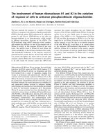
Immuno-localization and confocal microscopy observation showed co-localization of AtKinesin-13A and the Golgi stacks in Arabidopsis root tip cellsFigure 1
Immuno-localization and confocal microscopy observation showed co-localization of AtKinesin-13A and the
Golgi stacks in Arabidopsis root tip cells. (A) Nag-GFP showed the distribution of Golgi stacks in Arabidopsis root tip
cells. (B) Immunofluorescence labeling showed the distribution of AtKinesin-13A in Arabidopsis root tip cells. (C) A merged
image had AtKinesin-13A signal pseudo-colored in red and Nag-GFP in green, showing co-localization of AtKinesin-13A and
the Golgi apparatus in Arabidopsis root tip cells. Bar: 5 μm.
BMC Plant Biology 2009, 9:138 />Page 3 of 8
(page number not for citation purposes)
AtKinesin-13A is mainly localized on Golgi vesicles in
Arabidopsis root-cap cells
To determine the localization of AtKinesin-13A on Golgi
stacks at the ultra-structural level, ultra-thin sections were
immuno-gold labeled with anti-AtKinesin-13A antibody
in Arabidopsis root-cap cells. The immuno-gold labeling
with the affinity-purified anti-AtKinesin-13A antibody
and electron microscopy revealed that AtKinesin-13A was
specifically linked with the Golgi stacks of Arabidopsis
root cap cells. Electron microscopic observation detected
that gold particles were associated with the Golgi vesicles
in the root-cap cells (Fig. 2A). Quantitative analysis of the
gold particles distribution showed a preferential associa-
tion of AtKinesin-13A with the Golgi vesicle, accounting
for 55.6% of the total gold particles (Table 1). We addi-
tionally found that gold labeling was located mainly on
the margin of Golgi vesicles in Arabidopsis root cap cells
(Fig. 2B, arrows). This result suggests that AtKinesin-13A
may locate on membranes of Golgi vesicles in these cells.
Control sections, incubated with the secondary antibody
alone, did not show gold particles association with Golgi
vesicles (Fig. 2C). In addition, we also found that Atki-
nesin-13A antibody can not label Golgi vesicles in the
root cap cells of kinesin-13a-1 mutant line (Fig. 2D).
AtKinesin-13A gene inactivation caused obvious
structural changes of Golgi stacks in root cap peripheral
cells
Lu et al [8] reported two independent Arabidopsis T-DNA
insertion mutations at the AtKinesin-13A locus, which led
to the loss of function of Kinesin-13A in Arabidopsis. In
Lu et al. paper, it was concluded that two Atkinesin-13A
mutant lines (kinesin-13a-1 and kinesin-13a-2) exhibited
identical phenotypes. They have confirmed that the
mutant phenotypes were indeed caused by the T-DNA
insertion at the Kinesin-13A locus based on their comple-
mentation results [8]. The kinesin-13a-1 mutant line was
used for current study.
The Golgi apparatus is the main executer of secretory
activity in root-cap peripheral cells [24]. Root-cap periph-
eral cells of the kinesin-13a-1 mutant were compared with
those of wild-type Arabidopsis using transmission elec-
tron microscopy. Peripheral cells of the kinesin-13a-1
mutant lines contained a few large vacuoles, but few vesi-
cles (Fig. 3A). In contrast, numerous vesicles were found
in the peripheral cells of the wild type root cap (Fig. 3B).
In addition, Golgi-associated vesicles were also rare and
small in the peripheral cells of the kinesin-13a-1 mutant
(Fig. 3C, E), compared to how abundant secretory vesicles
around the Golgi stack in wild type root-cap peripheral
cells (Fig. 3D, F). A different morphology was also found
in cisternal morphology of Golgi stacks between wild and
mutant line. Normally, cisternae swell at the ends in Golgi
stacks of root-cap peripheral cells (Fig. 3D). However, this
does not occur in the kinesin-13a-1 mutant line (Fig. 3C).
Therefore, it appeared that the morphology of the Golgi
apparatus in the kinesin-13a-1 mutant line is significantly
different from that of the wild type for root-cap peripheral
cells.
In the meristematic cells and columella cells of the root
cap, however, the Golgi morphology of the kinesin-13a-1
mutant was not significantly different from that of wild
type (data no shown).
Discussion
Golgi apparatus is a vital organelle in the process of cellu-
lar secretion. In animal cells, the high level of activity at
the Golgi apparatus is sustained largely through the com-
bined effects of microtubules, actin-microfilaments, and
some intermediate filaments [31]. In plant cells, the Golgi
apparatus consists of a large number of small, independ-
ent Golgi stacks that appear to be randomly distributed
throughout the cytoplasm that take on rapid stop-and-go
movements [21,22,32]. The Golgi apparatus is a polar
organelle. From its cis-cisternae to the trans-network, there
are multi-compartments that carry out versatile functions.
Within different functional compartments there are also
special proteins that perform different biological func-
tions. Previous studies have shown that a number of
molecular motors are around Golgi apparatus and
involved in maintaining its proper structure and function
in animal cells [31]. However, few motors were found to
locate on plant Golgi apparatus before. Recently, Lu et al
reported that AtKinesin-13A decorated Golgi stacks of var-
ious Arabidopsis cells [8]. Results from immuno-gold
labeling and electron microscopy presented here further
indicated that AtKinesin-13A located to the margin of
Golgi vesicles in Arabidopsis root cap cells. This result sug-
gests that AtKinesin-13A may associate with membranes
of Golgi vesicles in Arabidopsis root cap cells. On the
Table 1: Sub-cellular distribution of AtKinesin-13A in root-cap cells of Arabidopsis (mean ± SD) (N = 15).
Golgi-associated vesicles (%) Other vesicles (%) Non-vesicles (%)
55.6 ± 1.6 20 ± 1.2 24.4 ± 2.1
Note: number represented the percentages (mean ± SD) of the total labeling in distinct locations in root-cap cells of Arabidopsis.
N: the number of cells analyzed. Golgi-associated vesicles: the vesicles around Golgi stacks. Other vesicles: the vesicles beyond Golgi stacks. Non-
vesicles: cytoplasmic labeling not associated with vesicles or Golgi vesicles.
BMC Plant Biology 2009, 9:138 />Page 4 of 8
(page number not for citation purposes)
other hand, there is no predicted trans-membrane
domain in the Atkinesin-13A protein sequence. Taken
together, these results imply that AtKinesin-13A may be a
cytoplasmic oriented peripheral membrane protein of
Golgi-associated vesicles in Arabidopsis.
The root cap consists of a large number of parenchyma
cells. During the growth of the root system, root-cap cells
initially stem from the root-cap meristem by mitosis, then
progress through a series of distinctive developmental
stages. Ultimately, these cells separate from the periphery
of the root cap to produce border cells [33]. During differ-
entiation from meristematic cells into peripheral cells, the
number of Golgi stacks per cell as well as the size and the
number of Golgi-associated vesicles per Golgi apparatus
increase visibly. In root-cap peripheral cells, there are
large populations of active secretory Golgi apparatus, and
the secretory vesicles around the Golgi are large and abun-
dant, while the size and number of Golgi-associated vesi-
cles in root-cap meristematic cells are relatively few and
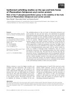
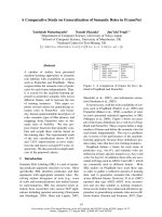
Immuno-gold labeling and electron microscopic observation showed that AtKinesin-13A was located on Golgi-associated vesi-cle in root cap cells of ArabidopsisFigure 2
Immuno-gold labeling and electron microscopic observation showed that AtKinesin-13A was located on Golgi-
associated vesicle in root cap cells of Arabidopsis. AtKinesin-13A was labeled with the purified AtKinesin-13A antibody.
The AtKinesin-13A antibody was detected with a secondary antibody with 10 nm gold particles. (A) Electron microscopic
observation showed that AtKinesin-13A was located mainly on Golgi-associated vesicle in root cap cells of Arabidopsis. (B)
Note the labeling on the margin of Golgi vesicles in Arabidopsis root cap cells (arrows). (C) Control section, incubated with
the secondary antibody alone, did not show gold particles association with Golgi vesicles. (D) Atkinesin-13A antibody could
not label Golgi vesicles in the section of root cap cells in kinesin-13a-1 mutant line. G: Golgi apparatus. SV: secretory vesicles.
Bars: 200 nm (A, D); 150 nm (B, C).
BMC Plant Biology 2009, 9:138 />Page 5 of 8
(page number not for citation purposes)
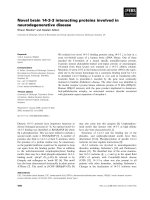
Electron microscopic observation showed obvious structural changes of Golgi apparatus in root cap peripheral cells of the kinesin-13a-1 mutant lineFigure 3
Electron microscopic observation showed obvious structural changes of Golgi apparatus in root cap peripheral
cells of the kinesin-13a-1 mutant line. (A) The peripheral cells of the kinesin-13a-1 mutant lines contained a few large vacu-
oles and few vesicles. (B) The peripheral cells of the wild type root cap contained numerous vesicles. (C), (E) Golgi-associated
vesicles were rare and small in the peripheral cells of the kinesin-13a-1 mutant. (D), (F) The wild type root-cap peripheral cells
contained abundant and bulky secretory vesicles around the Golgi stack. G: Golgi apparatus. P: peripheral cells. SV: secretory
vesicles. Bars: 1 μm (A, B); 200 nm (C, D); 150 nm (E, F).
BMC Plant Biology 2009, 9:138 />Page 6 of 8
(page number not for citation purposes)
small [24]. In this paper, electron microscopy observation
showed that the AtKinesin-13A gene inactivation induced
a significant decrease of the size and number of Golgi-
associated vesicles in root-cap peripheral cells. In addi-
tion, no swelling was observed at the ends of trans-cister-
nae of Golgi stacks in root-cap peripheral cells of the
mutant line. The large Golgi-associated vesicles often
come from trans-cisternae swelling and budding in root-
cap peripheral cells [22,34]. These results suggest that AtK-
inesin-13A may be involved in the trans-cisternae swelling
and budding of Golgi-associated vesicles in root-cap
peripheral cells, and then regulates the size and number of
Golgi-associated vesicles in these cells.
The expression of AtKinesin-13A was not tissue-specific in
Arabidopsis [30]. However, some unconventional Golgi
apparatus behaviors were only observed in the root-cap
peripheral cells of the Arabidopsis kinesin-13a-1 mutant
line. Recently, Poulsen et al also reported that Amino-
phospholipid ATPase3 (ALA3), a member of the P4-
ATPase subfamily in Arabidopsis, localizes to the Golgi
stacks and that mutations of ALA3 result in devoid of the
characteristic Golgi vesicles in only Arabidopsis root-cap
peripheral cells [35]. Taken together, these results indi-
cated that both AtKinesin-13A and ALA3 mutations have
similar phenotype of Golgi vesicles in root-cap peripheral
cells. The root-cap peripheral cells secrete mucilage to pro-
tect and lubricate root cap as they force their way between
soil particles. Hence the Golgi apparatus in root-cap
peripheral cells are very specialized and possess a high
secretory activity. So there may be some special Golgi-
associated vesicles or some special vesicle formation/bud-
ding mechanism in root-cap peripheral cells in which the
Atkinesin-13A and ALA3 play essential roles.
Conclusion
In this paper we found that AtKinesin-13A located on
Golgi-associated vesicles in Arabidopsis root-cap cells,
and the inactivation of the AtKinesin-13A gene caused a
sharp decrease of Golgi vesicles number and size in root-
cap peripheral cells. Based on these results, we speculate
that there may be a novel mechanism by which AtKinesin-
13A controls Golgi vesicles formation or budding in plant
root cap peripheral cells.
Methods
Plant materials
The Arabidopsis thaliana plants used were the ecotype
Columbia. The Arabidopsis kinesin-13a-1 mutant line and
the Arabidopsis line expressing N-acetylglucosaminyl
transferase I (Nag)-GFP used in our experiments were
described in Lu et al [8]. Arabidopsis seeds were germi-
nated on solid medium containing MS salt and 0.8% agar
under long day conditions (16 h of light/8 h of dark,
20°C) in Petri dish plates. The 5- to 6-day seedlings of the
Arabidopsis were used for the experiments.
Immunofluorescence labeling
The fixative procedure was similar to that in our previous
report [36]. The seedlings of the Arabidopsis line express-
ing Nag-GFP were fixed for 1 hour in freshly prepared 4%
paraformaldehyde in 50 mM Pipes (pH6.9). Following
three washes in 50 mM Pipes buffer, the samples were
incubated in an enzyme solution containing 1% cellulase
and 1% pectinase (50 mM Pipes buffer containing 40 μM
phenylmethylsulfonyl fluoride (PMSF) to inhibit the pro-
tease activity) at room temperature for 8 min. After further
three washes with 50 mM Pipes buffer, the release proce-
dure of root tip cells was conducted according to Liu et al
[37].
The immunofluorescence labeling of slides containing
Arabidopsis root tip cells was processed as described by
Lee and Liu [38] with slight modifications. In brief, the
cell was incubated in 1% Triton X-100 in PBS for 1 hour
at room temperature, followed by a rinse in PBS. The cells
were then treated with the purified AtKinesin-13A anti-
body (diluted at 1:60 in PBS) overnight at room tempera-
ture. The previous report has indicated that the purified
AtKinesin-13A antibody could label specific AtKinesin-
13A protein in Arabidopsis cells [8]. After a further wash-
ing, the secondary goat anti-rabbit TRITC-conjugated anti-
body (Sigma Company, diluted 1:100 in PBS) was added
and allowed to react for 1.5 hours at 37°C. In the control
treatment, the primary antibody was omitted. In that case,
no staining was detected.
Immuno-gold labeling and electron microscopic
observation
Arabidopsis root tips were processed for immuno-gold
labeling as described by Van den Bosch and Newcomb
[39], and modified as Chen et al [40]. In brief, Arabidop-
sis root tips were fixed and dehydrated. Then the materials
were embedded in L R White acrylic resin (Sigma Com-
pany). Polymerization of L R White was brought about by
heat-curing the resin at 46°C for 16 hours.
The sections were then placed in 3% (v/v) fish gelatin
(Sigma) in a PBS buffer for 1 hour, followed by primary
antibody incubation for 1 hour at room temperature.
Then after rinsing in PBS, secondary antibody was added
and incubated for 1 hour at room temperature. The sec-
tions were then rinsed in PBS. The purified rabbit anti-
AtKinesin-13A antibody diluted 1:60 in PBS containing
3% (v/v) fish gelatin (Sigma) served as the primary anti-
body [8]. The secondary antibody was a goat anti-rabbit
antibody conjugated with 10-nm colloidal gold particles
(Sigma Company, diluted 1:60 in PBS containing 3% fish
gelatin). For the controls, the primary antibody was omit-
BMC Plant Biology 2009, 9:138 />Page 7 of 8
(page number not for citation purposes)
ted, or the root tip cells of kinesin-13a-1 mutant line were
labeled. The samples were observed and photographed
under a JEM-100S or JEM 1230 electron microscope at 80
kV.
To estimate specificity of labeling, quantitative evalua-
tions were carried out on ultra-thin sections. The gold par-
ticles were counted and ascribed to one of the following
categories: Golgi-associated vesicles, vesicles, or non-vesi-
cles. The numbers in table 1 represented the percentage
(mean ± SD) of the total labeling in distinct locations in
whole cytoplasm.
Conventional transmission electron microscopic
observation
The Arabidopsis seedlings of wild and kinesin-13a-1
mutant line were harvested and Arabidopsis root tips were
fixed in 2.5% glutaraldehyde in 50 mM Pipes buffer, pH
6.8, for 1 hour at room temperature. Specimens were
washed in the Pipes buffer and post-fixed for 2 hours in
1% osmium tetroxide. Arabidopsis root tips were then
dehydrated in an acetone series and embedded in Spurr's
resin (SPI Supplies). Polymerization of the resin was con-
ducted by heat-curing the resin at 70°C for 18 hours. Thin
sections were then collected on formvar-coated gold grids.
The peripheral, columella, and root-cap meristematic cells
were observed. Both wild-type and kinesin-13a-1 mutant
line were processed and observed in the same condition.
Sections were stained with 2% uranyl acetate for 10 min
and 1% lead citrate for 20 min before being observed and
photographed at 80 kV with a JEM-100S or JEM-1230
electron microscope.
Authors' contributions
LW carried out the immuno-labeling and microscopy
observation, and drafted the manuscript. WZ carried out
the conventional transmission electron microscope obser-
vation. ZL participated in the conventional transmission
electron microscope observation. YL conceived of the
study, and participated in its design and coordination and
helped to draft the manuscript. All authors read and
approved the final manuscript.
Acknowledgements
We thank Dr. Bo Liu for providing AtKinesin-13A antibody and the Arabi-
dopsis seeds of Nag-GFP and kinesin-13a-1 T-DNA line. We also thank the
Arabidopsis Biological Research Center for services. This study was sup-
ported by the National Natural Science Foundation of China (30721062,
30570924 and 30870143)
References
1. Kim AJ, Endow SA: Kinesin family tree. J Cell Sci 2000,
113:3681-3682.
2. Dagenbach EM, Endow SA: A new kinesin tree. J Cell Sci 2004,
117:3-7.
3. Goldstein LSB, Philp AV: The road less traveled: Emerging prin-
ciples of kinesin motor utilization. Annu Rev Cell Dev Biol. 1999,
15:141-183.
4. Ovechkina Y, Wordeman L: Unconventional Motoring: An
Overview of the Kin C and Kin I Kinesins. Traffic 2003,
4:367-375.
5. Reddy ASN, Day IS: Kinesins in the Arabidopsis genome: a
comparative analysis among eukaryotes. BMC Genomics 2001,
2:2.
6. Lee YRJ, Liu B: Cytoskeletal Motors in Arabidopsis. Sixty-One
Kinesins and Seventeen Myosins. Plant Physiol 2004,
136:3877-3883.
7. Ovechkina Y, Wagenbach M, Wordeman L: K-loop insertion
restores microtubule depolymerizing activity of a "neckless"
MCAK mutant. J Cell Biol 2002, 159:557-562.
8. Lu L, Lee YRJ, Pan R, Maloof JN, Liu B: An internal motor kinesin
is associated with the Golgi apparatus and plays a role in tri-
chome morphogenesis in Arabidopsis. Mol Biol Cell 2005,
16:811-823.
9. Hirokawa N: Kinesin and dynein superfamily proteins and the
mechanism of organelle transport. Science 1998, 279:519-526.
10. Burkhardt JK: The role of microtubule-based motor proteins
in maintaining the structure and function of the Golgi com-
plex. Biochim Biophys Acta 1998, 1404:113-126.
11. Presley JF, Cole NB, Schroer TA, Hirschberg K, Zaal KJ, Lippincott-
Schwartz J: ER-to-Golgi transport visualized in living cells.
Nature 1997, 389:81-85.
12. Stephens DJ, PepperkÓk R: Imaging of procollagen transport
reveals COP I-dependent cargo sorting during ER-to-Golgi
transport in mammalian cells.
J Cell Sci 2002, 115:1149-1160.
13. Kondo S, Sato-Yoshitak R, Nod Y, Aizawa H, Nakata T, Matsuura Y,
Hirokawa N: KIF3A is a new microtubule-based anterograde
motor in the nerve axon. J Cell Biol 1994, 125:1095-1107.
14. Yamazaki H, Nakata T, Okada Y, Hirokawa N: KIF3A/B: A het-
erodimertic kinesin superfamily protein that works as a
microtubule plus end-directed motor for membrane
organelle transport. J Cell Biol 1995, 130:1387-1399.
15. Dorner C, Ciossek T, Muller S, Moller PH, Ullrich A, Lammers R:
Characterization of KIF1C, a new kinesin-like protein
involved in vesicle transport from the golgi apparatus to the
endoplasmic reticulum. J Biol Chem 1998, 273:20267-20275.
16. Le Bot N, Antony C, White J, Karsenti E, Vernos I: Role of Xklp3,
a subunit of the Xenopus kinesin II heterotrimeric complex,
in membrane transport between the endoplasmic reticulum
and the Golgi apparatus. J Cell Biol 1998, 143:1559-1573.
17. Yang Z, Goldstein LSB: Characterization of the KIF3C neural
kinesin-like motor from mouse. Mol Biol Cell 1998, 9:249-261.
18. Nakagawa T, Setou M, Seog DH, Ogasawara K, Dohmae N, Takio K,
Hirokawa N: A novel motor, KIF13A, transports mannose-6-
phosphate receptor to plasma membrane through direct
interaction with AP-1 complex. Cell 2000, 103:569-581.
19. Takeda S, Yamazaki H, Seog DH, Kanai Y, Terada S, Hirokawa N:
Kinesin superfamily protein 3 (KIF3) motor transports
fodrin-associating vesicles important for neurite building. J
Cell Biol 2002, 148:1255-1265.
20. Valderrama F, Babia T, Ayala I, Kok JW, Renau-Piqueras J, Egea G:
Actin microfilaments are essential for the cytological posi-
tioning and morphology of the Golgi complex. Eur J Cell Biol
1998, 76:9-17.
21. Andreeva AV, Kutuzov MA, Evans DE, Hawes C: The structure and
function of the Golgi apparatus: a hundred years of ques-
tions. J Exp Bot 1998, 49:
1281-1291.
22. Dupree P, Sherrier DJ: The plant Golgi apparatus. Biochim Biophys
Acta 1998, 1404:259-270.
23. Hawes C: Cell biology of the plant Golgi apparatus. New Phytol
2005, 165:29-44.
24. Iijima M, Kono Y: Development of Golgi apparatus in the root
cap cells of maize (Zea mays L.) as affected by compacted
soil. Ann Bot 1992, 70:207-212.
25. Iijima M, Higuchi T, Barlow PW: Contribution of Root Cap Muci-
lage and Presence of an Intact Root Cap in Maize (Zea mays)
to the Reduction of Soil Mechanical Impedance. Ann Bot 2004,
94:473-477.
26. Mizukami M, Wada S: Action spectrum for light-induced chlo-
roplast accumulation in a marine coenocytic alga, Bryopsis
plumosa. Plant Cell Physiol 1981, 22(7):1245-1255.
Publish with BioMed Central and every
scientist can read your work free of charge
"BioMed Central will be the most significant development for
disseminating the results of biomedical research in our lifetime."
Sir Paul Nurse, Cancer Research UK
Your research papers will be:
available free of charge to the entire biomedical community
peer reviewed and published immediately upon acceptance
cited in PubMed and archived on PubMed Central
yours — you keep the copyright
Submit your manuscript here:
/>BioMedcentral
BMC Plant Biology 2009, 9:138 />Page 8 of 8
(page number not for citation purposes)
27. Mineyuki Y, Furuya M: Involvement of colchicine-sensitive cyto-
plasmic element in premitotic nuclear positioning of Adian-
tum protonemata. Protoplasma 1986, 130:83-90.
28. Sato Y, Wada M, Kadota A: Choice of tracks, microtubules and/
or actin filaments for chloroplast photo-movement is differ-
entially controlled by phytochrome and a blue light recep-
tor. J Cell Sci 2001, 114:269-279.
29. Foissner I: Microfilaments and microtubules control the
shape, motility, and subcellular distribution of cortical mito-
chondria in characean internodal cells. Protoplasma 2004,
224:145-157.
30. Schmid M, Davison TS, Henz SR, Pape UJ, Demar M, Vingron M,
Schölkopf B, Weigel D, Lohmann JU: A gene expression map of
Arabidopsis thaliana development. Nat Genet 2005,
37:501-506.
31. Allan VJ, Thompson HM, McNiven MA: Motoring around the
Golgi. Nat Cell Biol 2002, 4:E236-E242.
32. Nebenführ A, Gallagher LA, Dunahay TG, Frohlick JA, Mazurkiewicz
AM, Meehl JB, Staehelin LA: Stop-and-Go movements of plant
golgi stacks are mediated by the acto-myosin system. Plant
Physiol 1999, 121:1127-1141.
33. Pan JW, Zhu MY, Peng HZ, Wang LL: Developmental regulation
and biological functions of root border cells in higher plants.
Acta Bot Sin 2002, 44:1-8.
34. Staehelin LA, Giddings TH, Kiss JZ, Sack FD: Macromolecular dif-
ferentiation of Golgi stacks in root tips of Arabidopsis and
Nicotiana seedlings as visualized in high pressure frozen and
freeze-substituted samples. Protoplasma 1990, 157:75-91.
35. Poulsen LR, Rosa Laura López-Marqués RL, McDowell SC, Okkeri J,
Licht D, Schulz A, Pomorski T, Harper JF, Palmgrena MG: The Ara-
bidopsis P4-ATPase ALA3 localizes to the Golgi and requires
a b-subunit to function in lipid translocation and secretory
vesicle formation. Plant Cell 2008, 20:658-676.
36. Li Y, Zee SY, Liu YM, Huang BQ, Yen LF: Circular F-actin bundles
and a G-actin gradient in pollen and pollen tubes of Lilium
davidii. Planta 2001, 213:722-730.
37. Liu B, Marc J, Joshi HC, Palevitz BA: A γ-tubulin-related protein
associated with the microtubules arrays of higher plants in a
cell cycle-dependent manner. J Cell Sci 1993, 104:1217-1228.
38. Lee YRJ, Liu B: Identification of a phragmoplast-associated
kinesin related protein in higher plants. Curr Biol 2000,
10:797-800.
39. Bosch KA Van den, Newcomb EH: Immunogold localization of
nodule-specific uricase in developing soybean root nodules.
Planta 1986, 167:425-436.
40. Chen Y, Zhang W, Zhao L, Li Y: AtGRIP protein locates to the
secretory vesicles of trans Golgi-network in Arabidopsis root
cap cells. Chinese Sci Bull 2008, 53:3191-3197.