THE PEDIATRICS CLERKSHIP - PART 4 potx
Bạn đang xem bản rút gọn của tài liệu. Xem và tải ngay bản đầy đủ của tài liệu tại đây (991.22 KB, 42 trang )
DIAGNOSIS
Ⅲ Upper GI endoscopy.
Ⅲ Barium meal not sensitive.
Ⅲ Plain x-rays may diagnose perforation of acute ulcers.
Ⅲ Angiography can demonstrate bleeding site.
T
REATMENT
Ⅲ Antacids, sucralfate, and misoprostol.
Ⅲ H
2
blockers and PPIs.
Ⅲ Give prophylaxis for peptic ulcer when child is NPO or is receiving
steroids.
Ⅲ Endoscopic cautery.
Ⅲ Surgery (vagotomy, pyloroplasty, or antrectomy) for extreme cases.
COLIC
DEFINITION
Ⅲ Frequent complex of paroxysmal abdominal pain, severe crying
Ⅲ Usually in infants < 3 months old
Ⅲ Etiology unknown
S
IGNS AND SYMPTOMS
Ⅲ Sudden-onset loud crying (paroxysms may persist for several hours)
Ⅲ Facial flushing
Ⅲ Circumoral pallor
Ⅲ Distended, tense abdomen
Ⅲ Legs drawn up on abdomen
Ⅲ Feet often cold
Ⅲ Temporary relief apparent with passage of feces or flatus
T
REATMENT
Ⅲ No single treatment provides satisfactory relief.
Ⅲ Careful exam is important to rule out other causes.
Ⅲ Passage of flatus or fecal material by aid of enema or suppository may
work.
Ⅲ Improve feeding techniques (burping).
Ⅲ Avoid over- or underfeeding.
PYLORIC STENOSIS
DEFINITION
Ⅲ Most common etiology is idiopathic.
Ⅲ Not usually present at birth.
Ⅲ Associated with exogenous administration of erythromycin, eosinophilic
gastroenteritis, epidermolysis bullosa, trisomy 18, and Turner’s syndrome.
S
IGNS AND SYMPTOMS
Ⅲ Nonbilious vomiting (projectile or not)
Ⅲ Usually progressive, after feeding
Ⅲ Usually after 3 weeks of age, may be as late as 5 months
128
HIGH-YIELD FACTSGastrointestinal Disease
Parents and caretakers of
children with colic are often
very stressed out, putting
the child at risk for child
abuse.
Ⅲ Hypochloremic, hypokalemic metabolic alkalosis (rare these days due
to earlier diagnosis)
Ⅲ Palpable pyloric olive-shaped mass in midepigastrium (difficult to find)
D
IAGNOSIS
Ⅲ Ultrasound (90% sensitivity)
Ⅲ Elongated pyloric channel (> 14 mm)
Ⅲ Thickened pyloric wall (> 4 mm)
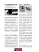
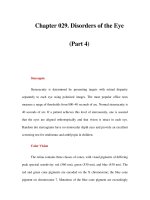
Ⅲ Radiographic contrast series (Figure 11-3)
Ⅲ String sign—from elongated pyloric channel
Ⅲ Shoulder sign—bulge of pyloric muscle into the antrum
Ⅲ Double tract sign—parallel streaks of barium in the narrow channel
T
REATMENT
Surgery—pyloromyotomy is curative.
DUODENAL ATRESIA
DEFINITION
Ⅲ Failure to recanalize lumen after solid phase of intestinal development
Ⅲ Several forms
S
IGNS AND SYMPTOMS
Ⅲ Bilious vomiting without abdominal distention (first day of life).
Ⅲ History of polyhydramnios in 50% of pregnancies.
Ⅲ Down’s syndrome seen in 20–30% of cases.
Ⅲ Associated anomalies include malrotation, esophageal atresia, and con-
genital heart disease.
D
IAGNOSIS
Ⅲ Clinical
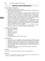
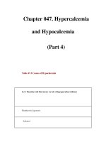
Ⅲ X-ray findings: Double bubble sign (Figure 11-4)
129
HIGH-YIELD FACTS
Gastrointestinal Disease
A 4-week-old male infant
has a 5-day history of
vomiting after feedings.
Physical exam shows a
hungry infant with
prominent peristaltic waves
in the epigastrium. Think:
Hypertrophic pyloric
stenosis.
Typical Scenario
FIGURE 11-3. Abdominal x-ray on the left demonstrates a dilated air-filled stomach with normal caliber bowel, consistent
with gastric outlet obstruction. Barium meal figure on the right confirms diagnosis of pyloric stenosis. The dilated duodenal
bulb is the “olive” felt on physical exam. Note how there is a paucity of contrast traveling through the duodenum. (Photo
courtesy of Drs. Julia Rosekrans and James E. Colletti.)
TREATMENT
Ⅲ Initially, nasogastric and orogastric decompression with intravenous
(IV) fluid replacement.
Ⅲ Treat life-threatening anomalies.
Ⅲ Surgery.
Ⅲ Duodenoduodenostomy.
VOLVULUS
DEFINITION
Ⅲ Gastric and intestinal:
Ⅲ Gastric: sudden onset of severe epigastric pain; intractable retching
with emesis; inability to pass gastric tube
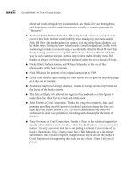
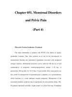
Ⅲ Intestinal: associated with malrotation (Figure 11-5)
S
IGNS AND SYMPTOMS
Ⅲ Vomiting in infancy
Ⅲ Emesis
Ⅲ Abdominal pain
Ⅲ Early satiety
D
IAGNOSIS
Ⅲ Plain abdominal films: characteristic bird-beak appearance
Ⅲ May also see air–fluid level without beak
T
REATMENT
Ⅲ Gastric: emergent surgery
Ⅲ Intestinal: surgery or endoscopy
130
HIGH-YIELD FACTSGastrointestinal Disease
FIGURE 11-4. Duodenal atresia. Gas-filled and dilated stomach show the classic “double
bubble” appearance of duodenal atresia. Note no distal gas is present. (Reproduced, with
permission, from Rudolph’s Pediatrics, 20th ed., Appleton & Lange, 1996.)
INTUSSUSCEPTION
DEFINITION
Invagination of one portion of the bowel into itself. The proximal portion is
usually drawn into the distal portion by peristalsis.
E
PIDEMIOLOGY
Ⅲ Incidence: 1 to 4 in 1,000 live births
Ⅲ Male-to-female ratio: 2:1 to 4:1
Ⅲ Peak incidence: 5 to 12 months
Ⅲ Age range: 2 months to 5 years
C
AUSES
Ⅲ Most common etiology is idiopathic.
Ⅲ Other causes:
Ⅲ Viral (enterovirus in summer, rotavirus in winter)
Ⅲ A “lead point” (or focus) is thought to be present in older children
2–10% of the time. These lead points can be caused by:
Ⅲ Meckel’s diverticulum
Ⅲ Polyp
Ⅲ Lymphoma
Ⅲ Henoch–Schönlein purpura
Ⅲ Cystic fibrosis
S
IGNS AND SYMPTOMS
Classic Triad
Ⅲ Intermittent colicky abdominal pain
Ⅲ Bilious vomiting
Ⅲ Currant jelly stool
Neurologic signs
Ⅲ Lethargy
Ⅲ Shock-like state
131
HIGH-YIELD FACTS
Gastrointestinal Disease
Ⅲ Most common cause of
acute intestinal
obstruction under 2
years of age
Ⅲ Most common site is
ileocolic (90%)
Intussusception is the most
common cause of bowel
obstruction in children ages
2 months to 5 years.
Intussusception and link
with rotavirus vaccine led to
withdrawal of vaccine from
the market.
FIGURE 11-5. Volvulus. 1st AP view done 6 weeks prior to the 2nd AP and corresponding lateral view. Note the markedly
dilated stomach above the normal level of the left hemidiaphragm, in the thoracic cavity. Also present is a large left-sided
diaphragmatic hernia. (Photo courtesy of Dr. Julia Rosekrans.)
Ⅲ Seizure activity
Ⅲ Apnea
Right Upper Quadrant Mass
Ⅲ Sausage shaped
Ⅲ Ill defined
Ⅲ Dance’s sign—absence of bowel in right lower quadrant
D
IAGNOSIS
Abdominal X-Ray
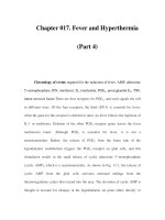
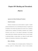
Ⅲ Paucity of bowel gas (Figure 11-6)
Ⅲ Loss of visualization of the tip of liver
Ⅲ “Target sign”—two concentric circles of fat density
Ultrasound
Ⅲ Test of choice
Ⅲ “Target” or “donut” sign—single hypoechoic ring with hyperechoic cen-
ter
Ⅲ “Pseudokidney” sign—superimposed hypoechoic (edematous walls of
bowel) and hyperechoic (areas of compressed mucosa) layers
Barium Enema
Ⅲ Not useful for ileoileal intussusceptions
Ⅲ Requires ingestion of barium, so more invasive than ultrasound
Ⅲ May note cervix-like mass
Ⅲ Coiled spring appearance on the evacuation film
Ⅲ Contraindications
Ⅲ Peritonitis
Ⅲ Perforation
Ⅲ Profound shock
Air Enema
Often provides the same diagnostic and therapeutic benefit of a barium
enema without the barium
132
HIGH-YIELD FACTSGastrointestinal Disease
FIGURE 11-6. Intussusception. Note the paucity of bowel gas in film A. Air enema partially reduces it in film B and then com-
pletely reduced it in film C.
Intussusception
Ⅲ Classic triad is present in
only 20% of cases.
Ⅲ Absence of currant jelly
stool does not exclude
the diagnosis.
Ⅲ Neurologic signs may
delay the diagnosis.
Contrast enema for
intussusception can be both
diagnostic and therapeutic.
Rule of threes:
Ⅲ Barium column should
not exceed a height of 3
feet.
Ⅲ No more than 3
attempts.
Ⅲ Only 3 minutes/attempt.
TREATMENT
Ⅲ Correct dehydration
Ⅲ NG tube for decompression
Ⅲ Hydrostatic reduction
Ⅲ Barium/air enema (see Figure 11–7)
R
ECURRENCE
Ⅲ With radiologic reduction: 7–10%
Ⅲ With surgical reduction: 2–5%
MECKEL’S DIVERTICULUM
DEFINITION
Persistence of the omphalomesenteric (vitelline) duct (should disappear by
seventh week of gestation).
S
IGNS AND SYMPTOMS
Usually in first 2 years:
Ⅲ Intermittent painless rectal bleeding
Ⅲ Intestinal obstruction
Ⅲ Diverticulitis
D
IAGNOSIS
Meckel’s scan (scintigraphy) has 85% sensitivity and 95% specificity. Uptake
can be enhanced with cimetidine, glucagons, or gastrin.
T
REATMENT
Surgical: diverticular resection with transverse closure of the enterotomy.
133
HIGH-YIELD FACTS
Gastrointestinal Disease
Meckel’s Rules of 2
Ⅲ 2% of population
Ⅲ 2 inches long
Ⅲ 2 feet from the ileocecal
valve
Ⅲ Patient is usually under
2 years of age
Ⅲ 2% are symptomatic
FIGURE 11-7. Abdominal x-ray following barium enema in a 2-month-old boy, consistent
with intussusception. Note paucity of gas in right upper quadrant and near obscuring of
liver tip.
Meckel’s diverticulum may
mimic acute appendicitis
and also act as lead point
for intussusception.
APPENDICITIS
DEFINITION
Ⅲ Most common cause for emergent surgery in childhood.
Ⅲ Perforation rates are greatest in youngest children (can’t localize symp-
toms).
Ⅲ Occurs secondary to obstruction of lumen of appendix.
Ⅲ Three phases:
1. Luminal obstruction, venous congestion progresses to mucosal is-
chemia, necrosis, and ulceration.
2. Bacterial invasion with inflammatory infiltrate through all layers.
3. Necrosis of wall results in perforation and contamination.
SIGNS AND SYMPTOMS
Ⅲ Classically: pain, vomiting, and fever.
Ⅲ Initially, pain periumbilical; emesis infrequent.
Ⅲ Anorexia.
Ⅲ Low-grade fever.
Ⅲ Diarrhea infrequent.
Ⅲ Pain radiates to right lower quadrant.
Ⅲ Perforation rate > 65% after 48 hours.
Ⅲ Rectal exam may reveal localized mass or tenderness.
D
IAGNOSIS
Ⅲ History and physical exam is key to rule out alternatives first.
Ⅲ Pain usually occurs before vomiting, diarrhea, or anorexia.
Ⅲ Labs helpful to rule other diagnosis.
Ⅲ Computed tomographic (CT) scan (Figure 11-8) indicated for patients
in whom diagnosis is equivocal—not a requirement for all patients.
T
REATMENT
Ⅲ Surgery as soon as diagnosis made.
Ⅲ Antibiotics are controversial in nonperforated appendicitis.
134
HIGH-YIELD FACTSGastrointestinal Disease
FIGURE 11-8. Abdominal CT of a 10-year-old girl demonstrating enlargement of the appen-
dix, some periappendiceal fluid, and an appendicolith (arrow), consistent with acute ap-
pendicitis.
Ⅲ Broad-spectrum antibiotics needed for cases of perforation (ampicillin,
gentamicin, clindamycin, or metronidazole × 7 days).
Ⅲ Laparoscopic removal associated with shortened hospital stay (nonper-
forated appendicitis).
CONSTIPATION
DEFINITION/SIGNS AND SYMPTOMS
Ⅲ Passage of bulky or hard stool at infrequent intervals.
Ⅲ During the neonatal period usually caused by Hirschsprung disease, in-
testinal pseudo-obstruction, or hypothyroidism.
Ⅲ Other causes include organic and inorganic (e.g., cow’s milk protein in-
tolerance, drugs).
Ⅲ May be metabolic (dehydration, hypothyroidism, hypokalemia, hyper-
calcemia, psychiatric).
T
REATMENT
Ⅲ Increase PO fluid and fiber intake.
Ⅲ Stool softeners (e.g., mineral oil).
Ⅲ Glycerin suppositories.
Ⅲ Cathartics such as senna or docusate.
Ⅲ Nonabsorbable osmotic agents (polyethylene glycol) and milk of mag-
nesia for short periods only if necessary—can cause electrolyte imbal-
ances.
HIRSCHSPRUNG’S MEGACOLON
DEFINITION
Ⅲ Abnormal innervation of bowel (i.e., absence of ganglion cells in
bowel)
Ⅲ Increase in familial incidence
Ⅲ Occurs in males more than females
Ⅲ Associated with Down’s syndrome
S
IGNS AND SYMPTOMS
Ⅲ Delayed passage of meconium at birth
Ⅲ Increased abdominal distention → decreased blood flow → deteriora-
tion of mucosal barrier → bacterial proliferation → enterocolitis
Ⅲ Chronic constipation and abdominal distention (older children)
D
IAGNOSIS
Ⅲ Rectal manometry: measures pressure of the anal sphincter
Ⅲ Rectal suction biopsy: must obtain submucosa to evaluate for ganglionic
cells
T
REATMENT
Surgery is definitive (usually staged procedures).
135
HIGH-YIELD FACTS
Gastrointestinal Disease
A 4-year-old girl has not
had a bowel movement for
a week and this has been a
recurrent problem. Various
laxatives and enemas have
been tried in the past. Prior
to toilet training, the girl
had one bowel movement a
day. Physical exam is
normal except for the
presence of stool in the
sigmoid colon and hard
stool on rectal examination.
After removing the
impaction, the next
appropriate step in
management would be to
administer mineral oil or
other stool softener.
Typical Scenario
Absence of Meissner’s and
Auerbach’s plexus in distal
colon leading to large
bowel obstruction. Think:
Hirschsprung’s.
IMPERFORATE ANUS
DEFINITION
Ⅲ Rectum is blind; located 2 cm from perineal skin
Ⅲ Sacrum and sphincter mechanism well developed
Ⅲ Prognosis good
T
REATMENT
Surgery (colostomy in newborn period).
ANAL FISSURE
DEFINITION
Painful linear tears in the anal mucosa below the dentate line induced by con-
stipation or excessive diarrhea.
S
IGNS AND SYMPTOMS
Pain with defecation, bright red blood on toilet tissue, markedly increased
sphincter tone, extreme pain on digital examination, visible tear upon gentle
lateral retraction of anal tissue.
D
IAGNOSIS
History and physical exam.
T
REATMENT
Sitz baths, fiber supplements, increased fluid intake.
INFLAMMATORY BOWEL DISEASE
DEFINITION
Idiopathic chronic diseases include Crohn’s disease and ulcerative colitis
(UC).
E
PIDEMIOLOGY
Ⅲ Common onset in adolescence and young adulthood.
Ⅲ Bimodal pattern in patients 15 to 25 and 50 to 80 years of age.
Ⅲ Genetics: increased concordance with monozygotic twins versus dizy-
gotic (increased for Crohn’s versus UC).
S
IGNS AND SYMPTOMS (SEE TABLE 11–1)
Ⅲ Crampy abdominal pain
Ⅲ Extraintestinal manifestations greater in Crohn’s than UC
Ⅲ Crohn’s: perianal fistula, sclerosing cholangitis, chronic active hepatitis,
pyoderma gangrenosum, ankylosing spondylitis
Ⅲ UC: bloody diarrhea, anorexia, weight loss, pyoderma gangrenosum,
sclerosing cholangitis, marked by flare-ups
T
REATMENT
Ⅲ Crohn’s: corticosteroids, aminosalicylates, methotrexate, azathioprine,
cyclosporine, metronidazole (for perianal disease), sitz baths, anti–
tumor necrosis factor-α, surgery for complications
Ⅲ UC: aminosalicylates, oral corticosteroids, colectomy
136
HIGH-YIELD FACTSGastrointestinal Disease
Imperforate anus is
frequently associated with
Down’s syndrome and
VACTERL.
A well-nourished 3-month-
old infant is brought to the
emergency department
because of constipation,
blood-streaked stools, and
excessive crying on
defecation. Think: Anal
fissure.
Typical Scenario
A 14-year-old girl has a 2-
month history of crampy
and diffuse abdominal pain
with anorexia and a 4.5-kg
weight loss. The pain is
unrelated to meals, and
there is no diarrhea or
constipation. Appropriate
initial management would
include all of the following:
rectal exam; stool exam for
ova, cysts, and parasites;
complete blood count (CBC)
and erythrocyte
sedimentation rate (ESR);
review of family emotional
stress, except referral to an
eating disorder clinic.
Typical Scenario
IRRITABLE BOWEL SYNDROME
DEFINITION
Ⅲ Abdominal pain associated with intermittent diarrhea and constipation
without organic basis
Ⅲ Approximately 10% in adolescents
S
IGNS AND SYMPTOMS
Ⅲ Abdominal pain
Ⅲ Diarrhea alternating with constipation
D
IAGNOSIS
Ⅲ Difficult to make, exclude other pathology
Ⅲ Obtain CBC, ESR, stool occult blood
T
REATMENT
Ⅲ None specific
Ⅲ Supportive with reinforcement and reassurance
Ⅲ Address any underlying psychosocial stressors
ACUTE GASTROENTERITIS AND DIARRHEA
DEFINITION
Ⅲ Diarrhea is the excessive loss of fluid and electrolytes in stool, usually
secondary to disturbed intestinal solute transport. Technically limited
to lower GI tract.
Ⅲ Gastroenteritis is an inflammation of the entire (upper and lower) GI
tract, and thus involves both vomiting and diarrhea.
E
PIDEMIOLOGY
Ⅲ Increased susceptibility seen in young age, immunodeficiency, measles,
malnutrition, travel, lack of breast-feeding, and contaminated food or
water.
Ⅲ Most common cause of diarrhea in children is viral—often rotavirus.
Ⅲ Children in developing countries often also get infected by bacterial
and parasitic pathogens.
S
IGNS AND SYMPTOMS
Ⅲ Important to obtain information regarding frequency and volume.
137
HIGH-YIELD FACTS
Gastrointestinal Disease
Table 11-1. Crohn’s disease versus ulcerative colitis.
Feature Crohn’s Disease Ulcerative Colitis (UC)
Depth of involvement Transmural Mucosal
Ileal involvement Common Unusual
Ulcers Common Unusual
Cancer risk Lower Higher
Pyoderma gangrenosum Slightly increased Greatly increased
Skip lesions Common Unusual
Fistula Common Unusual
Rectal bleeding Sometimes Common
Acute diarrhea is usually
caused by infectious agents,
whereas chronic persistent
diarrhea may be secondary
to infectious agents,
infection of
immunocompromised host,
or residual symptoms due
to intestinal damage.
Ⅲ General patient appearance important (well appearing versus lethargic).
Ⅲ Associated findings include cramps, emesis, malaise, and fever.
Ⅲ May see systemic manifestations, GI tract involvement, or extraintesti-
nal infections.
Ⅲ Extraintestinal findings include vulvovaginitis, urinary tract infection
(UTI), and keratoconjunctivitis.
Ⅲ Systemic manifestations: fever, malaise, and seizures.
Ⅲ Inflammatory diarrhea—fever, severe abdominal pain, tenesmus.
Ⅲ Noninflammatory diarrhea—emesis, fever usually absent, crampy ab-
dominal pain, watery diarrhea.
D
IAGNOSIS
Ⅲ Examine stool for mucus, blood, and leukocytes (colitis).
Ⅲ Fecal leukocyte—presence of invasive cytotoxin organisms (Shigella,
Salmonella).
Ⅲ Patients with enterohemorrhagic Entamoeba coli and Entamoeba histolyt-
ica—minimal to no fecal leukocytes.
Ⅲ Obtain stool cultures early.
Ⅲ Clostridium difficile toxins—test if recent antibiotic use.
Ⅲ Proctosigmoidoscopy—diagnosis of inflammatory enteritis.
T
REATMENT
Ⅲ Rehydration
Ⅲ Oral electrolyte solutions (e.g., Pedialyte).
Ⅲ Oral hydration for all but severely dehydrated (IV hydration).
Ⅲ Rapid rehydration with replacement of ongoing losses during first 4 to
6 hours.
Ⅲ Do not use soda, fruit juices, jello, or tea. High osmolality may exacer-
bate diarrhea.
Ⅲ Start food with BRAT diet.
Ⅲ Antidiarrheal compounds are not indicated for use in children.
Ⅲ See Table 11-2 for antibiotic treatment of enteropathogens.
138
HIGH-YIELD FACTSGastrointestinal Disease
TABLE 11-2. Antimicrobial treatment for bacterial enteropathogens.
Bacteria Treatment Comments
Aeromonas Trimethoprim–sulfamethoxazole Prolonged diarrhea
(TMP-SMZ)
Campylobacter Erythromycin Early in course of illness
Clostridium difficile Metronidazole or vancomycin Moderate to severe
diagnosis
Enterotoxigenic TMP-SMZ Severe or prolonged illness
Escherichia coli
Enteropathogenic TMP-SMZ Nursery epidemics
Enteroinvasive TMP-SMZ All cases
Salmonella Ampicillin or chloramphenicol Infants < 3 months,
or TMP-SMZ immunodeficient patients,
bacteremia
Shigella TMP-SMZ, ceftriaxone All susceptible organisms
Vibrio cholerae Tetracycline or doxycycline All cases
Ⅲ Diarrhea and emesis →
noninflammatory
Ⅲ Diarrhea and fever →
inflammatory process
Ⅲ Diarrhea and tenesmus →
large colon involvement
Diarrhea is a characteristic
finding in children poisoned
with bacterial toxin of
Escherichia coli, Salmonella,
Staphylococcus aureus, and
Vibrio parahemolyticus, but
not Clostridium botulinum.
A 9-month-old infant who
attends day care has a
temperature of 104°F
(40°C) rectally and diarrhea
× 2 days. The stools are
blood-streaked and contain
mucus. WBC count is 23,000
with 40% segmented
neutrophils and 20% band
forms. Sixty minutes earlier,
the patient had a brief
generalized seizure. Physical
and neurologic exams are
normal. Think: Shigella
sonnei.
Typical Scenario
PREVENTION
Ⅲ Hospitalized patients should be placed under contact precautions
(handwashing, gloves, gowns, etc.).
Ⅲ Education.
Ⅲ Exclude infected children from child care centers.
Ⅲ Report cases of bacterial diarrhea to local health department.
Ⅲ Vaccines for cholera and Salmonella typhi are available.
INTESTINAL WORMS
See Table 11-3 for common intestinal worm infestations.
PSEUDOMEMBRANOUS COLITIS
DEFINITION
Ⅲ Major cause of nosocomial diarrhea.
Ⅲ Rarely occurs without antecedent antibiotics (usually) penicillins,
cephalosporins, or clindamycin.
Ⅲ Antibiotic disrupts normal bowel flora and predisposes to C. difficile di-
arrhea.
S
IGNS AND SYMPTOMS
Classically, blood and mucus with fever, cramps, abdominal pain, nausea, and
vomiting days or weeks after antibiotics.
D
IAGNOSIS
Ⅲ Recent history of antibiotic use
Ⅲ C. difficile toxin in stool of patient with diarrhea
Ⅲ Sigmoidoscopy or colonoscopy
T
REATMENT
Ⅲ Discontinue antibiotics.
Ⅲ Oral metronidazole × 7 to 10 days.
ABDOMINAL HERNIAS
Umbilical
D
EFINITION
Ⅲ Occurs because of imperfect closure of umbilical ring
Ⅲ Common in low-birth-weight, female, and African-American infants
Ⅲ Soft swelling covered by skin that protrudes while crying, straining, or
coughing
Ⅲ Omentum or portions of small intestine involved
Ⅲ Usually 1 to 5 cm
T
REATMENT
Ⅲ Most disappear spontaneously by 1 year of age.
Ⅲ Strangulation rare.
Ⅲ “Strapping” ineffective.
Ⅲ Surgery not indicated unless symptomatic, strangulated, or grows larger
after age 1 or 2.
139
HIGH-YIELD FACTS
Gastrointestinal Disease
Do not treat E. coli
0157:H7 with antibiotics as
there is a higher incidence
of hemolytic uremic
syndrome with treatment.
The most frequent symptom
of infestation with
Enterobius vermicularis is
perineal pruritus. Can
diagnose with transparent
adhesive tape to area
(worms stick).
BRAT Diet for Diarrhea
Bread
Rice
Applesauce
Toast
Inguinal
D
EFINITION
Ⅲ Most common diagnosis requiring surgery
Ⅲ 10 to 20/1,000 live births (50% < 1 year)
Ⅲ Indirect > direct (rare) > femoral (even more rare)
Ⅲ Indirect secondary to patent processus vaginalis
Ⅲ Increased incidence with positive family history
S
IGNS AND SYMPTOMS
Ⅲ Infant with scrotal/inguinal bulge on straining or crying.
Ⅲ Do careful exam to distinguish from hydrocele (see Figure 11-9).
T
REATMENT
Ⅲ Surgery (elective).
Ⅲ Avoid trusses or supports.
140
HIGH-YIELD FACTSGastrointestinal Disease
TABLE 11-3. Common intestinal worms.
Mode of
Intestinal Nematodes Transmission Disease, Symptoms and Signs Treatment
Enterobius vermicularis Hand to Perianal itching, especially at night Albendazole or
(pinworm) mouth mebendazole or
pyrantel pamoate 11
mg/kg (max. dose, 1 g
PO × 1)
Trichuris trichuria Fecal–oral
Ⅲ Usually asymptomatic Albendazole or
(whipworm)
Ⅲ Mild anemia mebendazole
Ⅲ Abdominal pain
Ⅲ Diarrhea, tenesmus
Ⅲ Perianal itching
Ascaris lumbricoides Fecal–oral
Ⅲ Pneumonia Albendazole or
Ⅲ Loeffler’s pneumonitis mebendazole
Ⅲ Intestinal infection/obstruction
Ⅲ Liver failure
Necator Americanus Skin
Ⅲ Intense dermatitis Albendazole or
(new world hookworm) penetration
Ⅲ Loeffler’s pneumonitis mebendazole
and Ancylostoma
Ⅲ Significant anemia
duodenale (old world
Ⅲ GI symptoms
hookworm) Ⅲ Developmental delay in children (irreversible)
Strongyloides stercoralis Skin Same as for Necator, plus: Ivermectin 200
penetration
Ⅲ Diarrhea × 3–6 weeks µg/kg/day × 2 days
Ⅲ Superimposed bacterial sepsis
Trichinella spiralis Infected pork Trichinosis Albendazole 400 mg
Ⅲ Myalgias PO bid × 14 days +
Ⅲ Facial and periorbital edema prednisone 40–60 mg
Ⅲ Conjunctivitis PO qd
Ⅲ Pneumonia, myocarditis, encephalitis,
nephritis, meningitis
Usual albendazole dose is 400 mg PO × 1; usual mebendazole dose is 100 mg PO × 1 for 3 days.
Adapted, with permission, from Stead L. BRS Emergency Medicine. Lippincott Williams & Wilkins, 2000.
In inguinal hernia, processus
vaginalis herniates through
abdominal wall with
hydrocele into canal.
Ⅲ Contralateral hernia occurs in 30% after unilateral repair.
Ⅲ Antibiotics only in at-risk children (e.g., congenital heart disease).
Ⅲ Prognosis excellent (recurrence < 1%, complication rate approximately
2%, infection approximately 1%).
Ⅲ Complications include incarceration.
PEUTZ–JEGHERS SYNDROME
DEFINITION
Ⅲ Mucosal pigmentation of lips and gums with hamartomas of stomach,
small intestine, and colon
Ⅲ Rare; low malignant potential
S
IGNS AND SYMPTOMS
Ⅲ Deeply pigmented freckles on lips and buccal mucosa at birth
Ⅲ Bleeding and crampy abdominal pain
D
IAGNOSIS
Genetic and family studies may reveal history.
T
REATMENT
Excise intestinal lesions if significantly symptomatic.
GARDNER’S SYNDROME
DEFINITION
Multiple intestinal polyps, tumors of soft tissue and bone (especially
mandible).
S
IGNS AND SYMPTOMS
Ⅲ Dental abnormalities
Ⅲ Pigmented lesions in ocular fundus
Ⅲ Intestinal polyps (usually early adulthood) with high malignant potential
D
IAGNOSIS
Ⅲ Genetic counseling
Ⅲ Colon surveillance in at-risk children
141
HIGH-YIELD FACTS
Gastrointestinal Disease
Inguinal hernia increases
with straining, hydrocele
remains unchanged.
FIGURE 11-9. Inguinal hernia (slippage of bowel through inguinal ring) vs. hydrocele (collec-
tion of fluid in scrotum adjacent to testes).
HERNIA
HYDROCELE
A 15-year-old girl with
spots on her lips has some
crampy abdominal pain
associated with bleeding.
Think: Peutz–Jeghers
syndrome.
Typical Scenario
TREATMENT
Aggressive surgical removal of polyps.
CARCINOID TUMORS
DEFINITION
Tumors of enterochromaffin cells in intestine—usually appendix.
S
IGNS AND SYMPTOMS
Ⅲ May cause appendicitis
Ⅲ May cause carcinoid syndrome (increased serotonin, vasomotor distur-
bances, or bronchoconstriction) if metastatic to the liver
T
REATMENT
Surgical excision.
FAMILIAL POLYPOSIS COLI
DEFINITION/ETIOLOGY
Ⅲ Autosomal dominant
Ⅲ Large number of adenomatous lesions in colon
Ⅲ Secondary to germ-line mutations in adenopolyposis coli (APC) gene
S
IGNS AND SYMPTOMS
Ⅲ Highly variable
Ⅲ May see hematochezia, cramps, or diarrhea
Ⅲ Extracolonic manifestations possible
D
IAGNOSIS
Ⅲ Consider family history (strong).
Ⅲ Colonoscopy with biopsy (screening annually after 10 years old if posi-
tive family history).
T
REATMENT
Surgical resection of affected colonic mucosa.
JUVENILE POLYPOSIS COLI
DEFINITION
Ⅲ Most common childhood bowel tumor (3–4% of patients < 21 years)
Ⅲ Characteristically, mucus-filled cystic glands (no adenomatous changes,
no potential for malignancy)
E
PIDEMIOLOGY
Most commonly between 2 and 10 years; less common after 15 years; rarely
before 1 year.
S
IGNS AND SYMPTOMS
Ⅲ Bright red painless bleeding with bowel movement
Ⅲ Iron deficiency
D
IAGNOSIS
Ⅲ Colonoscopy
Ⅲ May use barium enema (not best test)
T
REATMENT
Surgical removal of polyp.
142
HIGH-YIELD FACTSGastrointestinal Disease
MALABSORPTION
Short Bowel Syndrome
D
EFINITION
Ⅲ Occurs with loss of at least 50% of small bowel (with or without loss of
large bowel)
Ⅲ Decreased absorptive surface and bowel function
E
TIOLOGY
Ⅲ May be congenital (malrotation, atresia, etc.)
Ⅲ Most commonly secondary to surgical resection
S
IGNS AND SYMPTOMS
Ⅲ Malabsorption and diarrhea
Ⅲ Steatorrhea (fatty stools): voluminous foul smelling stools that float
Ⅲ Dehydration
Ⅲ Decreased sodium and potassium
Ⅲ Acidosis (secondary to loss of bicarbonate)
T
REATMENT
Ⅲ Total parenteral nutrition (TPN).
Ⅲ Give small feeds orally.
Ⅲ Metronidazole empirically to treat bacterial overgrowth.
Celiac Disease
D
EFINITION
Ⅲ Sensitivity to gluten in diet
Ⅲ Most commonly occurs between 6 months and 2 years
E
TIOLOGY
Ⅲ Factors involved include cereals, genetic predisposition, and environ-
mental factors.
Ⅲ Associated with HLA-B8, -DR7, -DR3, and DQW2.
S
IGNS AND SYMPTOMS
Ⅲ Diarrhea
Ⅲ Failure to thrive
Ⅲ Vomiting
Ⅲ Pallor
Ⅲ Abdominal distention
Ⅲ Large bulky stools
D
IAGNOSIS
Ⅲ Antiendomysial and antitissue transglutaminase antibodies
Ⅲ Biopsy: most reliable test
T
REATMENT
Ⅲ Dietary restriction of gluten
Ⅲ Corticosteroids used rarely (very ill patients with profound malnutri-
tion, diarrhea, edema, and hypokalemia)
Tropical Sprue
D
EFINITION
Ⅲ Generalized malabsorption associated with diffuse lesions of small bowel
mucosa
Ⅲ Seen in people who live or have traveled to certain tropical regions—
some Caribbean countries, South America, Africa, or parts of Asia
143
HIGH-YIELD FACTS
Gastrointestinal Disease
A 5-year-old girl presents
with a protuberant
abdomen and wasted
extremities. Think: Gluten-
induced enteropathy.
Typical Scenario
SIGNS AND SYMPTOMS
Ⅲ Fever, malaise, and watery diarrhea, acutely
Ⅲ After 1 week, chronic malabsorption and signs of malnutrition includ-
ing night blindness, glossitis, stomatitis, cheliosis, muscle wasting
D
IAGNOSIS
Biopsy shows villous shortening, increased crypt depth, and increased chronic
inflammatory cells in lamina propia of small bowel.
T
REATMENT
Ⅲ Antibiotics × 3 to 4 weeks
Ⅲ Folate
Ⅲ Vitamin B
12
Ⅲ Prognosis excellent
Lactase Deficiency
D
EFINITION
Decreased or absent enzyme that breaks down lactose in the intestinal brush
border.
E
TIOLOGY
Ⅲ Congenital absence reported in few cases
Ⅲ Usual mechanism relates to developmental pattern of lactase activity
Ⅲ Autosomal recessive
Ⅲ Also decreases because of diffuse mucosal disease (can occur post viral
gastroenteritis)
S
IGNS AND SYMPTOMS
Ⅲ Seen in response to ingestion of lactose (found in dairy products)
Ⅲ Explosive watery diarrhea with abdominal distention, borborygmi, and
flatulence
Ⅲ Recurrent, vague abdominal pain
Ⅲ Episodic mid-abdominal pain (may or may not be related to milk in-
take)
T
REATMENT
Ⅲ Eliminate milk from diet.
Ⅲ Oral lactase supplement (Lactaid) or lactose-free milk.
Ⅲ Yogurt (with lactase enzyme producing bacteria tolerable in such pa-
tients).
HYPERBILIRUBINEMIA
Physiology—see chapter on gestation and birth.
D
EFINITION
Elevated serum bilirubin
E
PIDEMIOLOGY
Ⅲ Common and in most cases benign
Ⅲ If untreated, severe indirect hyperbilirubinemia neurotoxic
Ⅲ Jaundice in first week of life in 60% of term and 80% of preterm in-
fants—results from accumulation of unconjugated bilirubin pigment
S
IGNS AND SYMPTOMS
Ⅲ Jaundice at birth or in neonatal period
Ⅲ May be lethargic and feed poorly
144
HIGH-YIELD FACTSGastrointestinal Disease
DIAGNOSIS
Ⅲ Direct and indirect bilirubin fractions
Ⅲ Hemoglobin
Ⅲ Reticulocyte count
Ⅲ Blood type
Ⅲ Coombs’ test
Ⅲ Examine peripheral smear
T
REATMENT
Ⅲ Goal is to prevent neurotoxic range.
Ⅲ Phototherapy.
Ⅲ Exchange transfusion.
Ⅲ Treat underlying cause.
Gilbert’s Syndrome
Benign condition caused by missense mutation in transferase gene resulting in
low enzyme levels with unconjugated hyperbilirubinemia.
Crigler–Najjar I Syndrome
D
EFINITION
Ⅲ Autosomal recessive, secondary to mutations in glucuronyl transferase
gene.
Ⅲ Parents of affected children show partial defects but normal serum
bilirubin concentration.
S
IGNS AND
SYMPTOMS
Ⅲ In homozygous infants, will see unconjugated hyperbilirubinemia in first
3 days of life.
Ⅲ Kernicterus common in early neonatal period.
Ⅲ Some treated infants survive childhood without sequelae.
Ⅲ Stools pale yellow.
Ⅲ Persistence of increased levels of indirect bilirubin after first week of life
in absence of hemolysis suggests this syndrome.
D
IAGNOSIS
Ⅲ Based on early age of onset and extreme level of bilirubin in absence of
hemolysis
Ⅲ Definitive diagnosis made by measuring glucuronyl transferase activity
in liver biopsy specimen
Ⅲ DNA diagnosis available
T
REATMENT
Ⅲ Maintain serum bilirubin < 20 mg/dL for first 2 to 4 weeks of life.
Ⅲ Repeated exchange transfusion.
Ⅲ Phototherapy.
Ⅲ Treat intercurrent infections.
Ⅲ Hepatic transplant.
Crigler–Najjar II Syndrome
D
EFINITION
Ⅲ Autosomal dominant with variable penetrance
Ⅲ May be caused by homozygous mutation in glucuronyl transferase iso-
form I activity
145
HIGH-YIELD FACTS
Gastrointestinal Disease
Ⅲ Indirect
hyperbilirubinemia,
reticulosis, and red cell
destruction suggest
hemolysis.
Ⅲ Direct hyperbilirubinemia
may indicate hepatitis,
cholestasis, inborn errors
of metabolism, cystic
fibrosis or sepsis.
Ⅲ If reticulocyte count,
Coombs’, and direct
bilirubin are normal,
then physiologic or
pathologic indirect
hyperbilirubinemia is
suggested.
Children with cholestatic
hepatic disease need
replacement of vitamins A,
D, E, and K (fat soluble).
SIGNS AND SYMPTOMS
Ⅲ Unconjugated hyperbilirubinemia in first 3 days of life.
Ⅲ Concentration remains increased after third week of life.
Ⅲ Kernicterus unusual.
Ⅲ Stool normal.
Ⅲ Infants asymptomatic.
D
IAGNOSIS
Ⅲ Concentration of bilirubin nearly normal.
Ⅲ Decreased bilirubin after 7- to 10-day treatment with phenobarbital
may be diagnostic.
T
REATMENT
Phenobarbital for 7 to 10 days.
Alagille Syndrome
D
EFINITION
Ⅲ Absence or reduction in number of bile ducts
Ⅲ Results from progressive destruction of the ducts
S
IGNS AND SYMPTOMS
Ⅲ Variably expressed
Ⅲ Unusual facies (broad forehead, wide-set eyes, underdeveloped
mandible)
Ⅲ Ocular abnormalities
Ⅲ Cardiovascular abnormalities (peripheral pulmonic stenosis)
Ⅲ Tubulointerstitial nephropathy
Ⅲ Vertebral defect
P
ROGNOSIS
Long-term survival good but may have pruritis, xanthomas, and increased
cholesterol and neurologic complications.
Zellweger Syndrome
D
EFINITION
Ⅲ Rare autosomal recessive condition causing progressive degeneration of
liver and kidneys
Ⅲ 1/100,000 births
S
IGNS AND SYMPTOMS
Ⅲ Usually fatal within 6 to 12 months
Ⅲ Severe generalized hypotonia
Ⅲ Impaired neurologic function with psychomotor retardation
Ⅲ Abnormal head and unusual facies
Ⅲ Hepatomegaly
Ⅲ Renal cortical cysts
Ⅲ Ocular abnormalities
Ⅲ Congenital diaphragmatic hernia
D
IAGNOSIS
Ⅲ Absence of peroxisomes in hepatic cells (on biopsy)
Ⅲ Genetic testing available
146
HIGH-YIELD FACTSGastrointestinal Disease
Extrahepatic Biliary Atresia
D
EFINITION
Distal segmental bile duct obliteration with patent extrahepatic ducts up to
porta hepatis
E
PIDEMIOLOGY
Ⅲ Most common form (85%): obliteration of entire extrahepatic biliary
tree at/above porta
Ⅲ 1/10,000 to 1/15,000 live births
S
IGNS AND SYMPTOMS
Ⅲ Acholic stools
Ⅲ Increased incidence of polysplenia syndrome with heterotaxia, malrota-
tion, levocardia, and intra-abdominal vascular anomalies
D
IAGNOSIS
Ⅲ Ultrasound
Ⅲ Hepatobiliary scintigraphy
Ⅲ Liver biopsy
T
REATMENT
Ⅲ Exploratory laparotomy and direct cholangiography to determine pres-
ence and site of obstruction
Ⅲ Direct drainage—if lesion correctable
Ⅲ Surgery—if lesion not correctable (liver transplant)
HEPATITIS
Ⅲ Continues to be major problem worldwide.
Ⅲ Six known viruses cause hepatitis as their primary manifestation—A
(HAV), B (HBV), C (HCV), D (HDV), E (HEV), and G (HGV).
Ⅲ Many others cause hepatitis as part of their clinical spectrum—herpes
simplex virus (HSV), cytomegalovirus (CMV), Epstein–Barr virus
(EBV), rubella, enteroviruses, parvovirus.
Ⅲ HBV is a DNA virus, whereas HAV, HCV, HDV, HEV, and HGV are
RNA viruses.
Ⅲ HAV and HEV are not known to cause chronic illness, but HBV, HCV,
and HDV cause important morbidity and mortality through chronic in-
fection.
Ⅲ HAV causes most cases of hepatitis in children.
Ⅲ HBV causes one third of all cases; HCV found in 20%.
Hepatitis A
D
EFINITION
Ⅲ RNA-containing member of the Picornavirus family
Ⅲ Found mostly in developing countries
Ⅲ Causes acute hepatitis only
Ⅲ More likely symptomatic in children
Ⅲ Transmission by person-to-person contact; spread by fecal–oral route
Ⅲ Percutaneous transmission rare, maternal–neonatal not recognized
Ⅲ Increased risk in child care centers, contaminated food or water, or
travel to endemic areas
Ⅲ Mean incubation 4 weeks (15–50 days)
147
HIGH-YIELD FACTS
Gastrointestinal Disease
SIGNS AND SYMPTOMS
Ⅲ Abrupt onset with fever, malaise, nausea, emesis, anorexia, and abdomi-
nal discomfort.
Ⅲ Diarrhea common.
Ⅲ Almost all recover but may have relapsing course over several months.
Ⅲ Jaundice.
D
IAGNOSIS
Ⅲ Consider when history of jaundice in family contacts or child care play-
mates or travel history to endemic region
Ⅲ Serologic criteria:
Ⅲ IgM anti-HAV present at onset of illness and disappears within 4
months. May persist for more than 6 months (acute infection). IgG is
detectable at this point.
Ⅲ Increased alanine transaminase (ALT), aspartate transaminase
(AST), bilirubin, and gamma-glutamyl transpeptidase (GGT).
T
REATMENT
Ⅲ Careful handwashing
Ⅲ Vaccines available (preferred over immunoglobulin in children > 2
years)
Hepatitis B
D
EFINITION
Ⅲ DNA virus from the Hepadnaviridae family.
Ⅲ Most important risk factor for infants is perinatal exposure to hepatitis
B surface antigen (HbsAg)-positive mother.
S
IGNS AND SYMPTOMS
Ⅲ Many cases asymptomatic
Ⅲ Increased ALT prior to lethargy, anorexia, and malaise (6–7 weeks post
exposure)
Ⅲ May be preceded by arthralgias or skin lesions and rashes
Ⅲ May see extrahepatic conditions, polyarteritis, glomerulonephritis,
aplastic anemia
Ⅲ Jaundice—icteric skin and mucous membranes
Ⅲ Hepatosplenomegaly and lymphadenopathy common
D
IAGNOSIS
Ⅲ Routine screening requires assay of two serologic markers: HbsAg (all
infected persons, increased when symptomatic) and hepatitis b core
antigen (HbcAg) (present during acute phase, highly infectious state).
Ⅲ HbsAg fall prior to symptom resolution; IgMAb to HbcAg also required
because it is increased early after infectivity and persist for several
months before being replaced by immunoglobulin G (IgG) anti-HbcAg.
Ⅲ HbcAg most valuable; it is present as early as HbsAg and continues to
be present later when HBsAg disappears.
Ⅲ Only anti-HbsAg detected in immunized persons with hepatitis B vac-
cine whereas anti-HbsAb and anti-HBcAG seen in persons with re-
solved infection.
T
REATMENT
Ⅲ No available medical treatment effective in majority of cases.
Ⅲ Interferon-alpha (INF-α) is approved treatment in children.
Ⅲ Liver transplant for patients with end-stage HBV.
148
HIGH-YIELD FACTSGastrointestinal Disease
A 10-year-old boy is
diagnosed with acute
hepatitis A. How would you
treat the parents and
siblings who are doing
fine? Think: IV
immunoglobulin.
Typical Scenario
Hepatitis C
D
EFINITION
Ⅲ Single-stranded RNA virus
Ⅲ Perinatal transmission described but uncommon except with high-titer
HCV
S
IGNS AND SYMPTOMS
Ⅲ Acute infection similar to other hepatitis viruses.
Ⅲ Mild and insidious onset.
Ⅲ Fulminant liver failure rare.
Ⅲ After 20 to 30 years, 25% progress to cirrhosis, liver failure, or primary
hepatocellular carcinoma.
Ⅲ May see cryoglobulinemia, vasculitides, and peripheral neuropathy (ex-
trahepatic).
D
IAGNOSIS
Ⅲ Detection of antibodies to HCV or direct testing for RNA virus
Ⅲ PCR detection possible
Ⅲ Increased ALT
Ⅲ Confirmed by liver biopsy
T
REATMENT
Ⅲ Treat to prevent progression to future complications.
Ⅲ INF-α-2b for patients with compensated liver disease (response rate
long term approximately 25%)
Ⅲ May use with ribavirin for higher frequency of sustained response
Hepatitis D (Delta Agent)
D
EFINITION
Ⅲ Smallest known animal virus
Ⅲ Cannot produce infection without HBV infection (co-infective or su-
perinfection)
Ⅲ Transmission by intimate contact
S
IGNS AND SYMPTOMS
Ⅲ Similar to but more severe than other hepatitis viruses
Ⅲ In co-infection, acute hepatitis more severe, risk of developing chronic
hepatitis low; in superinfection, risk of fulminant hepatitis highest
D
IAGNOSIS
Detect IgM antibody to HDV (2–4 weeks after coinfection, 1 week after su-
perinfection).
P
REVENTION
No vaccine for hepatitis D, but can minimize against hepatitis B (needs hep-
atitis B to infect).
Hepatitis E
D
EFINITION
Ⅲ RNA virus with nonenveloped sphere shape with spikes (similar to cal-
civiruses)
Ⅲ Non-A, non-B hepatitis
149
HIGH-YIELD FACTS
Gastrointestinal Disease
SIGNS AND SYMPTOMS
Ⅲ Similar to HAV, but more severe
Ⅲ No chronic illness
Ⅲ High prevalence of fulminant hepatic failure and death in pregnant
women
D
IAGNOSIS
Ⅲ Antibody to HEV exists
Ⅲ IgM and IgG assays available
Ⅲ Can detect viral RNA in stool and serum by PCR
P
REVENTION
No vaccines available.
Hepatitis G
D
EFINITION
Ⅲ Single-stranded RNA virus of Flaviviridae family.
Ⅲ Virus not yet isolated.
Ⅲ Reported in all population groups in approximately 1.5% U.S. blood
donors.
Ⅲ 1% transmission through transfusions but also by organ transplant.
Ⅲ Vertical transmission occurs.
S
IGNS AND SYMPTOMS
Ⅲ Symptoms associated with hepatic inflammation.
Ⅲ Co-infection does not worsen course of HBV or HCV.
D
IAGNOSIS
Only PCR assays available for testing.
T
REATMENT
No method available.
Neonatal Hepatitis
D
EFINITION
Ⅲ Hepatic inflammation of unknown etiology.
Ⅲ Most result from systemic disease (e.g., sepsis).
Ⅲ Also caused by CMV, HSV, human immunodeficiency virus (HIV).
Ⅲ Nonviral causes include congenital syphilis and toxoplasmosis.
Ⅲ HBV results in asymptomatic infection.
S
IGNS AND SYMPTOMS
Ⅲ Jaundice
Ⅲ Vomiting
Ⅲ Poor feeding
Ⅲ Increased liver enzyme levels
Ⅲ Fulminant hepatitis
T
REATMENT
Ⅲ Antibiotics for bacteria-associated hepatitis
Ⅲ Acyclovir for HSV
Ⅲ Ganciclovir and foscarnet for CMV
150
HIGH-YIELD FACTSGastrointestinal Disease
Autoimmune (Chronic) Hepatitis
D
EFINITION
Ⅲ Hepatic inflammatory process manifested by increased serum amino-
transferase and liver associated autoantibodies
Ⅲ Variable severity
Ⅲ 15–20% of cases associated with HBV
Ⅲ Clinical constellation that suggests immune-mediated disease process
responsive to immunosuppressive treatment
S
IGNS AND SYMPTOMS
Ⅲ Variable.
Ⅲ May mimic acute viral hepatitis.
Ⅲ Onset insidious.
Ⅲ May be asymptomatic or may have fatigue, malaise, anorexia, or amen-
orrhea.
Ⅲ Extrahepatic signs include arthritis, vasculitis, and nephritis.
Ⅲ Mild to moderate jaundice.
D
IAGNOSIS
Ⅲ Made clinically.
Ⅲ Exclude other disease.
T
REATMENT
Ⅲ Corticosteroid
Ⅲ Azathioprine
REYE’S SYNDROME
DEFINITION
Ⅲ Acute encephalopathy and fatty degeneration
Ⅲ Decreased incidence secondary to awareness but relation to acetylsali-
cylic acid (ASA) ingestion.
Ⅲ Many other “Reye-like” syndromes exist.
S
IGNS AND SYMPTOMS
Ⅲ Stereotypic, biphasic course
Ⅲ Usually see prodromal illness, upper respiratory infection (URI), or
chickenpox initially, followed by a period of apparent recovery, then see
abrupt onset of protracted vomiting 5 to 7 days after illness onset
Ⅲ May see delirium, combative behavior, and stupor
Ⅲ Neurologic symptoms including seizures, coma, or death
Ⅲ Slight to moderate liver enlargement
D
IAGNOSIS
Ⅲ Based on clinical staging.
Ⅲ Liver biopsy may show yellow to white color because of high triglyc-
eride content.
T
REATMENT
Ⅲ Control intracranial pressure (ICP) secondary to cerebral edema.
Ⅲ Supportive management depending on clinical stage.
151
HIGH-YIELD FACTS
Gastrointestinal Disease
α
1
-ANTITRYPSIN DEFICIENCY
DEFINITION
Ⅲ α
1
-Antitrypsin is a major protease inhibitor (PI).
Ⅲ A small percentage of homozygous patients have neonatal cholestasis,
and later in childhood cirrhosis.
Ⅲ Present in > 20 codominant alleles; only a few associated with defective
PI.
Ⅲ PI ZZ usually predisposes to clinical deficiency (< 20% develop neona-
tal cholestasis).
S
IGNS AND SYMPTOMS
Ⅲ Variable course
Ⅲ Jaundice, acholic stools, and hepatomegaly in first week of life; jaundice
clears by second to fourth month.
Ⅲ May have complete resolution, persistent liver disease, or cirrhosis.
Ⅲ Older children may present with chronic liver disease.
D
IAGNOSIS
Ⅲ Determination of α
1
-antitrypsin phenotype
Ⅲ Confirmed by liver biopsy
T
REATMENT
Ⅲ Liver transplant curative
Ⅲ No other effective treatment
WILSON’S DISEASE
DEFINITION
Ⅲ Autosomal recessive disease characterized by excessive copper deposi-
tion in brain and liver
Ⅲ Worldwide incidence: 1/30,000
S
IGNS AND SYMPTOMS
Ⅲ Variable manifestations, including:
Ⅲ Asymptomatic in early stages
Ⅲ Jaundice, abdominal pain
Ⅲ Hepatomegaly, subacute/chronic hepatitis or fulminant liver failure
Ⅲ Portal hypertension, ascites, edema, esophageal bleeding
Ⅲ Delayed puberty, amenorrhea, or coagulation defect
Ⅲ Psychosis
Ⅲ Tremors
Ⅲ Kayser–Fleischer rings are greenish-brown rings of pigment seen at the
limbus of the cornea, reflecting deposits of copper in Descemet’s mem-
brane. They can be seen with the naked eye in patients with blue eyes.
In patients with dark eyes, a slit lamp is often needed to identify them.
Ninety percent of patients with Wilson’s disease have Kayser–Fleischer
rings.
D
IAGNOSIS
Ⅲ Copper indices reveal:
Ⅲ Low serum ceruloplasmin
Ⅲ High serum copper level
Ⅲ Liver biopsy for histochemistry and copper quantification
Ⅲ Genetic testing, including siblings
152
HIGH-YIELD FACTSGastrointestinal Disease
Consider ordering serum
ceruloplasmin for any
patient with an unexplained
elevation of liver function
tests (LFTs).
The most likely clinical
manifestation of α
1
-
antitrypsin deficiency in the
newborn is jaundice
(neonatal cholestasis).