Neonatal Formulary - part 7 pptx
Bạn đang xem bản rút gọn của tài liệu. Xem và tải ngay bản đầy đủ của tài liệu tại đây (216.78 KB, 32 trang )
OXYGEN
Use
Supplemental oxygen is used to correct hypoxia in babies with pulmonary problems, especially where this is causing a
mismatch between the ventilation and the perfusion of the lung.
Pathophysiology
Oxygen deserves its place in any pharmacopoeia because – like almost any other drug – oxygen can do a lot of harm as
well as a lot of good. It needs to be used with care; all use should be documented, and the ‘dose’ used recorded. While
lack of oxygen can be damaging, the body can manage with blood that is only about 50–60% saturated as long as the
quantity
of oxygen delivered to the tissues is adequate. Were this not true, the fetus would be in substantial trouble before
birth, as would the brain of the baby with cyanotic heart disease. Cardiac output and tissue perfusion matter more than
blood pressure, and anaemia can undermine oxygen delivery as much as overt cyanosis. While tissue hypoxia can be
damaging, it is the combined effect of CO
2
accumulation and lack of oxygen (asphyxia) that is most damaging, causing a
respiratory (carbonic acid) as well as a metabolic (lactic acid) acidosis.
Too much oxygen can also be damaging however. Prolonged exposure to more than ~60% oxygen can be toxic to the
pulmonary epithelium, and hyperbaric oxygen can cause convulsions. There is also evidence that a relatively high partial
pressure of oxygen in the blood is one of a range of factors that can interfere with the normal growth of blood vessels into
the retina at the back of the eye in the last 10 weeks of what should have been intrauterine life. In most cases the retinal
changes resolve spontaneously leaving no damage, but severe change can lead to permanent (cicatricial) scarring if it
involves more than the outer rim of the retina, and this scarring can sometimes progress to retinal detachment and com-
plete blindness. Good controlled trial evidence that excessive oxygen could cause blindness first appeared in 1952, but we
still do not know precisely what constitutes ‘excessive’ oxygen. Even the ‘routine’ use of 100% oxygen during resuscita-
tion at birth is now being questioned.
The more immature the baby the greater the risk to the eye, but changes take at least six weeks to develop, and most
severe disease develops at a postconceptional age of 33 to 40 weeks. Damage can be reduced by surgery to limit the
capillary proliferation that precedes permanent scarring, but the disease can progress quite rapidly. It is essential, there-
fore, for every baby born before 28 weeks gestation to be seen by an experienced ophthalmologist when they reach a
postmenstrual age of 31 weeks, and then serially every 7–14 days until any acute proliferative change has started to
regress. Babies of 28–32 weeks gestation first merit review when 4 weeks old. Review can be discontinued after 36
weeks if there is still no retinal abnormality because disease appearing for the first time after this is extremely unlikely to
progress to permanent scarring. Diode-laser treatment should be offered
immediately
if stage 3 change develops in zone I
(the central area of the retina), or if any change develops in this zone accompanied by ‘plus’ disease (vessel dilatation
and tortuosity involving two quadrants [usually 6 or more clock hours]). It is also indicated if stage 2 or 3 change with plus
disease develops in zone II. The recent ET-ROP trial showed that there was a 15% risk of the child becoming near blind
in that eye if nothing is done once the disease process had become that extensive, and that prompt intervention can
probably reduce this risk by a third.
Administration
Oxygen is usually given into an incubator, especially in small babies, but cot nursing using a nasal cannula is a valuable
(and economic) alternative that simplifies parental involvement when the concentration of oxygen called for does not
exceed 50%. A humidified head box (see below) is the only satisfactory way of providing more than 60% oxygen; oxygen
tents are seldom very satisfactory at any age. It is not generally recognised that substantial (but not very precisely con-
trolled) amounts of oxygen can also be given directly into any high-sided carry cot or basinette since oxygen, because of its
temperature and density, ‘layers’ immediately above the surface of the mattress; it is not necessary to put a plastic sheet
over the top of the basinette.
Measurement in air
The amount of oxygen each baby is breathing (as a percentage) should be recorded regularly, and those given oxygen via
a nasal catheter should have the ambient concentration needed to provide an equivalent arterial saturation documented
periodically, because the relation between catheter flow and the inspired concentration varies. Equipment needs daily
calibration against room air (20·9% oxygen).
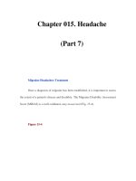
Measuring blood levels
What constitutes a safe range for arterial oxygen pressure is not known. It is said that there must be 50 g/l of desaturated
haemoglobin for cyanosis to be visible. Cyanosis is certainly difficult to detect by eye until 25% of the blood is desaturated,
and in the neonate this often only occurs when the arterial partial pressure (
P
aO
2
) is down to 35 mmHg or 4·7 kPa (the left
hand vertical line in Fig 1). There is no good controlled trial evidence that the use of arterial catheters improves outcome,
although their use can reduce trauma to the heels from repeated capillary sampling. Transcutaneous pressure and
saturation monitors are valuable but not free from error.
The largest cohort study ever mounted showed an association between the prevalence of acute retinopathy and the
duration of exposure to a transcutaneous oxygen (TcpO
2
) of more than 80 mmHg (~10·7 kPa). As a result it has long been
considered good practise to monitor all babies with a postmenstrual age of less than 37 weeks requiring supplemental
oxygen to prevent unnecessary hyperoxia, aiming for TcpO
2
levels of 6–10 kPa. Pulse oximeters are now widely used to
supplement, or replace, the monitoring of TcpO
2
even though the relation between
P
aO
2
and arterial saturation is quite
variable (Fig 1). In particular, blood that is cool, that contains relatively few hydrogen ions, little carbon dioxide, and a
181
Continued on p. 182
minimum of adult haemoglobin, remains well saturated at relatively low pressures. To be 98% certain of keeping
P
aO
2
below 80 mmHg, the
functional
saturation in babies has to be kept from exceeding 92% (Fig 2) – equivalent to a
frac-
tional
saturation of 90%. Given the variable performance of some monitors, even this probably leaves preterm babies at
some small risk of ‘hyperoxia’. Four linked international trials (SUPPORT, BOOST-II and COT) are currently looking to see
how to optimise oxygen delivery to the very preterm baby using a pulse oximeter to monitor saturation.
No such restriction needs to limit management in babies in whom retinal vascular development is complete (or in
whom retinopathy has already developed). Here monitoring is only necessary to identify hypoxia, and significant central
cyanosis is not difficult to detect (although badly chosen fluorescent lighting can affect assessment). Babies with chronic
lung disease are often given oxygen in the belief that this will improve weight gain and reduce emergency hospital
readmission, but there was no evidence of this in the recent Australian BOOST trial, and babies given enough supple-
mental oxygen to maintain a
fractional
saturation of 96–99% in the American STOP-ROP trial actually had
more
pulmonary problems than those only given enough to achieve a saturation of 89–94%. Views differ widely on how often
home use is necessary.
Supply
Piped hospital supplies result in our taking the provision of oxygen for granted: the same is not true in many developing
countries. Oxygen cylinders can be prescribed by GPs and provided for domiciliary use by UK community pharmacists.
Hospital cylinders and regulators can be loaned for portable use, while GPs can prescribe a concentrator in the UK for
patients requiring
continuous
supplemental home oxygen.
Humidification
Piped supplies and cylinders are devoid of water vapour, and humidification is essential when giving >50% oxygen to
avoid excessive drying of the respiratory tract. Bubbling through water at room temperature (25°C) adds 20 grams of
water to each cubic metre of gas (equivalent to 50% saturation at body temperature), and this is generally adequate
unless the baby has been intubated and the nose’s humidification system by-passed. Better humidification requires
the water itself to be fairly close to body temperature: for babies breathing high concentrations of head box oxygen in an
incubator this can be achieved without a heated humidifier by placing a humidification bottle in the incubator itself.
References See also the relevant Cochrane reviews
The STOP-ROP Multicenter Study Group. Supplemental therapeutic oxygen for prethreshold retinopathy of prematurity (STOP-ROP), a
randomised, controlled trial. I: Primary outcomes.
Pediatrics
2000;105:295–310. (See also 420–5.) [RCT]
Bohnhorst B, Peter CS, Poets CF. Detection of hyperoxaemia in neonates: data from three new pulse oximeters.
Arch Dis Child
2002;87:F217–9.
Chow LC, Wright KW, Sola A. Can changes in clinical practice decrease the incidence of severe retinopathy in very low birth weight infants.
Pediatrics
2003;111:339–45.
Tin W, Wariyar U. Giving small babies oxygen: 50 years of uncertainty.
Sem Neonatol
2002;7:361–7.
Frey B, Shann F. Oxygen administration in infants. [Review]
Arch Dis Child
2003;88:F84–8.
Gerstmann D, Berg R, Haskell R,
et al.
Operational evaluation of pulse oximetry in NICU patients with arterial access.
J Perinatol
2003;23:378–83.
Askie LM, Henderson-Smark DJ, Irwig L,
et al.
Oxygen saturation targets and outcomes of extremely preterm infants.
N Engl J Med
2003;349:959–67. [RCT]
Early Treatment for Retinopathy of Prematurity [ET-ROP] Cooperative Group. Revised indications for the treatment of retinopathy of
prematurity.
Arch Ophthalmol
2003;121:1684–96. [RCT] (See also 1697–1701 and 1769–71.)
Balfour-Lynn IM, Primahak RA, Shaw BNJ. Home oxygen for children: who, how and when?
Thorax
2005;60:76–81. (See also the related
British Thoracic Society guideline on home oxygen use [and its paediatric supplement]: www.brit-thoracic.org.uk)
Section of Ophthalmology, American Academy of Pediatrics. Screening examination of premature infants for retinopathy of prematurity.
Pediatrics
2006;117:572–6.
OXYGEN (
Continued
)
182
100
80
60
40
46
40 60
Arterial pO
2
80
Functional oxygen
saturation (%)
100 (mmHg)
8 10 12 14 (kPa)
100
96
92
88
86
46
Arterial pO
2 (
kPa)
95%
CONFIDENCE
INTERVALS
Functional oxygen
saturation (%)
810121416
18
Fig 1 Fig 2
OXYTOCIN
Use
Oxytocin is used (and misused) to induce or augment labour, and to reduce postpartum haemorrhage.
Pharmacology
Oxytocin is a synthetic octapeptide identical to the naturally occurring hypothalamic hormone. Crude pituitary extracts
were first used clinically in 1909, and became commercially available in 1928. Its structure was confirmed by synthesis in
1953. It is now widely used to initiate and augment labour, and given as a continuous IV infusion because uptake is erratic
from mucous membranes and the natural half life is only 3–4 minutes. A sudden bolus can cause transient vasodilatation
and tachycardia, and secondary hypotension can be dangerous in patients with underlying heart disease. Uterine hyper-
stimulation can also cause fetal hypoxia, but this can be reversed by stopping the infusion and/or giving a betamimetic
drug. There is some risk of uterine rupture, especially in patients with a uterine scar, even in the absence of cephalopelvic
disproportion. Effectiveness can be enhanced by prior cervical ‘priming’ with 1 or 2 mg of prostaglandin E
2
vaginal gel
(q.v.), and by amniotomy (which seems to stimulate local prostaglandin synthesis). Doses of more than 15 mU/min have
an antidiuretic effect, and the risk of symptomatic fetal and maternal hyponatraemia is compounded if the mother is given
a lot of 5% dextrose in labour. Such problems can be minimised by always using a motor-driven syringe pump to adminis-
ter IV oxytocin. Use marginally increases subsequent peak neonatal jaundice levels.
While use in mothers delivering under epidural anaesthesia can speed up the second stage of labour, there is no
controlled trial evidence that use (with or without early amniotomy) to ‘augment’ spontaneous labour is of any significant
clinical benefit. On the other hand, such augmentation can certainly cause increased pain and there is a significant risk
of uterine hyperstimulation. Oxytocin (10 units IV or IM) can also reduce the risk of postpartum haemorrhage, and a con-
tinuous infusion can be used if bleeding continues after the placenta is delivered. A combined IM injection of oxytocin
and ergometrine maleate (Syntometrine
®
), is marginally more effective in reducing blood loss, but can sometimes cause
nausea, vomiting, and other unpleasant symptoms together with a transient rise in blood pressure. Misoprostol (q.v.) is an
extremely effective way of containing excessive post-delivery blood when it does occur, especially in a setting where it is
difficult to keep supplies of oxytocin refrigerated. The inadvertent administration of Syntometrine to a baby (in mistake
for an injection of vitamin K) causes respiratory depression, seizures, and severe hyponatraemia. Ventilation and anticon-
vulsant treatment may well be needed for 1–3 days. Paralysis and a tolazoline infusion have sometimes been required.
Luckily, such errors of administration are compatible with complete recovery.
Units used when prescribing oxytocin
Oxytocin is such a potent drug that only a few nanograms are needed. Many staff feel insecure trying to use nanogram
units and, for this reason, oxytocin remains (like insulin) one of the few drugs still widely prescribed using the old pharma-
ceutical unit of potency – the ‘unit’ and, because of its short half life, prescribed in milliunits/min (often written as mU/min)
to avoid writing ‘start by giving 0·001 units/min’.
Treatment
Inducing and augmenting labour: Start with 1 or 2 mU/min and increase this by 1 mU/min every 30 minutes as
necessary using a motor-driven syringe. If more than 4 mU/min proves necessary increase the dose by 2 mU/min incre-
ments once every 30 minutes to a maximum of 20 mU/min.
Postpartum use: Give 10 units of oxytocin (or 1 ml of Syntometrine) IM once the anterior shoulder of the baby is safely
delivered. Continuous IV oxytocin will usually limit residual postpartum bleeding.
Supply and administration
Oxytocin comes in 1 ml ampoules containing 5 or 10 units/ml. 1 ml ampoules of Syntometrine contain 5 units of oxytocin
and 500 micrograms of ergometrine. Midwives can use these products on their own authority. All three products cost
approximately £1·30 per ampoule. Store in the dark at 4°C. For accurate, continuous, dose-adjusted IV administration,
dilute 3 units of oxytocin to 50 ml with 0·9% sodium chloride (or with Hartmann’s solution). This gives a solution
containing 60 mU/ml. Such a solution, when infused at a rate of 1 ml/hr, gives the patient 1 mU/min of oxytocin. (1 unit =
2·2 micrograms of oxytocin).
References See also the relevant Cochrane reviews
Irons DW, Thornton S, Davison JM,
et al
. Oxytocin infusion regimens: time for standardisation.
Br J Obstet Gynaecol
1993;100:786–7.
Soriano D, Dulitzki M, Schiff E,
et al.
A prospective cohort study of oxytocin plus ergometrine compared with oxytocin alone for prevention of
postpartum haemorrhage.
Br J Obstet Gynaecol
1996;103:1068–73. (See also 104:643–4.)
Daraville PA, Campbell NT. Overdose of ergometrine in the newborn infant: acute symptomatology and long-term outcome.
J Paediatr Child
Health
1998;34:83–9.
Royal College of Obstetricians and Gynaecologists.
Induction of labour.
National Evidence-based Clinical Guideline Number 9. London:
RCOG Press, 2001. [SR] (See www.rcog.org.uk)
Harrison K, Read MD, Woodman NM. Current practice for induction of labour in the United Kingdom: time for a review?
J Obstet Gynaecol
2003;23:138–42. (Reprinted in
MIDIRS Midwifery Digest
2003;13:350–3.)
Chelmow D, O’Brien B. Postpartum haemonhage: prevention.
Clin Evid
2006;15:1932–50 (and updates). [SR]
183
PALIVIZUMAB
Use
Prophylactic use of this monoclonal antibody can reduce the risk of a baby requiring hospital admission with bronchiolitis
as a result of respiratory syncytial virus (RSV) infection. Treatment is of no use in babies with established infection. Neither
is treatment with RSV immune globulin (RSV-IVIG).
Respiratory syncytial virus infection
Infection occurs in epidemic form every winter. Adults usually only get a mild cold, but babies can develop a chest infection
severe enough to need hospital admission, and a few need ventilation. Infection is rapidly diagnosed from a nasopharyn-
geal wash specimen using immunofluorescence or an ELISA test (though the latter is not always positive early on). Coryza
and/or apnoea may be the only symptoms in a preterm baby, but infants 2–9 months old can become seriously ill, particu-
larly if they have congenital heart disease or chronic lung disease. Much can be done to reduce these risks by making
parents more aware of the extent to which handwashing and limiting ‘social’ family exposure can lessen cross-infection.
Barrier nursing reduces the risk of infection spreading to other vulnerable inpatients. Most babies merely need brief help
with fluid intake and a little oxygen—support that may not always require hospital admission. Antibiotic treatment
can usually be reserved for babies with heart disease, and for those who need intensive care or become infected while in
hospital. Nebulised adrenaline (q.v.) lowered the number of children needing hospital admission in one recent trial, but it
does not modify the severity of symptoms, or the length of stay, in those who are admitted. Corticosteroids may benefit
a few of those starting to reveal early signs of asthma, but controlled trials have shown that it is of no general value.
Ribavirin (q.v.) and salbutamol (q.v.) are of no proven value.
Pharmacology
Palivizumab is a combined human and murine monoclonal antibody produced by recombinant DNA technology that
inhibits RSV replication. It has a 20 day half life. The first large placebo controlled trials were reported in 1998. A monthly
injection during the seasonal winter epidemic reduces the need for hospitalisation due to RSV infection in babies of less
than 36 weeks gestation. However, use does
not
reduce total health service costs, even when treatment is limited to
babies who are still oxygen dependent because of chronic lung disease, unless readmission rates are atypically high. The
risk of such babies becoming ill is further increased where there are other young school-age children in the house. Side
effects, other than pain and swelling at the injection site, are rare. Use does not interfere with the administration of other
vaccines. Monthly RSV-IVIG treatment (750 mg/kg IV) may be more appropriate in babies needing immunoglobulin for
other reasons, and it offers some protection from other viral illnesses, but it seems to do more harm than good in babies
with cyanotic heart disease.
Prophylaxis
Some babies who are, or were until recently, oxygen dependent because of post-ventilator lung scarring probably merit
treatment. So may a few babies with haemodynamically significant congenital heart disease (see web commentary). Give
15 mg/kg IM once a month for 3–5 months from the start of the winter RSV epidemic. Use the outer thigh (employing
2 sites where the injection volume exceeds 1 ml).
Supply and administration
The 50 mg and 100 mg vials of palivizumab (costing £360 and £600) should be stored at 4°C. Do not freeze. The small
50 mg vial actually contains more than 50 mg of palivizumab, but it is not possible to draw all the drug back out of the
vial after reconstitution. This is why the manufacturers recommend that the powder should be dissolved by running 0·6 ml
(50 mg vials) or 1 ml (100 mg vials) of water for injection slowly down the side of the vial. Rotate gently for 30 seconds
without shaking and then leave it at room temperature for at least 20 minutes until the solution clarifies (it will remain
opalescent). The resultant 100 mg/ml solution must be used within 6 hours. Cost can be reduced by using the larger vial,
and scheduling several babies for treatment on the same day. RSV-IVIG is only licensed in the USA.
References See also the Cochrane reviews of the management of bronchiolitis
Madge P, Paton JY, McColl JH,
et al.
Prospective controlled trial of four infection-control procedures to prevent nosocomial infection with
respiratory syncytial virus.
Lancet
1992;340:1079–83. [RCT]
Impact-RSV Study Group. Palivizumab, a humanized respiratory syncytial virus monoclonal antibody, reduces hospitalization from respiratory
syncytial virus infection in high-risk infants.
Pediatrics
1998;102:531–7. [RCT]
Garrison MM, Christakis DA, Harvey E,
et al.
Systemic corticosteroids in infant bronchiolitis: a meta-analysis.
Pediatrics
2000;105:e44. [SR]
Kumal-Bahl S, Doshi J, Campbell J. Economic analysis of respiratory syncytial virus immunoprophylaxis in high-risk infants.
Arch Pediatr
Adolesc Med
2002;156:1034–41. (See also 1180–1.) [SR]
Shireman TI, Braman KS. Impact and cost-effectiveness of respiratory syncytial virus prophylaxis for Kansas Medicaid’s high-risk children.
Arch Pediatr Adolesc Med
2002;156:1251–5.
King VJ, Viswanathan M, Bordley WC,
et al.
Pharmacologic treatment of bronchiolitis in infants and children. A systematic review.
Arch
Pediatr Adolesc Med
2004;158:127–37. [SR] (See also 119–26.)
Duttweiler L, Nadal D, Frey B. Pulmonary and systematic bacterial co-infection in severe RSV bronchiolitis.
Arch Dis Child
2004;89:1155–7.
Lorzano JM. Bronchiolitis.
Clin Evid
2006;15:355–67 (and updates). [SR]
184
PANCREATIN
Use
Pancreatic supplements are given to aid digestion in patients with cystic fibrosis.
Cystic fibrosis
Cystic fibrosis (CF) is a relatively common, recessively inherited, genetic disorder associated with abnormal mucus produc-
tion. It seems to be caused by a primary defect of chloride ion secretion. Pancreatic damage causes malabsorption, while
the production of viscid sputum renders patients vulnerable to recurrent bacterial infection. Thick meconium may cause
intestinal obstruction (meconium ileus) at birth. Other complications include liver disease (due to biliary tract obstruction)
and male infertility. The high chloride content of sweat is diagnostic, and a sample of sweat for laboratory analysis can be
obtained by pilocarpine iontophoresis in most term babies more than a few weeks old. Most defective mutant genes are
identifiable in the laboratory, and prenatal diagnosis is now possible. Lung damage, including bronchiectasis, used to limit
the number of patients reaching adult life, but survival has now improved significantly. Diagnosis and treatment should
start as soon after birth as possible to minimise lung scarring, and management should be supervised from a specialist
clinic. Nutritional support has played an important part in improving survival. Lung transplantation has been offered to
a few patients, but progressive liver disease remains an unsolved problem. Gene therapy offers hope for the future.
Neonatal screening (using an immunoreactive trypsin blood test) is about to be introduced in the UK, but its net value is
not yet entirely clear.
The condition, which affects about 1:2500 of all children born in Europe and North America, was rapidly fatal when
first recognised fifty years ago, but the median age of survival is now into the late 20s and still rising. Lower respiratory
tract infection needs prompt and vigorous treatment, and there is one small controlled trial to suggest that continuous
prophylaxis with 250 mg a day of oral flucloxacillin during the first two years of life may reduce the need for frequent hos-
pital admission. Only a few babies need pancreatic supplements at birth, but almost all need supplementation before they
are six months old.
Pharmacology
Pancreatin is an extract prepared from pancreatic tissue that is given by mouth to aid digestion in patients with cystic
fibrosis and pancreatic insufficiency. It contains protease enzymes that break protein down into peptides and proteases,
lipases that hydrolyse fats to glycerol and fatty acids, and amylases that convert starch into dextrins and sugars. It is avail-
able as a powder, in capsules containing powder, in capsules containing enteric-coated granules, as free granules, and as
a tablet. Pancreatin should be taken with food, or immediately before food, in order to speed transit into the small intes-
tine, because the constituent enzymes are progressively inactivated by stomach acid. The extent to which the enteric-
coated formulations actually improve intact passage into the duodenum is open to some doubt. Buccal soreness can occur
if the powdered product is not swallowed promptly. Perianal soreness can be helped by a zinc oxide barrier ointment, but
it may be a sign of excessive supplementation. High dose enteric-coated formulations have occasionally caused colonic
strictures in children 2–12 years old.
Treatment
Sprinkle the powder from one capsule of Pancrex V
®
‘125’ into each feed, and increase this dose cautiously as necessary,
as judged by the amount of undigested fat in the stool.
Vitamin supplements
The risk of subclinical vitamin A and D deficiency (the main fat soluble vitamins) can be eliminated by giving Abidec
®
drops
(as outlined in the monograph on multiple vitamins). Marginally low alpha tocopherol levels can persist, even in children
on a 25 mg daily oral supplement of vitamin E (q.v.), but whether this matters is far from clear. More seriously, suboptimal
vitamin K status frequently affects bone metabolism.
Supply
Pancrex V ‘125’ capsules are a convenient first preparation to use in the neonatal period. They contain a minimum of
160 protease units, 2950 lipase units and 3300 amylase units per capsule, and cost 3p each. Enteric-coated microspheres,
which deliver a higher proportion of the constituent enzymes intact into the small intestine, have completely replaced
powders for older children. Store all products in a cool place.
References See the relevant Cochrane reviews of CF care
Feranchak AP, Sontag MK, Wagener JS,
et al.
Prospective, long-term study of fat-soluble vitamin status in children with cystic fibrosis
identified by newborn screen.
J Pediatr
1999;135:601–10.
Littlewood JM, Wolfe SDP. Control of malabsorption in cystic fibrosis.
Paediatr Drugs
2000;2:205–22.
Farrell MH, Farrell PM. Newborn screening for cystic fibrosis: ensuring more good than harm.
J Pediatr
2003;143:707–12.
Parsons EP, Clarke AJ, Bradley DM. Implications of carrier identification in newborn screening for cystic fibrosis.
Arch Dis Child
2003;88:F467–71. (See also F448–9.)
Ratjen F, Döring G. Cystic fibrosis. [Seminar]
Lancet
2003;361:681–9.
Conway SP, Wolfe SP, Brownlee KG,
et al
. Vitamin K status among children with cystic fibrosis and its relationship to bone mineral density
and bone turnover.
Pediatrics
2005;115:1325–31.
Minasian C, McCullagh A, Bush A. Cystic fibrosis in neonates and infants. [Review]
Early Hum Devel
2005;81:997–1004.
185
PANCURONIUM BROMIDE
Use
Pancuronium causes sustained muscle paralysis. Ventilated babies should not be paralysed unless they are sedated, and
most sedated babies do not need paralysis. Sustained paralysis is usually only offered to babies needing major respiratory
support who continue to ‘fight’ the ventilator despite sedation.
Pharmacology
Pancuronium is a competitive non-depolarising muscle relaxant developed in 1966 as an analogue of curare
(tubocurarine), the arrow-tip poison used by South American Indians. Pancuronium competes (like tubocurarine) with
acetylcholine for the neuromuscular receptor sites of the motor end plates of voluntary muscles. It is partly metabolised by
the liver and then excreted in the urine with a half life that is variably prolonged in the neonatal period. Simultaneous
treatment with magnesium sulphate or an aminoglycoside will further prolong the period of blockade. Pharmacokinetic
information does not seem to have influenced the empirical dose regimens generally used in neonatal practice. Very little
crosses the placenta but doses of 100 micrograms/kg have been given into the fetal circulation to induce fetal paralysis
prior to intrauterine fetal transfusion. Larger doses cause paralysis for 2–4 hours.
Sedation or paralysis can reduce lung barotrauma in small babies requiring artificial ventilation, reducing the risk of
pneumothorax and prolonged oxygen dependency due to early bronchopulmonary dysplasia, but there are no grounds for
sedating or paralysing babies as a
routine
. Paralysis makes it much more difficult to judge whether a baby is in pain, and
sedation or paralysis both make it harder to watch for seizures or assess a baby’s neurological status. Rocuronium (q.v.) is
a related drug largely cleared from the body through the biliary tract rather than the renal tract; it may be a better drug
to use where there is renal failure. Atracurium (q.v.) may be the best drug to use in this situation; it is usually given as
a continuous infusion because it has a much shorter duration of action. Suxamethonium (q.v.) is the drug to use when
paralysis is only required for a few minutes.
Never paralyse a non-ventilated baby without first checking that you can achieve face-mask ventilation, and never
paralyse a ventilated baby without first checking whether pain, correctable hypoxia, respiratory acidosis, inadequate
respiratory support, or an inappropriate respiratory rate is the cause of the baby’s continued non-compliance. The prophy-
lactic use of pancuronium might theoretically reduce the risk of fluctuations in cerebral blood flow velocity, but only two
very small trials have, as yet, looked at this issue. Pancuronium sometimes produces a modest but sustained increase in
heart rate and blood pressure, but does not usually have any noticeable effect on gastrointestinal activity or bladder func-
tion, and its use does not preclude continued gavage feeding. Joint contractures responsive to gentle physiotherapy have
been reported in a few chronically paralysed babies but such problems seem to resolve spontaneously once the infant is no
longer paralysed.
Treatment
First dose: Give 100 micrograms/kg to obtain prompt paralysis. Take a blood gas sample 20–30 minutes later (or use
transcutaneous monitoring) to check for CO
2
accumulation. A restless baby who appears to be ’fighting the ventilator‘
may have been contributing to his own ventilation because of inadequate artificial ventilatory support, in which case
paralysis will only exacerbate the problem.
Further doses: Most babies continue to comply with the imposed ventilatory rate as they ‘wake’ from the first
paralysing dose (especially if a moderately fast rate and a relatively short (<0·7 sec) inspiratory time is used) but a few
require prolonged paralysis. The standard repeat dose is half the initial dose IV (or IM) every 4–6 hours as need arises, but
some larger and older babies seem to require a higher maintenance dose.
Antidote
Give a combination of 10 micrograms/kg of glycopyrronium (or 20 micrograms/kg of atropine) and 50 micrograms/kg of
neostigmine IV, as outlined in the monograph on glycopyrronium.
Supply
2 ml ampoules containing 4 mg of pancuronium cost 65p each. Dilute 0·5 ml from the ampoule with 0·5 ml of 0·9%
sodium chloride in a 1 ml syringe before use to obtain a preparation containing 100 micrograms in 0·1 ml. Pancuronium is
stable for up to 6 weeks at 25°C, but is best stored, wherever possible, at 4°C. Open ampoules should not be kept. The US
product contains 1% benzyl alcohol.
References See also relevant Cochrane reviews
Costarino AT, Polin RA. Neuromuscular relaxants in the neonate.
Clin Perinatol
1987;14:965–99.
Besunder JB, Reed MD, Blumer JL. Principles of drug biodisposition in the neonate. A critical evaluation of the pharmacokinetic-
pharmacodynamic interface (part ll).
Clin Pharmacokinet
1988;14:261–86.
Fanconi S, Ensner S, Knecht B. Effects of paralysis with pancuronium bromide on joint mobility in premature infants.
J Pediatr
1995;127:134–6.
186
PAPAVERINE
Use
Papaverine has been used experimentally in a few centres to reduce the risk of vasospasm and prolong the life of
peripheral arterial catheters. Glyceryl trinitrate ointment (q.v.) will sometimes correct any vasospasm that does occur.
Pharmacology
Papaverine is an alkaloid present in opium although it is not related, either chemically or pharmacologically, to the other
opium alkaloids. It was first isolated in 1848 and was briefly in vogue as a vasodilator and antispasmodic in the 1920s
prior to the development of synthetic analogues of atropine. It has a direct relaxant effect on smooth muscle, probably
because it inhibits phosphodiesterase, and it was frequently used for a time by intercavernosal injection in the treatment
of male impotence. It can, however, cause general vasodilatation, and it was shown, in a randomised controlled trial
involving over 200 children in 1993, to extend the functional life of peripheral arterial cannulae. Such lines also lasted
40% longer in a recent neonatal trial. However, since this study only involved 141 babies, more studies are needed before
we can be sure that this form of prophylaxis is not only effective but also safe when used in the preterm baby. Its use in the
first few days of life certainly needs to be approached with some caution because vasodilatation could have adverse
cerebrovascular consequences. A sustained low dose intra-arterial infusion of tolazoline (q.v.) has been used for the same
purpose, and has also been used to abolish the acute ‘white leg’ occasionally caused by femoral artery spasm following
umbilical artery catheterisation. Low dose heparin (q.v.) has been shown to extend the ‘life’ of intravascular lines in adults,
but the only neonatal trials done to date have been too small to show similar benefit with any certainty. The need for inva-
sive arterial sampling has been much reduced by recent developments in pulse oximetry, and systolic blood pressure can
also be monitored noninvasively using Doppler sphygmomanometry.
Adverse effects of papaverine are uncommon, but include flushing, hypotension and gastrointestinal disturbances.
High doses can cause cardiac arrhythmia. The drug is rapidly metabolised by the liver and excreted in the urine, the adult
half life being variable, but usually only a little more than one hour. Nothing is known about the time course of drug
elimination in the neonatal period, or the effect of maternal use during pregnancy or lactation.
Take care not to confuse papaveretum for papaverine.
Papaverine can be confused with papaveretum, a
preparation containing a mixture of opium alkaloids (including morphine and codeine as well as papaverine hydro-
chloride) with potentially fatal consequences.
Treatment
A slow syringe-controlled infusion can be used to help sustain catheter patency. 100 micrograms/ml of papaverine made
up as described below, and infused at a rate of 1 ml per hour (with or without additional heparin), can prolong the func-
tional life of a peripheral arterial line. This fluid must
not
be used to flush the catheter through after sampling: any such
bolus of papaverine could cause marked vasodilatation.
Compatibility
Papaverine was co-infused with heparin at a rate of 1 ml/hour in both the controlled trials referred to above.
Supply
Papaverine is an unlicensed product obtainable by the pharmacy to special order. Ampoules containing 30 mg in 2 ml cost
£2·20 each. To obtain a solution containing approximately 100 micrograms/ml take 5 mg (0·3 ml) of papaverine, dilute to
50 ml with dextrose, dextrose saline or saline, and infuse at a rate of not more than 1 ml per hour using a syringe pump.
While 0·9% sodium chloride is the most frequently used infusion fluid, the sodium this delivers to the baby needs to be
considered with some care when calculating a preterm baby’s total daily sodium intake – dextrose or dextrose saline may
often be a better option.
References
Heulitt MJ, Farrington EA, O’Shea TM,
et al
. Double blind, randomised, controlled trial of papaverine-containing infusions to prevent failure
of arterial catheters in pediatric patients.
Crit Care Med
1993;21:825–9. [RCT]
Griffin MP, Kendrick AS. Does papaverine prevent failure of arterial catheters in neonates? [Abstract]
Pediat Res
1995;37:207A.
Griffin MP, Siadaty MS. Papaverine prolongs patency of peripheral arterial catheters in neonates.
J Pediatr
2005;146:62–5. [RCT]
187
PARACETAMOL
=
Acetaminophen (USAN)
Use
Paracetamol is a useful analgesic also sometimes used to control fever. An IV formulation is now available.
Pharmacology
Paracetamol is an analgesic and anti-pyretic with no anti-inflammatory properties first marketed as an alternative to
phenacetin in 1953. Now that aspirin (q.v.) is no longer recommended for children under 16 (except as an anti-thrombotic
and in Kawasaki disease) because of its link to Reye’s syndrome, paracetamol has become the most widely used analgesic
for children (although dosage is often suboptimal). Intermittent (p.r.n.) administration in response to perceived pain
seldom provides optimal relief and, while anticipatory use (treatment started 1–2 hours before surgery) certainly helps
to control postoperative pain, visceral pain often needs opiate analgesia. Its value in babies with cerebral irritability
has never been properly evaluated. Tolerance does not develop with repeated use (as it does with opioid drugs), and
respiratory depression is not a problem, but there is an analgesic ceiling that cannot be overcome by using a higher dose.
Paracetamol is rapidly absorbed by mouth, widely distributed in the body, and mostly conjugated in the liver before
excretion in the urine. Optimum pain relief occurs over an hour after the blood level peaks. The main metabolite changes
during childhood, but elimination in babies over 3 months old (half life ~3 hours) is as rapid as in adults. It is a little slower
in term babies at birth (4 hours), and is initially 8 hours in babies born more than 8 weeks early. Rectal absorption is rapid
but incomplete, and influenced by the volume given. Toxicity is uncommon in infancy, possibly because reduced
cytochrome P450 activity limits toxic arene metabolite production, but an overdose could still cause late lethal liver failure
if not treated promptly. The IV formulation now available (see web commentary), renders rectal use unnecessary, but
the manufacturer has not yet endorsed IV use in babies less than a year old. Paracetamol is the analgesic of choice in
pregnancy, and the breastfed baby is exposed to less than 5% of the weight-related maternal dose.
Management of fever
While paracetamol, like ibuprofen (q.v.), can undoubtedly give symptomatic relief to a child with a severe flu-like illness
(just as an adult will sometimes take two aspirins and retire to bed), its use to control fever
per se
is usually uncalled for,
and animal evidence suggests that its use in infection can actually do harm. One oral 30 mg/kg dose often suffices.
Prophylactic use in children prone to febrile convulsions is of no proven value. Seizures usually occur while body temper-
ature is still rising, and are only hazardous if prolonged. Most feverish children merely need to be unwrapped. Forced
cooling does not work.
Treatment in the neonate
Oral pain relief: Give a 24 mg/kg loading dose (1 ml/kg of the 24 mg/ml oral elixir) and a maintenance dose of
12 mg/kg every 4 hours (every 8 hours in babies of less than 32 weeks postconceptional age).
IV administration: Give 20 mg/kg over about 15 minutes. Term babies should then be given a further 10 mg/kg main-
tenance dose IV once every 4 hours. Preterm babies should be given further IV doses every 6 hours (using a 10 mg/kg
maintenance dose in babies of 28 weeks postconceptional age rising incrementally to a maintenance dose of 15 mg/kg in
babies of 36 weeks postconceptional age).
Rectal administration: Give term babies a 36 mg/kg loading dose and then 24 mg/kg once every 8 hours
Sustained use: Because experience remains limited it is wise to check the trough blood level before continuing to give
high dose treatment by
any
route for more than 24 hours to a baby less than 3 months old.
Treatment in babies over 3 months old
Oral pain relief: Give a 24 mg/kg loading dose and then 18 mg/kg once every four hours.
IV pain relief: Give a 20 mg/kg loading dose and then 15 mg/kg once every 4–6 hours.
Toxicity
Lethal liver damage can occur in adults if the plasma level exceeds 150 mg/l four or more hours after ingestion (1 mg/l =
6·62 mmol/l). The safe threshold after repeated use is much less certain. Give 150 mg/kg of IV acetylcysteine
promptly
over 30 minutes, in a little 5% dextrose, if there is concern. Then give 12 mg/kg per hour for 4 hours, followed by 4 mg/kg
per hour for 48 hours. Later doses can be given orally.
Blood levels
Measurement requires 50 ml of plasma. Patients can be asymptomatic despite toxic blood levels, but relief of pain and
fever probably requires a peak plasma level of over 20 mg/l. Keep the trough level below 10 mg/l.
Supply
100 ml of the 24 mg/ml sugar-free elixir costs 41p. Parents can get this for a baby over 3 months old without a prescrip-
tion. Using this elixir rectally (instead of a suppository) speeds absorption. 100 ml (10 mg/ml) IV vials cost £1·50. 10 ml
ampoules of acetylcysteine (200 mg/ml) cost £2·50.
References See also the relevant Cochrane reviews
Arana A, Morton NS, Hansen TG. Treatment with paracetamol in infants. [Review]
Acta Anaesthesiol Scand
2001;45:20–29.
Allegaert K, Anderson BJ, Naulaers G,
et al.
Intravenous paracetamol (propacetamol) pharmacokinetics in term and preterm neonates.
Eur
J Clin Pharmacol
2004;60:191–7.
188
PARALDEHYDE
Use
Paraldehyde can be used to achieve the rapid short term control of persistent non-hypoglycaemic convulsions resistant to
full loading doses of IV phenobarbital (q.v.).
Pharmacology
Paraldehyde, a polymer of acetaldehyde, has been used for a century as a sedative-hypnotic and for seizure control. It is
a potent anticonvulsant capable of controlling seizures refractory to phenobarbital and phenytoin without causing
respiratory depression. It exerts its action rapidly and is then eliminated from the body with a half life that is rather
variable, but only a little shorter than that of most other anticonvulsants used in the neonatal period. It crosses the
placenta, but there is nothing to suggest that its use is hazardous in pregnancy.
Drug elimination is by oxidation to acetaldehyde and carbon dioxide in the liver and also by direct excretion through the
lungs. Dispersal into body tissues is very variable (V
D
~ 4 l/kg). The half life in babies is also very variable (8–27 hours) but
generally rather longer than in children (7
1
/2 hours) and adults (6 hours). The dose given does not need to be modified in
babies with kidney failure because renal clearance is negligible, but the drug’s variable and prolonged half life makes
repeated dosing unwise in the first few weeks of life. It has been suggested that high barbiturate levels can retard drug
clearance by the liver, probably because of competition for the liver’s oxidative pathways, but this remains to be
confirmed. It is equally possible that the prolonged half life often seen in the first week of life could be a consequence of
the impact of intrapartum asphyxia on liver metabolism. The management of babies in whom EEG evidence of seizure
activity persists despite treatment with both phenobarbital and phenytoin (q.v.) is in urgent need of further study.
Paraldehyde has fallen out of favour, but might well turn out to be quite effective if a blood level of 100 mg/l can be
achieved. Clonazepam (q.v.), lidocaine (q.v.) and valproate (q.v.) are alternatives currently under study.
The IM route has been widely used in babies: while standard texts now generally consider the rectal route safer,
absorption is then slower and rather less reliable. Large injections are painful and can cause an unpleasant sterile abscess
with subsequent muscle and/or nerve damage, but such problems are very uncommon following the deep intramuscular
injection of volumes not exceeding 1 ml. Rectal diazepam was once widely used to control seizures in a home setting, but
it is much more effective (and more acceptable) to give a dose of liquid lorazepam or midazolam (q.v.) into the nose or
mouth instead. Indeed, this approach provides an extremely effective way of controlling prolonged seizures in
any
setting
when IV access proves difficult to achieve.
Treatment
Intramuscular: Give 0·2 ml/kg
deep
IM. A second identical dose can be given if seizures persist or recur, but further
doses should probably not be given after that for 48 hours in the first month of life because of the drug’s unpredictable
neonatal half life. Undiluted paraldehyde can be given from a plastic syringe as long as it is injected as soon as it is drawn
up, but it should not be left in the syringe for more than 10 minutes because it reacts chemically with rubber and most
plastics (polythene or polypropylene syringes being more resistant than those made of polyvinyl chloride [PVC]).
Intravenous: Paraldehyde
can
be given as an IV infusion, but the use of this route is now generally discouraged, and
there is no need to use a continuous infusion in order to sustain satisfactory anticonvulsant levels for at least 24 hours
given the drug’s long neonatal half life. To give 0·4 ml/kg of paraldehyde (the maximum safe dose) as an IV infusion,
dilute 2·5 ml of paraldehyde to 50 ml with 5% dextrose and then give 4 ml/kg of this solution as a continuous infusion for
just two hours
. Such an infusion has to be given through a polypropylene (and not a PVC) syringe and infusion line.
Rectal: Give 0·4 ml/kg once only mixed in a syringe with an equal volume of olive oil (or mineral oil).
Supply
Stock 5 ml ampoules of paraldehyde (containing 1 g/ml) cost £9·50 each. Do not use the ampoule if there is evidence of
brown discolouration.
Most syringes and infusion sets are made with PVC. The Plastipak
®
syringes made by Becton Dickinson are made
of polypropylene, as are some of the extension sets marketed by Vygon.
References
Giacoia GP, Gessner IK, Zaleska MM,
et al
. Pharmacokinetics of paraldehyde disposition in the neonate.
J Pediatr
1984;104:291–6.
Koren G, Butt W, Tajchgot P,
et al.
Intravenous paraldehyde for seizure control in newborn infants.
Neurology
1986;36:108–11.
Armstrong DL, Battin MR. Pervasive seizures caused by hypoxic-ischaemic encephalopathy: treatment with intravenous paraldehyde.
J Child
Neurol
2001;16:915–7.
Ahmad S, Ellis JC, Kamwendo H,
et al
. Efficacy and safety of intranasal lorazepam versus intramuscular paraldehyde for protracted convul-
sions in children: an open randomised trial.
Lancet
2006;367:1591–7. [RCT]
189
PARENTERAL NUTRITION
Use
Amino acid solutions, together with glucose and other trace nutrients, are used with or without Intralipid
®
(q.v.), to
supplement or replace enteral feeding when milk feeds are contra-indicated or poorly tolerated.
Nutritional factors
Intravenous solutions are capable of providing every nutrient necessary for growth, although enteral feeding is always to
be preferred where it is possible. Serious progressive cholestatic jaundice can occur in the preterm baby who is not offered
at least a little milk by mouth, and sepsis can exacerbate this problem. Preterm babies not given at least 1 g/kg of protein
a day develop a progressive negative nitrogen balance, and an intake of at least 2–3 g/kg a day is necessary to support
growth.
The standard neonatal preparation that is most widely used in the north of England, for example, contains glucose and
a mixture of synthetic L-amino acids (Vaminolact
®
) with trace minerals (7·5 ml/l of Peditrace
®
) water soluble vitamins
(0·7 of a vial of Solivito N
®
) and an extra 30 mg ascorbic acid per litre, and a basic quantity of sodium (27 mmol/l), potas-
sium (20 mmol/l), calcium (12·5 mmol/l), magnesium (1·3 mmol/l) and phosphate (12·3 mmol/l). This provides either 2·7
or 3·5 g/l of nitrogen (17 or 22 g/l of protein), and is available formulated so that the final glucose concentration is 10%,
12·5% or 15% (providing 400, 500 or 600 kcal/l of energy). It contains no iron. Solutions containing more than 10% glu-
cose rapidly cause thrombophlebitis unless infused into a large vessel. Intralipid with Vitlipid N
®
infant should be added to
augment the calorie intake and provide the baby’s other nutritional needs. Amino acid solutions with a profile mimicking
that provided by the placenta or breast milk are now generally used. These contain taurine, and do not produce the high
plasma tyrosine and phenylalanine levels previously seen with egg protein based products. The acidosis that develops
when the intake of non-metabolisable chloride exceeds 6 mmol/kg per day can be reduced by substituting up to
6 mmol/kg of acetate. Aluminium (present as a contaminant in some ingredients – notably calcium gluconate) can cause
permanent neurological damage. One trial has suggested that additional selenium may reduce the risk of sepsis.
Intake
Babies taking nothing by mouth can usually be started on 5 ml/kg per hour of the standard 10% solution with 2·7 g/l of
nitrogen from birth (6 ml/kg in babies over 2 days old). Energy intake can then be increased further, once the baby is
stable, by using a formulation containing 12·5% or 15% glucose (if a central ‘long line’ is available), or by increasing the
infusion rate to 7 or 8 ml/kg per hour. Such a policy provides 2·4 g/kg of protein a day from the outset, but a higher protein
intake may better optimise growth if all nutrition needs to be given IV for many weeks. More phosphate (q.v.) may also
be needed. Some babies of <30 weeks gestation need another 2–3 mmol/kg of sodium a day to replace loss due to renal
immaturity.
Administration
Individually prepared infusions can be supplied, but their routine use causes much unnecessary blood sampling, the
results are no better, and any such policy doubles the total cost. Whether it is appropriate to add heparin (q.v.) remains
inadequately studied. A few other drugs (as noted in the relevant monographs in this compendium) can be co-infused
with the formulation specified here if lack of vascular access so demands, but this may increase the risk of sepsis. These
should be infused using a ‘Y’ connector sited as close to the patient as possible. Do not add
anything
to any amino acid
solution after it leaves the pharmacy.
Monitoring
Clinically stable children require only marginally more biochemical monitoring than bottle fed babies when on the stand-
ard formulation described here: it is the problem that made parenteral nutrition necessary that usually makes monitoring
necessary. Ignore urinary glucose loss unless it exceeds 1%. Liver function should be monitored. Sepsis is the main hazard
associated with any reliance on IV nutrition.
Tissue extravasation
’Tissue burns’ are much more serious than those caused by a comparable solution of glucose. A strategy for the early
treatment is described in the monograph on hyaluronidase (q.v.).
Supply
Pre-prepared standard nominal half-litre bags cost about £20 to produce and remain safe to use for month. Bags should
be changed aseptically after 48 hours; change the bag, filter
and
giving set every 96 hours.
References See also the relevant Cochrane reviews
Heird WC. Parenteral feeding. In: Sinclair JC. Bracken MB, eds.
Effective care of the newborn infant
. Oxford: Oxford University Press, 1992:
Chapter 8, pp 141–60. [SR]
Bishop NJ, Morley R, Day JP,
et al
. Aluminium neurotoxicity in preterm infants receiving intravenous-feeding solutions.
N Engl J Med
1997;336:1557–61. [RCT]
Beecroft C, Martin H, Puntis JWL. How often do parenteral nutrition prescriptions for the newborn need to be individualized?
Clinical
Nutrition
1999;18:83–5.
Yeung MY, Smyth JP, Maheshwari R,
et al.
Evaluation of standardized versus individualised total parenteral nutrition regime for neonates
less than 33 weeks gestation.
J Paediatr Child Health
2003;39:613–7.
Kaufman AA, Gondolesi GE, Fishbein TM. Parenteral nutrition associated liver disease.
Semin Neonatol
2003;8:375–81.
Lenclen R, Crauste-Manciet S, Narcy P,
et al
. Assessment and implementation of a standardized parenteral formulation for early nutritional
support of very preterm infants.
Eur J Pediatr
2006;165:512–8.
190
PENICILLAMINE
Use
Penicillamine is used to treat heavy metal poisoning, and in the long term management of severe rheumatoid arthritis and
Wilson’s disease. Two small studies of prophylaxis have suggested that is has the potential to reduce the risk of retino-
pathy of prematurity (ROP).
Pharmacology
Penicillamine is obtained by controlled hydrolysis of penicillin. It was discovered in 1943 and first came into clinical use in
1956, because of its ability to bind with (chelate) lead, copper, mercury, iron and other heavy metals to form a stable com-
plex that is then excreted in the urine. It is well absorbed when taken by mouth and mostly metabolised by the liver prior to
slow bi-phasic excretion in the urine (the plasma half life being 1–6 hours). No complications have been seen with short
term oral treatment, but sustained use has been associated with skin problems and marrow dysfunction, and with
nephrotic syndrome caused by a membranous nephropathy.
The drug is sometimes used in children with cystinuria (a recessively inherited defect of dibasic amino acid transport in
the proximal tubule) if simpler measures, such as a high fluid intake and the use of sodium bicarbonate to keep the urine
alkaline (pH predominantly ≥6), do not suffice to prevent stone formation. Use the minimum dose needed to keep the uri-
nary cystine concentration reliably below its solubility limit (300 mg/l). Treatment with 20 mg/kg a day is routinely used in
Wilson’s disease (a recessively inherited metabolic disorder associated with excessive copper accumulation) where life
long treatment has revolutionised the management of a previously fatal condition. A similar dose may counteract the cop-
per poisoning that seems to be responsible for Indian childhood cirrhosis, if started early enough. Variable amounts may
be needed in the management of rheumatoid factor positive juvenile chronic arthritis. Adverse effects are not uncommon,
and can be severe, but usually resolve when the drug is discontinued. Reports exist of the use of penicillamine in more
than 100 pregnancies. Most babies have been unaffected at birth, although a minority have shown signs of cutis laxa.
Treatment should certainly not be stopped in a woman with Wilson’s disease, although it may be wise to keep the daily
dose below 500 mg. There is no information on drug use during lactation.
Two small Hungarian trials, involving 281 preterm babies, have suggested that early, prophylactic, high dose adminis-
tration may significantly reduce the risk of retinopathy of prematurity (ROP), either by impeding new vessel growth by
reducing the bioavailability of vascular growth factors, or by acting as a free-radical oxygen scavenger (a property it shares
with vitamin E (q.v.), which has also been used in much the same way). Such treatment should only be contemplated at
present as part of a properly conducted, randomised, controlled trial, because safety needs to be established as systemat-
ically as efficacy before any drug as potent as penicillamine is used on the many in order to benefit the few. The same dose
has also been used to control jaundice in babies with haemolytic disease in Hungary.
Prophylaxis for retinopathy
The only trials have used 100 mg/kg of penicillamine IV once every 8 hours for 3 days, and then 50 mg/kg once a day for
2 weeks.
Monitoring long term treatment
The care of patients requiring sustained treatment with penicillamine should be supervised by a clinician experienced in
the management of metabolic disease. It is generally considered important to check the blood count initially once a week
and then monthly and to suspend treatment if the white cell count falls below 2.5 × 10
9
/l, or the platelet count falls below
120 × 10
9
/l. Nephrotoxicity with proteinuria is an occasional problem. Prednisalone has sometimes been given briefly if
toxic symptoms develop.
Supply
Penicillamine is usually supplied as 125 mg tablets costing 10p each, but the pharmacy can prepare a sugar-free
10 mg/ml suspension for oral use which is stable for 4 weeks if stored at 4°C. No commercial IV preparation is available
at present, and it would take time to develop an ‘in house’ formulation.
References See also Cochrane review of use to prevent ROP
Oga M, Matsue N, Anai T,
et al.
Copper disposition of the fetus and placenta in a patient with untreated Wilson’s disease.
Am J Obstet
Gynecol
1993;169:196–8.
Bavdekar AR, Bhave SA, Pradhan AM,
et al.
Long term survival in Indian childhood cirrhosis treated with D-penicillamine.
Arch Dis Child
1996;74:32–5.
Sanches-Albisua I, Gartde T, Hierro L,
et al.
A high index of suspicion: the key to the early diagnosis of Wilson’s disease in childhood.
J Pediatr
Gastroenterol Nutr
1999;28:18–90.
Chow GK, Streem SB. Medical treatment of cystinuria: results of contemporary clinical practice.
J Urol
1996;156:1579–80.
Cox DW, Tümer Z, Roberts EA. Copper transport disorders: Wilson Disease and Menkes Disease. In: Fernandes J, Saudubray J-M, van den
Berghe G, eds.
Inborn metabolic diseases. Diagnosis and treatment.
3rd edn. Berlin: Springer-Verlag, 2000: Chapter 33, pp 384–91.
Lakatos L, Csáthy L, Nemes E. ‘Bloodless’ treatment of a Jehovah’s witness infant with ABO haemolytic disease.
J Perinatol
1999;19:530–2.
191
PENICILLIN (Penicillin G; Benzylpenicillin)
Use
Benzylpenicillin is the treatment of choice for pneumococcal, meningococcal, aerobic and anaerobic streptococcal, and
gonococcal infection. It is also very adequate for
Listeria
infection, though ampicillin or amoxycillin (q.v.) is even better.
Flucloxacillin (q.v.) is more appropriate for staphylococcal infection because most strains produce penicillinase. Procaine
penicillin (q.v.) is sometimes used to treat syphilis.
Pharmacology
Benzylpenicillin is a naturally occurring, bactericidal substance, first used clinically in 1941, that acts by interfering with
bacterial cell wall synthesis. Fetal concentrations approach those in maternal serum, but extremely little is ingested in
breast milk. Since it is also destroyed by gastric acid and poorly absorbed by the gut, there is no contra-indication to its use
during lactation. Use phenoxymethylpenicillin (penicillin V), which is acid stable, when giving penicillin by mouth, giving
25 mg/kg doses at similar time intervals as for the IV or IM drug (although oral amoxicillin [q.v.] is a more widely used
alternative in this situation). Active excretion by the renal tubules is the most important factor affecting the serum half life,
which falls from 4–5 hours at birth to 1
1
/2 hours by one month (gestation at birth having only a modest influence on this).
Exposure may further stimulate tubular secretion. Very high levels are neurotoxic, making it important to reduce the dose
or choose a different drug when there is renal failure. Transient thrombocytopenia can also occur. Allergic reactions are
the main hazard in those with a history of prior exposure. Penetration into the CSF is limited even when the meninges are
inflamed, and the recommended dose regime takes this into account. Intrathecal injections are seldom necessary.
Intrapartum group B streptococcal (GBS) prophylaxis
Neonatal death from intrapartum-acquired GBS infection is commoner than death from surfactant deficiency in babies
weighing ≥1·5 kg, but prevalence in the UK does not seem to justify the universal screening policy advocated in North
America. Intermittent bowel carriage is common in adults; it seldom causes symptoms but can cause urinary infection
during pregnancy. However, half the babies born to carriers also become carriers for a time, and 1–2% develop life threat-
ening infection within hours of birth. Carriage cannot be eliminated by antenatal treatment and early neonatal infection
often spreads too rapidly for post-delivery treatment to be effective, but prophylaxis started at least 4 hours before
delivery can reduce the risk of neonatal illness. Current US guidelines recommend that ‘at risk’ mothers should have 3 g of
benzylpenicillin every 6 hours as a slow IV injection in labour. Women allergic to penicillin should receive IV erythromycin
or clindamycin (q.v.). Offer prophylaxis to known carriers, to mothers in active preterm labour, to mothers whose mem-
branes have been ruptured ≥18 hours, and to mothers with intrapartum pyrexia (≥38°C). Babies only require further
investigation or treatment after delivery if symptomatic, or born before 35 weeks gestation. An alternative strategy for
protecting babies from
all
early onset bacterial sepsis is outlined in the monograph on ampicillin.
Treatment
Dose: Give 60 mg/kg per dose IM or (slowly) IV when there is evidence of meningitis (especially group B streptococcal
meningitis); 30 mg/kg is more than adequate in all other circumstances. Consider giving gentamicin synergistically as well
for 48 hours for infection with group B streptococci or listeria.
Timing: Give one dose every 12 hours in the first week of life, one dose every 8 hours in babies 1–3 weeks old, and one
dose every 6 hours in babies 4 or more weeks old. The dose should be halved and the dosage interval doubled when there
is renal failure. Give treatment for at least 10 days in proven pneumonia and septicaemia and in the management of con-
genital syphilis. Treat meningitis for 3 weeks and osteitis for 4 weeks. Oral medication is sometimes used to complete a
course of treatment.
Supply and administration
A 600 mg (one million units or one ‘mega unit’) vial costs 43p. Add 5·6 ml of sterile water for injection to get a solution
containing 10 mg in 0·1 ml. Slow IV administration has been advocated, but there is no published evidence to support this
advice (see web commentary). A 60 mg/kg dose of the UK product contains 0·17 mmol/kg of sodium (most US products
contain the potassium salt). Staff handling penicillin regularly should avoid hand contact as this can cause skin sensitisa-
tion. Penicillin V (25 mg/ml) is available as a syrup (£1·70 per 100 ml) which is stable for 2 weeks after reconstitution if
stored at 4°C.
References See the relevant Cochrane reviews of GBS prophylaxis
American Academy of Pediatrics. Committee on Infectious Disease and Committee on Fetus and Newborn. Revised guidelines for prevention
of early onset group B streptococcal (GBS) infection.
Pediatrics
1997;99:489–96.
Mercer BM, Carr TL, Beazley DD,
et al.
Antibiotic use and drug-resistant infant sepsis.
Am J Obstet Gynecol
1999;181:816–21.
Schrag SJ, Zywicki S, Farkey MM,
et al.
Group B streptococcal disease in the era of intrapartum antibiotic prophylaxis.
N Eng J Med
2000;342:15–20.
Oddie S, Embleton ND. Risk factors for early onset neonatal group B streptococcal sepsis: case control study.
BMJ
2002;325:308–11.
Royal College of Obstetrics and Gynaecology.
Prevention of early onset neonatal group B streptococcal disease.
Guideline 36. London:
RCOG Press, 2003. [SR] (See www.rcog.org.uk)
192
PENTASTARCH (and GELATIN)
Use
Colloids can be used to expand intravascular volume in patients with shock or impending shock. Artificial products are
generally as effective as 4·5% human plasma albumin (q.v.) and significantly cheaper.
Pharmacology
Pentastarch and hexastarch are artificial colloids derived from etherified starch with a mean molecular weight
(200,000) three times above that of plasma albumin. The glucagon-like polymerised glucose units are of variable size.
While the smaller molecules are rapidly excreted in the urine, the larger molecules remain in the blood stream for some
days undergoing slow enzymatic degradation. While use is thought to cause a sustained expansion of the intravascular
volume, even when endothelial damage causes increased capillary permeability allowing smaller molecules (such as
plasma albumin) to leak rapidly out of the intravascular space, use of this product rather than gelatin was associated with
an
increased
risk of transient renal failure in adults with septic shock in one recent trial. Large volumes reduce platelet
aggregation, lower the factor VIII level, and increase the bleeding time. The manufacturers stress that little in known
about the use of any of these products during pregnancy or childhood.
Gelatin is a purified protein obtained by the partial hydrolysis of BSE-free bovine collagen. A sterile saline solution
(Gelofusine
®
) containing 40 g/l of modified gelatin has the same properties and uses as dextran 40 (a polymer of glucose)
but gelatin, unlike dextran, does not interfere with subsequent blood grouping and compatibility testing procedures. Gelatin
has also been used in some countries as a haemostatic film or sponge (Sterispon
®
) in surgical procedures. The gelatin in
Gelofusine, with an average molecular weight (30,000) almost half that of human plasma albumin, only has a 4 hour half
life and is rapidly excreted unchanged in the urine. Anaphylactic reactions have been described, but seem rare in young
children. Immediate and delayed-type hypersensitivity reactions have sometimes occurred, however, after immunisation
with vaccines containing gelatin in pre-sensitised children. The trivalent measles (MMR) vaccine is the only UK vaccine to
contain gelatin. Prior exposure to Gelofusine might nevertheless make a reaction to this vaccine marginally more likely.
Indications for use
A major systematic review in 1998 suggested that the indiscriminate use of
any
colloid in the management of hypo-
volaemia actually does more harm than good. However, this may be because the product is being used inappropriately
rather than because it is inherently dangerous. Gelatin can be used to reconstitute packed red cells. It may also be the best
colloid to use during routine surgery because this has the least effect on
in vitro
tests of coagulation, but 20 ml/kg is the
largest dose known to have been used in any one day in the neonatal period. Naturally, where blood has been lost, it will
often be more appropriate to replace this as soon as practicable. Early neonatal hypotension without hypovolaemia is
more appropriately treated with dobutamine and/or dopamine (q.v.), or hydrocortisone (q.v.), while fresh frozen plasma
(q.v.) should be used where there is a significant clotting factor deficiency.
Treatment
20 ml/kg of Gelofusine infused over 5–15 minutes should correct all but the most severe hypovolaemia. The effect of giv-
ing more than a total of 30 ml/kg in the first week of life has not been studied.
Supply
500 ml bags of 6% pentastarch in 0·9% sodium chloride cost £16·50. 500 ml bags of 4% gelatin (Gelofusine) in 0·9%
sodium chloride cost £4·60. Both products contain 154 mmol/l of sodium. They should not be kept once they have been
opened because they contain no preservative. Do not use any material that looks cloudy or turbid.
References See also the relevant Cochrane reviews
The NNNI Trial Group. A randomised trial comparing the effect of prophylactic early fresh frozen plasma, gelatin or glucose on early mortality
and morbidity in preterm babies.
Eur J Pediatr
1996;155:580–8. [RCT]
Hope P. Pump up the volume? The routine early use of colloid in very preterm infants. [Commentary]
Arch Dis Child
1998;78:F163–5.
Roberts JS, Bratton SL. Colloid volume expanders. Problems, pitfalls and possibilities.
Drugs
1998;55:621–30.
Shierhout G, Roberts I. Fluid resuscitation with colloid or crystalloid solutions in critically ill patients: a systematic review of randomised trials.
BMJ
1998;316:961–4. [SR]
Schortgen F, Lacherade J-C, Bruneel F,
et al.
Effects of hydoxyethylstarch and gelatin on renal function in severe sepsis: a multicentre ran-
domsed study.
Lancet
2001;357:911–6. [RCT] (See also 358:581–3.)
Wilkes MM, Navickis RJ, Sibbald WJ. Albumin versus hydroxyethyl starch in cardiopulmonary surgery: a meta-analysis of postoperative
bleeding.
Ann Thorac Surg
2001;72:527–33. [SR]
Wills BA, Dung NM, Loan HT,
et al.
Comparison of three fluid solutions for resuscitation in dengue shock syndrome.
N Engl J Med
2005;353:877–89. [RCT] (See also 941–4.)
193
PETHIDINE
=
Meperidine (USAN)
Use
Pethidine remains widely used to relieve pain during labour, although evidence of efficacy is limited. Use in infancy has
received little study, and toxic quantities of the active metabolite, norpethidine, can accumulate with repeated usage.
Morphine (q.v.) remains by far the best studied neonatal analgesic.
Pharmacology
Pethidine is a synthetic opioid developed in Germany during a review of the many analogues of atropine in 1939. The dose
required to provide analgesia is variable. It is only a tenth as potent as morphine and its analgesic effect is not as well sus-
tained. It was originally hoped that, because it bears no chemical similarity to morphine it would not be addictive, but this
is not so. Oral bioavailability is limited (about 50%) because of rapid first-pass clearance by the liver, where the drug
undergoes hydrolysis or demethylation and conjugation before excretion. Tissue levels markedly exceed plasma levels (V
D
~ 7 l/kg), and clearance in the first three months is much slower than later in infancy. The average half life in young babies
is about 11 hours and also
very
variable (range 3–60 hours), but in babies 3–18 months old may be even lower than it is
in adults (t
1/2
~ 3·5 hours). Similar half life changes have been documented for morphine. This variation between patients
and over time, and the lack of any clear evidence as to what constitutes an effective analgesic dose, makes it difficult to
recommend the use of pethidine in young children. The active metabolite, norpethidine, is renally excreted. It has an
extended half life, and neurotoxic quantities can accumulate with repeated usage, particularly if there is renal failure.
Increased scepticism is being voiced about the drug’s central place in the management of pain relief in labour but, at
the moment, it remains the only parenteral analgesic that midwives in the UK can give on their own authority. It often
causes more drowsiness, disorientation and nausea than genuine relief from pain. Morphine is no better. Sclerotic legisla-
tion denies midwives and their patients straight access to any other parenteral analgesic while the scope for nitrous oxide
analgesia (q.v.) remains undervalued.
Pethidine crosses the placenta rapidly, and cord levels in babies delivered 1–5 hours after the mother had an IM injec-
tion during labour are higher than the corresponding maternal levels. Neonatal respiratory depression is most often seen
2–3 hours after such an injection. Feeding may be slow, and some babies show impaired behavioural responses and EEG
abnormalities for 2–3 days after birth. Maternal use during lactation only exposes the baby to about 2% of the weight-
related maternal dose. There is no evidence of teratogenicity.
Pain relief
Maternal pain relief in labour: A single dose of 100 or 150 mg is usually administered IM. This may be repeated once
during labour but rarely, if ever, more often than this. Try to avoid using a total of more than 1·5 mg/kg.
Pain relief in infancy: A dose of 1 mg/kg IM or IV has been used, but usually only in babies receiving ventilatory sup-
port. No repeat dose should be given for 10–12 hours in babies less than 2 months old (or for 4–6 hours in infants more
than 3 months old) if drug accumulation is to be avoided.
Antidote
Opiate depression is readily reversed by naloxone (q.v.) although this antidote still costs eight times as much as the earlier
dose of pethidine. It does not seem to reverse the signs of neurotoxicity.
Supply and administration
1 and 2 ml ampoules containing 50 mg/ml are available. They cost approximately 50p each. Take 0·2 ml (10 mg) from
the ampoule and dilute to 1 ml with dextrose, saline or dextrose saline to obtain a preparation containing 10 mg/ml for
accurate IM or IV administration.
The storage and administration of pethidine is controlled under Schedule 2 of the UK Misuse of Drugs Regulations
1988 (Misuse of Drugs Act 1971). Midwives in the UK have the legal right to prescribe pethidine or pentazocine with or
without promazine, oxytocin or Syntometrine
®
, and naloxone, and to give lidocaine during labour, on their own authority
(as outlined in the web commentary). Other analgesics can be given if use is covered by a Patient Group Direction.
References See also the relevant Cochrane reviews
Armstong PJ, Bersten A. Normeperidine toxicity.
Anaesth Analg
1986;65:536–8.
Pokela M-L, Olkkola KT, Koivisto M,
et al
. Pharmacokinetics and pharmacodynamics of intravenous meperidine in neonates and infants.
Clin
Pharmacol Ther
1992;52:342–9.
Pokela M-L. Pain relief can reduce hypoxia in distressed neonates during routine treatment procedures.
Pediatrics
1994;93:379–83. [RCT]
Saneto RP, Fitch JA, Cohen BH. Acute neurotoxicity of meperidine in an infant.
Pediatr Neurol
1996;14:339–41.
Hunt S. Pethidine: love it or hate it.
MIDIRS Midwifery Digest
2002;12:363–5.
Bricker L, Laender T. Parenteral opioids for labour pain relief: a systematic review.
Am J Obstet Gynecol
2002;186:S94–109. [SR]
194
Phenobarbitone (former BAN)
=
PHENOBARBITAL
Use
Phenobarbital is widely used in the initial management of neonatal fits. It is seldom the most appropriate drug to use in
the longer term management of epilepsy.
Pharmacology
Phenobarbital, first marketed as a hypnotic in 1904, was widely used as an anticonvulsant for many years but, because of
its adverse effect on cognition and behaviour, its popularity has now declined sharply. There are, however, many adults
still on long term medication. Oral phenobarbital is only slowly absorbed, and IM absorption can take 2–4 hours, so the
drug must be given IV if a rapid response is required. An overdose can cause drowsiness, vasodilatation, hypotension and
dangerous respiratory depression. Hypothermia and hypoglycaemia have been reported. The drug is largely metabolised
by the liver, but a quarter is excreted unchanged in the urine in the neonatal period. The plasma half life is so long in the
neonatal period
(2
–
4 days)
that treatment once a day is perfectly adequate, but the half life decreases with age, and is
halved after 1–2 weeks of medication because the drug acts to induce liver enzymes. This enzyme-inducing property has
been used to speed the liver’s conjugation and excretion of bilirubin. It also influences the metabolism and half life of a
number of other drugs. Phenobarbital, phenytoin and carbamazepine all induce hepatic microsomal enzymes, speeding
the metabolism of oestrogens and progestogens and making it unwise for women to rely on any low dose oral contra-
ceptive when taking any of these anticonvulsants.
Maternal use
See the valproate website for a general discussion of anticonvulsant use during pregnancy and lactation.
Fetal consequences: Barbiturates rapidly cross the placenta, the fetal blood level being two thirds the maternal level.
There is little clear evidence of teratogenicity, but minor cardiac anomalies, skeletal defects and palatal clefts are more
common in the babies of mothers taking anticonvulsants for epilepsy. Phenytoin has been implicated more than pheno-
barbital in this regard and some of the reported defects may have more to do with the epilepsy than its treatment. Fetal
exposure to phenobarbital may, however, have some impact on later cognitive development. The hazards associated with
uncontrolled epilepsy are, however, almost certainly greater than the hazards associated with continued medication.
Neonatal consequences: The babies of mothers taking phenobarbital are occasionally hypoprothrombinaemic at
birth, but this bleeding tendency can be easily corrected by giving the baby 100 micrograms/kg of vitamin K (q.v.) IM at
birth. (A standard 1 mg dose is widely used.) Giving phenobarbital during labour can cause the baby to be rather sleepy,
and feed poorly for 2–3 days. Some authorities (including the
British National Formulary
) feel that breastfeeding may be
unwise in mothers taking phenobarbital on a regular basis, and calculations suggest that neonatal blood levels could
approach or exceed those seen in the mother. More information is needed, because few problems have been reported in
practice. Drowsiness has occasionally been alluded to however, and there is one report of a baby who appeared to
develop severe withdrawal symptoms when breastfeeding was stopped abruptly at 7 months.
Use to prevent IVH: While early reports that giving phenobarbital immediately after birth could reduce the incidence of
intraventricular haemorrhage (IVH) were not supported by later larger trials, there remained a belief that
antenatal
pro-
phylaxis (typically 10 mg/kg slowly IV to the mother, followed by an oral maintenance dose of 100 mg once or twice a day)
might be beneficial. Six trials involving over 1600 women have now been reported and it would seem that, yet again, the
benefits suggested by a number of small trials of variable quality have not been confirmed by subsequent larger studies.
Use to prevent neonatal jaundice: Maternal treatment (typically 100 mg per day) reduces the chance that neonatal
jaundice will need treatment. Neonatal treatment (typically 5–8 mg/kg per day for 2–7 days) also has a measurable effect,
but is not widely used. Phototherapy (q.v.) usually suffices.
Neonatal use
Intrapartum asphyxia: Animal evidence suggests that phenobarbital reduces the amount of damage caused by cere-
bral anoxia (independent of its anticonvulsant effect) and the evidence from one small trial using a prompt 40 mg/kg load-
ing dose suggests it may also be of clinical value, although another small study, and a small trial of the barbiturate
thiopental (q.v.), failed to find evidence of clinical benefit. Other possible strategies are discussed in the monograph on
mannitol (q.v.).
Cholestatic jaundice: Phenobarbital (5 mg/kg per day) will improve bile flow and can sometimes alleviate pruritis,
although ursodeoxycholic acid (q.v.) is usually more effective. Additional vitamin K will be required. Vitamins A, D and E
(q.v.) may be needed if jaundice is prolonged.
Maternal drug dependency: Babies of mothers who are dependent on other drugs as well as opiates who are suffer-
ing
serious
withdrawal symptoms sometimes benefit from a short 4–6 day course of phenobarbital. Start with the same
loading as for seizure control (see below).
Seizures: There is no evidence that failure to control
all
seizure activity puts the baby at increased risk of long term cere-
bral damage. However, it is now also becoming clear that EEG seizure activity often occurs in the absence of visible motor
activity in the newborn baby, and that when such activity is semi-continuous it is potentially damaging. Animal evidence
certainly points in that direction. Much remains to be learnt from conventional or amplitude-integrated (aEEG) examina-
tion. Although some babies who fail to respond to a standard loading dose of phenobarbital seem to respond clinically to
a higher loading dose, electrographic seizure activity often continues unabated. High dose treatment (up to 40 mg/kg)
also makes most babies drowsy enough to render neurological assessment difficult, and a few babies ventilator
dependent. Where a high loading dose
has
been used, no daily maintenance dose should be started for at least 3–4 days
(especially if there has been intrapartum asphyxia). Seizures that fail to respond to phenobarbital may respond to
195
Continued on p. 196
PHENOBARBITAL (
Continued
)
196
phenytoin (q.v.) or high dose lidocaine (q.v.), although some believe paraldehyde (q.v.) is a more appropriate first option.
Clonazepam and midazolam (q.v.) seldom arrest EEG evidence of seizure activity if phenobarbital has not been sucessful.
See the website for a longer discussion of what is currently known about the available options. Pyridoxine dependency
(q.v.) and biotin deficiency (q.v.)
must
be considered if unexplained seizures do not respond to phenobarbital.
The tonic posturing and motor automatisms, the repetitive stereotypic mouthing movements, rotatory arm movements,
pedalling and stepping activity that is seen in most encephalopathic babies is clearly abnormal. The background (inter-
ictal) EEG activity in these babies is also usually very abnormal.
Isolated seizures, in a baby who appears alert, awake and normal when not actually fitting, are usually well controlled
by phenobarbital. These babies usually have a normal inter-ictal EEG, and their long term prognosis is usually good. If phe-
nobarbital and phenytoin fail, carbamazepine (q.v.) valproate (q.v.) or vigabatrin (q.v.) may work. It is seldom necessary to
use more than one drug. Most babies given an anticonvulsant in the neonatal period can be weaned from all treatment
within 7–10 days, and few need medication at discharge from hospital.
Treatment
Give 20 mg/kg as a slow IV loading dose over 20 minutes to control seizures (once any biochemical disturbance, such
as hypoglycaemia, has been excluded or treated) followed by 4 mg/kg once a day IV, IM or by mouth. Increase this to
5 mg/kg once a day if treatment is needed for more than two weeks. Higher loading doses have been used (see above),
but can cause significant respiratory depression.
Blood levels
The therapeutic level in the neonatal period is 20–40 mg/l (1 mg/l = 4·42 mmol/l.). This is higher than the range generally
quoted for use in later childhood. Drowsiness is common, especially if levels exceed 50 mg/l, and respiratory depression
becomes progressively more likely, particularly in the preterm baby. Levels can be measured in 50 ml of plasma. Because of
the long half life, timing is not critical.
Supply and administration
IV ampoules contain viscid propylene glycol (80–90% w/v). 1 ml (30 mg) ampoules costing £1·70, are convenient
for neonatal use; dilution with an equal quantity of water (giving a 15 mg/ml solution) makes injection through a fine
(24 gauge) cannula easier. Greater dilution, though widely recommended, is not necessary with slow administration
when this strength ampoule is used, but slow administration is important to minimise the risk of shock, hypotension, or
laryngospasm. Extravasation is also damaging because the solution has a high osmolality and high pH (10–11). An oral
BNF elixir containing 3 mg/ml is available, but the alcohol content of this is potentially toxic. An aqueous, sugar-free, pre-
paration with a 2-week shelf life can be made in various strengths on request (100 ml for about 70p). Use is controlled
under Section 3 of the UK Misuse of Drugs Regulations 1985 (Misuse of Drugs Act 1971).
References See also the relevant Cochrane reviews
Gilman JT, Gal P, Duchowny MS,
et al
. Rapid sequential phenobarbital treatment of neonatal seizures.
Pediatrics
1989;83:674–8.
Hellstrom-Westas L, Blennow G, Londroth M,
et al
. Low risk of seizure recurrence after withdrawal of antiepileptic treatment in the neonatal
period.
Arch Dis Child
1995;72:F97–101.
Hall RT, Hall FK, Daily DK. High-dose phenobarbital therapy in term newborn infants with severe perinatal asphyxia: a randomised, prospect-
ive study with three-year follow-up.
J Pediatr
1998;132:345–8. [RCT]
Painter MJ, Scher MS, Stein AD,
et al.
Phenobarbital compared with phenytoin for the treatment of neonatal seizures.
N Engl J Med
1999;341:485–9.
Boylan GB, Rennie JM, Pressler RM,
et al.
Phenobarbitone, neonatal seizures, and video-EEG.
Arch Dis Child
2002;86:F165–70.
Toet MC, van der Meij W, de Vries LS,
et al.
Comparison between simultaneously recorded amplitude integrated electro-encephalogram
(cerebral function monitor) and standard electroencephalogram in neonates.
Pediatrics
2002;109:772–9.
Coyle MG, Ferguson A, Lagasse L,
et al.
Diluted tincture of opium (DTO) and phenobarbital versus DTO alone for neonatal opiate withdrawal
in term infants.
J Pediatr
2002;140:561–4. [RCT]
PHENYTOIN
Use
Phenytoin controls acute neonatal seizures as effectively as phenobarbital (q.v.), but phenytoin is seldom the first anti-
convulsant used because it has a rather unpredictable half life. Giving one or other of these drugs controls about 45% of
all neonatal seizures; giving both controls about 60%.
Pharmacology
Phenytoin was first developed and used as an anti-epileptic drug in 1936. Cosmetic changes, such as gum hypertrophy,
acne, hirsutism and facial coarsening have now reduced the popularity of phenytoin as a drug of first choice in the long
term management of epilepsy. Unwanted psychological changes, such as aggression, sedation, depression and impaired
memory, are also common, making carbamazepine (q.v.) and sodium valproate (q.v.) preferable first choice drugs.
Phenytoin may control the arrhythmia seen with digoxin toxicity. An overdose can cause restlessness or drowsiness, vom-
iting, nystagmus and pupilary dilatation, but symptoms resolve without specific intervention when treatment is stopped.
The related prodrug fosphenytoin (1·5 mg of fosphenytoin = 1 mg of phenytoin), is less irritant, but neonatal experience is
limited and prescribing this drug in ‘phenytoin equivalent’ units risks causing confusion.
Pharmacology in pregnancy
Phenytoin crosses the placenta freely and there is a slightly increased risk of congenital malformation (especially cleft
palate and congenital heart disease) in the babies of mothers with epilepsy which is thought to be at least partially due to
anticonvulsant medication. Fetal exposure can also occasionally affect the child’s appearance and measurably retard
growth and intelligence. The issues are more fully discussed in a website entry linked to the monograph on valproate.
While uncontrolled epilepsy is more of a hazard to the fetus than well controlled medication, many adult patients continue
to take medication unnecessarily for many years without review. Mothers who need to remain on medication during preg-
nancy may need to take more phenytoin in the third trimester because of pharmacodynamic changes.
In utero
exposure
can depress fetal vitamin K-dependent clotting factor levels, but the risk of haemorrhage can be controlled by giving IM
vitamin K (q.v.) at birth. Treatment during lactation will result in the baby receiving about a tenth of the mother’s dose on
a weight-related basis.
Pharmacology in the neonate
Oral treatment in babies is more reliable than most texts currently maintain. Phenytoin is excreted by the liver as a glu-
curonide, but elimination varies unpredictably with age, is influenced by many other drugs, and changes rapidly during the
neonatal period. The V
D
is 1·2 l/kg. The elimination process is also rapidly saturated at plasma levels near the upper end of
the therapeutic range. Small changes in the amount prescribed can have a disproportionate effect on the plasma level
once clearance exceeds half the maximum rate possible (the Michaelis constant), prolonging the half life (‘zero-order’
kinetics).
Treatment
A loading dose of 20 mg/kg given IV over at least 20 minutes (to avoid cardiac dysrhythmia) will usually control acute sta-
tus epilepticus at any age. The optimum maintenance dose is variable but 2 mg/kg IV every 8 hours will usually maintain a
therapeutic level in the first week of life, and the same maintenance dose usually works when given by mouth (at least in
babies over 2 weeks old). Older babies may require two or three times as much as this. Crystallisation makes the IM route
unsatisfactory.
Blood levels
The optimum plasma concentration is usually 10–20 mg/l (1 mg/l = 3·96 mmol/l), but 20% less than this in the first 3
months of life because of reduced protein binding. Levels must be measured if phenytoin is given for more than 2–3 days.
Collect 50 ml of plasma just before the drug is next due to be given.
Supply and administration
5 ml (250 mg) ampoules of phenytoin cost £3·40. Give IV through a filter
always
preceded and followed by a bolus of
0·9% sodium chloride because crystals form when phenytoin comes into contact with any solution containing dextrose.
To give IV maintenance treatment accurately, first draw 1 ml of fluid from the ampoule into a syringe and dilute to 10 ml
with 0·9% sodium chloride to get a solution containing 5 mg/ml. The fluid is very alkaline (pH 12). UK ampoules contain
2 g propylene glycol; the US product also contains 10% benzyl alcohol. An oral suspension in sucrose contains 6 mg/ml
(100 ml costs 85p). 750 mg (10 ml) vials of fosphenytoin (which can be given IV or IM) cost £40.
References See also the relevant Cochrane reviews
Bourgeois BFD, Dodson WE. Phenytoin elimination in newborns.
Neurology
1983;33:173–8
.
Takeoka M, Krishnamoorthy KS, Soman TB,
et al.
Fosphenytoin in infants.
J Child Neurol
1998;13:537–40,
Frey OR, von Brenndorff AI, Probst W. Comparison of phenytoin serum concentrations in premature neonates following intravenous and oral
administration.
Ann Pharmacother
1998;32:300–3.
Painter MJ, Scher MS, Stein AD,
et al.
Phenobarbital compared with phenytoin for the treatment of neonatal seizures.
N Engl J Med
1999;341:485–9. [RCT]
197
PHOSPHATE
Use
Supplemental phosphate (as oral sodium phosphate) can be used prophylactically to prevent neonatal rickets due to phos-
phate deficiency in the very low birth weight baby.
Nutritional factors
The transplacental fetal uptake of calcium and phosphate is high especially in the second trimester of pregnancy and com-
parable intakes are hard to achieve after birth in the preterm baby. The mineral content of breast milk is particularly inad-
equate but ordinary neonatal milk formulae (q.v.) are also deficient and most special preterm formulae contain additional
calcium and phosphate for this reason. Breast milk fortifiers (q.v.) contain calcium and phosphate for the same reason.
Deficient mineral intake after birth compromises subsequent bone growth. Poor bone mineralisation leads to osteo-
penia, and pathological fractures can develop once bone growth starts to accelerate after 6–8 weeks; severe deficiency
can also cause rickets with fraying and cupping of the bony metaphyses on X-ray. When breast milk is used, phosphate
deficiency is normally the limiting factor. Low plasma phosphate levels are associated with increased hydroxylation of
25-hydroxycholecalciferol to 1,25-dihydroxycholecalciferol (the metabolically active form of vitamin D), increased phosph-
ate absorption from the gut, maximum renal retention of phosphate, and hypercalciuria (which is corrected by phosphate
supplementation). Parenterally fed babies develop similar problems. Formula fed babies can, on the other hand, sometimes
develop a calcipenic type of rickets with marginal hypocalcaemia and no renal calcium spill, but secondary hyperparathy-
roidism with hyperphosphaturia. There is some evidence of a prenatal deficiency of phosphate in some very low birth
weight babies possibly as a result of pre-eclampsia and/or placental insufficiency. A controlled trial of oral phosphate sup-
plementation in those babies with a low plasma phosphate level and a high initial urinary calcium loss shortly after birth
found that early supplementation can prevent the development of osteopenia of prematurity. Post-discharge supple-
mentation does not seem necessary.
Treatment
Oral administration: Very low birth weight babies developing a plasma phosphate level of <1·5 mmol/l in the first few
weeks of life should be offered 250 micromol of extra phosphate twice a day by mouth. A few babies benefit from supple-
mentation three times a day.
IV administration: The low solubility of inorganic calcium and phosphorus can compromise bone growth in low birth-
weight babies needing prolonged parenteral nutrition (q.v.). Intake can be increased to 1·5 mmol/kg per day by using the
soluble organic salt, sodium glycerophosphate.
Monitoring
Treatment can be reduced or stopped when the plasma phosphate level exceeds 1.8 mmol/l and/or the tubular reabsorp-
tion of phosphate in the urine falls below 95% (in the absence of acute tubular necrosis). The renal tubular phosphate
resorption (% TPR) can be calculated from the formula:
%TPR = 1 −
Urine phosphate
×
Plasma creatinine
× 100
Urine creatinine Plasma phosphate
Supply
An oral solution containing 1 mmol/ml can be obtained by dissolving a 500 mg Phosphate-Sandoz
®
tablet (costing 16p)
in water, and then making the resultant solution up to 16 ml. Alternatively the formulation used in the study published in
the Lancet in 1990 containing 500 micromol/ml (50 mg/ml) of phosphate can be prepared by adding 94·5 g of disodium
hydrogen phosphate dodecahydrate and 41 g of sodium dihydrogen phosphate dihydrate to one litre of chloroform water.
10 ml ampoules containing 2·16 g of anhydrous sodium glycerophosphate suitable for continuous IV infusion are
available in the UK from the Queen’s Medical Centre pharmacy, Nottingham. These ’special order’ ampoules contain
1 mmol/ml of phosphate and 2 mmol/ml of sodium. They cost £1·10.
References
Holland PC, Wilkinson AR, Diaz J,
et al
. Prenatal deficiency of phosphate, phosphate supplementation, and rickets in very-low-birthweight
babies.
Lancet
1990;335:697–701. [RCT]
Pohlandt F. Prevention of postnatal bone demineralisation in very low-birth-weight infants by individually monitored supplementation with
calcium and phosphorus.
Pediatr Res
1994;35:125–9.
Costello I, Powell C, Williams AF. Sodium glycerophosphate in the treatment of neonatal hypophosphataemia.
Arch Dis Child
1995;73:F44–5.
Ryan S. Nutritional aspects of metabolic bone disease in the newborn.
Arch Dis Child
1996;74:F145–8.
Catache M, Leone CR. Role of plasma and urinary calcium and phosphorus measurements in early detection of phosphorus deficiency in very
low birthweight infants.
Acta Paediatr
2003;92:76–80.
Kurl S, Heinonen K, Lansimies E. Randomized trial: effect of short versus long duration of calcium and phosphate supplementation on bone
mineral content of very low birth weight (VLBW) infants born < 32 weeks gestation. [Abstract]
Pediatr Res
2004;55:448A.
198
PHOTOTHERAPY
Use
Effective phototherapy will immediately stop jaundice increasing unless there is abnormal haemolysis.
Physiology
Bilirubin is formed during the breakdown of the iron-
containing haem component of the haemoglobin molecule.
Biliverdin, the first product formed, is then converted to
bilirubin in the reticuloendothelial system. One gram of
haemoglobin yields 35 mg of bilirubin, and the newborn
baby normally produces 8–10 mg/kg of bilirubin a day.
Before birth this then crosses the placenta to be con-
jugated in the mother’s liver and excreted in the bile, a
task that the neonatal liver has to take on after birth.
Conjugated (direct acting) bilirubin is water soluble and
harmless, but excess unconjugated bilirubin is toxic to the
brain causing deafness, athetoid cerebral palsy and death
from ‘kernicterus’ so babies go through a vulnerable period
until their liver enzymes ‘switch on’ after birth. Normal
bilirubin levels in the healthy breastfed term baby are
shown (see Fig). Levels above the 97th centile at 24–36
hours do not always predict high levels at 4–5 days, but
levels above the dotted line suggest abnormal red cell
breakdown (haemolysis), requiring additional diagnostic
assessment. Values below 470 mmol/l (~27 mg/dl) are seldom damaging to the healthy term baby.
199
Photochemistry
Phototherapy causes photo-oxidation or bleaching (as recognised by a neonatal nursing sister in 1958), a reversible
configurational isomerisation (a change in molecular shape without any change in composition), and a non-reversible
structural isomerisation of bilirubin, to a product called lumirubin, which is rapidly excreted in the bile and the urine with-
out prior conjugation in the liver. The natural isomer is toxic and fat soluble, but not very water soluble. The products pro-
duced by phototherapy are non-toxic and water soluble. As a result, phototherapy starts to detoxify the bilirubin in the
blood stream even before any lumirubin is excreted into the gut, or any decline in the plasma bilirubin is detectable. The
bilirubin level will also fall within two hours, unless there is excess haemolysis, making early ‘just in case’ treatment of
moderate jaundice quite unnecessary. Skin bronzing can occur if biliary stasis causes a high
conjugated
bilirubin level,
from an effect of light on accumulating copper porphyrin.
Treatment
Use phototherapy to prevent the
total
plasma bilirubin (in mmol/l) rising above a value equal to ten times the gestational
age (in weeks). Lower this ceiling by 50 mmol/l if there is haemolysis, or the baby is ill. Remember that duplicate measure-
ments, even from the same laboratory, may differ by 10% (95% confidence limits). Some allowance can be made for con-
jugated bilirubin in babies over a week old, but such measurements only have limited accuracy. Exchange transfusion is
seldom needed. However, this does have a role where antibodies have developed in response to feto-maternal red cell
incompatibility, not so much to correct anaemia or jaundice, as to remove antibody-coated cells, especially where
anaemia (Hb <130 g/l) has developed before birth and no intrauterine transfusion has been undertaken.
Administration
Phototherapy only works when jaundice already exists, so there is little point starting treatment until the level approaches
170 mmol/l. The speed of decline is directly related to the amount of light used, until a plateau intensity is reached similar
to that achieved outdoors in the shade on a sunny day (an irradiance of about 2 mW/cm
2
). Unfortunately, much standard
treatment is ‘homeopathic’: a standard light cradle, with 4–8 white strip lights placed 50 cm above the baby, or a fibre-
optic BiliBlanket
®
, only provides about one fifth as much light as this. Halogen lights are even less effective. Halving the
distance between the cradle and the baby, or using both a blanket and a cradle (to give light from above and below), will
double the speed with which the bilirubin level falls. Doing both speeds the fall four fold. ‘Special blue’ (F20T12/BB) lights
are more effective than white lights. Skin exposure should be maximised, and the eyes covered to prevent retinal damage.
Treatment can be stopped while feeding. Extra fluid is not necessary. Skin colour cannot be used to judge jaundice in
babies once they have been started on phototherapy.
References See also the relevant Cochrane reviews
Hansen TWR. Acute management of extreme neonatal jaundice – the potential benefits of intensified phototherapy and interruption of
enterohepatic bilirubin circulation.
Acta Paediatr
1997;86:843–6.
Maisels MJ, Watchko JF. Treatment of jaundice in low birthweight babies. [Review]
Arch Dis Child
2003;88:F459–63. (See also F455–8.)
Newman TB, Lijestrand P, Jeremy RJ,
et al
. Outcomes among newborns with total serum bilirubin levels of 25 mg per deciliter or more.
N Eng
J Med
2006;354:1889–900. (See also 1947–9.)
400
300
200
100
0
Total serum bilirubin
(mmol/l) (mg/100ml)
97th centile
50th centile
3rd centile
20
15
10
5
12096
Age (hours)
7248240
PLASMA ALBUMIN (Human Albumin Solution)
Use
There are few established indications for using plasma albumin – 0·9% sodium chloride, gelatin or pentastarch (q.v.),
expand plasma volume at lower cost. Fresh frozen plasma (FFP) is more appropriate (q.v.) when there is a bleeding tend-
ency, and hypotension managed with an inotrope such as dobutamine (q.v.).
Blood levels
95% of normal babies have a plasma albumin of between 20 and 40 g/l at term, but values of between 10 and 30 g/l are
normal at 28 weeks gestation.
Products
Pooled plasma prepared from donated whole blood contains soluble proteins and a caprylate stabiliser, but no bactericide
or clotting factors. It is prepared by cold ethanol fractionation, sterilised by filtration, and heated to 60°C for 10 hours to
inactivate any contaminating viruses. An isotonic solution with a similar colloid osmotic pressure to plasma contains 4·5%
albumin. A hyperoncotic, isotonic 20% solution is also available. Some products contain significant amounts of alu-
minium. Albumin cost five times as much as pentastarch, and ten times as much as dextran and gelatin. The Australian
SAFE trial, involving 6933 adult patients requiring intensive care, did not confirm the danger associated with albumin use
that a Cochrane review had identified in 1998 (see web commentary). While it failed to identify any situation where giving
albumin improved outcome, many still believe that plasma has a role in treating burns and in severe sepsis.
Indications
Hypovolaemia: The value of plasma infusions in the neonatal period is very imperfectly established. Persisting hypoten-
sion immediately after birth, once acidosis has been corrected, can, rarely, be due to acute hypovolaemia, and this is best
treated by blood transfusion. Most cases are more appropriately treated with an inotrope such as dopamine and/or dobu-
tamine. Trials in adults with burns or trauma found that crystalloids (like Ringer lactate) reduce mortality more than an
albumin infusion. Some artificial colloids, such as pentastarch, may nevertheless be of value in selected patients with
anaphylaxis, peritonitis or septic shock when there are features suggesting increased capillary permeability (a capillary
‘leak’ syndrome), although trials to support such a view have not yet been done.
Hypoproteinaemia: Underproduction due to liver failure, or to excess gut or renal loss, can cause oedema and hypo-
volaemia, triggering a compensatory retention of salt and water. Where this does not respond to a diuretic, 20% albumin
may produce a diuresis, although the effect will be relatively short lived because most of the body’s albumin is in the
extravascular space, intercompartmental exchange is rapid (even when vascular permeability is normal), and plasma pro-
tein turnover is high (25% per day). The use of albumin to treat hypoproteinaemia actually
increased
the risk of death in
one recent systematic review.
Polycythemia: A partial (dilutional) exchange transfusion is sometimes done in a symptomatic child if the venous
haematocrit is 75% or more although this has not yet been shown to have any impact on long term outcome, and can
occasionally cause necrotising enterocolitis. Although colloid (20–30 ml/kg of gelatin or 4·5% albumin) is often used for
this purpose, 0·9% sodium chloride is just as effective.
Treatment and administration
20 ml/kg of 4·5% albumin or 5 ml/kg of 20% albumin may be pickabacked terminally into an existing glucose infusion:
stopping the glucose will merely precipitate reactive hypoglycaemia. Infusion (distal to any filter) into a line containing an
amino acid solution (TPN) increases the risk of bacterial proliferation, but may have to be accepted. Any 20% albumin
must
be given slowly to prevent vascular overload.
Supply
50 ml bottles of 4·5% human albumin solution cost £6·50, and 50 ml bottles of 20% human albumin solution cost
£22·40. Blood grouping is not necessary. Preparations contain 120–150 mmol/l of sodium and small amounts of potas-
sium and are stable for 3 years at room temperature. Do not use if turbid.
References See also the relevant Cochrane reviews
Reading RF, Ellis R, Fleetwood A. Plasma albumin and total protein in preterm babies from birth to eight weeks.
Early Hum Dev
1990;
22:81–7.
Cochrane Injuries Group Albumin Reviewers. Human albumin administration in critically ill patients: systematic review of randomised
controlled trials.
BMJ
1998;317:235–40. [SR]
Horsey P. Albumin and hypovolaemia: is the Cochrane evidence to be trusted?
Lancet
2002;359:70–2. (See also 72–3 and 2278.)
Haynes GR, Navickis RJ, Wilkes MM. Albumin administration – what is the evidence of clinical benefit? A systematic review of randomised
controlled trials.
Eur J Anaesthesiol
2003;20:771–93. [SR]
Schimmel MS, Bromiker R, Soll RF. Neonatal polycythemia: is partial exchange transfusion justified?
Clin Perinatol
2004;31:545–53. [SR]
Uhing MR. The albumin controversy.
Clin Perinatol
2004:31:475–88.
The SAFE Study Investigators. A comparison of albumin and saline for fluid resuscitation in the intensive care unit.
N Eng J Med
2004;350:2247–56. [RCT]
Dempsey EM, Barrington K. Short and long term outcomes following partial exchange transfusion in the polycythaemic newborn: a systemic
review.
Arch Dis Child
2006;91:F2–6. [SR] (See also the systematic review by de Wall on F7–10.)
200
PLATELETS
Use
Platelet concentrates are used in the management of severe thrombocytopenia with bleeding.
Pathophysiology
The risk of serious internal haemorrhage increases sign-
ificantly when the platelet count falls below 30 × 10
9
/l,
and the risk of intracranial haemorrhage may be particu-
larly high in the preterm baby shortly after birth. Always
check first that the ‘thrombocytopenia’ is not due to clots
in the sample.
A number of inherited conditions, and syndromes (such
as thrombocytopenia absent radius [TAR] syndrome) are
associated with thrombocytopenia. These seldom call for
active treatment. Ill babies can have sepsis or a consump-
tion coagulopathy (disseminated intravascular coagulation,
or DIC): the main need here is usually to treat the underly-
ing condition. Platelets can pool in the spleen in conditions
causing hypersplenism (such as rhesus isoimmunisation),
and exchange transfusion can further exacerbate throm-
bocytopenia. A low count may point to focal infection or to
thrombus formation on a long line. Marrow disorders will
reduce platelet production, but the results of a full blood
count and examination of a blood film will usually provide
a diagnostic clue in these situations. Heparin therapy (q.v.)
occasionally causes a dangerous thrombocytopenia that is
made worse if platelets are given.
Platelet antibodies cause most cases of severe
isolated
neonatal thrombocytopenia. Platelet transfusions are of
little value in
auto
immune thrombocytopenia because the
maternal antiplatelet antibodies also attack any trans-
fused platelets. Most of these mothers will have idiopathic
thrombocytopenia (ITP) or systemic lupus erythematosus
(SLE).
Allo
immune thrombocytopenia is more hazardous.
Here maternal antibodies, produced as a result of
201
Is there a
recognisable
congenital syndrome
(e.g. TAR; giant
haemangioma etc)?
No
Yes
Yes
Yes
No
Transfuse random
donor, group
compatible platelets
if necessary.
Treat underlying
disease and support
with random,group
compatible platelets
if necessary.
Treat as autoimmune
with IV immunoglobulin
(q.v.) if the neonatal
platelet count is
≤30 × 10
9
/l or there is
bleeding.
Get the red cells and the platelets of the mother
and baby ’typed‘ by the NBS without delay.
Give IV immunoglobulin and treat as
alloimmune with washed or compatible
platelets if the neonatal platelet count is
<50 × 10
9
/l.
Is there systemic
illness
(e.g. asphyxia,
infection, NEC,
or DIC etc)?
Is there maternal
thrombocytopenia
or SLE?
No
transplacental sensitisation, attack fetal platelets (in a process analogous to the red cell destruction that occurs in rhesus
haemolytic disease): treatment with immunoglobulin (q.v.) may be appropriate, and fully compatible platelets are
required (i.e. they must lack the antigen against which the antibodies are directed). The transfusion service can usually
provide platelets that are both HPA-1a and HPA-5b negative (the antibodies responsible for 95% of all problems). These
will almost always be suitable, and can be used if the situation is urgent before platelet grouping and any formal confirma-
tion of the diagnosis is possible. Maternal washed and irradiated platelets can be used on those rare occasions when the
blood transfusion service finds itself unable to provide suitable donor platelets.
Administration
10 ml/kg of platelets from a single ABO and Rh compatible CMV-negative donor will usually suffice unless there is alloim-
mune thrombocytopenia. Here more is given, and a higher minimum count aimed for, because platelet function is poorer.
To minimise loss, draw the contents of the pack into a 50 ml syringe through a special platelet or blood transfusion set
with a 170–200 mm filter and then infuse over 30 minutes, using a narrow bore extension set linked (near the patient) to
an IV line primed with 0.9% sodium chloride.
Always confirm compatibility by checking that the patient’s name
is on the pack
.
Supply
Leucodepleted 50 ml single-unit packs containing 60 × 10
9
platelets are available from hospital blood banks. They cost
about £70 to prepare and dispense. Packs for intrauterine use are irradiated and further concentrated before issue.
Platelets need to be stored under special conditions, kept at room temperature, and used
promptly
on receipt. They
quickly loose their therapeutic power if this is not done, and bacterial contamination also becomes increasingly likely.
Send 2 ml of blood for grouping.
References
Sola MC, Del Vecchio A, Rimsza LM. Evaluation and treatment of thrombocytopenia in the neonatal intensive care unit.
Clin Perinatol
2000;27:655–79.
Birchall JE, Murphy MF, Kaplan C,
et al.
European Fetomaternal Alloimmune Thrombocytopenia Study Group. European collaborative study
of the antenatal management of feto-maternal alloimmune thrombocytopenia.
Br J Haematol
2003;122:275–88.
Roberts I, Murray NA. Neonatal thrombocytopenia: causes and management.
Arch Dis Child
2003;88:F350–64.
British Society for Haematology. Guidelines for the use of platelet transfusions.
Br J Haematol
2003;122:10–23.
British Committee for Standards in Haematology, Blood Transfusion Task Force. Guidelines on the transfusion of neonates and older chil-
dren.
Br J Haematol
2004;124:433–53.
Chakravorty S, Murray N, Roberts I. Neonatal thrombocytopenia. [Review]
Early Hum Devel
2005;81:35–41.
PNEUMOCOCCAL VACCINES
Use
Two vaccines are now available offering protection from some, but not all, forms of pneumococcal meningitis, septi-
caemia, pneumonia and otitis media.
Pneumococcal infection
A range of potentially serious bacterial infections are caused by the encapsulated Gram-positive coccus
Streptococcus
pneumoniae;
84 capsular forms have been identified, but 8–10 of these are responsible for 85% of the cases seen in
childhood in the UK. The organism, which is becoming increasingly resistant to penicillin and erythromycin, often causes
community acquired pneumonia, and is now the commonest cause of lethal or disabling bacterial meningitis. Patients
with impaired immunity are at particular risk.
Infants at
high risk
include those with homozygous sickle cell disease, with no spleen (or a poorly functioning spleen),
or with congenital or acquired immunodeficiency (including HIV infection). Such patients should be offered prophylactic
antibiotics (see the monograph on immunisation), because the current vaccines only offer protection from
some
of the
capsular types of pneumococcal infection. They may also benefit from being given the multivalent plain polysaccharide
vaccine when two years old, and such immunisation should also be offered to patients two weeks ahead of any planned
splenectomy or chemotherapy.
Products
Plain polysaccharide vaccine: An unconjugated vaccine, active against 23 of the more commonly encountered
capsular types of pneumococcal infection, has been available for some years. Because this vaccine offers relatively little
protection when given to children under two years old, it has generally only been offered to adults, and to older children
considered to be at particularly high risk of infection.
New conjugate vaccine: A new 7-valent protein-polyaccharide vaccine (active against the 4, 6B, 9V, 18C, 19F and
23F strains) was licensed for use in infants and young children in the United States at the beginning of 2000, and its use
reduced the incidence of all invasive pneumococcal disease in young children by 70% within three years, and the incid-
ence due to vaccine-related serotypes by almost 80%.
Contra-indications
Avoid immunisation during an acute infection, and while pregnant. Patients already immunised with the plain 23-valent
vaccine (or the earlier 12- or 14-valent vaccines) do not need to be re-immunised with the present 23-valent vaccine for
3–5 years.
Interactions
The conjugate vaccine can be given (into a different limb) at the same time as any other childhood vaccine, but parents
who seem unhappy at the thought of their child facing more than one ‘needle’ at a single clinic visit can, if necessary, be
offered a different, staged, plan. The plain vaccine should not be given until at least 8 weeks after the new conjugate vac-
cine has been given. Anaphylaxis is extremely unlikely – its management is discussed in the monograph on immunisation.
Administration
Conjugate vaccine: Young children who have not yet started their primary course of immunisation should be offered
three 0·5 ml doses of the new conjugate 7-valent vaccine. Children in the UK are now offered this when 2, 4 and
13 months old. A very similar four dose policy is currently used in North America.
Plain vaccine: High risk children (see above) who are 2 or more years old should still be offered a single 0·5 ml
deep intramuscular injection of the plain 23-valent vaccine, because it provides broader protection from pneumococcal
infection.
Documentation
Record what has been given in the child’s own personal child health record (red book), and keep the community child
health department informed of all immunisation procedures.
Supply
0·5 ml vials of the plain polysaccharide vaccine (Pneumovax
®
or Pnu-Imune
®
) cost £10. 0·5 ml vials of the conjugate vac-
cine (Prevenar
®
) cost £39 (but are available cheaper on the NHS). Always store at 4°C.
References See also the relevant Cochrane reviews and UK guidelines
Bedford H, de Louvois J, Halket S,
et al.
Meningitis in infancy in England and Wales: follow up at 5 years.
BMJ
2001;323:533–6.
Whitney CG, Farley MM, Hadler J,
et al.
Decline in invasive pneumococcal disease after the introduction of protein-polysaccharide conjugate
vaccine.
N Eng J Med
2003;348:1737–46. (See also 1747–55.)
Lin PL, Michaels MG, Janosky J,
et al.
Incidence of invasive pneumococcocal disease in children 3 to 36 months of age at a tertiary care pedi-
atric center 2 years after licensure of the pneumococcal conjugate vaccine.
Pediatrics
2003;111:896–9.
Butler JRG, McIntyre P, MacInrye CR,
et al.
The cost-effectiveness of pneumococcal conjugate vaccination in Australia.
Vaccine
2004;22:1138–49.
Ispahani P, Slack RCB, Donald FE,
et al.
Twenty year surveillance of invasive pneumococcal disease in Nottingham: serogroups responsible
and implications for immunisation.
Arch Dis Child
2004;89:757–62.
202202
203
POLIO VACCINES
Use
Polio vaccine gives lasting immunity to the three polio viruses.
Poliomyelitis
Poliomyelitis is a notifiable infectious illness that has now been eradicated from most of the world, but cases were
still being recorded in Afganistan, Chad, Ethiopia, north India, Indonesia, Pakistan, Nigeria and the Yemen in 2005.
The WHO launched a global 15-year plan to rid the world of this disease in 1988 and one country (northern Nigeria) now
accounts for almost half of all the new cases being reported across the world each year. Infection may not be clinically
apparent, but may also produce aseptic meningitis and severe lasting paralysis. An injectable formaldehyde-inactivated
triple-strain (Salk) vaccine first became available in 1958, and a live, attenuated, triple-strain oral (Sabin) vaccine was
introduced in 1962. The Salk vaccine is now being used again with increasing frequency in most parts of Europe, and is
currently the only product used in North America. However, the Sabin vaccine was, until September 2004, still used to pro-
vide lasting immunity to paralytic poliomyelitis in the UK. These two products have, between them, made the eventual
global eradication of polio a realistic aim. Polio (and measles) could, with commitment and good management, soon go
the same way as smallpox did in 1980.
Indications
Inactivated parenteral vaccine (IPV): With the arrival of a combined, injectable, vaccine that also offers protection
from diphtheria, tetanus, whooping cough and haemophilus (Hib) infection, this is now becoming the product of choice
worldwide. Give 3 doses IM at monthly intervals, starting 2 months after birth. Because the live and inactivated products
are interchangeable, there is nothing to stop the inactivated vaccine being used to complete a course of treatment started
using the live, oral vaccine.
Live oral vaccine (OPV): Give 3 doses by mouth at monthly intervals (as with the inactivated vaccine). Remember how-
ever that children excrete the live virus in their stools for up to 6 weeks after immunisation, putting other unimmunised
and immunocompromised patients and family contacts at risk. This product should never, therefore, be used in a maternity
hospital setting. There is also a one in a million chance of the live, attenuated vaccine itself causing paralytic disease.
Contra-indications
Early pregnancy, immunodeficiency, immunosuppression, reticuloendothelial malignancy and high dose corticosteroid
treatment (the equivalent of more than 1 mg/kg prednisolone a day, or 2 mg/kg for more than one week in the last
6 weeks) are contra-indications to the use of any live vaccine (but
not
for the inactivated Salk [IPV] vaccine). Children
should not be immunised while febrile, or given the oral vaccine while suffering from diarrhoea or vomiting. For anaphy-
laxis (rare even with the IM product), see under immunisation.
Interactions
Polio vaccine can be given at the same time as other live and inactivated vaccines. The live, oral (Sabin) vaccine should
not, ideally, be given less than three weeks before or three months after any planned injection of normal immunoglobulin.
Administration
Inactivated vaccine: Give 0·5 ml by deep intramuscular injection into any limb not simultaneously being used to give
some other vaccine, using a fresh syringe and a 25 mm, 23 gauge, needle.
Oral live vaccine: The normal dose is 3 drops by mouth. Repeat if regurgitated. Older children have, traditionally, been
offered the drops on a sugar cube.
Documentation
Inform the district immunisation co-ordinator (see monograph on immunisation) when any UK child is immunised in
hospital, and complete the relevant section of the child’s own personal health record (red book).
Supply
The combined (DTaP/IPV/Hib) vaccine (Pediacel
®
) made by Aventis Pasteur, is the inactivated polio vaccine (IPV) now
used in the UK. Always shake each 0·5 ml vial before use. A monovalent inactivated vaccine is also available on request.
The live oral polio vaccine (OPV) remains available in some countries in 10-dose containers (which should be discarded at
the end of any session), and in 10 × 1-dose packs. Store all products in the dark at 2–8°C.
References See also full UK website guidelines
American Academy of Pediatrics. Committee on Infectious Diseases. Poliomyelitis prevention: revised recommendations for use of only inac-
tivated poliovirus vaccine for routine immunisation.
Pediatrics
1999;104:1404–6.
Sutter RW, Prevots R, Cochi SL. Poliovirus vaccines.
Pediatr Clin North Am
2000;47:287–308.
Bompart F. Vaccination strategies for the last stages of global polio eradication.
Indian Pediatr
2005;42:163–9.
MacLennan C, MacLennan J. What threat from persistent vaccine-related poliovirus?
Lancet
2005;366:351–3. (See also 359–60 and
394–6.)
204
POLYSTYRENE SULPHONATE RESINS
Use
Sodium and calcium polystyrene sulphonate are cation-exchange resins administered orally or rectally in the treatment of
severe hyperkalaemia (a plasma potassium level of ≥7·5 mmol/l). IV salbutamol (q.v.) seems to provide a more immedi-
ate, and an IV glucose infusion with added insulin (q.v.) a more reliable, way of achieving a sustained lowering of the
plasma potassium level in the neonatal period.
Pharmacology
Sodium and calcium polystyrene sulphonate are cation-exchange resins used to draw potassium out of the body and into
the gut in exchange for sodium or calcium, thus effecting the elimination of potassium from the body in the faeces. Faecal
impaction has been reported following rectal administration in children, as have gastrointestinal concretions when the
drug is given by mouth in early infancy, especially if there is already some degree of intestinal ileus for any reason.
Because none of the exchange resins are entirely selective for potassium it is best to choose a calcium resin if the
plasma calcium level is already low, since a sodium resin will inevitably draw further calcium out of the body. The calcium
resin is also to be preferred if the plasma sodium level is already high, because this will cause a further rise in the plasma
sodium level, and severe hypernatraemia (a plasma sodium level of ≥160 mmol/l) can cause serious neurological damage.
Each gram of sodium resin is capable, in practice, of extracting about 1 mmol of potassium from the body (as much as
3 mmol in theory). An equivalent weight of the calcium resin is marginally less effective.
Do not attempt
any
treatment for hyperkalaemia without first checking that the apparently high plasma potassium
level is not merely due to potassium leaking from damaged red cells (as a result of haemolysis) into the plasma sample
sent for laboratory analysis. Neonates seem to tolerate high plasma potassium levels much better than older patients, but
treatment should be considered, as a matter of urgency, if there are severe ECG changes. Treatment with 2 ml/kg of 10%
calcium gluconate IV (q.v.) can control cardiac excitability, at least briefly. Intravenous or nebulised salbutamol, and intra-
venous glucose and insulin, are both capable of lowering plasma potassium levels more rapidly than any cation-exchange
resin, while an exchange transfusion with
fresh
blood (or washed red cells), although it may take a little time to set up, is
probably better at achieving a
sustained
fall in the plasma potassium level. Peritoneal dialysis, or haemodialysis, may be
a better option in centres with the necessary expertise to do this, although this should only be necessary if there is renal
failure and/or fluid overload. Consider adrenal failure (usually due to congenital adrenal hyperplasia) if there is hypo-
natraemia, hypoglycaemia and/or hypotension, and treat as outlined in the monograph on hydrocortisone.
Treatment
Give 500 mg/kg as a retention enema. Ensure evacuation by colonic irrigation after 8–12 hours (preferably with the aid of
X-ray image intensification) in order to ensure complete recovery of the resin. Treatment may be repeated after 12 hours if
necessary. Double this dose can be employed at least once in severe hyperkalaemia. Do
not
give polystyrene sulphonate
resins by the oral route in the neonatal period. Monitor the plasma electrolytes to minimise the risk of overtreatment.
Supply and administration
Sodium polystyrene sulphonate (Resonium A
®
) can be provided as a powder by the pharmacy on request. Calcium
polystyrene sulphonate (Calcium Resonium
®
) can also be provided where the use of a sodium containing resin has to be
avoided because of latent hypocalcaemia or hypernatraemia. Both resins cost about 15p per gram. The sodium resin con-
tains approximately 4·5 mmol of sodium per gram. It is best to get the pharmacy to prepare the enema in advance using a
mixture of water and 9% methylcellulose (which acts as a faecal softener), but the resin can be prepared on the ward
immediately prior to use if necessary using 6 ml/kg of water. In the United States, polystyrene sulphonate resins are usu-
ally made up in a solution of 25% sorbitol rather than in a mixture of water and methylcellulose.
References
Malone TA. Glucose and insulin versus cation-exchange resin for the treatment of hyperkalaemia in very low birth weight infants.
J Pediatr
1991;118:121–3.
Hu P-S, Su B-H, Peng C-T,
et al.
Glucose and insulin infusion versus kayexalate for the early treatment of non-oliguric hyperkalaemia in
very-low-birth-weight infants.
Acta Paediatr Taiwan
1999;40:314–8.
Filippi L, Cecchi A, Dani C,
et al.
Hypernatraemia induced by sodium polystyrene sulphonate (Kayexalate
®
) in two extremely low birth weight
newborns.
Paediatr Anaesth
2004;14:271–5.
205
POTASSIUM CHLORIDE
Use
Potassium is an essential nutrient and potassium chloride is often used to correct bodily depletion.
Pathophysiology
An intake of 2 mmol/kg of potassium per day is more than enough to meet all the body’s normal needs. Breast milk,
artificial milk formulae (q.v.) and the standard neonatal parenteral nutrition solution (q.v.) all contain more than enough
potassium to meet basic needs, and a low plasma potassium level in the neonatal period (hypokalaemia) is more often the
result of potassium redistribution than any true body deficit.
While urinary sodium loss (as summarised in the monograph on sodium chloride) can vary widely in the neonatal
period, potassium loss seldom varies very much. Most healthy preterm babies remain in positive potassium balance
throughout the neonatal period. Stressed, ventilator dependent preterm babies sometimes show a raised renal potassium
loss during the first two days of life, although this almost always resolves spontaneously within 3–4 days and seldom
causes a serious fall in plasma level. Indeed urinary loss is almost always sufficiently small as to make supplementation
unnecessary in an unfed baby even if fluid support is limited to the provision of dextrose saline for up to a week after birth.
There are, however, a few conditions associated with excessive renal potassium loss that can produce severe
hypokalaemia. Some diuretics, if used for a sustained period, can cause significant urinary potassium loss (c.f. the mono-
graphs on chlorothiazide and furosemide), while chronic diarrhoea can also induce a significant body potassium deficit.
Potassium is the most important intracellular cation in the body, and a cellular deficit causes ileus, retention of urine,
neuromuscular weakness and ECG changes (including ST segment depression, a low-voltage T wave and U wave
changes). Alkalosis drives extracellular potassium into the cells, making the plasma level a poor marker of whole body
depletion. Insulin can have a similar effect. Compartmental shifts are the commonest cause of apparent neonatal hypo-
kalaemia: true depletion requiring replacement is really quite rare. Overtreatment, on the other hand, can easily cause
hyperkalaemia (a serious management problem discussed in the monograph on salbutamol). A dose of 3 mmol/kg has
been used to cause immediate cardiac asystole in those rare situations where deliberate fetocide is deemed necessary.
Treatment
Oral treatment: This is the preferred route for correcting any potassium deficit. Start with a total of 2 mmol/kg a
day given in a series of small divided doses with feeds to minimise gastric irritation. The oral rehydration fluid (q.v.)
recommended by the WHO provides both the simplest and the quickest way of correcting the salt and fluid loss caused
by diarrhoea.
Intravenous treatment: Correct any true body deficit slowly over 1–2 days, using a solution that does not contain
more than 40 mmol of potassium per litre, given at a rate of no more than 0·2 mmol/kg per hour (a higher rate of up to
0·5 mmol/kg per hour may rarely be justified if there is severe potassium depletion). ECG monitoring is recommended
during infusion in some centres. Concentrated solutions can cause thrombophlebitis and pain at the injection site, while
extravasation can cause tissue necrosis.
Always check the dose carefully: an overdose can be rapidly fatal.
Supply and administration
A sugar-free oral 7·5% solution of potassium chloride containing 1 mmol (75 mg) per ml is available from the pharmacy
on request (100 ml costs 70p).
10 ml ampoules of strong 15% potassium chloride (containing 1·5 g, or approximately 20 mmol, of potassium) for IV
use are available as stock costing 42p each. Note that ampoules are also available in a range of
other
strengths. Strong
potassium chloride must normally be diluted
at least fifty fold
with 0·9% sodium chloride (or a mixture of 0·9% sodium
chloride in dextrose) prior to administration, and the resultant solution mixed with some care in order to make quite sure
that the potassium does not separate or ‘layer’ out prior to administration.
The inadvertent use of potassium chloride instead of sodium chloride during the reconstitution of other IV drugs has
caused several deaths. There are strong grounds for insisting that all potassium chloride ampoules should be stored well
away from all other routinely used ampoules. Many hospitals keep all such ampoules with the controlled drugs.
References
Engle WD, Arant BS Jr. Urinary potassium excretion in the critically ill neonate.
Pediatrics
1984;74:259–64.
John E, Klavdianou M, Vidyasagar D. Electrolyte problems in neonatal surgical patients.
Clin Perinatol
1989;16:219–32.
Brem AS. Electrolyte disorders associated with respiratory distress syndrome and bronchopulmonary dysplasia.
Clin Perinatol
1992;19:223–32.
Tubman M, Majumdar SR, Lee D,
et al.
Best practices for safe handling of products containing concentrated potassium.
BMJ
2005;331:274–7.