Các Tái thiết và Ung thư ppsx
Bạn đang xem bản rút gọn của tài liệu. Xem và tải ngay bản đầy đủ của tài liệu tại đây (528.92 KB, 14 trang )
Adult Cavovarus Foot
Abstract
Cavovarus foot deformity, which often results from an imbalance
of muscle forces, is commonly caused by hereditary motor sensory
neuropathies. Other causes are cerebral palsy, cerebral injury
(stroke), anterior horn cell disease (spinal root injury), talar neck
injury, and residual clubfoot. In cavovarus foot deformity, the
relatively strong peroneus longus and tibialis posterior muscles
cause a hindfoot varus and forefoot valgus (pronated) position.
Hindfoot varus causes overload of the lateral border of the foot,
resulting in ankle instability, peroneal tendinitis, and stress
fracture. Degenerative arthritic changes can develop in overloaded
joints. Gait examination allows appropriate planning of tendon
transfers to correct stance and swing-phase deficits. Inspection of
the forefoot and hindfoot positions determines the need for soft-
tissue release and osteotomy. The Coleman block test is invaluable
for assessing the cause of hindfoot varus. Prolonged use of orthoses
or supportive footwear can result in muscle imbalance, causing
increasing deformity and irreversible damage to tendons and joints.
Rebalancing tendons is an early priority to prevent unsalvageable
deterioration of the foot. Muscle imbalance can be corrected by
tendon transfer, corrective osteotomy, and fusion. Fixed bony
deformity can be addressed by fusion and osteotomy.
C
avovarus foot can present in
childhood or adulthood as ei-
ther progressive or fixed, depending
on the underlying cause and its se-
verity. Cavovarus foot deformities
are categorized by etiology. The four
main causes of the adult cavovarus
foot are neurologic, traumatic, the
result of residual clubfoot, and idio-
pathic (Table 1).
Etiology
Neurologic
The hereditary motor and senso-
ry neuropathies (HMSNs) that cause
cavus foot deformity are mostly mo-
tor, rather than congenital or pro-
gressive.
1
Muscle imbalance usually
predominates in agonist-antagonist
pairs, such as a weak anterior tibial
muscle with a strong peroneus lon-
gus, or a weak peroneus brevis with
a strong tibialis posterior muscle.
Subgroups of the HMSNs have in-
cluded Charcot-Marie-Tooth (CMT)
disease and Dejerine-Sottas disease;
however, gene analysis is rapidly
changing the classification and un-
derstanding of HMSNs. For example,
17 variants of CMT disease have
been determined with gene map-
ping. Subgroups now include
demyelinating and axonal patholo-
gies subdivided into autosomal-
dominant, autosomal-recessive, and
X-linked transmission groups. Be-
cause there is no definitive diagnos-
tic technique for patients with an
HMSN, the diagnosis is made based
Alastair S. E. Younger, MB, ChB,
MSc, ChM, FRCSC, and
Sigvard T. Hansen, Jr, MD
Dr. Younger is Director, Foot and Ankle
Program, Providence Health Care, and
Clinical Associate Professor, The
Division of Lower Limb Reconstruction
and Oncology, Department of
Orthopaedics, University of British
Columbia, Vancouver, BC, Canada.
Dr. Hansen is Chief, Foot and Ankle
Service, and Professor and Chairman
Emeritus, Department of Orthopaedics,
University of Washington, Harborview
Medical Center, Seattle, WA.
None of the following authors or the
departments with which they are
affiliated has received anything of value
from or owns stock in a commercial
company or institution related directly or
indirectly to the subject of this article:
Dr. Younger and Dr. Hansen.
Reprint requests: Dr. Younger, Univer-
sity of British Columbia, 401-1160
Burrard Street, Vancouver, BC V6Z 2E8
Canada.
J Am Acad Orthop Surg 2005;13:302-
315
Copyright 2005 by the American
Academy of Orthopaedic Surgeons.
302 Journal of the American Academy of Orthopaedic Surgeons
on the appearance of the foot and a
positive family history.
Peripheral neuropathy usually
causes weakness of the intrinsic
muscles, followed by more proximal
involvement. The long flexor and ex-
tensor tendons overpower the lum-
brical and interosseus muscles, caus-
ing flexion at the interphalangeal
joints and hyperextension at the
metatarsophalangeal joints. Sublux-
ation followed by dislocation at the
metatarsophalangeal joint causes
the plantar pad to migrate distal to
the metatarsal head, bringing the
thinner, more proximal skin under
the weight-bearing metatarsal head.
Proximal weakness may affect pero-
neal and tibial nerve distribution.
2
Relative weakness of the anterior
tibialis and the peroneus longus
muscles causes plantar flexion of the
first ray relative to the lesser meta-
tarsal heads. In time, these deformi-
ties become fixed.
The combination of the relative-
ly strong tibialis posterior and pero-
neus longus muscles with the weak
peroneus brevis and tibialis anterior
muscles results in a hindfoot varus
and forefoot valgus (pronated) posi-
tion. In patients with CMT disease,
the peroneus longus is hypertro-
phied with normal muscle architec-
ture, creating imbalance in relation
to the tibialis anterior muscle.
3
Re-
cruitment of the extensor hallucis in
the absence of a functional tibialis
anterior further drives down the first
metatarsal head via the windlass
mechanism on the medial plantar
fascia. Patients also often present
with forefoot adduction and plantar
flexion of the first ray. When a sec-
ondary equinus deformity develops
at the ankle, the patient with ad-
vanced involvement walks with a
high-stepping drop-foot gait with hy-
perextension of the knee. Other neu-
rologic and congenital causes of
cavovarus foot include conditions
with more profound proximal in-
volvement (eg, amyotrophic lateral
sclerosis [Lou Gehrig’s disease],
Huntington’s chorea, Friedreich’s
ataxia) that often make addressing
foot deformity a lower priority.
Depending on the area of the mo-
tor cortex involved and the resultant
weakness and spasticity, patients
with cerebral palsy may have a
planovalgus (66%) or cavovarus de-
formity (34%) (23 of 35 and 12 of 35
patients, respectively).
4
Mulier et al
5
reported on 17 patients with tendon
transfer for equinovarus. Foot in-
volvement in cerebral palsy varies in
nature and presentation. Equinus is
rarely seen alone, and a varus or val-
gus component is almost always as-
sociated with a tight heel cord. The
deformity varies between the swing
and stance phases of gait, particularly
in patients with a flexible deformity .
Adult patients with intracerebral
bleeding or closed head injury may
develop subsequent equinus and
equinovarus deformities. The suc-
cess of treatment depends on the de-
gree and severity of central involve-
ment. Patients with poor cognition
or with extensive stroke-related
motor, proprioceptive, or sensory
deficits are poor candidates for re-
constructive surgery. In some
wheelchair-bound patients, tendon
releases may be indicated to assist in
shoe wear or transferring or to re-
solve pressure sores. A delay of 18 to
24 months between cerebral injury
and reconstructive surgery is advis-
able because of the possibility of var-
ious degrees of functional recovery.
Poliomyelitis affects the anterior
horn cells in the spinal cord and
causes a lower motor neuron paraly-
sis that affects specific spinal roots.
The level of root involvement deter-
mines whether patients develop a
cavovarus, planovalgus, or calcaneus
gait pattern. Depending on the ex-
tent and pattern of deformity, pa-
tients with poliomyelitis may bene-
fit from limited foot fusions or
osteotomies as well as in-phase and
out-of-phase muscle transfers.
Congenital multiple arthrogrypo-
sis is usually obvious by its other
manifestations, resulting in a stiff
fixed equinovarus foot deformity.
Amyotrophic lateral sclerosis and
spinal muscular atrophy also can re-
sult in progressive cavovarus foot
position. A unilateral progressive
cavovarus foot may be caused by an
instrinsic spinal cord lesion. In a re-
view of 43 patients with diastema-
tomyelia, 11 had a cavus foot and 4
had a clubfoot.
6
Progression of cavus
foot is an indication for tethered
cord release.
7
Pes cavus associated
with scoliosis suggests a neurologic
origin for both conditions.
7
Traumatic
The muscle contractures created
by deep posterior compartment syn-
drome cause the tibialis posterior
and the flexor digitorum longus
muscles to pull the foot into an equi-
nus and cavovarus position. Severe
scarring after burns, crush injuries,
or venous stasis may pull the foot
into the cavovarus position. A talar
neck fracture malunion can leave
the distal portion of the talar neck in
a shortened, dorsally and medially
translated position, resulting in a
fixed varus position of the subtalar,
talonavicular, and calcaneocuboid
joints.
8
Four Primary Causes of Adult
Cavovarus Foot
Neurologic
Hereditary motor and sensory
neuropathies
Cerebral palsy
Aftereffects of cerebral injury
(stroke)
Anterior horn cell disease (spinal
root injury)
Spinal cord lesions
Traumatic
Compartment syndrome
Talar neck malunion
Peroneal nerve injury
Knee dislocation (neurovascular
injury)
Residual clubfoot
Idiopathic
Table 1
Alastair S. E. Younger, MB, ChB, MSc, ChM, FRCSC, and Sigvard T. Hansen, Jr, MD
Volume 13, Number 5, September 2005 303
Injury to the deep branch of the
peroneal nerve or to the L5 nerve
root resulting in peroneal muscle
weakness leaves the action of the
tibialis posterior and long toe flexor
muscles unopposed, causing hind-
foot and forefoot varus. For example,
knee dislocation with permanent in-
jury to the peroneal nerve may lead
to an equinocavovarus position of
the ankle or foot, requiring multiple
tendon transfers early in treatment.
Heel varus may subject the peroneus
brevis tendon to repetitive injury, re-
sulting in a degenerative tear and
possible rupture. Loss of the pero-
neus brevis tendon can progress to a
significant cavovarus foot.
Residual Clubfoot
The untreated or partially treated
clubfoot can result in a persisting
cavovarus and equinus position in
adults, with the foot fixed in a char-
acteristic hindfoot and forefoot
varus position. Other residual prob-
lems may include an overlengthened
heel cord, causing a calcaneus gait or
restricted ankle motion secondary to
a flat-topped talus.
9
Idiopathic
In some patients, an underlying
cause is not found. The genetic pat-
terns of some HMSNs are still un-
known, offering promise that many
of the presently idiopathic causes
will be better understood in the fu-
ture. Idiopathic peripheral neuropa-
thy is the most distressing cause of a
cavus foot, often presenting early
with neuropathic ulcers and sensory
imbalance as well as motor involve-
ment. In all cases, early muscle re-
balancing by transfers and osteoto-
mies is required, as are ongoing
physiotherapy and shoe modifica-
tions.
Clinical Presentation
Patients often present with pain
caused by increased stress on one
part of the foot. Overload of the
cuboid region occurs with the hind-
foot and forefoot varus position seen
in patients with clubfoot. Patients
with CMT disease may overload the
lateral border of the foot, the first
metatarsal head,
10
or the lateral
metatarsal heads. This increased
load can cause stress fractures, most
commonly in the fifth metatarsal. In
runners, the cavus foot position
causes increased load on the meta-
tarsal heads and on the calcaneus.
11
The varus position of the hind-
foot also may result in lateral ankle
instability, with the lateral collater-
al ligaments being continuously
overloaded by the medially displaced
moment arm of the Achilles ten-
don. Symptomatic metatarsalgia is
caused by distal migration of the fat
pad underneath the metatarsal heads
in association with claw toe defor-
mity.
Prolonged weight bearing in the
cavus position may cause overload
of the ankle and of the joints of the
so-called triple-joint complex (ie, the
subtalar, talonavicular, and calca-
neocuboid joints). Secondary degen-
erative change occurs in the over-
loaded medial aspect of the ankle
joint, often associated with varus tilt
of the talus and concomitant lateral
ligament laxity.
A family history of similar defor-
mity indicates a hereditary cause.
Spontaneous occurrence of a unilat-
eral clubfoot, especially when ac-
companied by other neurologic
symptoms, suggests a spinal cord le-
sion, necessitating additional work-
up.
Physical Examination
Patients should be examined while
walking and standing, and limb
alignment and the weight-bearing
posture of the foot should be as-
sessed. The presence of foot drop,
hyperextension of the great or lesser
toes, and varus or valgus positioning
of the forefoot and hindfoot may be
appreciated during the swing phase
of gait. Stance phase is analyzed
from heel strike to toe-off. The posi-
tion of calluses should reinforce the
observations of gait. The range of
motion of all surrounding joints
should be measured. The heel cord is
tested for tightness with the knee
both flexed and extended. Lack of
change in equinus deformity be-
tween the two positions may indi-
cate a mechanical block to ankle
dorsiflexion from a tight posterior
capsule or anterior osteophytes. Oc-
casionally, an isolated contracture of
the soleus causes restriction of ankle
dorsiflexion with the knee in both
flexion and extension. Equinus with
the knee straight, and increased dor-
siflexion with the knee flexed, indi-
cate a tight gastrocnemius muscle.
Function and strength of all mus-
cles and nerve roots in the region
should be mapped at least twice. The
power or strength of each muscle is
tested against active resistance ap-
plied by the examiner, and the re-
sults are graded using the Medical
Research Council scale. The grading
of muscular response in this scale
ranges from 0 to 5: grade 0, no con-
traction; grade 1, flicker or trace of
contraction; grade 2, active move-
ment with gravity eliminated; grade
3, active movement against gravity;
grade 4, active movement against
gravity and resistance; and grade 5,
normal power. Tendons are palpated
to determine whether they are a
source of pain. Tendon imbalance
may cause dynamic deformity (eg,
the peroneus longus may be recruit-
ed to compensate for a weak pero-
neus brevis). The dynamic deformi-
ty causes a plantarflexed position of
the first ray and an increased cavus
deformity. Complete neurologic
examination also should include
reflexes, sensation, and vibratory
response.
A Coleman block test should be
performed to separate forefoot-
driven hindfoot varus from an intrin-
sic or tibialis posterior muscle–
driven hindfoot varus. A flexible
hindfoot with a fixed plantarflexed
first ray will correct with the Cole-
man block test, indicating that cor-
Adult Cavovarus Foot
304 Journal of the American Academy of Orthopaedic Surgeons
rection of the forefoot position
should correct the hindfoot varus
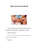
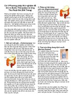
(Fig. 1). Failure of the hindfoot varus
to cor rect indicates a fixed hindfoot
deformity that may require both a
hindfoot procedure to correct the
varus (eg, calcaneal osteotomy or
subtalar fusion) and a dorsiflexion
osteotomy of the first ray.
10
The re-
sults of the Coleman block test must
be interpreted in the context of the
remainder of the physical examina-
tion because a patient with fixed
hindfoot varus and a valgus position
of the forefoot still requires correc-
tion of the forefoot position. When
in doubt, a lateralizing calcaneal os-
teotomy should be performed be-
cause the varus deformity of the
hindfoot remains undercorrected in
all but the mildest cases.
Radiographic
Evaluation
Weight-bearing anteroposterior and
lateral views of the foot and ankle
are required, along with a calcaneal
axial view. Oblique views of the foot
are occasionally helpful to visualize
changes at the tarsometatarsal joint
level. Patients with a cavus foot po-
sition often have a degenerative spur
in the posterior aspect of the subta-
lar joint. A lateral radiograph of the
patient performing a Coleman block
test will indicate the degree of cor-
rection obtainable with a first ray os-
teotomy. A modified Cobey view
may be a better guide for hindfoot
alignment than a calcaneal axial
view.
12
A Canale view of the talar
neck is best for finding talar neck
fracture and malalignment.
13
Com-
puted tomography (CT) scans also
may be of value in assessing hind-
foot position, but they provide only
a simulated weight-bearing view.
14
A
CT scan of the foot allows more ac-
curate assessment of degenerative
changes within the foot. Techne-
tium 99m bone scanning can assist
in identifying involved joints. Anes-
thetic blocks of symptomatic joints
can be identified fluoroscopically
and can assist in determining the
source of pain.
Nonsurgical Treatment
Orthoses
An orthotic device may be fash-
ioned to broaden the weight-bearing
area in the foot. Because custom
orthoses are expensive and the high-
arched foot is hard to fit, patients
initially may wish to modify an
off-the-shelf insert. Commercially
available metatarsal pads may be
added to a foam rubber insert. Pa-
tients with first metatarsal head
overload may need a cutout for the
first metatarsal head. Carpet felt also
can be added to the shoe, with a cut-
out for the first metatarsal head.
If the modified shoe insert works,
a custom-made tridensity or semi-
rigid orthosis can be fashioned using
the prefabricated insert as a tem-
plate. A metatarsal pad can be added
to the semirigid or tridensity ortho-
sis, with a metatarsal head cutout.
Hard orthoses are often poorly toler-
ated by patients with rigid cavus
feet. A high-arched orthosis actually
may increase ankle instability and
may need to be modified. A high
boot or an off-the-shelf ankle brace
may be used. Braces that lace up are
easier to fit inside a shoe or boot and
offer stabilization similar to that of
plastic upright ankle braces.
Patients with muscle weakness
often benefit from a full-length cus-
tom ankle-foot orthosis (AFO) to
prevent foot drop. Orthotic modifi-
cations can be integrated into the
AFO, providing more control over
ankle instability than would a brace
Figure 1
The Coleman block test. A, On initial examination, the hindfoot is in varus. B, The
patient stands with a book or block under the lateral side of the forefoot, and the
hindfoot is reexamined. Heel varus correction indicates that the hindfoot deformity
is flexible and that the varus position is secondary to the plantarflexed first ray, or
valgus position of the forefoot.
Alastair S. E. Younger, MB, ChB, MSc, ChM, FRCSC, and Sigvard T. Hansen, Jr, MD
Volume 13, Number 5, September 2005 305
alone. Patients with an equinus de-
formity may require a brace to pre-
vent its progression. A splint should
be worn every night when the equi-
nus contracture is progressing. Brac-
ing also is important for maintaining
correction after heel cord lengthen-
ing. Often a full clamshell brace or
bivalved cast is required because the
deformity will overcome the correc-
tion obtainable with a posterior
brace and anterior straps.
Shoe Wear
The high-arched foot may be dif-
ficult to fit inside a shoe, particular-
ly a slip-on style of shoe. Initially, an
off-the-shelf lace-up shoe with an
extra-deep toe box will accommo-
date the foot. A lace-up boot has the
added benefit of allowing more room
for the arch and providing some de-
gree of ankle stability. Extra depth
and custom shoes may be required
to fit insertable orthoses, AFOs, and
very-high-arched-foot or claw toe de-
formities.
Surgical Treatment
Surgical goals, expectations, and re-
covery times should be clearly out-
lined to the patient. All normal
joints should be preserved when pos-
sible. In patients with muscle imbal-
ance, well-planned osteotomies and
tendon transfers provide more reli-
able outcomes than does triple ar-
throdesis. Symptomatic degenera-
tive joints should be fused and
contracted soft tissues released.
Contracted tendons also should be
released or transferred, or the
muscle-tendon junction fractionally
lengthened. Osteotomies, tendon
transfers, or releases can correct
muscle imbalance in most patients
with a neurogenic cavovarus foot.
Although some surgeons recom-
mend initial treatment with
orthoses and bracing, others believe
that, in some cases, surgical inter-
vention is indicated as soon as the
diagnosis is made. A cavovarus
clawfoot is a progressive deformity
in the presence of muscle imbalance.
Therefore, the muscle imbalance
must be corrected to stop the pro-
gression before fixed deformity and
unsalvageable secondary joint de-
generation occur. Correction of mus-
cle imbalance requires transfer of
the two most deforming muscles—
the long peroneal and tibialis poste-
rior tendons. Consideration should
be given to a lateralizing calcaneal
osteotomy and dorsiflexion osteoto-
my of the first ray.
Soft-Tissue Release and
Tendon Lengthening
In the equinovarus foot, contrac-
tures affect the structures of the
plantar and medial aspects. Depend-
ing on the position and extent of the
contracture, a posteromedial release
will be required. A tight heel cord
can be addressed with either a gas-
trocnemius recession (slide) or heel
cord lengthening (Table 2). These
procedures are performed for hind-
foot equinus deformities, which
only are diagnosed by reviewing lat-
eral weight-bearing radiographs.
Forefoot equinus is measured using
the talo-first metatarsal angle (nor-
mal, 0° to 3°). Hindfoot equinus is
measured by a decreasing calcaneal
pitch angle as the ankle plantarflex-
es. For the patient with a unilateral
cavus foot, comparison with a
weight-bearing lateral radiograph of
the normal side is helpful.
When the gastrocnemius compo-
nent alone is tight, a gastrocnemius
recession can be performed. The
range of passive ankle dorsiflexion is
examined with the knee in both
flexion and extension. If the range
does not change and if the palpated
Achilles tendon does not feel tight,
then a mechanical block to dorsi-
flexion is likely present, caused by a
tight posterior capsule, anterior os-
teophytes, or a tight soleus compo-
nent. A weight-bearing lateral radio-
graph of the ankle may illustrate
impinging anterior osteophytes.
For open heel cord lengthening, the
incision is made just posterior and
medial to the ankle joint. The Achil-
les tendon is identified through this
incision by deep dissection. The neu-
rovascular bundle is identified when
an extensive release is planned.
15
A
percutaneous heel cord lengthening
also can be performed, although over-
lengthening may result in a calcaneus
gait and weakness in plantar flexion.
A split tibialis posterior tendon trans-
fer should be performed at the same
time if the hindfoot is in varus dur-
ing the stance phase of gait.
16
The
flexor digitorum longus is lengthened
Soft-Tissue Release and Tendon-Lengthening Procedures
Range of Forced
Ankle Dorsiflexion
(degrees) With the
Knee in Flexion
Range of Forced
Ankle Dorsiflexion
(degrees) With the
Knee Extended Procedure
>10 <5 Gastrocnemius recession
0 to 10 5 (dorsiflexion) to
20 (plantar flexion)
Gastrocnemius recession and/or
open heel cord lengthening
<0 20 (plantar flexion) Open heel cord lengthening or
percutaneous Achilles tendon
lengthening
<0 Does not change
with knee flexion;
heel cord not tight
Ankle joint débridement and
posterior release
<0 Does not change
with knee flexion;
heel cord tight
Percutaneous Achilles tendon
lengthening and posterior
release if the foot does not
correct
Table 2
Adult Cavovarus Foot
306 Journal of the American Academy of Orthopaedic Surgeons
or transferred if the toes are flexed
with the foot in a neutral position.
The flexor hallucis longus tendon
may be released at the knot of Henry,
transferred, or fractionally lengthened
at any level at which it can be safely
exposed.
17
Transfer into a very weak
or paralyzed peroneus brevis can be
very effective.
An isolated gastrocnemius reces-
sion is performed using a midcalf
medial incision over the palpable
junction of the gastrocnemius with
the heel cord. The tissue plane be-
tween the sural nerve, the fascia, and
the tendon is developed. The isolat-
ed gastrocnemius tendon is sec-
tioned, and adequate ankle dorsi-
flexion with the knee extended is
confirmed after the release.
Soft-tissue contractures may pre-
vent the ankle joint from correcting
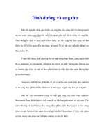
after tendon release (Fig. 2). In this
situation, the deltoid ligament can
be released at the posterior aspect of
the medial malleolus. The ankle and
subtalar joint capsule also may need
to be released. After isolating the
neurovascular bundle, the capsule is
identified anterior and posterior to
the tibialis posterior tendon. As with
a clubfoot, it may help to divide the
posterior aspect of the syndesmosis
between the tibia and fibula to allow
the talus to rotate posteriorly in the
ankle mor tise. The fat and fascia sur-
rounding the superficial and deep
compartments also can be scarred or
contracted and may need to be re-
leased (Fig. 2).
The midfoot may be held in the
equinovarus position. Release of the
plantar fascia will allow correction
and can be performed by multiple
small incisions, excision, or exten-
Figure 2
A, Weight-bearing lateral radiograph of a 44-year-old man with a cavovarus foot deformity associated with severe equinus
secondary to multiple sclerosis. B, Weight-bearing lateral radiograph of the normal contralateral foot confirmed that most of the
deformity was at the level of the ankle joint, with only a small portion secondary to midfoot cavus. C, Weight-bearing lateral
radiograph of the foot in panel A taken 6 months postoperatively. A posteromedial release was performed. Single-stage
correction was achieved with a cast change under anesthetic at 2 weeks. Claw toe deformities were treated by interphalangeal
fusions, and residual forefoot valgus deformity was corrected by a dorsiflexion osteotomy of the first ray. Calcaneal osteotomy
was not required because the foot corrected beyond neutral. Eight months postoperatively, the patient ambulated for 1 hour with
a single cane, which was used more for balance than to relieve pain.
Alastair S. E. Younger, MB, ChB, MSc, ChM, FRCSC, and Sigvard T. Hansen, Jr, MD
Volume 13, Number 5, September 2005 307
sile release from the calcaneus.
18
Midfoot cavus deformities should be
addressed by a plantar fascia release,
with releases of the deep muscles
and their tendon sheaths as neces-
sary.
19
An extensive release of the
talonavicular joint capsule also may
be required. If the soft-tissue releas-
es fail to correct the foot position,
then osteotomies or fusions will be
required to correct the foot to neu-
tral.
After trauma and a compartment
syndrome, correction may require
tendon releases, resection of infarct-
ed tissue within the muscle, and ad-
vancement of muscle tendon units
as well as tendon transfers or correc-
tive osteotomies and fusions.
20,21
In
Volkmann’s ischemic contracture, a
significant length of the necrotic and
scarred tendon and muscle may need
to be removed to ensure a perma-
nent release. This extensive surgical
procedure requires exacting preoper-
ative knowledge of the neurovascu-
lar anatomy of the extremity.
Tendon Transfer
Out-of-phase transfers (eg, tibialis
posterior to tibialis anterior ten-
don transfer) are recommended for
younger patients with lower motor
neuron pathology. The anterior
transfer of the flexor digitorum lon-
gus and flexor hallucis longus ten-
dons has been used as an out-of-
phase transfer for stroke patients.
22
Specific requirements must be met.
(1) The transferred muscle should
both be strong enough and have an
appropriate excursion to perform the
function of the substituted mus-
cle.
18
A grade of power will be lost af-
ter the transfer. (2) The transferred
tendon should be inserted close to
the substituted tendon and routed in
a comparatively direct line. (3) The
transferred tendon should be routed
in a tendon sheath, either its own or
the sheath of the substituted tendon,
or within tissues that will allow it to
glide. (4) The nerve and blood supply
of the transferred tendon should not
be damaged. (5) The joints on which
the tendon is to act must be func-
tional (ie, have a reasonable range of
motion, be stable, and have minimal
deformity). (6) The tendon should be
attached directly to bone, or indi-
rectly by another tendon using a ten-
don weave, and it should be in slight
to moderate tension. For example,
the tibialis posterior tendon can be
transferred through the interosseous
membrane and inserted into the me-
dial cuneiform via a weave into the
tibialis anterior. Agonists are prefer-
able to antagonists.
18
Tendon trans-
fers can be categorized according to
whether they affect gait in either the
swing phase or stance phase
5,23,24
(Ta-
bles 3 and 4).
Debate exists as to the donor
morbidity of the transferred tibialis
posterior tendon. In the cavus foot
position, the released tendon does
not cause a subsequent planovalgus
deformity because the bones and lig-
aments of the foot apparently are
able to maintain the medial arch. In
contrast, the tibialis posterior ten-
In-Phase Tendon Transfers for Swing Phase and Stance Phase
Phase Donor Recipient Indication Concomitant Procedure
Swing Extensor
hallucis longus
Tibialis anterior Clawed first ray;
weak dorsiflexion
First ray IP fusion;
MTP joint release
Extensor
hallucis longus
Peroneus tertius
(complete or split)
Weak dorsiflexion with
inversion on swing phase
First ray IP fusion;
MTP joint release
Extensor
digitorum brevis
Extensor digitorum
longus stump
Clawtoes IP fusions or excisions;
MTP joint releases
Extensor
digitorum longus
Peroneus tertius Clawed lesser toes;
weak dorsiflexion
IP fusions or excisions;
MTP joint releases
Tibialis anterior
(complete or split)
Peroneus tertius Excessive forefoot inversion
during swing phase
—
Stance Flexor hallucis longus Peroneus brevis Weak ankle eversion Calcaneal osteotomy
Flexor hallucis longus Peroneus longus Flexible forefoot varus Midfoot fusion
Peroneus longus Peroneus brevis Weak ankle eversion Calcaneal osteotomy
Peroneus brevis Peroneus longus Weak ankle eversion and
flexible forefoot varus
Calcaneal osteotomy
Tibialis posterior
(complete or split)
Peroneus brevis Weak ankle eversion Calcaneal osteotomy
Tibialis posterior
(complete or split)
Peroneus longus Forefoot varus and
weak ankle eversion
Calcaneal osteotomy
IP = interphalangeal, MTP = metatarsophalangeal
Table 3
Adult Cavovarus Foot
308 Journal of the American Academy of Orthopaedic Surgeons
don is an essential part of the medi-
al column in a flexible planovalgus
foot.
For stroke patients, a heel cord
lengthening on its own is rarely suf-
ficient because the tight tibialis pos-
terior tendon will cause a varus heel
position once the foot is correct-
ed.
16
Therefore, a tibialis posterior
tendon lengthening or transfer will
be required at the same time. Exces-
sive lengthening of the heel cord
should be avoided because increased
cavus deformity or a calcaneus gait
may develop.
25
An overlengthened
heel cord also will result in weak-
ness in plantar flexion, poor gait pro-
gression at toe-off, and, in some cas-
es, anterior impingement in the
ankle joint. Additionally, increased
energy may be required for gait be-
cause the quadriceps muscle is re-
cruited to prevent the patient from
falling forward. Failure to release
tight long toe flexors may result in a
poorer outcome and require a second
release.
26
Appropriate releases or
tendon transfers allow the foot to be
brought into the neutral position
and improve or prevent bracing.
Walking ability is related to the age
at surger y and the degree of paraly-
sis.
22
In patients with hemiplegia,
anterior transfer of the flexor digi-
torum longus and flexor hallucis
longus may assist dorsiflexion pow-
er.
27
Osteotomy
The lateralizing calcaneal osteot-
omy can effectively reduce the varus
moment arm of the Achilles tendon
at the ankle during stance phase;
this osteotomy also can reduce the
additive contribution of the Achilles
tendon toward the tibialis posterior
in favor of the peroneus brevis dur-
ing toe-off. The patient with an in-
ternally rotated distal tibia also
tends to have a varus moment of the
Achilles tendon at the ankle. The de-
gree of tibial rotation can be assessed
by CT.
28
An osteotomy is indicated for a
mild to moderate fixed deformity
that persists after appropriate tendon
releases in a patient without arthrit-
ic change in the surrounding joints.
Osteotomy also may be indicated in
combination with fusion when the
foot position cannot be corrected by
fusion alone. For example, after an
ankle fusion, the hindfoot may cor-
rect completely, but the forefoot
may be left with plantar flexion of
the first ray. Thus, a dorsiflexion os-
teotomy through the first tarsometa-
tarsal joint or in the proximal me-
taphysis would be indicated.
Distal tibial osteotomy may be of
value in a patient with forefoot
varus, hindfoot varus, and varus
alignment at the ankle joint. A su-
pramalleolar osteotomy with a later-
al closing wedge will bring the foot
flat to the ground and redistribute
the force within the ankle joint.
Supramalleolar derotational os-
teotomy also may be beneficial. Ro-
tating the distal tibia changes the di-
rection of the moment arm of the
Achilles tendon. External rotation
osteotomy of the distal tibia in-
creases the valgus moment at the
subtalar joint and unlocks the subta-
lar joint. McNicol et al
29
reported
successful outcomes in patients with
polio and other neurologic etiologies
who were treated with rotational os-
teotomy to externally rotate the foot.
In most cases of cavovarus foot, the
foot is correctly aligned on the tibia
after the talonavicular joint has been
released.
Calcaneal Osteotomy
When the hindfoot does not pas-
sively correct to neutral, a lateraliz-
ing calcaneal osteotomy must be
performed, with or without a subta-
lar fusion. Because hindfoot varus is
difficult to assess, calcaneal osteoto-
my should be done when there is
any residual hindfoot varus. The cal-
caneal osteotomy will correct the
foot during heel strike and at rest
and, more important, will lateralize
the moment arm o f the Achilles ten-
don during toe-off. In a patient with
a mobile midfoot and hindfoot, a lat-
eralizing calcaneal osteotomy in-
creases the load on the medial border
Out-of-Phase Tendon Transfers for Swing-Phase Deficit
Donor Recipient Indication Concomitant Procedure
Tibialis posterior Tibialis anterior and/or
peroneus tertius
Weak dorsiflexion caused by
lower motor neuron
pathology, or nerve or
muscle injury
Heel cord lengthening
Peroneus longus Peroneus tertius Weak dorsiflexion Tibialis posterior transfer; heel
cord lengthening
Flexor hallucis longus
and digitorum
longus
Fourth metatarsal through
interosseous membrane
Weak dorsiflexion caused by
stroke
Short flexor release;
lengthening of heel cord or
tibialis posterior
Flexor digitorum
longus
Extensor hood Intrinsic deformity of toes Interphalangeal joint fusion or
excision
Table 4
Alastair S. E. Younger, MB, ChB, MSc, ChM, FRCSC, and Sigvard T. Hansen, Jr, MD
Volume 13, Number 5, September 2005 309
of the foot during toe-off. A dorsi-
flexion osteotomy of the first ray or
a dorsiflexion fusion of the first tar-
sometatarsal joint should be per-
formed at the same time if the first
ray is plantarflexed, as is common in
CMT disease.
In many patients with a cavo-
varus foot requiring a lateralizing
calcaneal osteotomy, a posterior and
medial osteophyte is present in the
subtalar joint. If this is the only ev-
idence of degenerative change and is
mildly symptomatic, the osteophyte
can be excised medially by dissect-
ing just anterior to the flexor digi-
torum longus.
The Dwyer closing wedge osteot-
omy weakens the moment arm of
the Achilles tendon and often can-
not achieve full correction.
30
A slid-
ing calcaneal osteotomy without ex-
cision is usually preferable
17
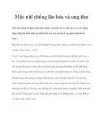
(Fig. 3).
In this procedure, a posterior lateral
incision is made. The calcaneal cut
is made transverse to the long axis of
the foot to prevent shortening or
lengthening of the osteotomy. The
medial cut should not penetrate
close to the sustentaculum tali be-
cause of the proximity of the neu-
rovascular bundle to this groove. A
skin mark can be made medially
halfway between the neurovascular
bundle and the tuberosity of the cal-
caneus. A finger of the surgeon’s
nondominant hand is placed on the
mark, and the saw is directed to this
point. After mobilizing both the tu-
berosity fragment and the deep
investing fascia surrounding the
Achilles tendon, lateral translation is
performed. The osteotomy is held
with two screws. Anteroposterior,
lateral, and calcaneal axial views of
the ankle should be taken intraoper-
atively. The lateral aspect of the tu-
berosity fragment is trimmed and
can be used for bone graft elsewhere.
Posterior calcaneal osteotomy
with plantar release has been used to
correct hindfoot cavus associated
with a weak gastrocnemius-soleus
complex. By sliding the tuberosity of
the calcaneus posteriorly and superi-
orly using an oblique osteotomy, the
position of the calcaneus is correct-
ed and the lever arm of the weak tri-
ceps surae muscle is augmented
31
(Fig. 4). Posterior calcaneal osteoto-
my should not be performed in the
presence of anterior ankle joint im-
pingement.
In this procedure, a lateral inci-
sion is made using a portion of the L-
Figure 3
Lateralizing sliding calcaneal osteotomy. A, A posterior lateral incision is made.
B, Once the soft tissues have been retracted, the calcaneus is cut with a saw.
C, The medial cut should not penetrate close to the sustentaculum tali. D and
E, The osteotomy is held with two proximal-distal transcalcaneal screws.
F and G, Alternative screw positions. (Adapted with permission from Hansen ST Jr
[ed]: Functional Reconstruction of the Foot and Ankle. Philadelphia, PA: Lippincott
Williams and Wilkins, 2000, p 369.)
Adult Cavovarus Foot
310 Journal of the American Academy of Orthopaedic Surgeons
or J-shaped calcaneal fracture inci-
sion. This incision can be combined
with a sinus tarsi incision if correc-
tion of varus is incomplete after a
subtalar fusion. The osteotomy be-
gins anterior to the Achilles tendon
insertion and the insertion of the
plantar fascia. An osteotomy poste-
rior to the plantar fascia will destabi-
lize the osteotomy. The osteotomy
should be transverse. An oblique os-
teotomy lengthens or shortens the
tuberosity of the calcaneus, and
lengthening the calcaneus may re-
strict the correction of var us because
the soft-tissue envelope may become
too tight. An oblique osteotomy ex-
iting anteriorly on the medial wall
may damage the neurovascular bun-
dle.
The osteotomy site is released
from the medial soft tissues using a
periosteal elevator or curved curet.
The Achilles tendon sheath is re-
leased to improve displacement of
the tuberosity fragment; the invest-
ing fascia will prevent translation of
the Achilles tendon. The tuberosity
fragment is held by two screws. Af-
ter placing the distal screw, the supe-
rior aspect of the posterior fragment
of the osteotomy is then rotated me-
dially and transfixed with a second
screw. Radiographic views (eg, later-
al ankle and Broden views) are ob-
tained to ensure that the screws do
not penetrate the subtalar joint.
Lateral Column Shortening
Because lateral column shorten-
ing corrects hindfoot varus, forefoot
varus, and forefoot abduction, it is
ideally suited to correct the position
of a residual clubfoot. Lateral col-
umn shortening can be performed
through the cuboid, the lateral as-
pect of the calcaneus, or the calca-
neocuboid joint.
18
This procedure is
indicated when the foot fails to cor-
rect after medial talonavicular re-
lease.
Talar Neck Osteotomy
A malreduced talar neck fracture
can result in dorsal and medial trans-
lation of the distal portion of the ta-
lar neck as well as shortening of it.
This results in a cavovarus position
of the foot. The malunion locks the
triple-joint complex, causing a pain-
ful rigid foot with overload of the lat-
eral border. Components of rotation
and translation also coexist within
the subtalar and midtarsal joints as
well as at the malunion, causing
overload of the triple-joint complex
(subtalar, talonavicular, and calca-
neocuboid joints).
8
A talar neck osteotomy may be
performed when the surrounding
joints are well preserved. A preoper-
ative CT scan is needed to assess the
amount of correction required. If
necessary, intact blood supply to the
body is confirmed with magnetic
resonance imaging. The risks of this
procedure include failure to correct
all components of the deformity,
nonunion of the distraction graft,
and osteonecrosis of the talar
body.
32
Dorsiflexion Osteotomy of
the First Ray
Dorsiflexion osteotomy of the
first ray is indicated for a symptom-
atic plantarflexed first ray with pain
in the forefoot secondary t o overload
of the first metatarsal head. It is also
indicated for a symptomatic plantar-
flexed first ray with pain over the
lateral border of the foot resulting
from supination caused by forefoot-
driven hindfoot varus. Clawing of
the first ray with a dorsal contrac-
ture of the metatarsophalangeal
joint and a tight extensor tendon are
often seen in conjunction. Dorsiflex-
ion osteotomy or fusion of the first
tarsometatarsal joint also is indicat-
ed if the hindfoot corrects to neutral
when a Coleman block test indicates
a forefoot-driven hindfoot varus.
Dorsiflexion fusion of the first
tarsometatarsal joint should be con-
sidered when the tarsometatarsal
joint is hypermobile and a strong
peroneus longus plantarflexes the
first ray. Care should be taken not to
over-shorten or over-elevate the first
ray. Excessive correction can result
in hallux rigidus and transfer meta-
tarsalgia.
23
A plantar fascia release
may be required at the same time to
allow elevation of the first ray.
The proximal cut of the tar-
sometatarsal fusion is almost paral-
lel to the joint and is perpendicular
to an imaginary line running
through the talus and the navicular
Figure 4
Posterior calcaneal osteotomy for
hindfoot cavus. A, Normal relationship
of the hindfoot bones. B, Position of the
hindfoot secondary to a weak triceps
surae. The osteotomy is made from the
lateral aspect. C, The posterior
tuberosity fragment is displaced in a
dorsal and posterior direction to
restore length, reduce the arch, and
improve the moment arm of the weak
triceps surae muscle. The screws are
placed in parallel across the osteotomy
site. (Adapted with permission from
Hansen ST Jr [ed]: Functional
Reconstruction of the Foot and Ankle.
Philadelphia, PA: Lippincott Williams
and Wilkins, 2000, p 373.)
Alastair S. E. Younger, MB, ChB, MSc, ChM, FRCSC, and Sigvard T. Hansen, Jr, MD
Volume 13, Number 5, September 2005 311
bone. The distal cut is made perpen-
dicular to the axis of the first meta-
tarsal. Very little bone should be re-
moved. Precise axial alignment
ensures straight postoperative align-
ment of the talus and first metatar-
sal bone. Ideally, the plantar liga-
ments are kept intact to act as a
tension band (Fig. 5, A). A Jones
transfer and dorsal capsulotomy to
release the metatarsophalangeal
joint may be required at the same
time. The wedge is closed and fixed
with a one-quarter tubular plate and
cortical screws to straighten the ta-
lar to first metatarsal alignment.
Care should be taken to match the
length of the metatarsals as well as to
ensure that the metatarsal heads lie
in the same transverse plane. A con-
comitant second metatarsal shorten-
ing osteotomy may be required when
the second metatarsal head articular
surface projects distal to the first by
more than 4 mm to 5 mm.
In an alternative technique, bone
from the proximal metatarsal me-
taphysis is removed (Fig. 5, C). (A
dorsal wedge of 5 mm of bone will
elevate the metatarsal head approx-
imately 12 mm.) Alignment of the
foot is corrected in a manner similar
to the first method. Using a fine
Kirschner wire to identify the plane
of the first tarsometatarsal joint as-
sists in the correct alignment of the
cut. Fixation is done with a two- or
three-hole one-third tubular plate
and 3.5-mm cortical screws. This os-
teotomy, like the first method, cor-
rects alignment of the talus to the
first metatarsal.
Midfoot Osteotomy
Correction of midfoot (global) ca-
vus can be achieved by a midfoot os-
teotomy with removal of a segment
of bone at multiple tarsometatarsal
joint levels (Jahss osteotomy
33
)orat
the navicular cuneiform joint level
(Cole and Japas osteotomies), fol-
lowed by a closing osteotomy and
fusion.
18
Common complications in-
clude residual opening of the osteot-
omy site and malunion. Surgeons are
wary o f this procedure because of the
high nonunion rate
10,33,34
(Fig. 6). Os-
teotomy of the metatarsal shafts also
can correct the deformity and be
held in reduction with a cast.
35
A
plantar fascia release may be re-
quired at the same time as the os-
teotomy to allow correction through
the osteotomy. The position of the
metatarsal heads should be level af-
ter closing multiple proximal meta-
tarsal osteotomies (Jahss osteotomy)
because metatarsalgia will develop
under a residual plantarflexed ray.
Figure 5
First metatarsal osteotomy for correction of a fixed first metatarsal cavus deformity. A, Medial view demonstrating a plantarflexed
first ray with the deformity in the first metatarsal. The shaded area depicts the wedge to be removed from the first tarsometatar-
sal joint. B, The wedge is closed and fixed with a four-hole one-quarter tubular plate and 2.7-mm or 3.5-mm cortical screws.
C, In an alternative technique, bone from the proximal metatarsal metaphysis (shaded area) is removed. Alignment of the foot is
corrected in a manner similar to that in panel B. D, Fixation is performed with a two- or three-hole one-third tubular plate and
3.5-mm cortical screws. (Adapted with permission from Hansen ST Jr [ed]: Functional Reconstruction of the Foot and Ankle.
Philadelphia, PA: Lippincott Williams and Wilkins, 2000, p 393.)
Adult Cavovarus Foot
312 Journal of the American Academy of Orthopaedic Surgeons
Fusion
Fusion is indicated in the
cavovarus foot after secondary de-
generative changes have occurred.
However, it should not be used to
compensate for muscle imbalance.
For example, a triple arthrodesis
done without correcting the pull of
the tibialis posterior muscle may fail
because the ankle joint can open
laterally, leading to recurrent defor-
mity. Therefore, muscle balancing
should be done at the same time as
the fusion to ensure correct position-
ing of the foot. Subtalar fusion is in-
dicated for subtalar degeneration and
can be done to correct hindfoot
varus by rotating the foot externally
on the talus to close the sinus tarsi
before fixation. Calcaneocuboid fu-
sion can be performed to correct de-
generative changes at the calca-
neocuboid joint or to shorten the
lateral column. A triple arthrodesis
is indicated for arthritis at the talo-
navicular joint or for the patient
with rigid deformity with arthritis at
Figure 6
Midfoot osteotomy to correct midfoot cavus. A, Transverse cross-sectional view of the midtarsus showing the relative anatomy
of the structures corrected by a dorsally based wedge. The cuneiforms are configured as a roman arch, with the second
cuneiform at the apex. Neurovascular structures and other soft tissues occupy the space under the cuneiforms. The inferior
surface of the first cuneiform is lower than the second, and the lowest point of the arch is the lateral plantar surface of the
cuboid. B, Medial view of the right foot depicts the area of bone (shaded area) to be removed. The two limbs of the osteotomy
are shown. C, Lateral view demonstrating that the two limbs of the cut meet at the lowest point (ie, the plantar cortex of the
cuboid). D, Dorsal view demonstrating the wedge (shaded area) and the skin incision (dotted line) through which the osteotomy
is made. Fixation is achieved using lag screws from the navicular to the cuneiforms. A plantar fasciotomy may be required to
achieve correction. Casting is required to close the osteotomy and to maintain correction and stretch in the heel cord. (Adapted
with permission from Hansen ST Jr [ed]: Functional Reconstruction of the Foot and Ankle. Philadelphia, PA: Lippincott Williams
and Wilkins, 2000, p 389.)
Alastair S. E. Younger, MB, ChB, MSc, ChM, FRCSC, and Sigvard T. Hansen, Jr, MD
Volume 13, Number 5, September 2005 313
any one of the three joints forming
the triple-joint complex.
36
Isolated
talonavicular fusion is rarely indicat-
ed because little residual motion re-
mains in the joints of the complex.
Multiple hindfoot joint fusion is the
only surgical option in some pa-
tients, such as those with secondary
degenerative changes at the ankle
and talonavicular joint or those with
involvement of both the ankle and
subtalar joints.
Fusions may be performed at one
surgery a s long as careful attention is
paid to the blood supply to the talus.
The ankle joint should be fused first,
with cor rection of as much of the de-
formity as possible. The subtalar,
talonavicular, and calcaneocuboid
joints (ie, the triple-joint complex)
are then reduced and arthrodesed as
needed to correct the remainder of
the deformity. A calcaneal osteoto-
my still may be required.
A staged approach that allows the
blood supply to the talus to stabilize
between the triple and ankle fusions
also is reasonable. The ankle joint
may become less symptomatic after
correction of the foot position by a
subtalar or triple arthrodesis. There
are advantages and disadvantages to
each fusion method, and the tech-
nique should be tailored to the clin-
ical situation.
Clawtoe Reconstruction
Patients with a cavovarus foot
often have an associated claw toe
deformity. The deformity may be
flexible or rigid with contracture,
subluxation, or dislocation at the
metatarsophalangeal joint level. Dy-
namic clawtoes may resolve with
correction of hindfoot deformity. For
dynamic clawing with flexible defor-
mities, flexor-to-extensor tendon
transfer with either the Girdlestone
or the Taylor procedure is useful.
18
Fixed clawing requires a closed os-
teoclasis or interphalangeal joint fu-
sion or excision. A Jones transfer or
extensor tendon transfer to the later-
al border of the foot may be required
at the same time.
Summary
Cavovarus foot covers a broad spec-
trum of etiologies and pathologies,
with the common features of an in-
verted hindfoot and a high arch.
Management requires identifying
treatable causes, followed by isolat-
ing and treating the symptoms with-
in the foot. A complete neurologic
assessment is required. If the cause
is not identified, the patient should
be referred to a neurologist. Non-
surgical treatment is done with
orthoses and/or braces. Early surger y
should be considered to correct mus-
cle imbalance and prevent the devel-
opment of a fixed deformity. Surgery
should address all components of the
cavovarus deformity, with tendon
transfer to treat muscle imbalance,
osteotomy to treat fixed deformities,
and fusions to treat severe deformi-
ty or secondary arthritic change. Sat-
isfactory outcomes can be expected
if a global assessment and treatment
plan is adopted and if all compo-
nents of the cavovarus foot deformi-
ty are corrected.
Acknowledgment
The authors would like to acknowl-
edge the editorial assistance pro-
vided by Colin D. Meakin, MSc,
Research Coordinator for the Uni-
versity of British Columbia Depart-
ment of Orthopaedics.
References
1. Holmes JR, Hansen ST Jr: Foot and an-
kle manifestations of Charcot-Marie-
Tooth disease. Foot Ankle 1993;14:
476-486.
2. Price AE, Maisel R, Drennan JC: Com-
puted tomographic analysis of pes ca-
vus. J Pediatr Orthop 1993;13:646-
653.
3. Tynan MC, Klenerman L, Helliwell
TR, Edwards RH, Hayward M: Inves-
tigation of muscle imbalance in the
leg in symptomatic forefoot pes ca-
vus: A multidisciplinary study. Foot
Ankle 1992;13:489-501.
4. Tenuta J, Shelton YA, Miller F: Long-
term follow-up of triple arthrodesis in
patients with cerebral palsy. J Pediatr
Orthop 1993;13:713-716.
5. Mulier T, Moens P, Molenaers G,
Spaepen D, Dereymaeker G, Fabry G:
Split posterior tibial tendon transfer
through the interosseus membrane in
spastic equinovarus deformity. Foot
Ankle Int 1995;16:754-759.
6. Miller A, Guille JT, Bowen JR: Evalu-
ation and treatment of diastematomy-
elia. J Bone Joint Surg Am 1993;75:
1308-1317.
7. Carpintero P, Entrenas R, Gonzalez I,
Garcia E, Mesa M: The relationship
between pes cavus and idiopathic
scoliosis. Spine 1994;19:1260-1263.
8. Sangeorzan BJ, Wagner UA, Har-
rington RM, Tencer AF: Contact char-
acteristics of the subtalar joint: The
effect of talar neck misalignment.
J Orthop Res 1992;10:544-551.
9. Haasbeek JF, Wright JG: A compari-
son of the long-term results of poste-
rior and comprehensive release in the
treatment of clubfoot. J Pediatr Or-
thop 1997;17:29-35.
10. Alexander IJ, Johnson KA: Assess-
ment and management of pes cavus in
Charcot-Marie-Tooth disease. Clin
Orthop 1989;246:273-281.
11. Sneyers CJ, Lysens R, Feys H, Andries
R: Influence of malalignment of feet
on the plantar pressure pattern in run-
ning. Foot Ankle Int 1995;16:624-632.
12. Saltzman CL, El-Khoury GY: The
hindfoot alignment view. Foot Ankle
Int 1995;16:572-576.
13. Canale ST, Kelly FB Jr: Fractures of the
neck of the talus: Long-term evalua-
tion of seventy-one cases. J Bone Joint
Surg Am 1978;60:143-156.
14. Van Bergeyk AB, Younger A, Carson B:
CT analysis of hindfoot alignment in
chronic lateral ankle instability. Foot
Ankle Int 2002;23:37-42.
15. Saraph V, Zwick EB, Uitz C, Linhart
W, Steinwender G: The Baumann pro-
cedure for fixed contracture of the gas-
trosoleus in cerebral palsy: Evaluation
of function of the ankle after multi-
level surgery. J Bone Joint Surg Br
2000;82:535-540.
16. Kagaya H, Yamada S, Nagasawa T,
Ishihara Y, Kodama H, Endoh H: Split
posterior tibial tendon transfer for
varus deformity of hindfoot. Clin Or-
thop 1996;323:254-260.
17. Hansen ST Jr: Functional Reconstruc-
tion of the Foot and Ankle. Philadel-
phia, PA: Lippincott Williams &
Wilkins, 2000.
18. Ingram AJ: Paralytic disorders, in
Crenshaw AH (ed): Campbell’s Or-
thopaedics, ed 7. St. Louis, MO: Mos-
by, 1987, vol 4, pp 2925-3061.
19. Sherman FC, Westin GW: Plantar re-
Adult Cavovarus Foot
314 Journal of the American Academy of Orthopaedic Surgeons
lease in the correction of deformities
of the foot in childhood. J Bone Joint
Surg Am 1981;63:1382-1389.
20. Santi MD, Botte MJ: Volkmann’s is-
chemic contracture of the foot and an-
kle: Evaluation and treatment of es-
tablished deformity. Foot Ankle Int
1995;16:368-377.
21. David A, Lewandrowski KU, Josten C,
Ekkernkamp A, Clasbrummel B,
Muhr G: Surgical correction of talipes
equinovarus following foot and leg
compartment syndrome. Foot Ankle
Int 1996;17:334-339.
22. Yamamoto H, Okumura S, Morita S,
Obata K, Furuya K: Surgical correc-
tion of foot deformities after stroke.
Clin Orthop 1992;282:213-218.
23. Breusch SJ, Wenz W, Doderlein L:
Function after correction of a clawed
great toe by a modified Robert Jones
transfer. J Bone Joint Surg Br 2000;82:
250-254.
24. Morita S, Muneta T, Yamamoto H,
Shinomiya K: Tendon transfer for
equinovarus deformed foot caused by
cerebrovascular disease. Clin Orthop
1998;350:166-173.
25. Johnson WL, Lester EL: Transposition
of the posterior tibial tendon. Clin Or-
thop 1989;245:223-227.
26. Keenan MA, Gorai AP, Smith CW,
Garland DE: Intrinsic toe flexion de-
formity following correction of spas-
tic equinovarus deformity in adults.
Foot Ankle 1987;7:333-337.
27. Morita S, Yamamoto H, Furuya K: An-
terior transfer of the toe flexors for
equinovarus deformity due to hemi-
plegia. J Bone Joint Surg Br 1994;76:
447-449.
28. Liggio FJ, Kruse R: Split tibialis poste-
rior tendon transfer with concomitant
distal tibial derotational osteotomy in
children with cerebral palsy. J Pediatr
Orthop 2001;21:95-101.
29. McNicol D, Leong JC, Hsu LC: Supra-
malleolar derotation osteotomy for
lateral tibial torsion and associated
equinovarus deformity of the foot.
J Bone Joint Surg Br 1983;65:166-170.
30. Dwyer FC: The present status of the
problem of pes cavus. Clin Orthop
1975;106:254-275.
31. Mitchell GP: Posterior displacement
osteotomy of the calcaneus. J Bone
Joint Surg Br 1977;59:233-235.
32. Monroe MT, Manoli A II: Osteotomy
for malunion of a talar neck fracture:
A case report. Foot Ankle Int 1999;20:
192-195.
33. Jahss MH: Tarsometatarsal truncated-
wedge arthrodesis for pes cavus and
equinovarus deformity of the fore part
of the foot. J Bone Joint Surg Am 1980;
62:713-722.
34. Watanabe RS: Metatarsal osteotomy
for the cavus foot. Clin Orthop 1990;
252:217-230.
35. Sammarco GJ, Taylor R: Cavovarus
foot treated with combined calcaneus
and metatarsal osteotomies. Foot An-
kle Int 2001;22:19-30.
36. Haritidis JH, Kirkos JM, Provellegios
SM, Zachos AD: Long-term results of
triple arthrodesis: 42 cases followed
for 25 years. Foot Ankle Int 1994;15:
548-551.
Alastair S. E. Younger, MB, ChB, MSc, ChM, FRCSC, and Sigvard T. Hansen, Jr, MD
Volume 13, Number 5, September 2005 315