Gãy xương chày trục ở trẻ em và thanh thiếu niên pdf
Bạn đang xem bản rút gọn của tài liệu. Xem và tải ngay bản đầy đủ của tài liệu tại đây (144.9 KB, 8 trang )
Tibial Shaft Fractures in
Children and Adolescents
Abstract
Tibial shaft fractures are among the most common pediatric
injuries managed by orthopaedic surgeons. Treatment is
individualized based on patient age, concomitant injuries, fracture
pattern, associated soft-tissue and neurovascular injury, and
surgeon experience. Closed reduction and casting is the mainstay
of treatment for diaphyseal tibial fractures. Careful clinical and
radiographic follow-up with remanipulation as necessary is
effective for most patients. Surgical management options include
external fixation, locked intramedullary nail fixation in the older
adolescent with closed physis, Kirschner wire fixation, and flexible
intramedullary nailing. Union of pediatric diaphyseal tibial
fractures occurs in approximately 10 weeks; nonunion occurs in
<2% of cases. Some clinicians consider sagittal deformity
angulation >10° to be malunion and indicate that 10° of valgus and
5° of varus may not reliably remodel. Compartment syndromes
associated with tibial shaft fractures occur less frequently in
children and adolescents than in adults. Diagnosis may be difficult
in a young child or one with altered mental status. Although the
toddler fracture of the tibia is one of the most common in children
younger than age 2 years, child abuse must be considered in the
young child with an inconsistent history or with suspicious
concomitant injuries.
F
ractures of the tibial shaft are
among the most common inju-
ries in children and adolescents and
account for approximately 15% of
long-bone fractures in that popula-
tion. Only femur and forearm frac-
tures are more common.
1
The mech-
anism of injury varies from minor
falls or twisting injuries in young
children to sports-related trauma
and motor vehicle accidents in older
children and adolescents. Injury to
the tibia is the second most com-
mon fracture resulting from inten-
tional trauma.
2
Tibial shaft fractures
are less commonly caused by nonac-
cidental trauma than are apophyseal
ring or metaphyseal corner fractures.
In the multiply traumatized child,
fracture of the tibia is the third most
common long-bone fracture, after
fractures of the femur and humer-
us.
3
The average age at injury is 8
years, and this injury occurs more
frequently in boys than in girls.
1
Most tibial fractures in children
are short oblique or transverse frac-
tures of the middle or distal third of
the shaft. Thirty-seven percent of
tibial fractures are comminuted.
1
Fractures of the tibial shaft occur in
association with fibular fractures in
Rakesh P. Mashru, MD,
Martin J. Herman, MD, and
Peter D. Pizzutillo, MD
Dr. Mashru is Trauma Fellow, Campbell
Clinic, University of Tennessee College
of Medicine, Memphis, TN. Dr. Herman
is Assistant Professor, Orthopedics and
Pediatrics, Orthopedic Center for
Children, St. Christopher’s Hospital for
Children, Philadelphia, PA. Dr. Pizzutillo
is Chief, Orthopedic Surgery Section,
Director, Orthopedic Center for
Children, and Professor, Pediatrics and
Orthopedic Surgery, St. Christopher’s
Hospital for Children, Philadelphia.
None of the following authors or the
departments with which they are affili-
ated has received anything of value from
or owns stock in a commercial company
or institution related directly or indirectly
to the subject of this article: Dr. Mashru,
Dr. Herman, and Dr. Pizzutillo.
Reprint requests: Dr. Herman,
Orthopedic Center for Children, St.
Christopher’s Hospital for Children, Erie
Avenue at Front Street, Philadelphia, PA
19134.
J Am Acad Orthop Surg 2005;13:345-
352
Copyright 2005 by the American
Academy of Orthopaedic Surgeons.
Volume 13, Number 5, September 2005 345
30% of affected children.
1,4
Both tib-
ial and fibular fractures are com-
monly complete, displaced fractures
caused by high-energy trauma. Val-
gus angulation of the distal fragment
and shortening are caused by over-
pull of anterior and lateral compart-
ment muscle groups. T ibial fractures
with an intact fibula occur in 70% of
affected children and usually are the
result of torsional forces.
4,5
Although
isolated tibial fractures are often
minimally displaced at presentation,
varus angulation without shortening
often occurs in the first few weeks
after injury as a result of posterior
compartment muscular forces on
the distal fragment. Concomitant
plastic deformation of the fibula
may cause valgus displacement and
malrotation in some children.
5
Clinical Presentation
Children and adolescents common-
ly present with pain, tenderness, or
deformity of the lower leg after an
acute injury. The young child may
present with a limp, diminished
movement of the affected limb, or
refusal to bear weight (often without
a distinct history of injury, in the
case of a toddler fracture of the tibia).
A complete clinical history is re-
quired, including a detailed descrip-
tion of an observed traumatic event
to exclude the existence of other se-
rious injury involving the remainder
of the musculoskeletal system,
head, thorax, abdomen, or pelvis.
Chronic and recent illnesses as well
as the use of regular medications
should be noted. When no traumat-
ic event is witnessed or an inconsis-
tent history is provided, the physi-
cian must obtain a detailed social
history, including a diary of the
child’s most recent caregivers and
family contacts.
Primary assessment and cardio-
respiratory stabilization is the first
priority in the child or adolescent
presenting with potential multisys-
tem injury.
6
The surgeon may facil-
itate effective trauma resuscitation
and diagnostic evaluation by realign-
ing gross tibial deformity using gen-
tle longitudinal traction and tem-
porary splint immobilization.
6
A
complete musculoskeletal survey
may be completed once the child is
stabilized. Thorough examination of
the injured extremity includes
assessment of the hip, knee, and an-
kle joints; concomitant soft-tissue
injury; compartment tension; and
neurovascular status. Frequent re-
evaluation of the injured limb is nec-
essary in the unconscious or uncoop-
erative patient; signs and symptoms
of compartment syndrome should be
documented after each evaluation
(Table 1).
Radiographic
Assessment
Anteroposterior (AP) and lateral ra-
diographs of the tibia and fibula, in-
cluding the knee and ankle, are re-
quired to assess lower leg injuries.
Especially in patients with low-
energy injuries, careful assessment of
the fracture configuration is neces-
sary to make certain that there are no
missing areas of bone suggesting a
pathologic origin. Technetium bone
scanning is helpful in the diagnosis of
occult fractures or stress reactions
when radiographs of the lower leg are
normal. A large percentage of toddler
fractures are radiographically normal,
and it is often prudent for the clini-
cian to empirically immobilize these
children and follow up with weekly
serial radiographic examinations.
When neoplasm is suspected, mag-
netic resonance imaging provides
more comprehensive assessment of
pathologic fractures of the tibia and
surrounding soft tissues.
Treatment
Closed reduction with cast immobi-
lization is the mainstay of ortho-
paedic management of diaphyseal
tibial shaft fractures in children and
adolescents. Nondisplaced fractures
of the tibia without significant soft-
tissue injury or swelling should be
immobilized in a long leg cast for 4
to 6 weeks, followed by progressive
weight bearing in a short leg cast
(with a patellar tendon–bearing mod-
ification for fractures of the proximal
shaft) for an additional 4 to 6 weeks.
The toddler fracture of the tibia re-
quires only 4 weeks of immobiliza-
tion. Patient activity is allowed
when the fracture site is not tender
to palpation and follow-up radio-
graphs document healing.
Closed manipulation and casting
under conscious sedation or general
anesthesia is indicated for displaced
tibial fractures. A short leg cast is ap-
plied first to control the fracture re-
duction. The ankle is positioned in
gentle plantar flexion to prevent
apex posterior angulation of the frac-
ture. This technique is most helpful
in fractures involving the most
distal third of the tibial shaft. After
the short leg portion is set, the cast
is extended to the groin with the
knee flexed 30° to 60°. During cast
application, the surgeon should care-
fully mold about the tibial fracture
site, avoiding pressure over the fibu-
lar head and soft-tissue compar t-
ments. Molding to the supracondy-
lar anatomy of the distal femur helps
control rotation within the cast and
enhances control of fracture align-
Symptoms and Signs of
Compartment Syndrome
Symptoms
Pain out of proportion to injuries
Persistent pain following removal of
constrictive dressings/splints
Paresthesias in the injured extremity
Signs
Swollen and tense compartment
Pain on palpation of compartment
Pain on passive stretch of muscles
in the involved compartment
Prolonged capillary refill and loss of
palpable pulse (late finding)
Increased pressure measurements
(>30 mm Hg)
Table 1
Tibial Shaft Fractures in Children and Adolescents
346 Journal of the American Academy of Orthopaedic Surgeons
ment. Immediate bivalving of the
cast is indicated in the uncoopera-
tive or obtunded child, or in one
with soft-tissue swelling. Once the
cast is bivalved, the child must be
monitored for continued swelling or
changes in neurovascular status. Af-
ter reduction and casting, the patient
is observed for compartment syn-
drome.
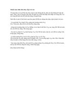
AP and lateral radiographs of the
lower leg, including the knee and an-
kle joints, should be obtained imme-
diately after reduction to verify
alignment (Fig. 1). Acceptable pa-
rameters of reduction are up to 5° of
varus or valgus angulation, <5° of
sagittal angulation, and 1 cm of
shortening. Translation of the entire
shaft may be tolerated in a child
younger than 8 years; 50% transla-
tion is acceptable in older children
and adolescents. Up to 10° of varus
and 10° of sagittal deformity are ac-
ceptable in children younger than
age 8 years. Maintenance of reduc-
tion is monitored for 3 weeks with
weekly radiographs of the lower leg.
Wedging of the cast or repeat manip-
ulation of the fracture with recasting
can improve angulation within 3
weeks of injury, often without the
need for sedation or anesthesia.
Wedging of the cast can be per-
formed by either an opening or clos-
ing wedge technique. In a closing
wedge technique, a 1- to 2-cm wedge
of cast material is removed from the
same side of the leg as the apex of
the fracture. The wedge is then
closed, correcting fracture angula-
tion. Because this technique may
cause the fracture to shorten or the
skin to impinge in the wedge, close
clinical and radiographic observa-
tion is required. In an opening wedge
technique, small blocks of varying
sizes may be inserted into the cast.
The cast is cut perpendicular to the
axis of the tibia on the side opposite
the apex of the fracture. Once the ap-
propriate size blocks are chosen,
fracture reduction should be exam-
ined radiographically.
Closed tibial osteoclasis or open
reduction of the tibia, with or with-
out fibular osteotomy, may be per-
formed in the operating room under
anesthesia to realign more rigid mal-
reduced fractures. Excessive short-
ening requires alternative tech-
niques, such as external fixation or
intramedullary rodding, to reestab-
lish and maintain tibial length. Cast
management for displaced fractures
of the tibia is similar to that for non-
displaced fractures. Tibial fractures
requiring repeated manipulation or
open reduction, or fractures that are
severely comminuted, should be im-
mobilized for longer periods to
achieve clinical and radiographic
healing.
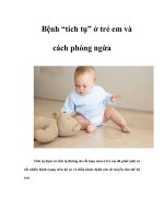
External fixation is most com-
monly used to stabilize severely
comminuted and unstable tibial
fractures and those associated with
severe soft-tissue injury
7-11
(Fig. 2).
Because of its ease of application and
adjustability, external fixation is an
excellent option for stabilizing tibi-
al fractures in children with head or
multisystem injuries. It also offers
improved access to and nursing care
of the lower leg compartments.
12
Management of these injuries in a
closed fashion with a long leg cast
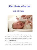
requires very close observation. Sim-
ple anteromedial frames using two
half-pins above and below the tibial
fracture site provide adequate stabil-
ity (Fig. 3). Surgeons may wish to
augment external fixation with min-
imal internal fixation, as per their
preference case by case. Early weight
bearing (within 4 weeks) and judi-
cious dynamization of the external
fixator may hasten healing.
Once clinical and radiographic
healing is complete, the external fix-
ation frame may be removed in the
clinic or the operating room. Early
removal of the frame and conversion
to a cast within 4 to 6 weeks may be
necessary in younger children or in
patients unable to tolerate the frame
or appropriately care for it. Pin tract
infection and refracture of the tibia
after frame removal are the most
common complications in these pa-
tients.
11
Although intramedullary fixation
is the treatment of choice for adults
Figure 1
A, Anteroposterior initial injury radiograph demonstrating marked displacement with
valgus angulation and shortening in a 16-year-old boy with a tibial and fibular shaft
fracture. B, Anteroposterior radiograph demonstrating acceptable alignment after
application of a long leg cast.
Rakesh P. Mashru, MD, et al
Volume 13, Number 5, September 2005 347
with fractures of the tibial shaft, its
use in children and adolescents has
been limited.
13
Rigid, interlocked
nails introduced through the proxi-
mal metaphysis of the tibia can
cause inadvertent injury to the phy-
sis or the anterior tibial tubercle.
The risk of growth disturbance of
the proximal tibia, manifested as
limb-length discrepancy and recur-
vatum of the proximal tibia, pre-
cludes the use of rigid, interlocked
nails in children.
Flexible intramedullary rod fixa-
tion is gaining in popularity for man-
agement of stable tibial fractures in
children and growing adolescents.
Intramedullary Kirschner wires are
effective for maintaining alignment
and length in stable fractures of the
tibia in the absence of severe com-
minution or fracture obliquity.
14
Un-
stable fractures with comminution
may require supplemental use of a
cast to hold the reduction. Elastic ti-
tanium nails, commonly used in the
forearm and femur, also can provide
stable fixation for unstable tibial
shaft fractures.
15
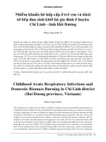
The elastic nails are
introduced through small drill holes
in the proximal or distal tibial me-
taphyses (Fig. 4). The flexible, elastic
nails are cut outside the bone be-
neath the skin, thereby eliminating
the need for pin care. Access to the
soft tissues of the leg for examina-
tion, débridement, or reconstruction
thus is unimpeded.
For fractures that are rotationally
unstable, a period of splint or cast
immobilization is required when
using constructs that do not impart
rotational control. Such immobiliza-
tion also functions as added protec-
tion for fractures in young or non-
compliant children. Range of motion
of the knee and ankle joints may be
initiated immediately after fixation,
and protected weight bearing on the
involved limb is progressed within 2
to 3 weeks postoperatively. The flex-
ible nails are removed in the operat-
ing room according to surgeon pref-
erence, usually within 4 to 6 months
of injury.
Other fixation options include
percutaneous pin fixation and plate-
screw constructs.
15
In younger chil-
dren with noncomminuted, unstable
oblique fractures, closed manipula-
tion of the fracture with percutane-
Figure 2
A, Anteroposterior radiograph of an open segmental grade IIIC (Gustilo-Anderson
classification) tibial fracture in a child who was hit by a car. B, Along with vascular
repair, the patient was treated with an external fixator to allow minimal fixation and
access to soft tissues.
Figure 3
A, Immediate postoperative anteroposterior radiograph of comminuted unstable
tibial and fibular shaft fractures in acceptable alignment with external fixation in a
12-year-old child with a closed head injury. B, Anteroposterior radiograph taken
6 months after injury, demonstrating a healed fracture.
Tibial Shaft Fractures in Children and Adolescents
348 Journal of the American Academy of Orthopaedic Surgeons
ous pin fixation under fluoroscopic
guidance provides sufficient stabili-
ty to maintain reduction in a cast.
However, this option can introduce
the possibility for infection in an
otherwise closed injury. This tech-
nique also is useful in conjunction
with débridement of open tibial
shaft fractures.
15
Standard open re-
duction and plate fixation, which re-
quires a large exposure with soft-
tissue stripping, usually is not
indicated in children.
Open Fractures of the
Tibia
To diminish the risk of infection and
enhance healing, urgent stabiliza-
tion and aggressive débridement of
contaminated and devitalized soft
tissue and bone should be done
within 8 hours of injury. Repeated
débridement is performed as neces-
sary. Guidelines for antibiotic cover-
age and tetanus prophylaxis are the
same as those for adults with open
fractures.
7-10,15-17
Prolonged delay in
wound closure or coverage decreases
the chance for a successful outcome.
Although small, clean wounds may
be closed primarily over a drain, de-
layed primary closure and vacuum-
assisted closure are preferred for
managing larger or contaminated
wounds.
18
Skin grafting, rotational
flaps, or free tissue transfers are nec-
essary for coverage of extensive soft-
tissue defects.
19-21
Vascular injuries are uncommon
in open tibial shaft fractures in chil-
dren and adolescents. Unlike those
in adults, grade IIIC injuries in the
pediatric population rarely require
amputation. Fractures of the proxi-
mal tibial metaphysis are most com-
monly associated with vascular
injuries, most notably disruption of
the anterior tibial artery. Injuries in-
volving the posterior tibial and
popliteal arteries have a poorer prog-
nosis than those involving the ante-
rior tibial and peroneal arteries.
22
Stabilization of the fracture before
revascularization prevents later dis-
ruption of the repair.
23
In limbs with
prolonged ischemia, temporary arte-
rial and venous shunting may be
necessary before bone stabilization.
To diminish the risk of compart-
ment syndrome, four-compartment
fasciotomy is recommended after
restoration of blood flow.
24
Complications
Compartment Syndrome
Multiple studies have shown that
the incidence of compartment syn-
drome in adults with open tibial
fractures ranges from 6% to
9%.
17,24,25
By comparison, acute com-
partment syndromes occur less fre-
quently in children and adolescents
with tibial shaft fractures, with most
of them developing in adoles-
cents.
26
Prolonged periods of elevat-
ed intracompartmental pressure (>30
mm Hg) may cause irreversible dam-
age to muscle and nerves. Serial
physical examinations, measure-
ment of compartment pressures, and
a high index of suspicion are neces-
sary for early diagnosis of compart-
ment syndrome. Fasciotomy of the
involved compartments of the lower
leg improves outcome. With timely
diagnosis and decompression of in-
tracompartmental pressures, most
children and adolescents have no
long-term sequelae.
26
Failure to rec-
ognize and aggressively treat com-
partment syndromes in children and
adolescents may result in severe per-
manent disability and limb amputa-
tion.
Delayed Union or
Nonunion
With appropriate treatment,
union of closed tibial shaft fractures
usually occurs within 8 to 12 weeks
after injury. Delayed union or non-
union has been observed in nearly
25% of immature patients with
open tibial shaft fractures.
27
The risk
of delayed union rises with increas-
ing age and increasing severity of the
open wound.
28,29
Concurrent wound
infection and instability at the frac-
ture site may contribute to the de-
velopment of delayed union. Elevat-
ed erythrocyte sedimentation rate
and C-reactive protein level suggest
infection of the fracture site.
Figure 4
A, Anteroposterior radiograph of a transverse tibial diaphyseal fracture in an 11-
year-old child. B, Postoperative anteroposterior radiograph demonstrating accept-
able reduction and alignment after stabilization with an elastic intramedullary nail.
Rakesh P. Mashru, MD, et al
Volume 13, Number 5, September 2005 349
Progressive angulation of the frac-
ture, minimal callus formation, and
radiographic lucency about fixator pin
sites indicate fracture site instability.
Radiographic evaluation, including
computed tomography scans of the
fracture site, is useful to assess pro-
gression of healing. As in the adult
population, protected weight bearing
on the involved limb may enhance
healing of delayed union in children.
Despite anecdotal reports, no pub-
lished data indicate that bone stim-
ulators have been successful in treat-
ing tibial nonunion in children and
adolescents. Excision of atrophic cal-
lus as well as iliac crest bone grafting,
fibular osteotomy, and cast immobi-
lization or revision of fixation may be
required in patients for whom non-
surgical treatment is ineffective. The
Ilizarov fixator also has been reported
to be useful in the management of
these complications,
28
especially for
fractures with segmental defects. The
Ilizarov frame may be used with dis-
traction histogenesis techniques to
manage complicated defects and re-
store leg length. In addition, appropri-
ate antibiotic treatment is necessary
for patients with concomitant frac-
ture sepsis.
Malunion
Remodeling of angular deformity
of the tibial shaft is relatively reliable
in children younger than age 8 years.
Ten degrees of coronal or sagittal
plane angulation will remodel pre-
dictably in children aged 8 years and
younger.
2
After age 12 years, angular
deformity of the tibial shaft usually
improves <25%. Single plane defor-
mities, apex anterior angulation, and
varus alignment are more likely to
remodel than complex defor mity,
apex posterior angulation, and valgus
alignment.
1
Most remodeling occurs
in the first 2 years after injury. Al-
though correcting single- plane defor-
mity is controversial, residual limb
malalignment may be clinically sig-
nificant and result in pain and prema-
ture symptomatology of the ankle
and knee joints. In symptomatic chil-
dren or those at risk for premature
joint degeneration, corrective osteot-
omy of the tibia and fibula is indi-
cated to restore the normal mechan-
ical axis of the limb.
Rotational malunion does not re-
model with growth. Malrotation be-
yond 10° may result in functional
impairment or unacceptable cosme-
sis. Distal derotational osteotomy of
the tibia and fibula is indicated for
children with rotational malunion
who experience gait disturbance or
abnormal limb appearance.
Growth Disturbance
Accelerated longitudinal growth
of the femur is expected in the young
child who sustains a fracture of the
femoral shaft, but it is not consis-
tently observed after tibial shaft frac-
ture. In children, overgrowth usual-
ly does not exceed 5 mm after
healing of a tibial shaft fracture.
1
Fractures in children younger than
age 10 years and those with commi-
nution are at greatest risk of over-
growth. Mild growth inhibition may
be seen after tibial shaft fractures in
children age 8 years and older.
Growth disturbance of the proximal
tibial physis, resulting in recurva-
tum deformity of the proximal tibia,
may occur after injury of the tibial
shaft.
30
The most likely explanations
for this phenomenon are unrecog-
nized injuries of the proximal tibial
physis or the anterior tibial tubercle
at the time of the original trauma, or
iatrogenic injury from traction pin or
fixator screw placement.
Related Clinical Entities
Child Abuse
Tibial shaft fractures are rarely
found in abused children. The diag-
nosis of child abuse must be consid-
ered when tibial fractures are discov-
ered in the nonambulatory child, the
clinical history is inconsistent with
the injury, and other physical findings
are suggestive of abuse. A complete
investigation for suspected abuse in-
cludes a thorough physical examina-
tion, skeletal survey, and evaluation
by social services personnel.
Toddler Fracture
Toddler fractures of the tibia,
which are caused by low-energy
twists and falls, are minimally dis-
placed short spiral or oblique frac-
tures without fracture of the fibu-
la.
31
The onset of limping after a
minor event, or without an obvious
injury in a young ambulatory child,
warrants a detailed search for local
tenderness of the tibia with radio-
graphic evaluation to rule out a tod-
dler fracture. However, these inju-
ries may be radiographically silent.
As a result, prolonged immobiliza-
tion in a long leg cast may not be
necessary for such injuries. These
fractures rarely displace, and healing
is often complete after 4 weeks of
cast immobilization. Radiographs
taken at the fourth week after inju-
ry often reveal periosteal reaction in-
dicative of fracture healing.
Insufficiency Fracture
Insufficiency fractures of the tib-
ia occur in the nonambulatory child
with neuromuscular disease, such as
spastic quadriplegia or spina bifida.
These fractures are caused by unrec-
ognized or minor trauma. Limb
swelling and hyperemia may be con-
fused with osteomyelitis or celluli-
tis. Children with osteogenesis im-
perfecta commonly sustain fractures
of the tibial shaft as a result of di-
minished bone density and progres-
sive bowing deformity. It is impor-
tant to attempt to align these
fractures anatomically, if possible, to
avoid the possibility of deformity.
Two to 4 weeks of cast immobiliza-
tion followed by weight bearing in a
long leg brace or ankle-foot orthosis
will promote healing of the injured
tibia and prevent worsening osteope-
nia from disuse. Children with os-
teogenesis imperfecta and multiple
tibial fractures with deformity may
benefit from realignment osteotomy
of the tibia and intramedullary rod
fixation.
32-34
Tibial Shaft Fractures in Children and Adolescents
350 Journal of the American Academy of Orthopaedic Surgeons
Floating Knee
A tibial shaft fracture that occurs
with an ipsilateral femur fracture (ie,
floating knee) is uncommon in chil-
dren. Multiple treatment combina-
tions, including cast immobilization
of both fractures, femoral traction
and tibial casting, and fixation of one
fracture with cast immobilization of
the other fracture may be used suc-
cessfully.
35
However, stable fixation
of both long-bone fractures allows
early range of motion of the knee
and earlier weight bearing, and it im-
proves outcomes in children aged 7
and 8 years.
35
Stress Fracture
Stress fractures of the tibia usual-
ly involve the proximal third of the
tibia. They occur in active children
older than age 10 years with a histo-
ry of insidious onset of pain that
worsens with activity, but with no
history of trauma.
36
The patient may
report a change in exercise pattern
related to sports training. AP, lateral,
and oblique radiographic views re-
veal localized periosteal reaction or
endosteal thickening of the involved
area. Technetium bone scanning is
useful to confirm the diagnosis.
Most children and adolescents with
stress fractures of the tibia improve
after a short period of immobiliza-
tion or limited weight bearing fol-
lowed by gradual reintroduction of
impact activities. External bone fix-
ation and iliac crest bone grafting
may be used for managing stress
fracture nonunions.
Summary
Treating a child or adolescent with a
tibial shaft fracture may be challeng-
ing for the orthopaedic surgeon. Al-
though there are some similarities
between adult and pediatric frac-
tures, the treatment algorithm dif-
fers. Each patient must be given in-
dividualized care based on the
clinical presentation. Age is one of
the differentiating criteria used in
the management of these injuries.
The great majority of children are
best treated with closed reduction
and a long leg cast. Close follow-up
with repeat radiographs increases
the likelihood of a successful out-
come. External fixation is reserved
for patients with unstable or commi-
nuted fracture patterns and those
with soft-tissue compromise. Mo-
dalities such as intramedullary fixa-
tion should be reserved for cases that
specifically warrant them.
Although most tibial fractures ul-
timately end in uncomplicated out-
comes, possible complications in-
clude compartment syndrome,
nonunion or malunion, and growth
disturbance. Urgent fasciotomies for
compartment syndrome must be
performed to relieve pressure inside
the myofascial compartments to
prevent muscle necrosis. As in the
adult, secondary closure and soft-
tissue reconstruction procedures are
used to cover any defect in the low-
er limbs. Although not always
pathognomonic for child abuse, the
surgeon must be cognizant of the
possibility of intentional trauma
with a tibial shaft fracture. The ap-
propriate social services should be-
come involved when the clinical
scenario warrants. Toddler fractures
of the tibia should be included in the
differential diagnosis of an ambula-
tory child who refuses to bear
weight. With proper initial care and
prevention of complications, a good
outcome can be expected in most
children and adolescents with tibial
shaft fracture.
References
1. Shannak AO: Tibial fractures in chil-
dren: Follow-up study. J Pediatr Or-
thop 1988;8:306-310.
2. King J, Diefendorf D, Apthorp J, Ne-
grete VF, Carlson M: Analysis of 429
fractures in 189 battered children.
J Pediatr Orthop 1988;8:585-589.
3. Buckley SL, Gotschall C, Robertson
W Jr, et al: The relationships of skele-
tal injuries with trauma score, injury
severity score, length of hospital stay,
hospital charges, and mortality in
children admittedto aregional pediat-
ric trauma center. J Pediatr Orthop
1994;14:449-453.
4. Yang JP, Letts RM: Isolated fractures
of the tibia with intact fibula in chil-
dren: A review of 95 patients. J Pediatr
Orthop 1997;17:347-351.
5. Teitz CC, Carter DR, Frankel VH:
Problems associated with tibial frac-
tures with intact fibulae. J Bone Joint
Surg Am 1980;62:770-776.
6. Moulton SL: Early management of the
child with multiple injuries. Clin Or-
thop 2000;376:6-14.
7. Grimard G, Naudie D, Laberge LC,
Hamdy RC: Open fractures of the tib-
ia in children. Clin Orthop 1996;332:
62-70.
8. Cramer KE, Limbird TJ, Green NE:
Open fractures of the diaphysis of the
lower extremity in children: Treat-
ment, results, and complications.
J Bone Joint Surg Am 1992;74:218-232.
9. Kreder HJ, Armstrong P: A review of
open tibiafractures inchildren. J Pedi-
atr Orthop 1995;15:482-488.
10. Buckley SL, Smith G, Sponseller PD,
Thompson JD, Griffin PP: Open frac-
tures of the tibia in children. J Bone
Joint Surg Am 1990;72:1462-1469.
11. Norman D, Peskin B, Ehrenraich A,
Rosenberg N, Bar-Joseph G, Bialik V:
The use of external fixators in the im-
mobilization of pediatric fractures.
Arch Orthop Trauma Surg 2002;122:
379-382.
12. Wood D, Hoffer MM: Tibial fractures
in head-injured children. J Trauma
1987;27:65-68.
13. Qidwai SA: Intramedullary Kirschner
wiring for tibia fractures in children.
J Pediatr Orthop 2001;21:294-297.
14. Pankovich AM, Tarabishy IE, Yelda S:
Flexible intramedullary nailing of
tibial-shaft fractures. Clin Orthop
1981;160:185-195.
15. Cullen MC, Roy DR, Crawford AH,
Assenmacher J, Levy MS, Wen D:
Open fracture of the tibia in children.
J Bone Joint Surg Am 1996;78:1039-
1047.
16. Song KM, Sangeorzan B, Benirschke S,
Browne R: Open fractures of the tibia
in children. J Pediatr Orthop 1996;16:
635-639.
17. Larsson K, van der Linden W: Open
tibial shaft fractures. Clin Orthop
1983;180:63-67.
18. Webb LX: New techniques in wound
management: V acuum-assisted wound
closure. J Am Acad Orthop Surg 2002;
10:303-311.
19. Weiland AJ, Moore JR, Hotchkiss RN:
Soft tissue procedures for reconstruc-
tion of tibial shaft fractures. Clin Or-
thop 1983;178:42-53.
20. Stompro BE, Stevenson TR: Recon-
Rakesh P. Mashru, MD, et al
Volume 13, Number 5, September 2005 351
struction of the traumatized leg: Use
of distally based free flaps. Plast Re-
constr Surg 1994;93:1021-1027.
21. Koladi J, Gang RK, Hamza AA, George
A, Bang RL, Rajacic N: Versatility of
the distallybased superficial sural flap
for reconstruction of lower leg and
foot in children. J Pediatr Orthop
2003;23:194-198.
22. Waikakul S, Sakkarnkosol S, Vanadu-
rongwan V: Vascular injuries in com-
pound fractures of the leg with initial-
ly adequate circulation. J Bone Joint
Surg Br 1998;80:254-258.
23. Brinker MR, Bailey DE Jr: Fracture
healing in tibia fractures with an asso-
ciated vascular injury. J Trauma 1997;
42:11-19.
24. DeLee JC, Stiehl JB: Open tibia frac-
ture with compartment syndrome.
Clin Orthop 1981;160:175-184.
25. Blick SS, Brumback RJ, Poka A, Bur-
gess AR, Ebraheim NA: Compart-
ment syndrome in open tibial frac-
tures. J Bone Joint Surg Am 1986;68:
1348-1353.
26. Bae DS, Kadiyala RK, Waters PM:
Acute compartment syndrome in
children: Contemporary diagnosis,
treatment, and outcome. J Pediatr Or-
thop 2001;21:680-688.
27. Hope PG, Cole WG: Open fractures of
the tibia in children. J Bone Joint Surg
Br 1992;74:546-553.
28. Liow RY, Montgomery RJ: Treatment
of established and anticipated non-
union of the tibia in childhood. J Pedi-
atr Orthop 2002;22:754-760.
29. Arslan H, Subas¸ý M, Kesemenli C, Er-
suz H: Occurrence and treatment of
nonunion in long bone fractures in
children. Arch Orthop Trauma Surg
2002;122:494-498.
30. Navascués JA, González-López JL,
López-Valverde S, Soleto J, Rodriguez-
Durantez JA, García-Trevijano JL:
Premature physeal closure after tibial
diaphyseal fractures in adolescents.
J Pediatr Orthop 2000;20:193-196.
31. Tenenbein M, Reed MH, Black GB:
The toddler’s fracture revisited. Am J
Emerg Med 1990;8:208-211.
32. Stockley I, Bell MJ, Sharrard WJ: The
role of expanding intramedullary rods
in osteogenesis imperfecta. J Bone
Joint Surg Br 1989;71:422-427.
33. Gamble JG, Strudwick WJ, Rinsky
LA, Bleck EE: Complications of in-
tramedullary rods in osteogenesis im-
perfecta: Bailey-Dubow rods versus
nonelongating rods. J Pediatr Orthop
1988;8:645-649.
34. Falk MJ, Heeger S, Lynch KA, et al: In-
travenous bisphosphonate therapy in
children with osteogenesis imperfec-
ta. Pediatrics 2003;111:573-578.
35. Yue JJ, Churchill RS, Cooperman DR,
Yasko AW, Wilber JH, Thompson GH:
The floating knee in the pediatric pa-
tient: Nonoperative versus operative
stabilization. Clin Orthop 2000;376:
124-136.
36. Walker RN, Green NE, Spindler KP:
Stress fractures in skeletally imma-
ture patients. J Pediatr Orthop 1996;
16:578-584.
Tibial Shaft Fractures in Children and Adolescents
352 Journal of the American Academy of Orthopaedic Surgeons