Chi-Chiều dài bất bình đẳng pdf
Bạn đang xem bản rút gọn của tài liệu. Xem và tải ngay bản đầy đủ của tài liệu tại đây (347.98 KB, 11 trang )
Vol 7, No 3, May/June 1999
143
Assessment of a skeletally imma-
ture patient with a limb-length dis-
crepancy and formulation of a treat-
ment plan require an understanding
of the etiology of the disorder and
the natural history of the condition,
as well as the ability to predict the
ultimate discrepancy at maturity.
The purpose of this article is to pro-
vide a systematic approach to the
patient with limb-length inequality
and to discuss the potential pitfalls
of assessment and the options for
treatment.
Mechanisms of
Compensation
Limb-length inequality is common
in the general population.
1
A vari-
ety of mechanisms are used to
compensate for the resultant gait
asymmetry.
2-4
Adults tend to walk
in a plantigrade fashion, ÒvaultingÓ
over the long leg. Children may
use either this mechanism or toe
walking on the short side, which
levels the pelvis and decreases the
effective trunk sway during gait.
Despite the prevalent belief that
limb-length discrepancy may be
deleterious to the spine or the hip,
there is little evidence to support
this assumption. While increased
trunk shift, vaulting, and toe-walking
all increase the energy expenditure
involved in walking, these mecha-
nisms appear to have little effect on
otherwise-healthy individuals. The
data relating to the possibility that
limb-length discrepancy causes low
back pain in adults are contradic-
tory.
5,6
Back pain is usually not a
complaint in children with limb-
length discrepancy. The effect of
limb-length inequality on spinal
alignment and the hip can be noted
only when the individual is bearing
weight equally on both legs. The
effort to produce an erect trunk
results in functional scoliosis.
7
According to the literature, howev-
er, the convexity of the curve is vari-
able.
8
The center-edge angle of the
hip of the long leg will be decreased
due to the compensatory pelvic
obliquity. The long-term effects of
these functional changes are undoc-
umented and largely speculative. In
the course of normal daily activity,
most people spend little time stand-
ing on both legs with their weight
evenly distributed.
The significance of limb-length
differences remains controversial.
In general, individuals with con-
genital or acquired inequalities that
have developed over the course of
many years accommodate more
readily than those with acute ac-
quired differences due, for exam-
ple, to trauma.
2,3
The literature
suggests that individuals with
limb-length disparities less than 2.0
to 2.5 cm usually require no active
intervention or, at most, a shoe lift.
1
Dr. Stanitski is Professor, Department of
Orthopaedic Surgery, Medical University of
South Carolina, Charleston.
Reprint requests: Dr. Stanitski, Department of
Orthopaedic Surgery, Medical University of
South Carolina, Suite 708, 96 Jonathan Lucas
Street, Charleston, SC 29425.
Copyright 1999 by the American Academy of
Orthopaedic Surgeons.
Abstract
Assessment and treatment of limb-length inequality, particularly in the grow-
ing child, is a challenging task. Evaluation of the discrepancy requires an
understanding of the significance of the disparity, as well as the natural history
of the disorder, before formulation of a treatment plan. In the immature patient,
consistent longitudinal data are essential to avoid pitfalls in the projection of
ultimate length difference. Therapeutic options range from no treatment or use
of a simple shoe lift to a surgical shortening or lengthening procedure. The cur-
rent indication for lengthening is a disparity exceeding 5 to 6 cm. Epiphys-
iodesis or femoral shortening is useful for smaller discrepancies or for residual
differences following a contralateral lengthening. Lengthening is done with a
circular or cantilever external fixator, which may be combined with an
intramedullary rod.
J Am Acad Orthop Surg 1999;7:143-153
Limb-Length Inequality:
Assessment and Treatment Options
Deborah F. Stanitski, MD
Clinical Assessment
The causes of limb-length inequality
are summarized in Table 1. While
not exhaustive, this list includes the
most commonly seen entities.
The patientÕs history will most
often elucidate the etiology of the
limb-length discrepancy, whether
congenital or acquired. The family
history may be helpful in identifying
inherited disorders, such as neurofi-
bromatosis and multiple hereditary
exostoses. The birth history and time
of onset may be important. Dis-
crepancies noted at birth are most
commonly due to the congenital
hypoplasia syndromes. Hemihyper-
trophy, Klippel-Trenaunay-Weber
syndrome, Proteus syndrome, and
neurofibromatosis are frequently
noted in the perinatal period. The
occurrence of generalized sepsis, a
septic joint, or osteomyelitis can be a
contributing factor. Other common
causes of acquired deformities are
trauma, inflammatory disorders, and
neurologic injury.
Skin examination may reveal
vascular or pigmentation abnor-
malities or scarring. Abnormalities
overlying the spine, such as a dim-
ple, sinus, or hairy patch, should
prompt investigation of the under-
lying spine and spinal cord. Ex-
amination of the limbs should re-
veal differences in size and muscle
strength. In hemihypertrophy and
hemangiomatous conditions, ab-
normalities may be confined to the
lower extremity or may involve the
entire side of the body.
With the patient supine, the
lower extremities should be fully
extended with the pelvis level to
best assess the relative amount of
shortening. Tape measurement is
generally useless due to the im-
precision of finding reproducible
landmarks, particularly at the
anterior superior iliac spine.
5
If
no difference in the limbs can be
appreciated clinically by noting
the relative relationship of the
medial malleoli, the difference is
usually small and may be insignif-
icant. The Galeazzi test should be
performed by flexing the hips 90
degrees and noting relative knee
height (Fig. 1). This will elucidate
whether limb-segment involve-
ment is femoral or tibial.
The patient with limb-length
inequality should then be examined
while standing with blocks placed
under the short leg to level the
pelvis. This gives the examiner a
reasonably accurate clinical mea-
surement of limb-length inequality,
including the potential contribution
of the foot height. Palpation of the
iliac wings and observation of the
two posterior dimples overlying the
sacrum can also be helpful. With
the pelvis level, the spine is exam-
ined for evidence of frontal- or
sagittal-plane deformity. Coexistent
spinal deformity can be identified
by examining the spine with the
patient seated, which eliminates
any potential contribution of limb-
length difference. The contribution
of foot height to limb-length dis-
crepancy is assessed clinically by
measuring the distance from the
floor to the medial malleolus with
the patient standing. This is espe-
cially helpful in virtually all con-
genital conditions distal to the knee
in which foot height is reduced on
the affected side. Examples of this
are the fibular hypoplasia syn-
dromes and congenital posterome-
dial bowing of the tibia.
Motor and sensory examinations
should be performed to rule out
any neuromuscular abnormalities.
Joint range of motion and stability
should be assessed clinically and
abnormalities, such as contractures,
noted. A flexion contracture of the
knee or hip will produce a func-
tional limb-length inequality. Hip
adduction or abduction contrac-
tures will also produce a functional
limb-length inequality.
The patientÕs gait should be
examined while walking bare-
foot and also with any shoes,
lifts, or orthoses. Observation of
rapid walking or running is use-
ful to magnify mild gait asym-
metries.
Limb-Length Inequality
Journal of the American Academy of Orthopaedic Surgeons
144
Table 1
Causes of Limb-Length Discrepancy
Congenital causes
Limb hypoplasia syndromes
Proximal
Proximal femoral focal
deficiency
Congenital short femur
Hypoplastic femur
Distal
Fibular hemimelia
Tibial hemimelia
Congenital posteromedial
bowing
Hemihypertrophy or atrophy
Idiopathic
Klippel-Trenaunay-Weber
syndrome
Proteus syndrome
Skeletal dysplasias
Ollier disease
Fibrous dysplasia
Multiple hereditary exostoses
Neurofibromatosis
Chondrodysplasia punctata
Acquired causes
Trauma
Acute bone loss
Physeal fracture
Burns
Irradiation
Iatrogenic
Infection
Osteomyelitis
Septic arthritis
Purpura fulminans
Inflammation
Juvenile rheumatoid arthritis
Hemophilia
Pigmented villonodular
synovitis
Neurologic
Closed head injury
Polio
Spinal cord injury or tumor
Peripheral nerve injury
Myelomeningocele
Cerebral palsy
Radiologic Assessment
A variety of radiologic techniques
are available for the assessment of
limb-length discrepancy. Past stan-
dards have been scanography,
orthoradiography, and teleradiogra-
phy. The teleradiograph is a single
exposure on a long 14×36- or 14×54-in
film, taken from a 6-ft distance with
the patient standing with a ruler
placed (ideally) in the center of the
cassette. It has the advantage of
demonstrating axial deformity but
is subject to magnification error. In
the authorÕs experience, this aver-
ages 6% and can be easily calculated
by using a magnification marker of
known size on the film. Another
advantage of this technique is the
demonstration of frontal-plane
deformity as well as limb-length
discrepancy on one film. Ortho-
radiography avoids magnification
by using separate exposures of the
hip, knee, and ankle.
9
Scanography
follows the same technique as
orthoradiography, but the film size
is reduced by moving the cassette
beneath the patient between expo-
sures. The difficulty with the latter
two techniques is that patient move-
ment between exposures produces
measurement error. All three tech-
niques are inaccurate if there is a
fixed hip- or knee-flexion contrac-
ture.
In the past decade, computed
tomographic (CT) scanogram tech-
niques have been reported by a
number of centers. The images ob-
tained entail considerably less radi-
ation exposure than conventional
radiographs,
10,11
but they have not
been shown to be more accurate,
except in patients with a significant
knee-flexion contracture.
10
De-
pending on the institutional avail-
ability of CT, the study may need to
be scheduled for a second visit.
The CT study is more expensive
than a standard radiographic exam-
ination (e.g., approximately $620 in
our institution for technical and
interpretation fees, compared with
$120 for a teleradiographic study).
Ultrasound has been utilized as a
tool for assessment of limb lengths.
Although it has the benefit of being
performed without the use of ioniz-
ing radiation, it is less accurate than
standard radiologic techniques and
may be useful only as a screening
tool.
12
A variety of pitfalls are present in
the projection of limb-length dis-
crepancy in a child. Many of these
are directly related to the vagaries
of the various radiologic techniques.
Regardless of the method chosen,
the same type of examination (e.g.,
scanography) should be performed
at each visit, preferably in the same
radiographic suite to provide stan-
dardization of technique.
Skeletal age determination based
on comparison with the Greulich
and Pyle atlas has traditionally been
used along with lower-extremity
radiographs to predict ultimate
limb-length discrepancy in children.
This technique has two inherent
flaws. The first is that the bone age
obtained is accurate only within
approximately 12 months, and bone
ages are notoriously inaccurate
before the age of 6 years. Ulti-
mately, however, if evaluations are
done sequentially over a number of
years, the intrinsic inaccuracy is
reduced. For example, if distal
femoral and proximal tibial epi-
physiodeses were performed in an
adolescent and the bone age deter-
mination was in error by 12 months
either way, the maximum resulting
disparity would likely be no more
than 16 mm (10 mm/yr for the dis-
tal femur, 6 mm/yr for the proxi-
mal tibial physis). From a practical
point of view, this is probably not a
serious concern.
The second flaw is related to
bone age determination on the
basis of measurement of the left
hand and wrist. In conditions in
which the left is the abnormal side
(e.g., hemihypertrophy and hemi-
atrophy), there may be a consequen-
tial difference between the bone
Deborah F. Stanitski, MD
Vol 7, No 3, May/June 1999
145
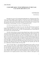
Fig. 1 The Galeazzi sign signifies shortening of the thigh segment, which may be sec-
ondary to hip dislocation or femoral shortening. The Galeazzi test is performed with the
patient supine, hips flexed 90 degrees, and knees flexed. The relative relationship of the
knee heights can then be assessed. (Adapted with permission from Tachdjian MO:
Pediatric Orthopaedics, 2nd ed. Philadelphia: WB Saunders, 1990, vol 1, p 326.)
ages as determined on the left and
right sides. Radiographs of both
hands should be obtained and
compared in this situation.
Prediction of Discrepancy
In the skeletally mature individual,
there is no need to analyze sequen-
tial data, as the situation is static.
The growing child, however, pre-
sents a challenge in predicting the
need for treatment and selecting
from the variety of treatment op-
tions. The importance of obtaining
reproducible data cannot be over-
emphasized. Currently, there are
four different methods incorporat-
ing three techniques for the predic-
tion of limb-length discrepancy: the
arithmetic method, the Eastwood-
Cole method, the Green-Anderson
growth-remaining method, and the
Moseley straight-line graph meth-
od.
13-16
The potential accuracy of any of
these methods is enhanced by hav-
ing longitudinal data. Obtaining
data at 6-month to yearly intervals
over a number of years is much
more helpful than using numerous
data points over a relatively short
time frame. The same technique
should be used for each radiographic
assessment to avoid the vagaries of
magnification. The fact that there are
a number of recognized patterns of
limb-length discrepancy, as de-
scribed by Shapiro,
17
further empha-
sizes the importance of minimizing
error.
The arithmetic method, or rule-
of-thumb method, was first de-
scribed by White and evaluated by
Westh and Menelaus.
13
It is based
on four assumptions about growth:
(1) boys stop growing at age 16;
(2) girls stop growing at age 14;
(3) the distal femoral physis grows
10 mm yearly; and (4) the proximal
tibia grows 6 mm yearly. This
method is useful only during the
later years, not in young children.
A potential disadvantage lies in
using chronologic rather than skele-
tal age, which may present prob-
lems in assessing individuals who
mature very early or very late.
Eastwood and Cole
16
published
a scheme using a graphic arith-
metic method. These data were
confirmed with CT scanning and
skeletal age measurements in mid-
dle and late childhood. Reference
slopes indicate the most appropri-
ate time for epiphysiodesis. Using
this technique, the authors pre-
dictably achieved limb-length
equality within 1 cm.
The growth-remaining method
is based on growth tables pub-
lished by Green and Anderson.
15
Graphs relate the limb lengths of
boys and girls to chronologic age
and can be used to determine a
childÕs growth percentile. Other
graphs demonstrate the remaining
proximal tibial and distal femoral
physeal growth and can be used to
predict the effect of epiphysiodesis.
Because only the most recent skele-
tal age determination is used, any
innaccuracy in its assessment will
cause the resultant estimation of
limb-length discrepancy to be
prone to imprecision. This method
has the greatest longevity of use
but is cumbersome due to the ne-
cessity of referring to two sets of
graphs.
The straight-line graph method
described by Moseley
14
is a distilla-
tion of the Green-Anderson data
graphically displayed in a straight
line over time on a single graph. It
is based on two principles: (1) a
nomogram can be used to deter-
mine the growth percentile from
limb length and skeletal age, and
(2) the growth of both limbs can be
represented graphically by two
straight lines. The difference in
slope between the long and short
limbs indirectly represents the
growth inhibition (or stimulation)
of the abnormal extremity. An
advantage of this method is that a
single-page graph represents the
entire limb-growth history. In addi-
tion, the vagaries of interpreting
skeletal age studies and their intrin-
sic inaccuracy become less impor-
tant over a number of estimations.
In a recent study, Little et al
18
compared the accuracy of the
Anderson-Green, Westh-Menelaus,
and Moseley methods of predicting
limb-length discrepancy. No im-
portant differences were revealed.
Disparities of up to 2.5 cm in the
foot height itself can be seen in
patients with congenital limb
shortening. To date, no radio-
graphic measurement method that
provides a reproducible standing
foot height has been described.
Any clinical measurement discrep-
ancy should be added to the ulti-
mate projected limb discrepancy.
Accuracy is enhanced by having
a single observer remeasure values
on all radiographs, regardless of
the source. Using measurements
taken from different reference
points creates unnecessary errors.
With accurate longitudinal data,
the goal of producing reasonable
limb symmetry with accuracy with-
in 1 cm should be readily achiev-
able. If the data are inadequate,
inaccurate, or confusing, an epi-
physiodesis should be avoided, and
another method of limb equaliza-
tion should be selected at skeletal
maturity.
Treatment Options
The broad spectrum of therapeutic
options available for the patient
with a limb-length discrepancy
includes no treatment at all; simple
shoe modification; shortening proce-
dures, such as percutaneous epi-
physiodesis (Fig. 2) and intra-
medullary shortening (Fig. 3);
lengthening procedures, and combi-
nations thereof. It is essential to
establish the goals of treatment
before embarking on any of these
Limb-Length Inequality
Journal of the American Academy of Orthopaedic Surgeons
146
options. In general, these goals are
equal limb lengths, normal axial
alignment with a level pelvis, and
enhanced function. These goals
may be modified, depending on var-
ious clinical variables. The patient
with a stiff knee or hip or weakness
of the involved extremity should be
left slightly short on that side to
allow the foot to clear the floor in
swing phase without the need for
circumduction or excessive Òhip
hike.Ó In patients with a fixed pelvic
obliquity, functional and actual limb
lengths may differ significantly. If
the pelvic obliquity cannot be elimi-
nated, functional limb-length equali-
ty should be the goal.
Data obtained by Gross
1
and
others suggest that projected dis-
crepancies of less than 2 cm require
no treatment. In a recent article,
Kaufman et al
2
demonstrated by
gait analysis that subjects with a
limb-length disparity of less than
2.0 cm had no greater gait asymme-
try than the general population.
Song et al
3
reported increased work
done by the long side and greater
vertical displacement of the center
of body mass in patients with dis-
crepancies greater than 5.5% com-
pared with the opposite limb.
In general, patients whose ulti-
mate inequality will be in the range
of 2 to 6 cm should undergo a short-
ening procedure, either by epiphys-
iodesis or femoral shortening.
There are several potential excep-
tions. One is the patient in whom
the short extremity has a major
angular deformity. In such a case,
simultaneous deformity correction
and lengthening should be consid-
ered. Another possible exception is
the patient with pathologically short
stature in whom further height
reduction would compromise func-
tion. Yet another potential excep-
tion is the patient with shortening
below the knee who presents either
at maturity or too late for an epi-
physiodesis and in whom contralat-
eral femoral shortening would pro-
Deborah F. Stanitski, MD
Vol 7, No 3, May/June 1999
147
A B C
D E
Fig. 2 A, Percutaneous drilling of the dis-
tal femur is performed from both the medi-
al and the lateral sides. B, Curettage is
then performed to remove all growth carti-
lage. C, An anterior approach to the proxi-
mal fibular physis provides direct visual-
ization and avoids potential peroneal nerve
injury. The incision can then be utilized to
drill and curette the lateral proximal tibial
physis. D, As in the distal femur, both
medial and lateral approaches to the proxi-
mal tibial physis are recommended to
ensure symmetrical growth arrest. E,
Introduction of contrast material confirms
adequate physeal excision.
duce additional knee-height asym-
metry. A review of the literature
indicates that there is no functional
or cosmetic disability as a result of
knee-height disparities of less than 4
cm. If the difference is greater than
this, lengthening of the involved
tibia may be preferable.
The patient with a discrepancy
exceeding 5 to 6 cm is best treated
by limb lengthening or a combina-
tion of limb lengthening and con-
tralateral shortening. Limb abla-
tion and/or prosthetic fitting
should be reserved for patients
whose problems are unmanageable
by current surgical techniques.
Shoe Modification
A shoe lift remains an excellent
treatment for small discrepancies.
Unfortunately, even with the new
lightweight orthotic materials, all
shoe lifts render the sole stiff.
Tapering at the toe is necessary to
approximate normal gait. This is
the least morbid and least expen-
sive method of limb-length equal-
ization and is preferable for patients
with discrepancies of less than 2.0
to 2.5 cm. Nearly half of the dispar-
ity can be accommodated inside the
shoe, which may be sufficient to
provide adequate patient comfort.
Although modern orthotic technol-
ogy has decreased shoe-lift weight,
most patients with larger discrepan-
cies shun the lift because of cosme-
sis and prefer a surgical option
despite the potential morbidity.
Shortening Procedures
Epiphysiodesis and acute femoral
shortening are both length-reducing
procedures. In the growing child
with adequate longitudinal data,
normal axial alignment, and a pro-
jected discrepancy of between 2
and 5 cm, epiphysiodesis remains
the procedure of choice. Various
techniques have been described,
including epiphyseal stapling and
the Blount and Phemister tech-
niques.
Epiphyseal stapling should be
used cautiously. In order to pro-
duce physeal arrest, three medial
and three lateral staples are placed
in the distal femur and the proximal
tibia. The most common complica-
tion reported is staple extrusion.
19
The method currently preferred
is the percutaneous technique ini-
tially reported by Canale et al.
20,21
Small medial and lateral physeal
incisions allow percutaneous
drilling, followed by physeal curet-
tage under image intensifier con-
trol (Fig. 2). Postoperative immobi-
lization is not required. Excellent
and reproducible results have been
achieved with this technique.
21-23
The choice of limb segment (i.e.,
distal femur or proximal tibia or
both) should be selected primarily
on the basis of the location of the
contralateral shortening. If the
shortening is idiopathic, both limb
segments will be involved. Under
these circumstances, knee height
Limb-Length Inequality
Journal of the American Academy of Orthopaedic Surgeons
148
A B C
D E F
Fig. 3 In intramedullary shortening, the intramedullary canal is first reamed over a guide
wire. A cam saw of appropriate size is then introduced into the femoral diaphysis (A) and
deployed gradually while being rotated to produce an osteotomy (B). The saw is then
moved the appropriate distance to achieve the amount of shortening desired proximally,
and the procedure is repeated (C). The saw is removed, and a J-shaped osteotome is
inserted to split the intercalary segment (D). This must be done twice, ideally at 180
degrees with respect to each longitudinal osteotomy (E). The guide wire is reintroduced,
the femur is shortened, and the intramedullary nail is inserted (F).
symmetry will be maintained if
epiphysiodesis is performed on
both the distal femur and the proxi-
mal tibia.
Acute tibial shortening has major
potential complications, including
nonunion and compartment syn-
drome,
24,25
which preclude its com-
mon use for limb-length equaliza-
tion. Femoral shortening is useful
for patients who present after matu-
rity and for those with insufficient
data or inadequate growth remain-
ing for an epiphysiodesis. The two
basic described techniques are closed
intramedullary shortening, as de-
scribed by Winquist
26
and Kempf et
al,
27
and open subtrochanteric short-
ening performed with use of either a
blade plate or large-fragment plate
fixation. An intramedullary saw is
used for the first technique, with dia-
physeal osteotomies, splitting of the
intercalary segment, and insertion of
a locked intramedullary rod (Fig. 3).
This method is technically de-
manding, requiring familiarity with
the instrumentation. Its success
depends on several anatomic as-
sumptions that may not be true. The
cam-deployed saw works in a circu-
lar fashion, but the femur is not
always cylindrical and of uniform
thickness throughout its circumfer-
ence. A small incision may be re-
quired to complete the osteotomy.
There are concerns as well about the
use of this technique in adolescents
because of reports of osteonecrosis of
the hip after femoral nailing.
28,29
The open subtrochanteric tech-
nique is generally easier than the
diaphyseal one. Fixation can be
achieved by using either a blade
plate or a contoured conventional
plate (Fig. 4). Nordsletten et al
30,31
have demonstrated a possible max-
imum of 10% length reduction in
middiaphyseal shortening as
opposed to subtrochanteric short-
ening. In their experience, thigh
muscle strength never returned to
normal in patients with diaphyseal
shortening greater than 10%. This
suggests that the open proximal
technique of shortening may be a
more physiologically sound proce-
dure than closed intramedullary
diaphyseal shortening.
Limb Lengthening
Lengthening has significantly
evolved over the past decade in
North America due to the introduc-
tion of the Ilizarov technique.
32,33
The biologic principles of gradual
incremental distraction have con-
tributed greatly to the ability to
form excellent bone in the distrac-
tion gap while avoiding the prob-
lems of the need for bone graft and
plate fixation, which plague the
Wagner and other techniques.
Despite the improvements in
gradual-distraction lengthening
techniques, the complications of
limb lengthening exceed those of
epiphysiodesis or acute shortening.
These include joint contracture,
joint subluxation or dislocation,
muscle weakness, vascular injury,
nerve palsy, bone regenerate defor-
mation, and pin-site infection.
34,35
Limb lengthening is indicated
for length discrepancies exceeding
5 to 6 cm and those associated with
significant angular and/or rotation-
al deformity of the short extremity.
Limb lengthening can be easily
combined with epiphysiodesis as
part of the overall strategy for man-
agement of limb-length inequality.
For example, if a patient with con-
genital limb hypoplasia has a pro-
jected discrepancy of 18 to 20 cm
and is a reasonable candidate for
limb elongation, two lengthenings
plus a contralateral epiphysiodesis
may be a more reasonable strategy
than three lengthening procedures.
Deborah F. Stanitski, MD
Vol 7, No 3, May/June 1999
149
Fig. 4 A, Preoperative scanogram of a skeletally mature 28-year-old woman with 3.3 cm
of left femoral shortening due to a previous fracture. B, Open subtrochanteric shortening
of the right femur was performed. Fixation was achieved with use of a 90-degree adoles-
cent blade plate.
A B
The currently utilized technique
involves a percutaneous osteotomy,
with care to avoid periosteal strip-
ping, followed by gradual incremen-
tal distraction.
32,33
This is accom-
plished with the use of external
skeletal fixation. Lengthening with
temporary external fixation over an
intramedullary nail may be used in
selected circumstances (Fig. 5).
36
The external fixator may be either a
multiplanar (circular) or a uniplanar
(cantilever) type. Bone fixation may
be achieved with transosseous ten-
sioned wires, half pins, or a combi-
nation of both, depending on the fix-
ator type.
Circular, Ilizarov-type fixators
allow application to almost any
limb segment or size and can be
adjusted to correct angular, rota-
tional, and translational deformi-
ties as well as to achieve lengthen-
ing (Fig. 6). The devices can be
extended to adjacent limb seg-
ments when necessary to protect
potentially unstable joints during
lengthening and to avoid tendon
contracture. However, Ilizarov fix-
ators are neither user- nor patient-
friendly. There is a steep learning
curve before one can consistently
avoid iatrogenic errors and major
complications related to their use.
34
Uniplanar devices are easier to
apply and are usually well tolerated
by the patient. Due to their configu-
ration, they have some limitations
in application in small patients and
in patients with multifocal or multi-
planar deformities (Fig. 7). Align-
ment adjustment in the pediatric
patient usually requires general
anesthesia. Lengthening of the fe-
mur with a uniplanar device causes
elongation along the anatomic bone
axis, producing medialization of the
knee.
33,36
Because the extent to
which this occurs is dependent on
the extent of lengthening, this factor
should be considered before choos-
ing a cantilever device.
Lengthening over an intra-
medullary nail probably has its
greatest application in the mature
patient. The advantage of this tech-
nique is limiting the time of exter-
nal fixation.
36
Once the desired
length has been achieved, the nail is
locked distally, and the external fix-
ator is removed. The most signifi-
cant potential risk is intramedullary
sepsis due to communication of
Limb-Length Inequality
Journal of the American Academy of Orthopaedic Surgeons
150
Fig. 5 A, Radiographs of a 21-year-old man who was injured in a lawn-mower accident at age 2. Multiple surgical procedures, including
a left knee arthrodesis, resulted in an 8-cm limb-length inequity. B, Lengthening of the femur over a proximally locked femoral nail was
initiated through a subtrochanteric osteotomy. A cantilever fixator was used. C, Radiographic appearance at the conclusion of gradual
distraction to achieve lengthening by 6 cm. The nail was locked distally, and the external fixator was removed. D, Radiographic appear-
ance after consolidation of the distraction gap. (Courtesy of John E. Herzenberg, MD, Baltimore.)
A B C D
external fixator pins with the intra-
medullary device. Juxta-articular
deformity (such as in the distal
femoral metaphysis) cannot be easi-
ly corrected with this technique
because the nail ascends within the
femur during lengthening. How-
ever, diaphyseal deformity can be
easily corrected acutely prior to nail
insertion.
A completely implantable inter-
nal lengthening device would be
ideal. The Albizzia nail works by a
ratchet mechanism.
37
The nail is
implanted and locked proximally
and distally. Rotation of the pa-
tientÕs lower extremity creates dis-
traction with an audible click. This
device is currently under develop-
ment and is considered experimen-
tal in North America. A hydraulic
mechanism would, in theory, be
advantageous to eliminate the rota-
tion necessary with this system.
Controversies in Limb
Lengthening
The ability to lengthen a limb is
now no longer limited by the abili-
ty to produce bone that will heal
reliably. Soft tissues and joint sta-
bility currently limit the ability to
lengthen a limb and produce a
functionally as well as cosmetically
acceptable result. The prior histori-
cal constraints of 15 to 18 cm of
maximum lengthening may no
longer be valid.
Patients with severe fibular and
tibial hemimelia are probably still
best treated by limb ablation in in-
fancy. For the patient with fibular
hemimelia and a foot with fewer
than three rays, there are currently
no effective means of producing a
reasonably functional weight-bear-
ing foot. Patients with acceptable
foot function and a moderately
mobile ankle can be treated with
soft-tissue releases, resection of the
fibrous fibular anlage (when pres-
ent), Achilles tendon lengthening,
and subsequent use of an articulated
ankle-foot orthosis until the length
discrepancy becomes unmanageable
(6 to 8 cm), at which time the first
tibial lengthening is performed. The
child can be reevaluated to plot the
developing discrepancy, and a sec-
ond lengthening and contralateral
epiphysiodesis can be done if neces-
sary. The important feature of these
patients is not the presence or ab-
sence of the fibula, but rather the
morphology and potential function
of the foot and ankle.
A patient with tibial hemimelia
and an absent or dysfunctional
knee extensor mechanism is best
treated by an early knee disarticu-
lation. Limb reconstruction can be
a viable option if the proximal tibia
is present (as determined by clini-
cal examination and ultrasound or
magnetic resonance imaging), the
knee actively extends and is rea-
sonably stable, and the foot can be
made functional by early compre-
hensive soft-tissue release. Ulti-
mately, symptomatic ankle insta-
bility in either tibial or fibular
hemimelia can be managed with an
ankle arthrodesis without sacrific-
ing the foot.
Severe forms of proximal fe-
moral focal deficiency in which
there is little femur present (type D
in the Aitken classification system)
or in which the hip cannot be ren-
dered stable are still not amenable
to lengthening. However, if hip
stability can be achieved, femoral
lengthening can be done. If the
foot is at the level of the contralat-
Deborah F. Stanitski, MD
Vol 7, No 3, May/June 1999
151
A B
Fig. 6 A, Preoperative radiograph of a skeletally mature woman with Ollier disease and a
14-cm limb-length inequality. Circular external fixation was used to gradually correct the proxi-
mal and distal tibial deformities and to lengthen the tibia by 8 cm. B, Teleradiograph at the
completion of the tibial lengthening. (Subsequent femoral lenghtening is illustrated in Figure 7.)
eral knee and the ankle has a func-
tional range of active motion, a Van
Nes rotationplasty may provide an
alternative to foot ablation.
Each limb-lengthening proce-
dure should probably be confined
to no more than 15% to 20% of the
limb-segment length. The rate of
distraction should be adjusted
according to the appearance of the
regenerate bone formation as well
as the range of motion of adjacent
joints. The Ò0.25 mm four times a
dayÓ guideline need not be fol-
lowed rigidly. The potential com-
plications of joint stiffness due to
cartilage injury and/or musculo-
tendinous contracture can be
avoided by careful assessment dur-
ing the distraction phase.
38,39
Limb
function should not be sacrificed in
the attempt to gain excessive length.
Residual discrepancy can be treated
by additional lengthening at a later
date, shortening of the contralateral
extremity, or both.
Summary
The management of the growing
patient with limb-length inequality
requires careful assessment, se-
quential limb-length evaluations,
and formation of a strategy based
on the individual patientÕs needs.
Treatment may involve a single
procedure or a series of proce-
dures, depending on the etiology
and magnitude of the discrepancy
and associated deformities.
Limb-Length Inequality
Journal of the American Academy of Orthopaedic Surgeons
152
A
E
B C D
Fig. 7 A, Photograph of the patient in
Figure 6 after tibial lengthening and
deformity correction but before initiation
of femoral lengthening and correction of
the distal femoral valgus deformity. B,
Anteroposterior and lateral radiographs
of the femur after acute correction of the
distal femoral deformity and initiation of
gradual distraction. C and D, Antero-
posterior and lateral radiographs at the
completion of treatment. E, Final appear-
ance of the patient at the completion of
limb-length equalization. The original
shoe lift is shown on the right.
References
1.Gross RH: Leg length discrepancy:
How much is too much? Orthopedics
1978;1:307-310.
2.Kaufman KR, Miller LS, Sutherland
DH: Gait asymmetry in patients with
limb-length inequality. J Pediatr
Orthop1996;16:144-150.
3.Song KM, Halliday SE, Little DG: The
effect of limb-length discrepancy on
gait. J Bone Joint Surg Am1997;79:
1690-1698.
4.Goel A, Loudon J, Nazare A, Rondi-
nelli R, Hassanein K: Joint moments in
minor limb length discrepancy: A pilot
study. Am J Orthop1997;26:852-856.
5.Grundy PF, Roberts CJ: Does unequal
leg length cause back pain? A case-
control study. Lancet1984;2:256-258.
6.Gofton JP: Persistent low back pain
and leg length disparity. J Rheumatol
1985;12:747-750.
7.Papaioannou T, Stokes I, Kenwright J:
Scoliosis associated with limb-length
inequality. J Bone Joint Surg Am1982;
64:59-62.
8.Morscher E: Etiology and pathophysi-
ology of leg length discrepancies.
Orthopade1972;1:1-8.
9.Green WT, Wyatt GM, Anderson M:
Orthoroentgenography as a method of
measuring the bones of the lower
extremities. J Bone Joint Surg1946;28:
60-65.
10.Aaron A, Weinstein D, Thickman D,
Eilert R: Comparison of orthoroent-
genography and computed tomogra-
phy in the measurement of limb-
length discrepancy. J Bone Joint Surg
Am1992;74:897-902.
11.Helms CA, McCarthy S: CT scano-
grams for measuring leg length dis-
crepancy. Radiology1984;151:802.
12.Terjesen T, Benum P, Rossvoll I, et al:
Leg-length discrepancy measured by
ultrasonography. Acta Orthop Scand
1991;62:121-124.
13.Westh RN, Menelaus MB: A simple
calculation for the timing of epiphysial
arrest: A further report. J Bone Joint
Surg Br1981;63:117-119.
14.Moseley CF: A straight-line graph for
leg-length discrepancies. J Bone Joint
Surg Am1977;59:174-179.
15.Green WT, Anderson M: Experiences
with epiphyseal arrest in correcting
discrepancies in length of the lower
extremities in infantile paralysis: A
method of predicting the effect. J Bone
Joint Surg1947;29:659-675.
16.Eastwood DM, Cole WG: A graphic
method for timing the correction of
leg-length discrepancy. J Bone Joint
Surg Br1995;77:743-747.
17.Shapiro F: Developmental patterns in
lower-extremity length discrepancies.
J Bone Joint Surg Am1982;64:639-651.
18.Little DG, Nigo L, Aiona MD: Defi-
ciencies of current methods for the
timing of epiphysiodesis. J Pediatr
Orthop1996;16:173-179.
19.May VR Jr, Clements EL: Epiphyseal
stapling: With special reference to
complications. South Med J 1965;58:
1203-1207.
20.Canale ST, Russell TA, Holcomb RL:
Percutaneous epiphysiodesis: Experi-
mental study and preliminary clinical
results. J Pediatr Orthop1986;6:150-156.
21.Canale ST, Christian CA: Techniques
for epiphysiodesis about the knee.
Clin Orthop1990;255:81-85.
22.Porat S, Peyser A, Robin GC: Equali-
zation of lower limbs by epiphysiode-
sis: Results of treatment. J Pediatr
Orthop1991;11:442-448.
23.Horton GA, Olney BW: Epiphysio-
desis of the lower extremity: Results of
the percutaneous technique. J Pediatr
Orthop1996;16:180-182.
24.Broughton NS, Olney BW, Menelaus
MB: Tibial shortening for leg length
discrepancy. J Bone Joint Surg Br1989;
71:242-245.
25.Kenwright J, Albinana J: Problems
encountered in leg shortening. J Bone
Joint Surg Br1991;73:671-675.
26.Winquist RA: Closed intramedullary
osteotomies of the femur. Clin Orthop
1986;212:155-164.
27.Kempf I, Grosse A, Abalo C: Locked
intramedullary nailing: Its application
to femoral and tibial axial, rotational,
lengthening, and shortening osteoto-
mies. Clin Orthop1986;212:165-173.
28.Astion DJ, Wilber JH, Scoles PV:
Avascular necrosis of the capital
femoral epiphysis after intramedullary
nailing for a fracture of the femoral
shaft: A case report. J Bone Joint Surg
Am1995;77:1092-1094.
29.Beaty JH, Austin SM, Warner WC,
Canale ST, Nichols L: Interlocking
intramedullary nailing of femoral-
shaft fractures in adolescents: Prelimi-
nary results and complications. J
Pediatr Orthop1994;14:178-183.
30.Nordsletten L, Holm I, Steen H,
Bjerkreim: Muscle function after fe-
moral shortening osteotomies at the
subtrochanteric and mid-diaphyseal
level: A follow-up study. Arch Orthop
Trauma Surg1994:114:37-39.
31.Holm I, Nordsletten L, Steen H,
Folleras G, Bjerkreim I: Muscle func-
tion after mid-shaft femoral shorten-
ing: A prospective study with a two-
year follow-up. J Bone Joint Surg Br
1994;76:143-146.
32.Ilizarov GA: The tension-stress effect
on the genesis and growth of tissues:
Part II. The influence of the rate and
frequency of distraction. Clin Orthop
1989;239:263-285.
33.Paley D: Current techniques of limb
lengthening. J Pediatr Orthop1988;8:
73-92.
34.Velazquez RJ, Bell DF, Armstrong PF,
Babyn P, Tibshirani R: Complications
of use of the Ilizarov technique in the
correction of limb deformities in chil-
dren. J Bone Joint Surg Am1993;75:
1148-1156.
35.Herzenberg JE, Scheufele LL, Paley D,
Bechtel R, Tepper S: Knee range of
motion in isolated femoral lengthen-
ing. Clin Orthop1994;301:49-54.
36.Paley D, Herzenberg JE, Paremain G,
Bhave A: Femoral lengthening over
an intramedullary nail: A matched-
case comparison with Ilizarov femoral
lengthening. J Bone Joint Surg Am
1997;79:1464-1480.
37.Saleh M: The gradual elongation nail
for femoral shortening following intra-
medullary nailing. J Bone Joint Surg Br
1996;78(suppl II-III):134.
38.Stanitski DF, Rossman K, Torosian M:
The effect of femoral lengthening on
knee articular cartilage: The role of
apparatus extension across the joint. J
Pediatr Orthop1996;16:151-154.
39.Stanitski DF: The effect of limb
lengthening on articular cartilage: An
experimental study. Clin Orthop
1994;301:68-72.
Deborah F. Stanitski, MD
Vol 7, No 3, May/June 1999
153