Các biến chứng thường gặp nhất của các dây chằng pot
Bạn đang xem bản rút gọn của tài liệu. Xem và tải ngay bản đầy đủ của tài liệu tại đây (254.54 KB, 9 trang )
Vol 7, No 2, March/April 1999
119
Anterior cruciate ligament (ACL)
reconstruction is a commonly per-
formed orthopaedic procedure that
generally results in good to excel-
lent functional outcomes. Loss of
extension has been reported by
many authors to be the most com-
monly encountered complication
after ACL reconstruction, with an in-
cidence as high as 59%
1-10
(Table 1).
Loss of flexion, although common
after posterior cruciate ligament
reconstruction, is rare after ACL
reconstruction.
3,11
The clinical experience of many
authors indicates that a small loss
of extension is functionally signifi-
cant to athletically active individu-
als. Loss of extension is often more
detrimental to the patientÕs func-
tional capability than preoperative
instability.
3,6,12
In 1989, Sachs et al
8
reported that the three most com-
mon complications after ACL re-
construction were flexion contrac-
ture, patellofemoral pain, and
quadriceps weakness. They main-
tained that a loss of 5 degrees of
extension or more directly causes
an abnormal gait, leading to patello-
femoral pain and quadriceps weak-
ness. Since then, other authors
have agreed with this conclu-
sion.
3,5,6,12,13
Many factors have been associ-
ated with a high rate of loss of
extension, and most of them are
preventable. With the use of the
modern operative and postopera-
tive techniques reviewed in this
article, the incidence and severity
of loss of extension after ACL
reconstruction should be dramati-
cally reduced.
Etiology of Loss of
Extension
Impingement
The etiology of loss of extension
after ACL reconstruction is multi-
factorial. Anterior-intercondylar-
notch scar tissue, which prevents
full extension by mechanically
impinging on the roof of the notch
(Fig. 1), is the most commonly
reported cause of loss of exten-
sion.
1,4,9,13-15
Jackson and Schaefer
4
Dr. Petsche is a fifth-year resident in
orthopaedic surgery, University of Illinois at
Chicago College of Medicine. Dr. Hutchinson
is Assistant Professor of Orthopaedic Surgery,
University of Illinois at Chicago College of
Medicine.
Reprint requests: Dr. Hutchinson, University
of Illinois at Chicago, Department of
Orthopaedics, 901 S. Wolcott (M/C 844),
Chicago, IL 60612-7342.
Copyright 1999 by the American Academy of
Orthopaedic Surgeons.
Abstract
The most common complication of anterior cruciate ligament (ACL) reconstruc-
tion is loss of extension, which is often functionally worse for patients than their
preoperative instability. Many preventable surgical and nonsurgical etiologic
factors have been identified. Accurate placement of the tibial tunnel, adequate
notchplasty, and the routing of the femoral side of the graft are all critical fac-
tors. Several studies report that early range-of-motion therapy emphasizing
immediate postoperative "hyperextension" and avoiding immobilization in flex-
ion reduces the rate of loss of extension. Initial studies investigating the effect
of acute versus chronic ACL reconstruction suggested that acute reconstruction
is associated with a higher rate of loss of extension. However, the authors of
two recent studies in which modern techniques were used have disputed this
conclusion. It is likely that the loss of extension historically seen with acute
ACL reconstructions was related to tibial tunnel placement and postoperative
immobilization. It is possible that the timing of acute ACL reconstruction has
less of an effect than originally postulated. On the basis of the results of several
biomechanical studies, it appears that ACL reconstruction may be performed
with the knee in full extension during graft placement with excellent results
and a very low rate of loss of extension. Use of the descriptive term "loss of
extension" is preferred to the often misleading terms "arthrofibrosis" and "flex-
ion contracture."
J Am Acad Orthop Surg 1999;7:119-127
Loss of Extension After Reconstruction of
the Anterior Cruciate Ligament
Timothy S. Petsche, MD, and Mark R. Hutchinson, MD
Loss of Extension After ACL Reconstruction
Journal of the American Academy of Orthopaedic Surgeons
120
referred to this tissue as a Òcyclops
lesionÓ in 1990. They reported on
a series of 13 patients with loss of
extension after intra-articular ACL
reconstruction. All 13 underwent
arthroscopy, and all were found to
have anterior-intercondylar-notch
scar tissue arising anterior and lat-
eral to the tibial insertion of the
ACL graft. The cyclops nodule
was found to act as a mechanical
block to extension by impinging
on the roof of the notch with ter-
minal extension. Microscopically,
the cyclops nodule contained cen-
tral granulation tissue with peri-
pheral fibrous tissue; in three spec-
imens, cartilaginous tissue was
also found.
In 1992, Marzo et al
16
reported
on 21 patients with loss of exten-
sion after ACL reconstruction with
either a boneÐpatellar tendonÐbone
autograft or a hamstring tendon
autograft. All 21 patients under-
went arthroscopy, and all were
found to have a fibrous nodule
causing a mechanical block to
extension.
In 1993, Fisher and Shelbourne
2
reported on loss of extension that
necessitated reoperation on 42 of
959 consecutive ACL reconstruction
patients. Arthroscopy revealed
Òhypertrophy of the ligament or
abundant tissue formationÓ in the
anterior notch.
In 1994, Shelbourne and Johnson
15
reported on 9 patients referred for
Òarthrofibrosis (loss of more than
15 degrees of extension)Ó after ACL
reconstruction with boneÐpatellar
tendonÐbone autograft. At arthros-
copy, all patients were found to
have anterior-intercondylar-notch
scar tissue.
Capsulitis
Capsulitis is inflammation of the
capsule, characterized by abnormal
periarticular inflammation and
edema. Capsulitis may be either a
focal or a diffuse process. Focal
capsulitis involves an isolated
region of the capsule secondary to
localized trauma, such as a sympto-
matic plica, a contusion, or a unilat-
eral ligament injury (e.g., a medial
collateral ligament tear). Focal cap-
sulitis may cause pain with motion
but rarely leads to a passive loss of
flexion and extension.
Diffuse capsulitis is an excessive
inflammatory reaction to a stimulus
such as surgery, trauma, or infec-
tion. Focal capsulitis may progress
to total capsular involvement, but
the cause of this transition is
unclear. Prolonged immobilization
may be related. What is clear, how-
ever, is that diffuse capsulitis may
progress to arthrofibrosis, in which
intra-articular scar tissue restricts
both flexion and extension.
13
Arthrofibrosis may involve the fat
pad, leading to patella infera, or
may diffusely involve the entire
patellofemoral articulation, leading
to patellar entrapment.
7
These are
particularly debilitating problems.
Table 1
Loss of Extension After ACL Reconstruction
*
Study Date Treatment Incidence of Loss
of Extension >5
degrees, %
Sachs et al
8
1989 Mixed techniques 25
Strum et al
10
1990 Surgery within 21 days
after injury 35
Surgery 21 days or more
after injury 12
Jackson and
Schaefer
4
1990 BPTB repair 5.7
Shelbourne et al
9
1991 Surgery within 1 week
of injury 17
Surgery 2 to 3 weeks
after injury 11
Surgery 21 days after injury 0
Fisher and
Shelbourne
2
1993 BPTB repair 4.4
Dandy and 1994 BPTB repair, immobilization
Edwards
1
in cast 59
Nabors et al
6
1995 BPTB repair, tensioned
in extension 1.8
*
Over the years, the incidence of loss of extension has tended to diminish with the
institution of early mobilization, delay of surgery, and graft-tensioning techniques.
(The apparent exception is the results of Dandy and Edwards,
1
but in that study
patients were immobilized in a cast.) BPTB = boneÐpatellar tendonÐbone.
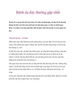
Fig. 1 Inadequate debridement of the old
ACL stump or immobilization after recon-
struction in flexion can allow the develop-
ment of scar tissue, which fills the notch
and prevents extension.
Timothy S. Petsche, MD, and Mark R. Hutchinson, MD
Vol 7, No 2, March/April 1999
121
Although diffuse capsulitis is
referred to by some authors as a
cause of loss of extension after ACL
reconstruction, our review of the lit-
erature indicates that diffuse cap-
sulitis or arthrofibrosis is a rare
cause of loss of extension. The most
common cause is focal anterior-
intracondylar-notch scar tissue (a
cyclops lesion).
4,9,11,13,15
Immobilization in Flexion
In 1994, Dandy and Edwards
1
reported on ACL reconstruction and
the causes of loss of extension. In
their study, 34 patients underwent
reconstruction with boneÐpatellar
tendonÐbone autograft, with cast
immobilization in flexion postopera-
tively. In 59% of cases, loss of exten-
sion necessitated reoperation. All of
these patients underwent arthro-
scopic surgery, and all were found
to have a mechanical block (a nod-
ule of anterior-intercondylar-notch
scar tissue) that prevented full
extension. The authors concluded
that postoperative immobilization in
flexion greatly increases loss of
extension, and that a cyclops lesion
is usually the cause. They also
found that flexion contracture and
arthrofibrosis were rare.
Other authors have found simi-
larly high rates of loss of extension
with postoperative immobilization
in flexion. Cosgarea et al
14
reported
a decrease in the rate of loss of
extension from 23% to 3% when
they changed from postoperative
bracing in 45 degrees of flexion to
bracing in full extension. Of the
nine patients referred to Shelbourne
and Johnson
15
for loss of extension
greater than 15 degrees after ACL
reconstruction, all had been immo-
bilized in flexion postoperatively.
Nonanatomic Graft Placement
Current operative techniques
used in ACL reconstruction are
based on placing the graft in an
anatomic location. Extra-articular,
nonanatomic reconstructions have
been abandoned by most authors
because of their high rate of recur-
rent instability and late failures.
With intra-articular reconstruction,
stability has been more successfully
achieved; however, nonanatomic
placement of the graft with intra-
articular reconstruction will often
lead to loss of motion, usually
extension.
11-13
With placement of
the femoral graft in the Òover the
topÓ position, the graft is tighter in
extension, which may lead to loss
of extension.
12
The ideal femoral
tunnel is placed in the posterior
quartile of the femoral notch, leav-
ing only 1 to 2 mm of posterior
wall remaining when the tunnel is
drilled (Fig. 2). If the over-the-top
position must be used, forming a
trough in the condyle is now rec-
ommended by most authors.
Graft impingement and loss of
extension as a result of anterior
placement of the tibial tunnel (Fig. 3)
have been observed by a number of
authors.
16-19
Marzo et al
16
reported
that anterior placement of the tibial
tunnel for the graft results in a
greater incidence of loss of exten-
sion due to formation of a fibrous
nodule. They postulated that the
anterior graft impinged on the
intercondylar roof, injuring the
graft and stimulating the formation
of the fibrous nodule. Microscopic
examination of the nodules re-
vealed findings similar to those
reported by Jackson and Schaefer.
4
In 1991, Howell et al
18
published
a study investigating the relation-
ship between tibial tunnel place-
ment and graft impingement. On
the basis of an analysis of magnetic
resonance (MR) images of 19 knees
with normal ACLs, the authors
suggested that placing the tibial
tunnel in the posterior aspect of the
original ACL insertion would re-
quire little to no notchplasty to pre-
vent impingement. Placing the tib-
ial graft farther anteriorly increased
the amount of bone that would
have to be removed during notch-
plasty (up to 6 mm) to prevent
impingement. The authors recom-
mended notchplasty with more
bone resection for all ACL recon-
structions performed with an ante-
riorly placed tibial tunnel. In our
opinion, notchplasties may not be
necessary if tunnels are appropri-
ately placed, and notchplasties that
exceed the space required by the
ACL will grow back. Also, the
notchplasty may fill in if patients
are not allowed to attain immediate
full extension to prevent regrowth.
In 1992, Howell and Clark
17
reported on 56 ACL-reconstructed
knees that were examined with MR
imaging 6 months postoperatively.
Thirty demonstrated increased sig-
nal in the graft due to impingement;
the other 26 did not. Lateral radio-
graphs were taken of all 56 knees to
define the location of the tibial tun-
nel. In the 30 knees with impinge-
ment, all the tibial tunnels were
placed between 12 and 23 mm from
the anterior edge of the tibia. Tun-
nel placement 22 to 28 mm from the
anterior edge of the tibia resulted in
10-mm-diameter
femoral tunnel
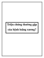
Fig. 2 Poor placement of the femoral tun-
nel can lead to nonisometric placement of
the graft and restricted motion. The ideal
placement is at the origin of the ACL on
the femur in the posterior quartile of the
lateral femoral notch in the 11-oÕclock
(right knee) or 1-oÕclock (left knee)
position.
Loss of Extension After ACL Reconstruction
Journal of the American Academy of Orthopaedic Surgeons
122
26 impingement-free knees. In those
26 knees, the tibial tunnels were
approximately 3 mm posterior to the
center of the original ACL, resulting
in improved extension and stability
(by KT-1000 arthrometer testing).
In 1993, Romano et al
19
reviewed
the radiographs of 111 patients
who had undergone ACL recon-
struction to determine whether tib-
ial tunnel placement affected final
range of motion. Logistic regres-
sion analysis showed that loss of
extension increased the farther
anterior the tibial tunnel was
placed. Furthermore, excessive
medial tibial tunnel placement was
correlated with loss of flexion.
Timing of Surgery
Many articles have evaluated the
effect of the time between knee
injury and ACL reconstruction on
the ultimate range of motion, with
most showing increased loss of
motion with early reconstruction.
In 1990, Strum et al
10
reported on
the rate of loss of motion requiring
lysis of adhesions after ACL recon-
struction. The incidence was 35%
for reconstructions done within 3
weeks of the injury versus 12% for
those done after 3 weeks. In 1991,
Shelbourne et al
9
reported on 169
ACL reconstructions. Patients who
underwent reconstruction within 1
week of the injury were found to
have a higher rate of loss of exten-
sion and decreased strength at 13
weeks postoperatively compared
with patients who underwent re-
construction 3 weeks or more after
injury.
In 1991, Mohtadi et al
5
reported
on loss of motion necessitating
manipulation under anesthesia in 37
of 527 patients (7%) following ACL
reconstruction. The only variable
associated with a higher rate of knee
stiffness was reconstruction within
2 weeks of injury. These results
have led many authors to recom-
mend delaying reconstruction until
acute edema has resolved and range
of motion is at least 0 to 120 degrees.
Despite these recommendations,
many authors have continued to
perform acute ACL reconstructions
with good results. Marcacci et al
20
reported on ACL reconstruction
with fascia lata grafts with a liga-
ment augmentation device in 1995.
Twenty-three patients were treated
within 15 days of injury, and 59
were treated 3 or more months
after injury. No difference in the
rate of loss of extension was found;
however, the early reconstruction
group had better results on clinical
evaluation and KT-2000 arthrome-
ter laxity testing.
Majors and Woodfin
21
recently
reported a retrospective review of
A B C
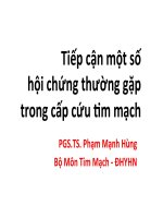
Fig. 3 A, Axial view of the knee demonstrates the normal ÒfootprintÓ of the tibial insertion of the ACL and the optimal position of the
tibial tunnel (1). Anterior tunnel placement (2) leads to anterior impingement on the roof of the intercondylar notch. B, Lateral view
demonstrates the ideal tibial tunnel placement (1) in the second quartile of the tibia as measured from anterior to posterior, with the graft
lying posterior to the roof of the femoral notch (arrow). Anterior tunnel placement (2) leads to impingement on the roof of the intercondy-
lar notch. C, Anteroposterior view of a left knee demonstrates the ideal placement of the tibial tunnel (3) and the femoral tunnel (4).
Lateral tunnel placement (5) can lead to impingement on the lateral condyle. Vertical femoral tunnel placement (6) leads to poor rotation-
al control and recurrent instability.
1
2
1
2
6
3
4
5
Timothy S. Petsche, MD, and Mark R. Hutchinson, MD
Vol 7, No 2, March/April 1999
123
111 arthroscopic intra-articular
ACL reconstructions with boneÐ
patellar tendonÐbone grafts. Full
extension was obtained in 21 of 21
acute (<2 weeks after injury) recon-
structions, 22 of 22 delayed (2 to 4
weeks) reconstructions, and 64 of
68 late (>4 weeks) reconstructions.
All 111 were determined to be sta-
ble by physical examination and
testing with a KT-1000 arthrometer.
The authors concluded that the
timing of ACL reconstruction does
not affect postoperative range of
motion, and that a strictly applied
program of physical therapy with-
out accelerated rehabilitation is
adequate to achieve full range of
motion.
Graft Tension
In the eighth edition of CampbellÕs
Operative Orthopaedics, 14 authors
describe ACL reconstruction tech-
niques.
22
Thirteen of the 14 recom-
mend tensioning and securing the
graft with the knee in varying
degrees of flexion. Most of these
authors recommend tensioning the
graft in the Lachman position (30
degrees of flexion) while exerting a
posterior force on the tibia, despite
biomechanical evidence that the
ACL is not isometric.
Recent studies have confirmed
earlier findings showing that the
ACL lengthens 1 to 3 mm in the ter-
minal 30 degrees of extension. In
1990, Bylski-Austrow et al
23
report-
ed on the biomechanics of ACL
reconstruction in cadaver knees.
Their data showed that knees ten-
sioned in 30 degrees of flexion were
overconstrained regardless of the
amount of tension at fixation.
Reconstructed knees were closest to
intact knees when the graft was
placed with an initial tension of 44
N while the knee was in full exten-
sion during tensioning and fixation.
In 1991, Melby et al
24
also re-
ported on the biomechanics of ACL
reconstruction in cadaver knees.
They concluded that tensioning at
30 degrees overconstrained the
knees. Their data showed that
greater initial tension at 30 degrees
required greater quadriceps force
(up to 26%) to achieve full exten-
sion.
Additional studies of anatomic
intra-articular ACL reconstructions
in cadaver knees have confirmed
these results, showing that tension-
ing at 30 degrees of flexion over-
constrains the knee regardless of
the amount of force used during
tensioning. On the basis of these
biomechanical studies, some au-
thors have recommended tension-
ing and securing the graft with the
knee held at full extension.
25
Despite the multiple biomechani-
cal studies confirming the 1- to 3-
mm lengthening of the ACL in ter-
minal extension, and despite the
recommendation by some authors
that the graft be tensioned in exten-
sion, only one clinical study has
been reported in which the ACL
was tensioned in full extension. In
1995, Nabors et al
6
reported on the
clinical results obtained with arthro-
scopically assisted ACL reconstruc-
tion with boneÐpatellar tendonÐ
bone graft. In a prospective study
of 57 consecutive patients, the graft
was tensioned with maximal one-
hand force and secured with the
knee in full extension. At the 2-year
minimum follow-up, instrumented
postoperative laxity testing with a
KT-1000 arthrometer revealed an
average side-to-side difference of
0.8 mm with a force of 89 N versus
7.5 mm preoperatively. Pivot shift
testing was positive in all 57
patients preoperatively. Postopera-
tively, 51 of 57 (89%) had a negative
pivot shift test, 4 (7%) had a pivot
glide, and 2 (3.5%) had a true pivot
shift. Only 1 patient had loss of
extension greater than 3 degrees
(specifically, 5 degrees), despite the
fact that an accelerated rehabilita-
tion protocol was not used and a
brace with a 10-degree extension
block was worn for the first 4 weeks
during ambulation. However, the
authors did allow immediate active
range of motion as tolerated.
Rehabilitation Protocol
A variety of postoperative tech-
niques have been developed to
decrease the rate of loss of exten-
sion. In 1987, Noyes et al
26
reported
on early knee motion after ACL
reconstruction and concluded that
the reconstructed ligament did not
stretch out with early motion, and
that range of motion was not affect-
ed. In 1990, Shelbourne and Nitz
27
published their results in 450
patients who underwent accelerated
rehabilitation after ACL reconstruc-
tion. They encouraged immediate
full weight bearing, immediate full
extension, early muscle strengthen-
ing, and an early return to activity
and sports. Only 11 of 247 patients
(4%) required reoperation for loss of
extension, compared with 16 of 138
patients (12%) in the control group.
Long-term evaluation of stability
and strength showed no clinically
significant differences. In 1993, Fu
et al
11
reported a reduction in occur-
rence of loss of extension from
11.1% to 1.7% with aggressive post-
operative physical therapy empha-
sizing early full extension.
A review of the long-term follow-
up data on the accelerated rehabili-
tation protocol disclosed excellent
results with regard to preventing
anterior knee pain. In 1997, Shel-
bourne and Trumper
28
reviewed the
results in 602 patients who under-
went ACL reconstructions with
boneÐpatellar tendonÐbone auto-
grafts between 1987 and 1992. The
accelerated rehabilitation protocol
was used with emphasis on obtain-
ing immediate postoperative knee
hyperextension. The authors exam-
ined all 602 patients as well as a con-
trol group of 122 patients who had
Loss of Extension After ACL Reconstruction
Journal of the American Academy of Orthopaedic Surgeons
124
no prior knee injury. The results
showed no difference in the rate of
anterior knee pain in the two
groups. The authors concluded that
emphasizing immediate postopera-
tive knee hyperextension will pre-
vent anterior knee pain while not
compromising long-term knee sta-
bility.
Treatment of Loss of
Extension
Early diagnosis and treatment of
loss of extension may prevent the
need for a second operation. In rare
instances, capsulitis develops after
ACL reconstruction. When this oc-
curs, patients present with diffuse
edema, warmth, constant pain, limi-
tation of patellar mobility, and limi-
tation of both extension and flex-
ion.
13
Late presentation of capsulitis
may result in patella infera.
7
Treat-
ment is usually with nonsteroidal
anti-inflammatory agents or a ta-
pered course of methylprednisolone
in refractory cases. Gentle physical
therapy is indicated, with early
efforts directed toward improving
extension and quadriceps function.
Early manipulation under anesthe-
sia and surgical debridement will
only further aggravate the inflam-
matory process. If loss of extension
persists after the inflammation has
resolved (which usually takes about
6 months), surgical lysis of adhe-
sions may be considered.
13
Patients with loss of extension
usually have impingement due to
anterior-intercondylar-notch scar-
ring. Patients may present asymp-
tomatically or complain of anterior
knee pain and loss of extension.
12
Physical examination shows that
flexion is unaffected. Early treat-
ment is with aggressive physical
therapy emphasizing extension and
quadriceps-strengthening exercises.
The use of an extension drop-out
cast at night has been recommended
by some authors.
13
If there is no
improvement after several weeks of
conservative treatment, arthroscopic
debridement is indicated. Excellent
results have been reported with
notchplasty enlargement combined
with debridement of anterior-
intercondylar-notch scar tissue.
Jackson and Schaefer
4
treated 13
patients with loss of extension. All
underwent arthroscopy, all had
cyclops lesions, and all improved
with arthroscopic debridement and
manipulation. Postoperatively, the
average loss of extension improved
from 16.0 to 3.8 degrees. There were
no complications with this treat-
ment. In 1991, Cannon and Vittori
29
found a clinically significant benefit
with arthroscopic debridement after
ACL reconstruction.
In the series of Dandy and Ed-
wards,
1
all 34 cases of loss of exten-
sion were due to anterior scar tissue
and were relieved with arthroscop-
ic debridement. There were no
cases of arthrofibrosis or flexion
contracture. The incidence of loss
of extension was lowered with
notch widening and immediate full
extension. The authors concluded
that the incidence of loss of exten-
sion is increased with immobiliza-
tion in flexion and is usually due to
anterior-intercondylar-notch scar
tissue.
In the series reported by Marzo et
al,
16
loss of extension due to a fibrous
nodule in 21 patients was treated
with arthroscopic debridement. The
average loss of extension improved
from 11 degrees to 3 degrees with
surgery and further improved to 0
degrees at 1-year follow-up.
Fisher and Shelbourne
2
excised
the Òoffending tissueÓ arthroscopi-
cally in 42 ACL-reconstruction
patients with loss of extension. The
25 patients available for follow-up
at 28 months were all found to have
improvement in function and symp-
toms. Shelbourne and Johnson
15
treated an additional group of 9
patients with arthroscopic anterior
scar resection, notchplasty, manipu-
lation, and extension casting; 8 of
the 9 achieved near-normal exten-
sion. Although these authors refer
to the cause of loss of extension as
arthrofibrosis, this is misleading
because the term ÒarthrofibrosisÓ
denotes the presence of diffuse scar
tissue or fibrous adhesions within
the joint, which does not appear
consistent with the findings in their
studies.
Terminology
A review of the literature shows
that failure to regain full extension
after ACL reconstruction is the most
common complication. Authors
have referred to loss of extension by
many different terms, but perhaps
the two most misleading terms are
ÒarthrofibrosisÓ and Òflexion con-
tracture.Ó The term ÒarthrofibrosisÓ
is correctly used to describe the for-
mation of diffuse scar tissue or
fibrous adhesions within a joint
after capsulitis.
7,13
This usually
causes a loss of both extension and
flexion. Shelbourne and Johnson
15
have used the term arthrofibrosis to
mean loss of more than 15 degrees
of extension after ACL reconstruc-
tion. We consider this to be mis-
leading because their patients did
not have either loss of flexion or dif-
fuse intra-articular fibrosis. We pre-
fer the term Òloss of extension,Ó
which is a generic descriptive term
that neither implies nor excludes
any etiologic possibility. ÒArthro-
fibrosisÓ implies a specific cause
and should be used only to describe
capsulitis leading to diffuse intra-
articular scarring that restricts both
flexion and extension.
The term Òflexion contractureÓ
has also been used by some authors
to describe loss of extension; how-
ever, flexion contracture means
there is high resistance to lengthen-
ing of the flexor muscles or other
posterior structures of the knee
preventing full extension. In our
Timothy S. Petsche, MD, and Mark R. Hutchinson, MD
Vol 7, No 2, March/April 1999
125
review of the literature, neither of
these conditions is a common cause
of loss of extension after ACL re-
construction; in fact, they occur
very rarely. Again, flexion contrac-
ture is a specific cause of loss of
extension, and it is misleading to
use the term generically to refer to
loss of extension regardless of
cause. Because the terms Òarthrofi-
brosisÓ and Òflexion contractureÓ
imply a specific cause, we believe
that the use of these terms has con-
tributed to the failure of many sur-
geons to recognize that intercondy-
lar-notch scarring is by far the most
common cause of loss of extension
after ACL reconstruction.
ÒCyclops lesionÓ is the term
used by Jackson and Schaefer
4
to
refer to anterior-intercondylar-
notch scar tissue that prevents full
extension by impinging on the roof
of the notch. The expression is
easy to remember and emphasizes
the singular nature of the common-
ly found nodule of scar tissue.
Unfortunately, the term is not
descriptive and has no meaning to
a surgeon unfamiliar with it.
Prevention of Loss of
Extension
Many of the identified factors asso-
ciated with loss of extension after
ACL reconstruction are easily pre-
ventable. Reconstructions per-
formed at least 1 month after injury
have been shown by several au-
thors to have a decreased rate of
loss of extension. This has led
some authors to recommend wait-
ing for acute edema to resolve, for
quadriceps function to improve,
and for range of motion to be at
least 0 to 120 degrees before under-
taking surgery. However, there are
many confounding variables in
these preliminary studies, and two
recently published reports dispute
those recommendations.
20,21
A
large prospective study with iden-
tical surgical and rehabilitation
techniques for both groups is nec-
essary before any clinical recom-
mendations can be made.
Intraoperatively, the key to
avoiding loss of extension is careful
anatomic placement of the graft
tunnels. It has been proved that
placement of the tibial tunnel ante-
rior to the center of the original
ACL insertion site will cause im-
pingement and loss of extension.
16-19
Furthermore, inadvertent anterior
drilling of the tibial tunnel despite
accurate placement of the guide
wire has been described.
12
Thus, it
is imperative that great care be
taken during placement of the tib-
ial tunnel, and that adequate notch-
plasty be performed as needed for
all reconstructions.
Techniques to ensure proper tib-
ial tunnel positioning include refer-
encing anatomic landmarks, includ-
ing the posterior cruciate ligament,
the posterior horn of the meniscus,
the medial tibial eminence, and the
roof of the notch; preoperative
x-ray evaluation of the tibia-notch
relationship; and intraoperative
radiography or other imaging.
Testing for impingement before
graft insertion and fixation is valu-
able.
17
A large roof notchplasty
may compensate for far-anterior
placement of the tibial tunnel; how-
ever, this may not be ideal and can
be associated with degenerative
joint disease. A femoral tunnel is
preferable to placing the graft over
the top of the condyle because of
the tensioning issues discussed pre-
viously.
12
Another intraoperative
technique associated with very low
rates of loss of extension is tension-
ing the graft with the knee in full
extension. Several biomechanical
studies and one clinical study
strongly support this technique.
6,10,23,24
One study showed a higher
rate of loss of extension with use of
autograft versus allograft.
3
It was
hypothesized that boneÐpatellar
tendonÐbone harvest-site pain pre-
vents full early extension; however,
this was the only study in which
this conclusion was drawn.
There are several postoperative
techniques for the prevention of
loss of extension. It has been defin-
itively proved that postoperative
immobilization in any amount of
flexion is deleterious.
1,7,12,14
Im-
mediate emphasis on obtaining full
extension is clearly the most impor-
tant factor in preventing loss of
extension.
30
It has been hypothe-
sized that immediate full extension
engages the ACL graft in the notch
and, by occupying this space, pre-
vents the formation of anterior-
intercondylar-notch scar tissue.
Postoperative immobilization in
extension may prevent fibrin clot
from forming in the notch and thus
prevent scar tissue formation.
Accelerated rehabilitation has
been shown by a large number of
authors to decrease the rate of loss
of extension. Additionally, longer
follow-up of ShelbourneÕs original
group of patients treated with
accelerated rehabilitation
27
has
shown that function and stability
are not adversely affected by
immediate postoperative full-knee
hyperextension.
28,30
Other authors
have applied ShelbourneÕs acceler-
ated rehabilitation protocol
27
to
patients undergoing ACL recon-
struction with semitendinosus and
gracilis tendon grafts. The results
have shown similarly decreased
rates of loss of extension with no
loss of stability.
Continuous-passive-motion
machines are used by a number of
authors in the early postoperative
stage. One study found no benefit
from routine use after ACL recon-
struction.
31
Others argue that con-
tinuous passive motion may help to
improve flexion in patients at risk
for loss of flexion but is of little use
in improving extension.
13
In gener-
al, as the continuous-passive-motion
device reaches full extension, the
restricted knee simply remains
Loss of Extension After ACL Reconstruction
Journal of the American Academy of Orthopaedic Surgeons
126
slightly flexed. New machines have
been designed with anterior straps
or hinges locked to the machine to
achieve complete extension, but no
study has been performed on
patients after ACL reconstruction to
confirm their efficacy.
Summary
Loss of extension is the most com-
mon complication of ACL recon-
struction. Various intraoperative
and postoperative techniques are
useful in markedly decreasing the
rate of loss of extension: careful
anatomic placement of graft tunnels;
strict avoidance of anterior place-
ment of the tibial tunnel; avoidance
of over-the-top placement of the
femoral graft; utilization of a trough
in the condyle if over-the-top place-
ment must be employed; use of the
intraoperative impingement test
before graft tensioning; tensioning
the graft with the knee in full exten-
sion; encouragement of immediate
postoperative full-knee hyperexten-
sion; strict avoidance of immobiliza-
tion in flexion or restriction of full
hyperextension in any way; and
early diagnosis and appropriate
treatment of loss of extension. It is
recommended that, for greater clari-
ty of expression, authors should
adopt the term Òloss of extension,Ó
rather than ÒarthrofibrosisÓ or Òflex-
ion contracture.Ó
References
1.Dandy DJ, Edwards DJ: Problems in
regaining full extension of the knee
after anterior cruciate ligament recon-
struction: Does arthrofibrosis exist?
Knee Surg Sports Traumatol Arthrosc
1994;2:76-79.
2.Fisher SE, Shelbourne KD: Arthro-
scopic treatment of symptomatic
extension block complicating anterior
cruciate ligament reconstruction. Am J
Sports Med1993;21:558-564.
3.Harner CD, Irrgang JJ, Fu FH:
Prevention and management of loss of
motion after arthroscopic anterior cru-
ciate ligament reconstruction. Compli-
cations Orthop1993;Spring:5-8.
4.Jackson DW, Schaefer RK: Cyclops
syndrome: Loss of extension following
intra-articular anterior cruciate liga-
ment reconstruction. Arthroscopy 1990;
6:171-178.
5.Mohtadi NGH, Webster-Bogaert S,
Fowler PJ: Limitation of motion fol-
lowing anterior cruciate ligament re-
construction: A case-control study.
Am J Sports Med1991;19:620-625.
6.Nabors ED, Richmond JC, Vannah
WM, McConville OR: Anterior cruci-
ate ligament graft tensioning in full
extension. Am J Sports Med1995;23:
488-492.
7.Paulos LE, Rosenberg TD, Drawbert J,
Manning J, Abbott P: Infrapatellar
contracture syndrome: An unrecog-
nized cause of knee stiffness with
patella entrapment and patella infera.
Am J Sports Med1987;15:331-341.
8.Sachs RA, Daniel DM, Stone ML,
Garfein RF: Patellofemoral problems
after anterior cruciate ligament recon-
struction. Am J Sports Med1989;17:
760-765.
9.Shelbourne KD, Wilckens JH, Molla-
bashy A, DeCarlo M: Arthrofibrosis in
acute anterior cruciate ligament recon-
struction: The effect of timing of recon-
struction and rehabilitation. Am J
Sports Med 1991;19:332-336.
10.Strum GM, Friedman MJ, Fox JM, et
al: Acute anterior cruciate ligament
reconstruction: Analysis of complica-
tions. Clin Orthop1990;253:184-189.
11.Fu FH, Irrgang JJ, Harner CD: Loss of
motion following anterior cruciate lig-
ament reconstruction, in Jackson DW,
Arnoczky SP, Woo SLY, Frank CB,
Simon TM (eds): The Anterior Cruciate
Ligament: Current and Future Concepts.
New York: Raven Press, 1993, pp 373-
380.
12.Johnson DL, Fu FH: Anterior cruciate
ligament reconstruction: Why do fail-
ures occur? Instr Course Lect1995:44:
391-406.
13.Irrgang JJ, Harner CD: Loss of motion
following knee ligament reconstruc-
tion. Sports Med1995;19:150-159.
14.Cosgarea AJ, Sebastianelli WJ, De-
Haven KE: Prevention of arthrofibro-
sis after anterior cruciate ligament
reconstruction using the central third
patellar tendon autograft. Am J Sports
Med1995;23:87-92.
15.Shelbourne KD, Johnson GE: Out-
patient surgical management of arth-
rofibrosis after anterior cruciate liga-
ment surgery. Am J Sports Med1994;
22:192-197.
16.Marzo JM, Bowen MK, Warren RF,
Wickiewicz TL, Altchek DW: Intra-
articular fibrous nodule as a cause of
loss of extension following anterior
cruciate ligament reconstruction.
Arthroscopy1992;8:10-18.
17.Howell SM, Clark JA: Tibial tunnel
placement in anterior cruciate ligament
reconstructions and graft impingement.
Clin Orthop1992;283:187-195.
18.Howell SM, Clark JA, Farley TE: A
rationale for predicting anterior cruci-
ate graft impingement by the inter-
condylar roof: A magnetic resonance
imaging study. Am J Sports Med1991;
19:276-282.
19.Romano VM, Graf BK, Keene JS,
Lange RH: Anterior cruciate ligament
reconstruction: The effect of tibial tun-
nel placement on range of motion. Am
J Sports Med1993;21:415-418.
20.Marcacci M, Zaffagnini S, Iacono F,
Neri MP, Petitto A: Early versus late
reconstruction for anterior cruciate lig-
ament rupture: Results after five years
of followup. Am J Sports Med1995;
23:690-693.
21.Majors RA, Woodfin B: Achieving full
range of motion after anterior cruciate
ligament reconstruction. Am J Sports
Med1996;24:350-355.
22.Sisk TD: Knee injuries, in Crenshaw
AH (ed): CampbellÕs Operative Ortho-
paedics, 8th ed. St Louis: Mosby-Year
Book, 1992, vol 3, pp 1564-1686.
23.Bylski-Austrow DI, Grood ES, Hefzy
MS, Holden JP, Butler DL: Anterior
cruciate ligament replacements: A
mechanical study of femoral attach-
ment location, flexion angle at tension-
ing, and initial tension. J Orthop Res
1990;8:522-531.
24.Melby A III, Noble JS, Askew MJ,
Boom AA, Hurst FW: The effects of
graft tensioning on the laxity and kine-
matics of the anterior cruciate liga-
ment reconstructed knee. Arthroscopy
1991;7:257-266.
25.Hardin GT, Bach BR Jr, Bush-Joseph
CA, Farr J: Endoscopic single-incision
anterior cruciate ligament reconstruc-
Timothy S. Petsche, MD, and Mark R. Hutchinson, MD
Vol 7, No 2, March/April 1999
127
tion using patellar tendon autograft:
Surgical technique. Am J Knee Surg
1992;5:144-155.
26.Noyes FR, Mangine RE, Barber S: Early
knee motion after open and arthroscop-
ic anterior cruciate ligament reconstruc-
tion. Am J Sports Med1987;15:149-160.
27.Shelbourne KD, Nitz P: Accelerated
rehabilitation after anterior cruciate
ligament reconstruction. Am J Sports
Med1990;18:292-299.
28.Shelbourne KD, Trumper RV: Pre-
venting anterior knee pain after anteri-
or cruciate ligament reconstruction.
Am J Sports Med1997;25:41-47.
29.Cannon WD Jr, Vittori JM: The role of
arthroscopic debridement after anteri-
or cruciate ligament reconstruction.
Arthroscopy1991;7:344-349.
30.Rubinstein RA Jr, Shelbourne KD,
VanMeter CD, McCarroll JR, Rettig
AC, Gloyeske RL: Effect on knee sta-
bility if full hyperextension is restored
immediately after autogenous bone-
patellar tendon-bone anterior cruciate
ligament reconstruction. Am J Sports
Med1995;23:365-368.
31.Irrgang JJ, Fu FH, Sawhney R, Dearwater
S, Paul J: Comparison of continuous pas-
sive motion to standard physical therapy
in rehabilitation of patients following
anterior cruciate ligament reconstruction
[abstract]. Orthop Trans1992-1993;16:723.