Báo cáo y học: " Variability in a dominant block to SIV early reverse transcription in rhesus monkey cells predicts in vivo viral replication and time to death" pot
Bạn đang xem bản rút gọn của tài liệu. Xem và tải ngay bản đầy đủ của tài liệu tại đây (470.59 KB, 11 trang )
Rogers et al. Virology Journal 2010, 7:79
/>Open Access
RESEARCH
BioMed Central
© 2010 Rogers et al; licensee BioMed Central Ltd. This is an Open Access article distributed under the terms of the Creative Commons
Attribution License ( which permits unrestricted use, distribution, and reproduction in
any medium, provided the original work is properly cited.
Research
Variability in a dominant block to SIV early reverse
transcription in rhesus monkey cells predicts
in vivo
viral replication and time to death
Thomas F Rogers*, So-Yon Lim, TJ Sundsvold, Tiffany Chan, Ariel Hsu and Norman L Letvin
Abstract
While it has long been appreciated that there is considerable variability in host containment of HIV/SIV replication, the
determinants of that variability are not fully understood. Previous studies demonstrated that the degree of permissivity
of a macaque's peripheral blood mononuclear cells (PBMC) for infection with simian immunodeficiency virus (SIV) in
vitro predicted that animal's peak plasma virus RNA levels following SIV infection in vivo. The present study was
conducted to define the mechanisms underlying the variable intrinsic susceptibility of rhesus monkey PBMC to
SIVsmE660 infection. In a cohort of 15 unrelated Indian-origin rhesus monkeys, infectability of PBMC of individual
animals with SIVsmE660, as defined by tissue culture infectious dose (TCID
50
), varied by more than 3 logs and was a
stable phenotype over time. Susceptibility of a monkey's PBMC to wild type SIVsmE660 infection correlated with the
susceptibility of that monkey's PBMC to infection with VSV-G pseudotyped SIVsm543-GFP. Moreover, the permissivity
of an individual monkey's PBMC for infection with this construct correlated with the permissivity of a B-lymphoblastoid
cell line (B-LCL) generated from PBMC of the same animal. We found that the degree of intrinsic resistance of monkey
B-LCL correlated with the copy number of early reverse transcription (ERT) SIV DNA. The resistance of monkey B-LCL to
SIVsmE660 replication could be abrogated by preincubation of cells with the SIV virus-like particles (VLPs) and SIV
resistance phenotype could be transferred to a SIV susceptible B-LCL through cell fusion. Finally, we observed a positive
correlation between susceptibility of monkey B-LCL to SIV infection with a VSV-G pseudotyped SIV-GFP construct in
vitro and both the peak plasma virus RNA levels in vivo and time to death following wild type SIV infection. These
findings suggest that a dominant early RT restricting factor that can be saturated by SIV capsid may contribute to the
variable resistance to SIV infection in rhesus monkey B-LCL and that this differential intrinsic susceptibility contributes
to the clinical outcome of an SIV infection.
Introduction
Humans vary in their susceptibility to human immunode-
ficiency virus type 1 (HIV-1) acquisition and in the level
of HIV-1 replication following infection [1,2]. Virus phe-
notype, the magnitude of cytotoxic T lymphocyte
response, major histocompatability complex (MHC) class
I haplotype, and chemokine receptor polymorphisms
have all been shown to contribute to this variability [3-5].
Recent studies suggest that further undefined host factors
are also contributing to the level of virus control in the
HIV-1-infected individual [6].
Some of the undefined host factors contributing to
HIV-1 containment may be responsible for the variable
ability of cells to be infected by and sustain replication of
these viruses. It has been shown that permissiveness for
HIV-1 varies substantially between isolated primary cells
Permissiveness, the ability of cells to be infected and sus-
tain the replication of HIV-1, also varies substantially
between isolated primary cells of individuals [7]. It has
been shown that the permissiveness of isolated primary
rhesus monkey lymphocytes for SIV infection correlates
with in vivo viral set point [8].
The variability in permissiveness observed in rhesus
monkey PBMC for SIV replication can be shaped by
dominant and nondominant mechanisms: through the
altered expression of required host factors and/or virus
* Correspondence:
1
Division of Viral Pathogenesis, Beth Israel Deaconess Medical Center, Harvard
Medical School, Boston, Massachusetts 02115, USA
Full list of author information is available at the end of the article
Rogers et al. Virology Journal 2010, 7:79
/>Page 2 of 11
restricting molecules [9]. The ability of a cell to become
infected by HIV/SIV requires cellular expression of
diverse proteins and HIV/SIV replication can be
repressed by the cellular expression of a number of
restriction factors including TRIM5α and APOBEC3G
[10,11]. While the restriction factor TRIM5α appears to
be under positive selection and is highly polymorphic in a
given species, there is little evidence that this genetic
variability has functional consequences [12-15].
We set out to identify genetic mechanisms underlying
the variable susceptibility to lentivirus replication in a
primate species through an analysis of the differential
rhesus monkey permissivity of lymphocytes for SIV repli-
cation. The SIVsmE660-infected rhesus monkey provides
a powerful model system for elucidating the genetic con-
tribution to virus control. Studies can be done with a sin-
gle, defined challenge stock of virus, and monkeys can be
selected for evaluation with specified genetic characteris-
tics. There is a well-documented variability in both peak
and set point replication of this virus in genetically dispa-
rate rhesus monkeys. Moreover, there is a clear correla-
tion between the in vitro susceptibility of PBMC of rhesus
monkeys to infection (TCID
50
) with this virus isolate and
the extent of early virus replication in these animals fol-
lowing experimental infection [8]. In the present study,
we explored the mechanisms resulting in the variation of
rhesus monkey PBMC permissiveness for SIVsmE660
replication and examined the contribution of this process
to the pathogenicity of the virus in vivo.
Methods
In vitro susceptibility assay
PBMC were separated by centrifugation through Ficoll
and CD8+ cells were depleted from PBMC cultures using
CD8 Dynabeads (Dynal). CD8+- depleted PBMC were
resuspended in RPMI 1640 with 6.25 ug of ConA per ml,
10% fetal calf serum (FCS), and 10% interleukin-1 (IL-2)
at a density of 2 × 10
6
PBMC per ml. Three days after ini-
tial culture, aliquots of 40,000 PBMC were incubated for
4 hours with 150 ul of serial 10-fold dilutions of a cell-free
virus stock of SIVsmE660. PBMC were washed with
Hanks balanced salt solution to remove residual virus,
resuspended in RPMI 1640 with 10% FCS and 10% IL-2,
and cultured in 96-well plates. Cultural supernatant were
collected at days 0,3,7,10, and 13 after infection and mon-
itored for the presence of virus by antigen capture assay
for SIV p27 antigen (Coulter Corp.) The minimal TCID
or endpoint titer of virus required to infect the cells of
each donor was determined as the last dilution in which
virus was detected by 13 days after infection.
Viruses and cell lines
The majority of studies were conducted using
SIVsmE660[16]. The molecular clone of SIVsmE660,
SIVsmE543-3 was used to generate VSV-G pseudotyped
SIVsm543-GFP. The SIVsmE543 vector was created by
inserting SalI-NotI fragment of pSIV.GFP into the env
gene of SIVsmE543-3. VSV-G pseudotyped SIVsm543-
GFP, HIV-GFP, Murine Leukemia Virus (MLV-GFP), and
SIVmac-GFP were produced as previously described [17].
SIV virus like particles (VLP) were generated by insertion
of a stop codon into the ORF of SIV reverse transcriptase
(RT). B-lymphoblastoid cell lines (B-LCL) were generated
by incubating rhesus monkey PBMC with 100 ul herpes
papio, washed and seeded into 96-well plates in RPMI
1640 with 20% FCS. PBMC were fed twice weekly with
50% medium changes and transferred to T25 flasks after
4 weeks.
Real-time RT-PCR quantification
The quantification of early reverse transcription, late
reverse transcription, and integrated SIV DNA copy
number was modified from a previously described
method [18]. At 2, 3, 6, 12, 24, and 48 hours following
infection of B-LCL with VSV-G pseudotyped SIVsm543-
GFP, cells were collected and DNA was isolated with
DNAeasy Tissue kit (Qiagen). The primer sets used to
detect each sequence were as follows: early RT forward,
5'-AAGCAAGTGTGTGTTCCCATCT-3'; early RT
reverse, 5'-CCTCGGTTTCCCAAAGCAGAA-3'; late RT
forward, 5'-AAGCAAGTGTGTGTTCCCATCT-3'; late
RT reverse, 5'-CACTTACCTGCAACCGGAGG-3'; inte-
grated forward, 5'-GCTGCCGATTGGGATTTACAAC-
3'; integrated reverse, 5'-AATGTCTGATCCTCTTG-
GCTCTC-3'. Quantification was performed with an
Applied Biosystems 7300 real-time PCR system (Foster
City, CA). GAPDH control primers and probe was pur-
chased through Applied Biosystems.
Fusion assay
B-LCL were stained with 1 ul of Oregon Green or 2 ul of
Vybrant DiD (Invitrogen) for 1 hour. Cells were washed
with RPMI 1640 and allowed to rest for 30 minutes. B-
LCL populations were mixed together and added to 2 ml
of 50% PEG for 1 minute. B-LCL were washed and allow
to rest in RPMI 1640 with 10% FCS for twelve hours.
Double positive fusion events were sorted using flow
cytometry and infected with VSV-G pseudotyped
SIVsm543-GFP.
Results
Variation in rhesus monkey PBMC susceptibility to SIV
replication
We established a cohort of 15 unrelated Indian-origin
rhesus monkeys to investigate the impact of cellular SIV
permissivity on in vivo viral replication. CD8+ T lympho-
cyte-depleted PBMC from each monkey were evaluated
for their relative ability to sustain SIVsmE660 replication
Rogers et al. Virology Journal 2010, 7:79
/>Page 3 of 11
in vitro. Lectin-stimulated CD8+ lymphocyte-depleted
PBMC were exposed to serial dilutions of SIVsmE660.
Replication of virus was assessed by measuring SIV p27
antigen in culture supernatants and a minimal tissue cul-
ture infectious dose (TCID
50
) of virus was defined for
each population of PBMC. We observed a broad range of
permissiveness of these PBMC for SIVsmE660 replica-
tion, with rhesus PBMC populations differing by as much
as 4 logs of the virus needed to initiate infection (Fig. 1A).
To determine whether this PBMC permissiveness for
SIV replication is a stable phenotype, the TCID
50
assay
was repeated after a 3 month interval on CD8+ T lym-
phocyte-depleted PBMC of the same 15 monkeys. The
relative susceptibility of these PBMC populations was
remarkably similar, with a correlation of data demon-
strating a Spearman p = 0.0002 and ρ = 0.8345 (Fig. 1B).
To assess the nature of this variable susceptibility phe-
notype in these PBMC, we examined the kinetics of viral
replication in each PBMC population by sequential analy-
sis of p27 production over time. We observed very differ-
ent kinetics of SIV replication in these rhesus monkey
PBMC, with susceptible cell lines supporting robust viral
replication and resistant cells supporting little to no viral
replication (Fig 1C and 1D). Rhesus monkey PBMC that
exhibited low TCID50 values did not have delayed repli-
cation kinetics; rather, they showed a persistent resis-
tance to viral replication.
Rhesus monkey PBMC susceptibility to SIV replication is
independent of entry
Since genetic polymorphisms have been described that
impact HIV-1 replication through modulation of viral
entry, we sought to determine whether this variable sus-
Figure 1 Susceptibility of PBMC of 14 rhesus monkeys to in vitro SIVsmE660 infection. (A) CD8+ T lymphocyte-depleted PBMC were infected
with SIVsmE660 and p27 production was assayed in the culture supernatant by ELISA. Susceptibility to in vitro infection is expressed as the relative
TCID
50
of the SIVsmE660 virus stock using the Spearman-Karber method. (B) PBMC of the same animals were assayed again for in vitro susceptibility
to SIVsmE660 after a three month interval. A significant positive correlation was observed between the values obtained from PBMC of each monkey
in these repeated assays. (C, D) CD8+ T lymphocyte-depleted PBMC were infected with SIVsmE660, samples collected serially and grouped for display
as (C) sensitive or (D) resistant to SIV replication on the basis of their TCID50 values.
1
2
3
4
log SIVsmE660 TCID
50
A
1
2 3 4
1
2
3
4
p=0.0002
U
U
=0.83
log SIVsmE660 TCID
50
- exp #1
log SIVsmE660 TCID
50
- exp #2
B
Sensitive phenotype
4 6 8 10
1
2
3
4
02N016
01N005
03N006
Days
log p27 (ng/ml)
Resistant phenotype
4 6 8 10
0
1
2
3
4
PH0952
02N011
04N005
04N004
03N001
02N020
02N012
Days
log p27 (ng/ml)
C
D
Rogers et al. Virology Journal 2010, 7:79
/>Page 4 of 11
ceptibility to SIV replication was a consequence of differ-
ential SIV entry or represented a post-entry
phenomenon. To explore these issues, we created a VSV-
G-pseudotyped SIVsmE660 construct that expressed
GFP. The molecular clone of SIVsmE660, SIVsmE543,
was engineered to produce VSV-G pseudotyped
SIVsme543 through the deletion of the SIV env gene and
complementation with a VSV-G-encoding vector. Fur-
thermore, the SIVsme543 plasmid was manipulated by
insertion of GFP ORF into the env gene of SIV543
(SIVsm543-GFP) to allow the expression of GFP follow-
ing SIV integration, transcription and translation of viral
proteins. This construct allowed a simple and rapid quan-
tification of SIV infected cells by flow cytometric analysis.
Moreover, since the VSV-G protein mediated viral entry
into a broad range of cell types, variability in infection by
this construct must reflect a gp120-CD4 independent
phenomenon.
To determine if rhesus PBMC susceptibility to wild
type SIVE660 is associated with susceptibility of these
cells to a single-cycle pseudotyped SIV construct, rhesus
monkey PBMC were infected with the VSV-G pseudo-
typed SIVSM543-GFP and analyzed by flow cytometry
for % GFP-positive cells (Figure 3A). Rhesus monkey
PBMC susceptibility to wild type SIVsmE660 virus was
defined by TCID
50
, while susceptibility to the single cycle
SIVsm543-GFP construct was defined as the % GFP+
PBMC 48 hrs post-infection. Rhesus monkey PBMC
exhibited varied susceptibility to the single cycle VSV-G
pseudotyped SIVSsm543-GFP, and a significant correla-
tion was observed for each cell population between %
SIVsm543-GFP+ cells and the TCID
50
of these cells for
wild type SIVsmE660 (Figure 3A). These data suggest that
the differential permissivity of rhesus monkey PBMC to
SIV replication is entry independent. Additionally,
because the permissivity phenotype was manifested by
the expression of GFP following a single cycle of replica-
tion, the relative blockage of viral replication must occur
in the virus life cycle between early reverse transcription
and post-integration viral expression. Moreover, the
expression of this phenotype does not require multiple
rounds of viral replication. Only one monkey's PBMC
demonstrate a disparity between their permissivity to
wild type virus and VSV-G pseudotyped SIVsm543-GFP.
Rhesus monkey PBMC susceptibility to SIV infection
correlates with B-lymphoblastoid cell line susceptibility to
SIV infection
The susceptibility of rhesus monkey CD4+ T lympho-
cytes to SIV infection is dependent on the activation state
of the T cells, the replication rate of the cells, as well as
the expression level of CCR5, which reflects the memory
phenotype of these cells (7,8,9). Additionally, the relative
representation of CD4+ T cells in a PBMC population
could impact the quantitation of the relative susceptibility
of PBMC to SIV infection. To rule out a contribution of
these well-defined factors to the variability of rhesus
monkey lymphocyte susceptibility to SIV replication, we
assessed the permissivity of rhesus monkey B-lympho-
blastoid cell lines (B-LCL) to VSV-G pseudotyped
SIVsm543-GFP infection. In addition to facilitating rapid
screening of large numbers of cell populations, analysis of
B-LCL would establish if this differential permissivity for
SIV replication is T cell-specific or is manifested in other
cell types.
We generated B-LCL from 15 unrelated Indian-origin
rhesus monkeys and analyzed B-LCL and PBMC from
the same animals for susceptibility to infection by VSV-G
pseudotyped SIVsm543-GFP. Following infection of rhe-
sus monkey B-LCL with this construct, we observed a
broad range in B-LCL susceptibility to SIV infection,
varying from 1% to 30% of target cells infected (Fig. 2A).
To determine whether B-LCL permissivity for SIV repli-
cation is a stable phenotype, B-LCL from the same ani-
mals were infected a second time. There was a significant
correlation between susceptibility phenotypes for experi-
ment #1 and #2 for all 15 animals (data not shown). Addi-
tionally, B-LCL were generated from the same monkeys
two months later and assayed for susceptibility using the
VSV-G pseudotyped SIVsm543-GFP construct. There
was a significant correlation between the relative suscep-
tibility to SIV infection of the two populations of B-LCL
for all animals (data not shown).
The differential susceptibility of B-LCL populations
from different monkeys for SIV replication could be a
consequence of two possible mechanisms: a subpopula-
tion of variable size in a B-LCL population could be per-
missive or the total B-LCL population could be uniform
in its relative resistance or susceptibility to SIV infection.
To examine these two possibilities, B-LCL were infected
with the VSV-G pseudotyped SIVsm543-GFP, GFP nega-
tive cells were sorted, and the GFP negative cells were
reinfected with the same viral construct. No significant
change in relative susceptibility to SIV infection was
observed following the second round of infection (Fig.
2B). This observation suggests that the total population
of B-LCL from each animal exhibits a uniform suscepti-
ble or resistant phenotype.
To determine the relationship between the susceptibil-
ity of a given population of B-LCL to VSV-G pseudotyped
SIVsm543-GFP infection and the susceptibility of PBMC
from the same monkey to wild type virus replication, we
assayed these cell populations from a cohort of rhesus
monkeys. In these experiments, we defined PBMC sus-
ceptibility to wild type SIVsmE660 by TCID
50
. To repre-
sent SIV permissivity of each cell line accurately over a
range of multiplicities of infection (MOIs), B-LCL sus-
ceptibility was defined as the area under the curve (AUC)
Rogers et al. Virology Journal 2010, 7:79
/>Page 5 of 11
of % GFP+ cells over 4 serial viral dilutions following
infection with the single cycle VSV-G pseudotyped
SIVsm543-GFP. We observed a clear positive correlation
between PBMC susceptibility to wild type SIVsmE660
infection and the permissivity of B-LCL from the same
monkey to single cycle VSV-G pseudotyped SIVsm543-
GFP (Spearman p value = 0.0004, ρ = 0.83(Fig. 3B). Simi-
larly, there was a significant correlation between permis-
sivity of PBMC to infection with wild type SIVmac239
and B-LCL to infection with VSV-G pseudotyped
SIVmac239-GFP (Fig. 3C). These data indicate that dif-
ferential intraspecies permissivity for SIV replication in
rhesus monkey lymphocytes is not dependent on the acti-
vation or memory state of CD4+ T cells, is not entry
dependent, does not reflect the susceptibility of a sub-
population of cells, is not dependent on multiple rounds
of infection, is not specific for SIVsmE660 and is mani-
fested at a stage of viral replication prior to viral gene
expression.
SIV resistance in rhesus monkey cells correlates with
resistance to primate immunodeficiency viruses, but not to
other viruses
To determine the range of viruses whose replication is
associated with SIV replication in rhesus monkey B-LCL,
we infected 15 B-LCL populations with VSV-G pseudo-
typed SIVmac239-GFP, HIV-GFP, N-MLV-GFP, and B-
MLV-GFP at varying dilutions. B-LCL infected with
SIVmac239-GFP and SIVsm543-GFP exhibited differen-
tial inflexibility which was significantly correlated (Fig.
4A). Furthermore, we found that HIV-GFP susceptibility
correlated with SIVsm543-GFP susceptibility (Fig. 4B).
However, rhesus monkey B-LCL permissivity for adeno-
v i r u s - G F P, V S V- G F P, H S V- GFP, N - M LV- G F P and B -
MLV-GFP did not correlate with permissivity for SIV or
HIV (Fig 4C, D, E, F, G). This study suggests that the rela-
tive block to SIVsmE660 infection in B-LCL acts through
a mechanism that can also restrict other SIVs and HIV,
but allows the replication of other viruses.
Figure 2 Rhesus monkey B-LCL exhibit differential susceptibility to SIVsmE660 infection. (A) B-LCL were generated from PBMC of 15 rhesus
monkeys and infected with varying dilutions of VSV-G pseudotyped SIVsmE543-GFP. SIVsmE543-GFP susceptibility was defined by flow cytometry as
% GFP+ B-LCL following infection (B) B-LCL were infected with SIVsmE660-GFP and sorted for the GFP negative cell population. These GFP negative
cells were reinfected with SIVsmE543-GFP and, again, analyzed by flow cytometry.
0 25 50 75
100 125 150 175
0
10
20
30
40
ul of SIVsmE543-GFP virus
%SIVsmE543-GFP+ B-LCL
A
0 25 50 75 100 125 150 175
0
10
20
30
40
ul of SIVsmE543-GFP
% SIVsmE543-GFP+ B-LCL
0 25 50 75
100 125 150 175
0
10
20
30
40
ul of SIVsmE543-GFP
% SIVsmE543-GFP+ B-LCL
1
st
round 2
nd
round
B
03N006
04N003
04N005
Rogers et al. Virology Journal 2010, 7:79
/>Page 6 of 11
Rhesus monkey cells exhibit a variable block to early
reverse transcription of SIV
We then sought to determine whether intraspecies host
genetic variability can influence HIV/SIV replication at
steps in the viral life cycle other than viral entry. Fifteen
B-LCL with variable resistance to SIV replication were
infected to assess the contributions of specific intracellu-
lar blocks to SIV replication on the resistance phenotype
of these cells. By assaying the progression of VSV-G
pseudotyped SIVsmE660 through its life cycle, we evalu-
ated key replication stages that might correlate with host
SIV permissiveness. DNA was collected at 5 time points
following infection for quantitation of early reverse tran-
scription (early RT), late reverse transcription (late RT),
and integrated viral DNA copy number using quantitative
real time PCR. Spearman correlations were performed to
evaluate the relationship between copy number/cell of
each viral DNA species and the SIVsmE660 permissivity
of that cell. A significant positive correlation was
observed between copy number of early RT, late RT, and
integrated copy number and SIV permissivity (Fig. 5).
These data suggest that the relative resistance to SIV
infection is manifest before early reverse transcription,
but after viral entry. Additionally, they suggest that there
is a wide differential in the ability of different rhesus
monkey cell populations to mediate an early reverse tran-
scription blockade.
Rhesus monkey cells can block early RT of SIV replication
through a dominant, saturable restriction factor
On the basis of these data, we hypothesized that rhesus
monkey B-LCL express a protein or variable forms of a
protein that inhibit early reverse transcription by inter-
acting with incoming lentiviral capsid. To test this
hypothesis, we first examined the dominance of the SIV
resistance phenotype in B-LCL. To determine if the resis-
tance of B-LCL derived from some rhesus monkeys to
SIV replication is a dominant or non-dominant pheno-
type, we performed SIV infection assays on cells that
were fusions of SIV-resistant and SIV-sensitive B-LCL. By
examining the SIV susceptibility phenotype in a cell that
is a fusion between a resistant and a susceptible B-LCL,
we could determine if resistance to SIV replication in B-
LCL is mediated by a factor that is present in resistant
cells or reflects the absence of a factor necessary for early
reverse transcription. Since nonfused cells and
homokaryotypic fusions would impact the MOI of the
VSV-G pseudotyped SIVsm543-GFP infection, we gener-
ated PEG-mediated fusions of resistant and susceptible
cell lines that were stained with intracellular dyes. Heter-
okaryotypic fusion products could then be isolated
through detect of the intermixing of the dyes by flow
cytometry. Using this strategy, we showed that a sensi-
tive/sensitive cell fusion maintained a sensitive pheno-
type, while a resistant/sensitive cell fusion was resistant
to SIV infection (Fig. 6). These data suggest that a domi-
nant factor exists in resistant B-LCL that can inhibit SIV
replication.
To determine if a protein or variable forms of a protein
inhibit early reverse transcription by interacting with
incoming lentiviral capsid, we examined the impact of
Figure 3 Positive correlation between rhesus monkey PBMC sus-
ceptibility to infection with wild type SIVsmE660 and a single cy-
cle VSV-G pseudotyped SIVsmE660-GFP construct. (A) PBMC
susceptibility to SIVsmE660 replication was defined by TCID
50
, and
SIVsmE543-GFP infection was defined as % GFP+ PBMC following in-
fection with VSV-G pseudotyped SIVsmE660-GFP determined using
flow cytometric analysis. (B) Positive correlation between rhesus mon-
key PBMC susceptibility to SIVsmE660 infection, as defined by TCID
50
,
and area under the curve (AUC) of % GFP+ B-LCL following infection
with serial dilutions of VSV-G pseudotyped SIVsmE543-GFP. (C) Positive
correlation between rhesus monkey PBMC susceptibility to SIVmac239
infection, as defined by TCID
50
, and area under the curve (AUC) of %
GFP+ B-LCL following infection with serial dilutions of VSV-G pseudo-
typed SIVmac239-GFP.
1 2 3 4
2
3
4
p= .0004
U
= .8308
log SIVsmE660 TCID
50
% SIVsmE543-GFP+ B-LCL
(log AUC)
A
3 4 5 6
9
10
11
12
p = 0.05
U
= 0.60
log SIVmac239 TCID
50
%SIVmac239-GFP+ B-LCL
(log AUC)
B
1 2 3 4
0
1
2
3
4
p=.019
U
=.594
log SIVsmE660 TCID
50
% SIVsmE543-GFP+ PBMC
C
Rogers et al. Virology Journal 2010, 7:79
/>Page 7 of 11
Figure 4 Rhesus monkey B-LCL susceptibility to SIVsmE543 infection correlates with susceptibility to SIVmac239 and HIV-1 infection. (A)
Positive correlation between B-LCL susceptibility to VSV-G pseudotyped SIVsmE543-GFP infection and VSV-G pseudotyped SIVmac239-GFP infection.
(B) Positive correlation between B-LCL susceptibility to VSV-G pseudotyped SIVsmE543-GFP infection and VSV-G pseudotyped HIV-GFP infection. No
positive correlation with susceptibility to VSV-G psuedotyped SIVsmE543-GFP was observed when rhesus monkey B-LCLs were infected with serial
dilutions of N-MLV-GFP (C), B-MLV-GFP (D), Ad-GFP (E), VSV-GFP (F), or HSV-GFP (G).
2 3 4 5
1
2
3
p= 0.86
U
U
= 0.056
% SIVsmE543-GFP+ B-LCL
(log AUC)
% N-MLV-GFP+ B-LCL
(log AUC)
2 3 4 5
2
3
4
p= 0.888
U
= -0.045
% SIVsmE543-GFP+ B-LCL
(log AUC)
% B-MLV-GFP+ B-LCL
(log AUC)
2 3 4 5
1
2
3
4
p= 0.86
U
= 056
% SIVsmE543-GFP+ B-LCL
(log AUC)
% VSV-GFP+ B-LCL
(log AUC)
2 3 4 5
1
2
3
p= 0.68
U
= -0.13
% SIVsmE543-GFP+ B-LCL
(log AUC)
% HSV-GFP+ B-LCL
(log AUC)
2 3 4 5
1
2
3
4
p= 0.16
U
= 0.428
% SIVsmE543-GFP+ B-LCL
(log AUC)
% Ad-GFP+ B-LCL
(log AUC)
E
C
D
B
F
G
2 3 4
2
3
4
p= .019
U
= .637
% SIVsmE543-GFP+ B-LCL
(log AUC)
% SIVmac239-GFP+ B-LCL
(log AUC)
A
2 3 4 5
1
2
3
4
p= 0.03
U
= 0.614
% SIVsmE543-GFP+ B-LCL
(log AUC)
% HIV-GFP+ B-LCL
(log AUC)
Rogers et al. Virology Journal 2010, 7:79
/>Page 8 of 11
virus-like particles on SIV resistance in B-LCL popula-
tions. The strategy we adopted for this study was to pre-
incubate resistant B-LCL with nonreplicating virus-like
particles (VLPs) which contain intact viral capsid. In
doing so we should saturate a capsid binding factor and
reverse the dominant block of early reverse transcription
and SIV replication. We engineered an SIVsmE660 con-
struct with a deletion in reverse transcriptase and pro-
duced VLPs through transfection of the plasmid into
293T cells. We preincubated rhesus monkey B-LCL with
SIVsmE660 VLPs and then infected the cells with
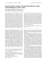
Figure 5 Rhesus monkey cells exhibit a variable block to early re-
verse transcription of SIV. Rhesus monkey B-LCLs were infected with
serial dilutions of VSV-G pseudotyped SIVsmE543-GFP and DNA was
collected a 5 time points. The quantity of early reverse transcription,
late reverse transcription, and integrated viral DNA was assessed by
real time PCR.
C
B
A
% SIVsmE543-GFP+ B-LCL
p= 0.0061
U= 0.7143
p= 0.0235
U= 0.6209
p= 0.0103
U= 0.6813
Figure 6 Fusion of SIV resistant and susceptible rhesus monkey
B-LCLs demonstrates a dominant SIV resistance phenotype. SIV
susceptible and resistant B-LCLs were labeled with either Vybrant DID
(Invitrogen) or Oregon Green (Invitrogen). Susceptible and resistant
cell lines were fused using PEG incubation and double positive fusion
events were sorted by flow cytometry. Fused cells were subsequently
infected with SIVsmE543-GFP and quantified for % GFP+ by flow cy-
tometry.
0 10 20 30 40 50 60
70 80 90 100
0.0
2.5
5.0
7.5
10.0
12.5
15.0
17.5
ul of SIVsmE543-GFP
% SIVsmE543-GFP+ B-LCL
Sensitive cell
Resistant cell
Sensitive cell + Resistant cell
Sensitive/Sensitive cell
Sensitive/Resistant cell
Figure 7 Enhancement of susceptibility of B-LCL to SIV infection
following incubation with SIV virus-like particles. Rhesus monkey
B-LCLs were incubated with media only, uncleaved Gag protein or SIV
virus like particles (VLPs) for four hours followed by infection with VSV-
G pseudotyped SIVsmE543-GFP. Preincubation of B-LCL with VLPs aug-
mented the % GFP+ B-LCL following SIVsmE543-GFP infection.
0 25 50 75 100 125 150 175
0
5
10
15
20
25
30
35
SIV VLP (ng)
% SIVsmE543-GFP+ B-LCL
resistant B-LCL+SIV VLP
resistant B-LCL+control gag only
resistant B-LCL+media only
susceptible B-LCL+SIV VLP
Rogers et al. Virology Journal 2010, 7:79
/>Page 9 of 11
SIVsm543-GFP (Fig. 7). We observed an increase in
SIVsmE660 replication in resistant cell lines as we added
increasing quantities of VLPs. In fact, the addition of 150
ng of VLPs led to the complete loss of resistance to SIV
replication in the cell line. These findings suggest that a
dominant early RT restricting factor that can be saturated
by SIV capsid may contribute to differential resistance to
SIV infection in rhesus monkey B-LCL.
Rhesus monkey B-LCL susceptibility to SIV-GFP correlates
with in vivo viral load and time to death following infection
of rhesus monkeys with SIV
To determine if this variable intracellular blockade of pri-
mate immunodeficiency virus replication impacts in vivo
viral replication and clinical outcome in SIV-infected rhe-
sus monkeys, we evaluated the permissivity of B-LCL
generated from 14 monkeys for VSV-G pseudotyped
SIVsm543-GFP infection. Then, following infection of the
monkeys with SHIV89.6P, we assessed the correlation
between the in vitro permissiveness of these B-LCL for
replication of this virus and the in vivo peak plasma virus
RNA levels and time to death following infection of rhe-
sus monkeys with wild type virus (Fig. 8A, B). Addition-
ally, we generated B-LCL from prechallenge PBMC of
another cohort of rhesus monkeys that were infected
with wild type SIVmac251 and assessed these B-LCL for
susceptibility to SIVmac239-GFP replication. We
observed a significant positive correlation and positive
trend between in vitro B-LCL susceptibility to infection
by these VSV-G pseudotyped SIV constructs and both
peak viremia and time to death in the wild type SIV-
infected monkeys, respectively (Fig. 8C, D). Monkeys
whose B-LCL demonstrated a relative block to early RT
exhibited a lower set point viral load following challenge
in vivo with SIVmac251. These data indicate that in vitro
permissivity of B-LCL for SIV replication is a reliable pre-
dictor of in vivo viral replication and clinical outcome.
Discussion
The variability in the clinical course of HIV-infected
humans and SIV-infected monkeys is likely a conse-
quence of both host and viral factors. The present study
was initiated to begin an exploration of the impact of the
intrinsic immune response to lentiviral infection on clini-
cal course in rhesus monkeys. We attempted to deter-
mine the importance of CD4+ T cell permissiveness for
SIV replication on clinical outcome in monkeys and
define a mechanism for the variability of this permissivity.
The present experiments built upon earlier studies of this
phenomenon which showed that in vitro CD4+ T cell
susceptibility to SIV infection is correlated with in vivo
peak virus load [8]. We observed that differential suscep-
tibility to SIV replication is a stable phenotype and was
reproducible with a single cycle VSV-G pseudotyped viral
construct. Moreover, B-LCL generated from a PBMC
population exhibited a relative permissiveness for SIV
replication that is similar to the relative permissiveness of
those PBMC. We also demonstrated a significant correla-
tion between the susceptibility of B-LCL to a VSV-G
pseudotyped SIVmac construct and the in vivo virus set
point and time to death in rhesus monkeys infected with
SIVmac251. The fact that B-LCL susceptibility can pre-
dict the variability observed in PBMC permissiveness for
lentivirus replication indicates that the intracellular con-
trol of retrovirus replication is of central importance in
determining the infectability of CD4+ T cells and the
clinical outcome of SIV infections. This positive correla-
tion between PBMC and B-LCL permissiveness for SIV
replication also indicates that the variability in virus repli-
cation between rhesus monkeys results from a post-entry
block of SIV replication that is manifested in diverse lym-
phocyte populations.
The infection of rhesus monkeys with SIV is a powerful
model for HIV infection in humans and is of critical
importance for drug and vaccine development. The vari-
ability in the level of virus replication in monkeys follow-
ing SIV infection necessitates the use of large numbers of
animals to appropriately power vaccine trials. Prescreen-
ing monkeys' B-LCL for susceptibility to the replication
of VSV-G-pseudotyped SIV-GFP should allow a predic-
tion of the relative susceptibility of monkeys to SIV repli-
cation following in vivo virus challenge. This should
facilitate the preselection of animals for a study with sim-
ilar permissivities, improving the power of the study and
clarifying the impact of the evaluated intervention.
Following the identification of this phenotype of vari-
able permissiveness for SIV replication in lymphocytes of
rhesus monkeys, we conducted a series of studies to
begin defining the mechanism underlying this observed
intraspecies variability. We established that the relative
block in SIV replication was not dependent on multiple
cycles of SIV replication and was due to a differential
ability of monkey lymphoctyes to block early reverse
transcription of SIV. Moreover, we demonstrated that the
relative block in early RT was a dominant phenotype.
This dominant block to SIV replication could be trans-
ferred to a highly susceptible monkey B-LCL population
by cell fusion. Additionally, the block to early RT could be
overcome by preincubation of SIV resistant cell lines with
virus-like particles. These data suggest that the block to
early RT may involve the binding of capsid of incoming
virions.
Several gene products have been implicated to date in
the control of HIV/SIV entry and cellular immunity,
including CCR5, MHC, and KIR. The present study dem-
onstrates another mechanism that contributes to SIV
control in monkeys. Several intrinsic anti-viral immune
molecules have been shown to inhibit retroviral replica-
Rogers et al. Virology Journal 2010, 7:79
/>Page 10 of 11
tion by preventing early reverse transcription in vitro.
APOBEC-3G, a cellular cytidine deaminase, induces C to
U mutations in the negative strand of the HIV DNA,
which results in a reduced number of infectious HIV
progeny virions[11]. Products of several members of the
trim gene family have the capacity to inhibit virus replica-
tion. Transfection of rhesus monkey TRIM5α into feline
fibroblast cells potently blocks early reverse transcription
of HIV-1, but only modestly alters SIV replication kinet-
ics[10]. Although findings in the present study suggest
that there is significant variability in the early reverse
transcription block between individuals in rhesus mon-
key populations, there is little evidence of rhesus monkey
APOBECs or TRIM5alpha alleles exhibiting a differential
ability to block SIV replication in vitro nor in vivo [14,15].
Our data demonstrate that lymphocytes of rhesus mon-
keys express an inhibitor of SIV early reverse transcrip-
tion that is associated with a reduced in vivo viral set
point, CD4+ T cell decline, and a delay in the time to
death following SIV infection. Whether differences in
lymphocyte susceptibility to SIV represent consequences
of allelic forms or variable expression levels of an SIV
restricting molecule, our findings underscore that an
innate antiviral response, which is capable of inhibiting
early RT, can impact the in vivo clinical outcome of the
animals infected with SIV. A complete understanding of
the host immune mechanisms that have a significant
impact on in vivo viral replication is critically important
to aid in our design and implementation of preventative
and therapeutic interventions to reduce HIV acquisition
and viral burden.
Competing interests
The authors declare that they have no competing interests.
Authors' contributions
TR conceived of and designed the study as well as participated in all assays. SL
participated in B-LCL phenotyping and staging assay. TS participated in B-LCL
phenotyping, staging, and fusion assay. TC participated in staging and fusion
assay. AH participated in in vivo correlation studies. All authors have read and
approved the manuscript.
Acknowledgements
The authors thank Professors Barton Hayes of Duke Human Vaccine Institute,
and David Goldstein of Center for Population Genomics and Pharmacogenet-
ics, Duke University for their valuable suggestions and critical reading of this
Figure 8 B-LCL susceptibility to VSV-G pseudotyped SIV-GFP correlates with in vivo plasma viremia and time to death following rhesus
monkey challenge with SIVmac251 or SHIV-89.6P. Positive correlation between % GFP+ B-LCL following VSV-G pseudotyped SIVsmE543-GFP in
vitro infection of rhesus monkey B-LCL and (A) in vivo peak plasma viremia of monkeys (day 14 post-infection) as measured by RT assay, and (B) time
to death following in vivo infection with SHIV-89.6P. Positive correlation between % GFP+ B-LCL following VSV-G pseudotyped SIVmac239-GFP in vitro
infection of rhesus monkey B-LCL and (C) in vivo peak plasma virus RNA levels of monkeys (day 14 post-infection) and positive trend with (D) time to
death following in vivo infection with SIVmac251.
2 3 4
6
7
8
9
p= 0.013
U
= 0.644
% SIVsmE543-GFP+ B-LCL
(log AUC)
log plasma virus RNA (copies/ml)
A
2 3 4
0.00
0.05
0.10
0.15
p= 0.05
U
= 0.68
% SIVsmE543-GFP+ B-LCL
(log AUC)
1 / Time to death (weeks)
B
1 2 3 4
3
5
7
9
p= 0.011
U
= 0.55
% SIVmac239-GFP+ B-LCL
(log AUC)
log plasma viral RNA (copies/ml)
D
C
1 2 3 4
0.00
0.01
0.02
0.03
0.04
0.05
0.06
p= 0.08
U
= 0.449
% SIVmac239-GFP+ B-LCL
(log AUC)
1 / Time to death (weeks)
Rogers et al. Virology Journal 2010, 7:79
/>Page 11 of 11
manuscript. We thank D. Knipe, J. Sodroski, D. Barouch, and S. Whelan for the
gift of HSV-GFP, SIV-GFP, Ad-GFP, and VSV-GFP, respectively. This work was sup-
ported by the NIAID Center for HIV/AIDS Vaccine Immunology grant AI067854.
Author Details
Division of Viral Pathogenesis, Beth Israel Deaconess Medical Center, Harvard
Medical School, Boston, Massachusetts 02115, USA
References
1. Michael NL, et al.: Rapid disease progression without seroconversion
following primary human immunodeficiency virus type 1 infection
evidence for highly susceptible human hosts. JInfect Dis 1997,
175(6):1352-9.
2. Cao Y, et al.: Virologic and immunologic characterization of long-term
survivors of human immunodeficiency virus type 1 infection. N Engl J
Med 1995, 332(4):201-8.
3. Schmitz JE, et al.: Control of viremia in simian immunodeficiency virus
infection by CD8+ lymphocytes. Science 1999, 283(5403):857-60.
4. Kawashima Y, et al.: Adaptation of HIV-1 to human leukocyte antigen
class I. Nature 2009, 458(7238):641-5.
5. Liu R, et al.: Homozygous defect in HIV-1 coreceptor accounts for
resistance of some multiply-exposed individuals to HIV-1 infection.
Cell 1996, 86(3):367-77.
6. Fellay J, et al.: A whole-genome association study of major
determinants for host control of HIV-1. Science 2007, 317(5840):944-7.
7. Ciuffi A, et al.: Entry and transcription as key determinants of differences
in CD4 T-cell permissiveness to human immunodeficiency virus type 1
infection. J Virol 2004, 78(19):10747-54.
8. Goldstein S, et al.: Intrinsic susceptibility of rhesus macaque peripheral
CD4(+) T cells to simian immunodeficiency virus in vitro is predictive of
in vivo viral replication. J Virol 2000, 74(20):9388-95.
9. Brass AL, et al.: Identification of host proteins required for HIV infection
through a functional genomic screen. Science 2008, 319(5865):921-6.
10. Stremlau M, et al.: The cytoplasmic body component TRIM5alpha
restricts HIV-1 infection in Old World monkeys. Nature 2004,
427(6977):848-53.
11. Huthoff H, Towers GJ: Restriction of retroviral replication by APOBEC3G/
F and TRIM5alpha. Trends Microbiol 2008, 16(12):612-9.
12. Johnson WE, Sawyer SL: Molecular evolution of the antiretroviral TRIM5
gene. Immunogenetics 2009, 61(3):163-76.
13. Stremlau M, et al.: Species-specific variation in the B30.2(SPRY) domain
of TRIM5alpha determines the potency of human immunodeficiency
virus restriction. J Virol 2005, 79(5):3139-45.
14. Wilson SJ, et al.: Rhesus macaque TRIM5 alleles have divergent
antiretroviral specificities. J Virol 2008, 82(14):7243-7.
15. Goldschmidt V, et al.: Role of common human TRIM5alpha variants in
HIV-1 disease progression. Retrovirology 2006, 3:54.
16. Hirsch V, et al.: A molecularly cloned, pathogenic, neutralization-
resistant simian immunodeficiency virus, SIVsmE543-3. J Virol 1997,
71(2):1608-20.
17. Hofmann W, et al.: Species-specific, postentry barriers to primate
immunodeficiency virus infection. J Virol 1999, 73(12):10020-8.
18. Butler SL, Hansen MS, Bushman FD: A quantitative assay for HIV DNA
integration in vivo. Nat Med 2001, 7(5):631-4.
doi: 10.1186/1743-422X-7-79
Cite this article as: Rogers et al., Variability in a dominant block to SIV early
reverse transcription in rhesus monkey cells predicts in vivo viral replication
and time to death Virology Journal 2010, 7:79
Received: 26 October 2009 Accepted: 26 April 2010
Published: 26 April 2010
This article is available from: 2010 Rogers et al; licensee BioMed Central L td. This is an Open Access article distributed under the terms of the Creative Commons Attribution License ( ), which permits unrestricted use, distribution, and reproduction in any medium, provided the original work is properly cited.Virology Journal 2010, 7:79