Báo cáo y học: " Human herpesvirus 6 infection impairs Toll-like receptor signaling" pptx
Bạn đang xem bản rút gọn của tài liệu. Xem và tải ngay bản đầy đủ của tài liệu tại đây (760.25 KB, 5 trang )
Murakami et al. Virology Journal 2010, 7:91
/>Open Access
SHORT REPORT
BioMed Central
© 2010 Murakami et al; licensee BioMed Central Ltd. This is an Open Access article distributed under the terms of the Creative Commons
Attribution License ( which permits unrestricted use, distribution, and reproduction in
any medium, provided the original work is properly cited.
Short report
Human herpesvirus 6 infection impairs Toll-like
receptor signaling
Yuichi Murakami
1
, Kazushi Tanimoto
1
, Hiroshi Fujiwara
1,2
, Jun An
1
, Koichiro Suemori
1
, Toshiki Ochi
1
,
Hitoshi Hasegawa
1,2
and Masaki Yasukawa*
1,2
Abstract
Human herpesvirus 6 (HHV-6) has a tropism for immunocompetent cells, including T lymphocytes, monocytes/
macrophages, and dendritic cells (DCs) suggesting that HHV-6 infection affects the immunosurveillance system. Toll-
like receptor (TLR) system plays an important role in innate immunity against various pathogens. In the present study,
we investigated the effect of HHV-6 infection on the expression and intracellular signaling of TLRs in DCs. Although
expression levels of TLRs were not decreased or slightly elevated following HHV-6 infection, the amounts of cytokines
produced following stimulation with ligands for TLRs appeared to be dramatically decreased in HHV-6-infected DCs as
compared to mock-infected DCs. Similarly, phosphorylation levels of TAK-1, IκB kinase, and IκB-α following stimulation
of HHV-6-infected DCs with lipopolysaccharide, which is the ligand for TLR4, appeared to be decreased. These data
show that HHV-6 impairs intracellular signaling through TLRs indicating the novel mechanism of HHV-6-mediated
immunomodulation.
Findings
Human herpesvirus 6 (HHV-6) is known as a causative
agent of exanthem subitum, and reactivation of HHV-6 in
adults causes various clinical manifestations [1,2]. HHV-6
can preferentially infect immunocompetent cells and
induces various immunobiological alterations [3-12].
Therefore, HHV-6 is recognized as one of the important
viruses that modulate immune responses.
Toll-like receptors (TLRs) are key molecules of the
innate immune system [13]. A subset of TLRs recognizes
components of microorganisms and induces innate
immune responses. After recognition of ligands, TLRs
activate their intrinsic signaling pathways, resulting in
activation of the transcription factor nuclear factor-κB
(NF-κB), which controls the expression of inflammatory
cytokine genes [14,15]. HHV-6 alters the regulation of
innate immunity as well as adaptive immunity. In the
light of these facts, it seems important to clarify the
effects of HHV-6 infection on the TLR system. We there-
fore investigated the effects of HHV-6 infection on the
expression and functions of TLRs in DCs.
The Z29 strain of HHV-6B was mainly used in the pres-
ent study, because HHV-6B is more prevalent than HHV-
6A in the general population. Immature DCs were gener-
ated from peripheral blood monocytes by culturing them
in the presence of GM-CSF and IL-4, as described previ-
ously [8]. Immature DCs were inoculated with HHV-6 at
an approximate multiplicity of infection of 1 50% tissue
culture infective dose. HHV-6-inoculated DCs were cul-
tured for 3 days and used for experiments. More than
95% of HHV-6-infected and mock-infected DCs were via-
ble when used for experiments.
Expression of mRNA for TLRs1-10 in HHV-6-infected
and mock-infected DCs was examined by semi-quantita-
tive reverse transcription-polymerase chain reaction (RT-
PCR) [16]. Sequences of the primers for PCR are shown
in the additional file 1.
Cytokine production by DCs was examined as follows.
After 3 days of HHV6 inoculation, DCs were cultured for
24 hours in RPMI 1640 medium supplemented with 10%
fetal calf serum and poly(I:C) (a ligand for TLR 3; Invitro-
gen, San Diego, CA, USA) at 25 μg/ml, lipopolysaccha-
ride (LPS) (a ligand for TLR 4; Sigma, St Louis, MO, USA)
at 100 ng/ml, or imidazoquinoline (a ligand for TLR7;
Invitrogen) at 5 mg/ml. The culture supernatants were
then harvested, and the amounts of cytokines they con-
* Correspondence:
1
Departmemt of Bioregulatory Medicine, Ehime University Graduate School of
Medicine, Toon, Ehime 791-0295, Japan
Full list of author information is available at the end of the article
Murakami et al. Virology Journal 2010, 7:91
/>Page 2 of 5
tained were measured by flow cytometry using a Cyto-
metric Bead Array System (BD Biosciences, San Diego,
CA, USA) and enzyme-linked immunosorbent assay
(Biosource Europe S.A., Nivelles, Belgium).
The binding of LPS to HHV-6-infected and mock-
infected DCs was examined quantitatively by flow cytom-
etry using fluorescent LPS conjugate (Alexa Fluor
®
488)
(Molecular Probes, Eugene, OR, USA).
Western blotting was performed by a standard method
using the following antibodies; anti-TLR4 (BioChain,
Hayward, CA, USA), anti-MyD88 (ProSci, Poway, CA,
USA), anti-TRAF6 (Santa Cruz Biotechnology, Santa
Cruz, CA, USA), anti-TAK-1 (Cell Signaling Technology,
Danvers, MA, USA), anti-phosphorylated IκB kinase α/β
(IKKα/β) (Cell Signaling Technology), anti-phosphory-
lated IκB-α (Cell Signaling Technology), and anti-β-actin
(Sigma).
We first confirmed HHV-6 infection in DCs. We and
other investigators previously reported that HHV-6 can
infect human DCs and modulates the expression of vari-
ous surface molecules including CD80, CD83, CD86, and
DC-SIGN [8,9,17]. As shown in the additional file 2,
expression of HHV-6 immediate early and late genes was
detected in HHV-6-inoculated DCs. In addition, two-
color flow cytometry showed that HHV-6 antigen expres-
sion was present in more than half of the DCs inoculated
with HHV-6. HHV-6 antigen expression was detected in
DCs in which CD80 expression was up-regulated, as we
have reported previously [8] (Additional file 3). These
data confirmed that HHV-6 was able to infect DCs under
our experimental conditions.
We screened the TLR1-10 expression in HHV-6-
infected DCs and compared it with that in mock-infected
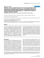
DCs. As shown in Figure 1, semi-quantitative RT-PCR
revealed that expression of mRNAs for TLR3, TLR4, and
TLR7 appeared to be slightly increased in HHV-6-
infected DCs as compared with mock-infected DCs.
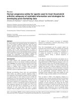
We next examined cytokine production by HHV-6-
infected and mock-infected DCs in response to stimula-
tion with TLR ligands. TLR3, TLR4, and TLR7 were
selected for this experiment, because expression of these
TLRs seemed to be increased after infection with HHV-6,
as shown in Figure 1. As shown in Figure 2, the amounts
of IL-6 and IL-8 produced by DCs stimulated with
poly(I:C), a TLR3 ligand, after infection with HHV-6
appeared to be significantly lower than those produced
by mock-infected DCs. Similarly, the production of IL-10
and IL-8 by HHV-6-infected DCs in response to stimula-
tion with LPS, a TLR4 ligand, was markedly impaired in
comparison with mock-infected DCs. The amount of IL-
8 produced by HHV-6-infected DCs stimulated with a
TLR7 ligand, imidazoquinoline, was also decreased as
compared with that produced by TLR7 ligand-stimulated
mock-infected DCs. The same experiments were per-
formed three times and similar data were obtained.
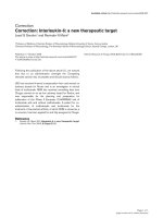
We further examined the mechanisms of impaired
cytokine production by HHV-6-infected DCs, focusing
on TLR4. First, expression of the TLR4 molecule on
HHV-6-infected and mock-infected DCs was examined
by Western blotting of cell lysates and flow cytometry to
detect the binding of fluorescent LPS conjugate. As
shown in Figures 3A and 3B, the level of TLR4 expression
on HHV-6-infected DCs unstimulated with LPS
appeared to be slightly higher than that on mock-infected
DCs.
The intracellular signaling system of TLR4 in HHV-6-
infected and mock-infected DCs was further examined.
First, it appeared that the amount of MyD88, an adaptor
molecule required for signal transduction through TLRs,
was slightly higher in HHV-6-infected DCs than in mock-
infected DCs, parallel to the TLR4 expression level. Simi-
larly, the expression level of TRAF6, another TLR adaptor
molecule, did not differ significantly, or was slightly
increased, in HHV-6-infected as compared with mock-
infected DCs (Figure 3C). In contrast, phosphorylation
levels of TAK-1, IKKα/β, and IκB-α, which are important
molecules for NF-κB activation [18], in HHV-6-infected
DCs after stimulation with LPS appeared to be signifi-
cantly lower than those in LPS-stimulated mock-infected
DCs (Figure 3D). The same experiments were performed
twice and similar data were obtained. These data reveal
that HHV-6 impairs signal transduction of TLRs.
In the present study, we demonstrated that although
the expression of TLRs and their adaptor molecules was
only slightly increased, cytokine production by DCs in
response to stimulation with TLR ligands was severely
impaired after infection with HHV-6. In contrast, phos-
phorylation levels of TAK-1, IKKα/β and IκB-α appeared
Figure 1 RT-PCR analysis of TLR mRNAs in mock-infected and
HHV-6-infected DCs. Semi-quantitative RT-PCR reveals that expres-
sion levels of mRNAs for TLR3, TLR4, and TLR7 are slightly higher in
HHV-6-infected DCs than in mock-infected DCs.
Murakami et al. Virology Journal 2010, 7:91
/>Page 3 of 5
to be significantly decreased in HHV-6-infected DCs as
compared with mock-infected DCs. NF-κB activation
resulting in production of inflammatory cytokines
depends on phosphrylation of IκB, which is induced by
activation of the IKK complex [18,19]. Activation of IKKs
depends on their phosphorylation, which results in con-
formational change and kinase activity [20-22]. It is also
noteworthy that impaired cytokine production is not
restricted in the TLR4 system but is also detected in the
signal pathways of other TLRs. Therefore, these findings
suggest that impairment of TLR4 signaling in HHV-6-
infected DCs is due to blocking not upstream, but down-
stream in the signal pathway.
Recently, various effects of viral infection on expression
and signaling of TLRs have been reported. Chen et al.
have recently reported that vaccinia virus virulence factor
B14 can directly bind to the IKK complex and inhibit
phosphorylation of IKKβ[23]. This results in impairment
of IκB-α degradation and inhibition of NF-κB activation.
In the present study, it was found that inoculation with
inactivated HHV-6 did not induce impairment of TLR
signaling, i.e., the amounts of cytokines produced by
HHV-6-infected and inactivated HHV-6-inoculated DCs
following stimulation with TLR ligands were not signifi-
cantly different (data not shown). Therefore, HHV-6 gene
product(s) produced de novo in HHV-6-infected DCs
might associate with the TAK-1 or IKK complex directly
or indirectly, resulting in inhibition of IKK activation, as
is the case for vaccinia virus infection. It has also been
reported that M45 protein of murine cytomegalovirus,
which, like HHV-6, is a β-herpesvirus, inhibits the RIP1-
mediated activation of NF-κB in response to TLR3 stimu-
lation [24]. This finding suggests that impairment of TLR
signaling might be the common strategy of immune eva-
sion by β-herpesviruses.
Viruses alter cell functions via mainly direct infection;
however, indirect mechanisms are also responsible for
virus-mediated immune-modulation. We previously
reported that HHV-6 infection mediates apoptosis in
HHV-6-uninfected T cells through a bystander effect
Figure 2 Downregulation of cytokine production by stimulation with TLR ligand in DCs after infection with HHV-6. The production of cytok-
ines by HHV-6-infetced DCs and mock-infected DCs was examined as detailed in the text. The amounts of cytokines produced by HHV-6-infected DCs
after stimulation with the TLR3 ligand poly(I:C), the TLR4 ligand LPS, and the TLR7 ligand imidazoquinoline are all lower than those produced by mock-
infected DCs that were stimulated with TLR ligands.
Murakami et al. Virology Journal 2010, 7:91
/>Page 4 of 5
[25]. In the present study, it was not clarified whether a
direct or a bystander effect plays an important role in
impairment of the innate immune response in HHV-6
infection. Further study will be needed to clarify this
issue.
In summary, we have demonstrated for the first time
that the intracellular signaling pathway through TLRs is
severely impaired by HHV-6 infection. Since the TLR sys-
tem is essential for recognition of various pathogens and
generation of innate immunity, disruption of TLR-medi-
ated signaling seems to be an effective strategy by which
viruses can evade the immunosurveillance system.
Additional material
Competing interests
The authors declare that they have no competing interests.
Authors' contributions
YM, KT, JA, KS, and TO carried out the experiments. HF and HH participated in
the design of the study and supported performing experiments. MY designed
the research, wrote and edited the paper, and provided financial support. All
authors read and approved the final manuscript.
Acknowledgements
We are grateful for the skilled technical assistance of Ms. Junko Mizumoto and
Dr. Kenji Kameda, Ehime University, Japan. We also thank Dr. Yasuko Mori, Kobe
University Graduate School of Medicine for kindly supplying anti-HHV-6 anti-
body. This work was supported in part by grants from the Ministry of Educa-
tion, Culture, Sports, Science and Technology of Japan.
Author Details
1
Departmemt of Bioregulatory Medicine, Ehime University Graduate School of
Medicine, Toon, Ehime 791-0295, Japan and
2
Proteo-Medicine Research
Center, Ehime University, Toon, Ehime 791-0295, Japan
Additional file 1 Sequences of the primers for RT-PCR. Expression of
mRNA for TLRs1-10 and β-actin in HHV-6-infected and mock-infected DCs
was examined by RT-PCR using the primers shown here.
Additional file 2 Expression of HHV-6 mRNA in DCs. cDNAs synthesized
from HHV-6-infected cord blood cells (lane 1), distilled water only (lane 2),
mock-infected DCs (lane 3), and HHV-6-infected DCs on day 5 after inocula-
tion (lane 4) were amplified using primers corresponding to the HHV-6
immediate-early and late genes and primers corresponding to the β-actin
gene.
Additional file 3 Flow cytometric analysis of HHV-6 antigen expres-
sion in DCs. Mock-infected DCs and HHV-6-infected DCs on day 5 after
inoculation were stained with anti-HHV-6 gB monoclonal antibody and
anti-CD80 monoclonal antibody.
Received: 11 March 2010 Accepted: 10 May 2010
Published: 10 May 2010
This artic le is available fro m: http://www.v irologyj.com/co ntent/7/1/91© 2010 Murakami et al; licensee BioMed Central Ltd. This is an Open Access article distributed under the terms of the Creative Commons Attribution License ( which permits unrestricted use, distribution, and reproduction in any medium, provided the original work is properly cited.Virology Journal 2010, 7:91
Figure 3 Impairment of TLR4 signaling in HHV-6-infected DCs. (A) Western blotting reveals that the expression level of TLR4 protein in HHV-6-
infected DCs, which were not stimulated with LPS, is slightly higher than that in mock-infected DCs. (B) Flow cytometric analysis using fluorescent LPS
conjugate reveals that the amount of LPS bound to HHV-6-infected DCs is slightly higher than that on mock-infected DCs, suggesting that the expres-
sion level of TLR4 molecules on DCs is increased after infection with HHV-6. (C) Western blotting reveals that the expression levels of MyD88 and TRAF6
proteins in HHV-6-infected DCs, which are not stimulated with LPS, are slightly higher than those in mock-infected DCs. (D) Western blotting reveals
that phosphorylation levels of TAK-1, IKKα/β and IκB-α in HHV-6-infected DCs after stimulation with LPS are significantly lower than those in LPS-stim-
ulated mock-infected DCs.
(A) (B)
(D)
(C)
Murakami et al. Virology Journal 2010, 7:91
/>Page 5 of 5
References
1. De Bolle L, Naesens L, De Clercq E: Update on human herpesvirus 6
biology, clinical features, and therapy. Clin Microbiol Rev 2005,
18:217-245.
2. Zerr DM: Human herpesvirus 6: a clinical update. Herpes 2006, 13:20-24.
3. Furukawa M, Yasukawa M, Yakushijin Y, Fujita S: Distinct effects of human
herpesvirus 6 and human herpesvirus 7 on surface molecule
expression and function of CD4
+
T cells. J Immunol 1994,
152:5768-5775.
4. Hasegawa A, Yasukawa M, Sakai I, Fujita S: Transcriptional down-
regulation of CXCR4 induced by impaired association of transcription
regulator YY1 with c-Myc in human herpesvirus 6-infected cells. J
Immunol 2001, 166:1125-1131.
5. Lusso P, De Maria A, Malnati M, Lori F, DeRocco SE, M Baseler M, Gallo RC:
Induction of CD4 and susceptibility to HIV-1 infection in human CD8
+
T
lymphocytes by human herpesvirus 6. Nature 1991, 349:533-535.
6. Lusso P, Malnati M, De Maria A, Balotta C, DeRocco SE, Markham PD, Gallo
RC: Productive infection of CD4
+
and CD8
+
mature human T cell
populations and clones by human herpesvirus 6. Transcriptional
down-regulation of CD3. J Immunol 1991, 147:685-691.
7. Lusso P, Malnati MS, Garzino-Demo A, Crowley RW, Long EO, Gallo RC:
Infection of natural killer cells by human herpesvirus 6. Nature 1993,
362:458-462.
8. Kakimoto M, Hasegawa A, Fujita S, Yasukawa M: Phenotypic and
functional alterations of dendritic cells induced by human herpesvirus
6 infection. J Virol 2002, 76:10338-10345.
9. Niiya H, Azuma T, Jin L, Uchida N, Inoue A, Hasegawa H, Fujita S, Tohyama
M, Hashimoto K, Yasukawa M: Transcriptional downregulation of DC-
SIGN in human herpesvirus 6-infected dendritic cells. J Gen Virol 2004,
85:2639-2642.
10. Niiya H, Lei J, Guo Y, Azuma T, Yakushijin Y, Sakai I, Hato T, Tohyama M,
Hashimoto K, Yasukawa M: Human herpesvirus 6 impairs differentiation
of monocytes to dendritic cells. Exp Hematol 2006, 34:642-653.
11. Smith A, Santoro F, Di Lullo G, Dagna L, Verani A, Lusso P: Selective
suppression of IL-12 production by human herpesvirus 6. Blood 2003,
102:2877-2884.
12. Yasukawa M, Hasegawa A, Sakai I, Ohminami H, Arai J, Kaneko S, Yakushijin
Y, Maeyama K, Nakashima H, Arakaki R, Fujita S: Down-regulation of
CXCR4 by human herpesvirus 6 (HHV-6) and HHV-7. J Immunol 1999,
162:5417-5422.
13. Takeda K, Kaisho T, Akira S: Toll-like receptors. Annu Rev Immunol 2003,
21:335-376.
14. Kawai T, Akira S: Signaling to NF-κB by Toll-like receptors. Trends Mol
Med 2007, 13:460-469.
15. Trinchieri G, Sher A: Cooperation of Toll-like receptor signals in innate
immune defense. Nat Rev Immunol 2007, 7:179-190.
16. Kadowaki N, Ho S, Antonenko S, Malefyt RW, Kastelein RA, Bazan F, Liu YJ:
Subsets of human dendritic cell precursors express different toll-like
receptors and respond to different microbial antigens. J Exp Med 2001,
194:863-869.
17. Takemoto M, Imasawa T, Yamanishi K, Mori Y: Role of dendritic cells
infected with human herpesvirus 6 in virus transmission to CD4
+
T
cells. Virology 2009, 385:294-302.
18. Baeuerle PA, Baltimore D: I kappa B: a specific inhibitor of the NF-kappa
B transcription factor. Science 1988, 242:540-546.
19. Zandi E, Rothwarf DM, Delhase M, Hayakawa M, Karin M: The IκB kinase
complex (IKK) contains two kinase subunits, IKKα and IKKβ, necessary
for IκB phosphorylation and NF-κB activation. Cell 1997, 91:243-252.
20. Delhase M, Hayakawa M, Chen Y, Karin M: Positive and negative
regulation of IκB kinase activity through IKKβ subunit
phosphorylation. Science 1999, 284:309-313.
21. DiDonato JA, Hayakawa M, Rothwarf DM, Zandi E, Karin M: A cytokine-
responsive IκB kinase that activates the transcription factor NF-κB.
Nature 1997, 388:548-554.
22. Mercurio F, Zhu H, Murray BW, Shevchenko A, Bennett BL, Li J, Young DB,
Barbosa M, Mann M, Manning A, Rao A: IKK-1 and IKK-2: cytokine-
activated IκB kinases essential for NF-κB activation. Science 1997,
278:860-866.
23. Chen RA, Ryzhakov G, Cooray S, Randow F, Smith GL: Inhibition of IκB
kinase by vaccinia virus virulence factor B14. PLos Pathog 2008, 4:e22.
24. Mack C, Sickmann A, Lembo D, Brune W: Inhibition of proinflammatory
and innate immune signaling pathways by a cytomegalovirus RIP1-
interacting protein. Proc Natl Acad Sci USA 2008, 105:3094-3099.
25. Inoue Y, Yasukawa M, Fujita S: Induction of T-cell apoptosis by human
herpesvirus 6. J Virol 1997, 71:3751-3759.
doi: 10.1186/1743-422X-7-91
Cite this article as: Murakami et al., Human herpesvirus 6 infection impairs
Toll-like receptor signaling Virology Journal 2010, 7:91