Báo cáo y học: "Proteome changes of lungs artificially infected with H-PRRSV and N-PRRSV by two-dimensional fluorescence difference gel electrophoresis" pps
Bạn đang xem bản rút gọn của tài liệu. Xem và tải ngay bản đầy đủ của tài liệu tại đây (1.32 MB, 17 trang )
Xiao et al. Virology Journal 2010, 7:107
/>Open Access
RESEARCH
© 2010 Xiao et al; licensee BioMed Central Ltd. This is an Open Access article distributed under the terms of the Creative Commons At-
tribution License ( which permits unrestricted use, distribution, and reproduction in any
medium, provided the original work is properly cited.
Research
Proteome changes of lungs artificially infected
with H-PRRSV and N-PRRSV by two-dimensional
fluorescence difference gel electrophoresis
Shuqi Xiao
†
, Qiwei Wang
†
, Jianyu Jia, Peiqing Cong, Delin Mo, Xiangchun Yu, Limei Qin, Anning Li, Yuna Niu,
Kongju Zhu, Xiaoying Wang, Xiaohong Liu and Yaosheng Chen*
Abstract
Background: Porcine reproductive and respiratory syndrome with PRRS virus (PRRSV) infection, which causes
significant economic losses annually, is one of the most economically important diseases affecting swine industry
worldwide. In 2006 and 2007, a large-scale outbreak of highly pathogenic porcine reproductive and respiratory
syndrome (PRRS) happened in China and Vietnam. However little data is available on global host response to PRRSV
infection at the protein level, and similar approaches looking at mRNA is problematic since mRNA levels do not
necessarily predict protein levels. In order to improve the knowledge of host response and viral pathogenesis of highly
virulent Chinese-type PRRSV (H-PRRSV) and Non-high-pathogenic North American-type PRRSV strains (N-PRRSV), we
analyzed the protein expression changes of H-PRRSV and N-PRRSV infected lungs compared with those of uninfected
negative control, and identified a series of proteins related to host response and viral pathogenesis.
Results: According to differential proteomes of porcine lungs infected with H-PRRSV, N-PRRSV and uninfected negative
control at different time points using two-dimensional fluorescence difference gel electrophoresis (2D-DIGE) and mass
spectrometry identification, 45 differentially expressed proteins (DEPs) were identified. These proteins were mostly
related to cytoskeleton, stress response and oxidation reduction or metabolism. In the protein interaction network
constructed based on DEPs from lungs infected with H-PRRSV, HSPA8, ARHGAP29 and NDUFS1 belonged to the most
central proteins, whereas DDAH2, HSPB1 and FLNA corresponded to the most central proteins in those of N-PRRSV
infected.
Conclusions: Our study is the first attempt to provide the complex picture of pulmonary protein expression during H-
PRRSV and N-PRRSV infection under the in vivo environment using 2D-DIGE technology and bioinformatics tools,
provides large scale valuable information for better understanding host proteins-virus interactions of these two PRRSV
strains.
Background
Porcine reproductive and respiratory syndrome (PRRS)
has become one of the most economically important dis-
eases affecting swine industry worldwide, causing signifi-
cant economic losses each year[1]. The disease was
initially found in North America in 1987[2], Europe in
1990[3], China in 1996[4], and Sweden in 2007[5]. PRRS
results in both reproductive failure in pregnant sows and
respiratory distress in young pigs, such as late-term abor-
tions and stillbirths, premature farrowing, mummified
pigs, interstitial pneumonia, respiratory difficulties, high
mortality in piglets, and so on[2]. The etiologic agent of
PRRS is PRRS virus (PRRSV), a small enveloped, linear,
single, positive-stranded RNA virus, which is a member
of the family Arteriviridae which includes lactate dehy-
drogenase-elevating virus (LDV), equine arteritis virus
(EAV), and simian hemorrhagic fever virus (SHFV) and
enters in the newly established order of the Nidovirales
together with the Coronaviridae and Roniviridae fam-
ily[6]. According to genomic and antigenic differences,
* Correspondence:
1
State Key Laboratory of Biocontrol, School of Life Sciences, Sun Yat-sen
University, Guangzhou 510006, China
†
Contributed equally
Full list of author information is available at the end of the article
Xiao et al. Virology Journal 2010, 7:107
/>Page 2 of 17
and different geographic origins, PRRSV can be classified
into two major genotypes: the North American type (NA
PRRSV) and the European type (EU PRRSV)[7,8]. To
date, PRRSV strains characterized in China are all the NA
PRRSV. In 2006 and 2007, the unparalleled large-scale
outbreaks of highly pathogenic PRRS (H-PRRS) affected
over 2,000,000 pigs with about 400,000 fatal cases and at
least 65,000 pigs in China[9,10] and Vietnam[10,11],
respectively, which posed great concern to the global
swine industry and to public health. Studies showed that
highly virulent Chinese-type PRRSV (H-PRRSV) is the
major causative pathogen of H-PRRS[9].
Preliminary results indicated that PRRSV strongly
modulates the host's immune responses. Studies showed
that the virus was able to inhibit IFN-a responses in the
lungs of pigs, and may significantly increase IL-10, IFN-γ,
IFN-β, TNF-α, MX1, RHIV1, and USP mRNA expres-
sion[12-15]. However, mRNA abundance is not always
consistent with the protein level[16], factors including
post-transcriptional changes in mRNA, post-transla-
tional modifications of proteins and microRNAs, which
regulate the conversion of mRNAs to proteins[17].
Therefore, information about proteins changes during
PRRSV infection may be crucial for us to understand host
response to virus and viral pathogenesis. Proteomics
analysis is a powerful tool for global evaluation of protein
expression, and gaining better insight into the host
response to PRRSV. Proteomics has been initially used
successfully in the pathogenesis studies, biomarker iden-
tification, and protein-protein interaction studies in
human disease processes[18]. This approach has been
recently applied in animal viral diseases, such as the dif-
ferential proteomes of chicken embryo fibroblasts after
Infectious bursal disease virus (IBDV) infection[19], the
cellular changes in Vero cells infected with African swine
fever virus[20], proteomic alteration of PK-15 cells after
infection by classical swine fever virus[21]. Haiming
Zhang and his colleagues identified 23 cellular proteins of
PAMs infected with PRRSV in vitro with significant alter-
ation in different courses post-infection by proteomic
approaches. Heat shock 27 kDa protein (HSP27) and
superoxide dismutase 2 (SOD2), involved in stress
response or ubiquitin-proteasome pathway, were
observed to be up-regulated[22]. The primary cellular
target of PRRSV is the alveolar macrophage of lung and
PRRSV infection results in widespread apoptosis in the
lungs and lymphoid tissues [23]. However, host response
to highly virulent Chinese-type PRRSV (H-PRRSV) and
non-high-pathogenic North American-type PRRSV
strains (N-PRRSV) in porcine lungs has not been ana-
lyzed by comparative proteomics profiling which may be
very critical to better understand novel characters of H-
PRRSV.
Two-dimensional gel electrophoresis (2-DE) is widely
used for proteomics research. However, integral variation
and excessive time/labor costs have been common prob-
lems with standard 2-DE[24].Two-dimensional fluores-
cence difference gel electrophoresis (2D-DIGE)
technology has recently been implemented as a quantita-
tive alternative to conventional 2-DE [25]. 2D-DIGE
enables the labeling of 2-3 samples with different dyes
(Cy2, Cy3 and Cy5) and electrophoresis of all the samples
on the same 2D gel, reducing spot pattern variability and
the number of gels in an experiment and yielding simple
and accurate spot matching[17]. Besides, an internal
standard labeled with Cy2 dye is used in every gel that
reduces inter-gel variation and false positives and
increases the robustness of statistical analysis. 2D-DIGE
system allows accurate detection of minor differences of
protein expression across multiple samples simultane-
ously with statistical confidence by using the DeCyder
software. The comparison of spot intensities using the
2D-DIGE approach and DeCyder software is more objec-
tive than the conventional approach based on the com-
parison of the brightness of gel images obtained by
conventional staining and thus has been applied to pro-
teomics studies[24,26]. Using 2D-DIGE followed by
MALDI-TOF or MALDI-TOF/TOF identification and
bioinformatics methods, we conducted an extensive anal-
ysis of proteomes in H-PRRSV and N-PRRSV infected
lungs compared with uninfected negative control lungs.
In this manuscript we discuss host response to these two
viruses through the altered proteins which were identi-
fied by comparative analysis of proteomes.
Results
Animal model construction
After infection, both H-PRRSV affected pigs and N-
PRRSV affected pigs exhibited common clinical symp-
toms within 3-7 days, including anorexia, rough hair
coats, dyspnoea, reddening of skin, oedema of the eyelids,
conjunctivitis, mild diarrhoea, shivering, lamping, etc.
However, the body temperatures of pigs inoculated with
H-PRRSv and N-PRRSV are different. The results are
showed as mean ± s.e. H-PRRSV affected pigs exhibited
persistently a higher body temperature (41.37 ± 0.23°C)
than those N-PRRSV affected (40.43 ± 0.076°C) from 3d
pi to 7d pi. Pigs in the uninfected negative control group
did not show any obvious changes in body temperature
(39.77 ± 0.042°C) and clinical signs. Histopathology
examination showed an interstitial pneumonia and
emphysema in lungs with thickening of alveolar septa
accompanied with infiltration of mononuclear cells from
both H-PRRSV affected pigs and N-PRRSV affected pigs
compared to lungs of uninfected negative control pigs
(Figure 1a). Lungs from all H-PRRSV and N-PRRSV
Xiao et al. Virology Journal 2010, 7:107
/>Page 3 of 17
affected pigs were positive for PRRSV by RT-PCR (data
not shown). Control pigs lungs were negative for PRRSV
by RT-PCR. Subsequently, viral re-isolates were success-
fully recovered from the infected pigs and confirmed by
RT-PCR detection, IFA, and EM. The sequences of NSP2
gene from the re-isolated virus were completely identical
with those of the inoculated virus by sequencing. Specific
immunofluorescence (Figure 1b) and PRRSV particles
(Figure 1c) in MARC-145 cells infected with re-isolated
either H-PRRSV or N-PRRSV was observed by IFA and
EM, respectively, but not from those of uninfected nega-
tive control group.
Analysis of Differentially Expressed Proteins by 2D-DIGE
A representative picture of an overlay of three dye scan-
images Cy2, Cy3, and Cy5 between samples was showed
in Figure 2. The estimated number of protein spots was
set at 1600 in the pH range of 3-10. From this initial
point, the software detected 1465.8 ± 105.75 spots (mean
± SD, n = 8 gel images). 2D-DIGE analyses rendered 14
and 26 spots that exhibited statistically significant expres-
sion changes across H-PRRSV infected groups (unin-
fected negative control; 96 h post H-PRRSV-inoculation,
H96; 168 h post H-PRRSV-inoculation, H168) and N-
PRRSV infected groups (uninfected negative control; 96 h
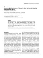
Figure 1 Identification of lungs infected with H-PRRSV and N-PRRSV. Lungs of uninfected negative control and experimentally infected pigs
were processed routinely for haematoxylin and eosin (H&E) staining and were re-isolated of H-PRRSV and N-PRRSV viruses and then were identified
by IFA and EM. Histopathology examination showed an interstitial pneumonia and emphysema in the lungs with thickening of the alveolar septa ac-
companied with infiltration of mononuclear cells from both H-PRRSV affected pigs and N-PRRSV affected pigs compared to the lungs of negative con-
trol pigs. Viral re-isolates were successfully recovered from lungs of the infected pigs, but not from those of uninfected negative control pigs. Specific
immunofluorescence and PRRSV particles in MARC-145 cells infected with re-isolated either H-PRRSV or N-PRRSV was observed by IFA and EM, respec-
tively, but not from those of uninfected negative control group. a. Representative images of HE stained lungs sections from H-PRRSV infected(C), N-
PRRSV infected(E), and uninfected negative control (A), original magnifications: ×40.; b. Assessment of H-PRRSV(B) or N-PRRSV(C) re-isolated infected
MARC-145 cells or negative control(A) by IFA staining at 48 h; c. H-PRRSV particle(A) and N-PRRSV particle(B) under the electron microscopy (EM).
Xiao et al. Virology Journal 2010, 7:107
/>Page 4 of 17
post N-PRRSV-inoculation, N96; 168 h post N-PRRSV-
inoculation, N168), respectively (ONE-ANOVA, p <
0.01). 19 and 8 protein spots differentially expressed
between different conditions (H96 vs N96, and H168 vs
N168) were obtained by Independent Student's t-test
contrast (Average Ratio > 1.5 or Average Ratio < -1.5, p <
0.05).
Identification of Differentially Expressed Proteins
As shown in Tables 1, 2 and 3, 48 differentially expressed
spots were successfully identified as 45 proteins. The
majority of spots contained only single proteins but in
some cases multiple spots flagged the same protein iden-
tity, such as three of spots (460, 481, and 484) were all
identified as lamin C, thus indicating the existence of
post-translational modifications or different isoforms.
GO enrichment and pathway analysis
These identified proteins were sorted by the enrichment
of GO categories (Additional file 1). 12 and 18 proteins
were revealed as differentially expressed across H-PRRSV
infected groups (uninfected negative control, H96, H168)
and N-PRRSV infected groups (uninfected negative con-
trol, N96, N168), respectively (Tables 1, 2 and Additional
file 2). The high-enrichment GOs targeted by H-PRRSV
infected groups proteins were ferric iron transport, posi-
tive regulation of myelination, response to organic cyclic
substance, pinocytosis, nitric oxide transport, positive
regulation of phagocytosis, regulation of inflammatory
response, acute-phase response, response to stress, etc
(Additional file 2). In contrast, significant GOs corre-
sponding to N-PRRSV infected groups proteins appeared
to be actin crosslink formation, ameboidal cell migration,
cytoplasmic sequestering of protein, T cell proliferation,
anti-apoptosis, oxidation reduction, etc (Additional file
2). 19 proteins were revealed as differentially expressed
between H-PRRSV infected lungs and N-PRRSV infected
lungs (Table 3). The high-enrichment GOs targeted by N-
PRRSV vs H-PRRSV infected groups proteins were ame-
boidal cell migration, myelin maintenance in the periph-
eral nervous system, myeloid cell homeostasis,
intermediate filament-based process, negative regulation
of cholesterol biosynthetic process, regulation of T cell
differentiation in the thymus, T cell proliferation,
response to superoxide, response to heat, activation of
MAPK activity, response to stress, etc (Additional file 2).
Pathway analysis was mainly based on the KEGG, Bio-
Carta and REATOME bioinformatics database. These
identified proteins were sorted by the enrichment of sig-
naling pathway categories. (Additional file 3). The signifi-
cant signaling pathways of these identified proteins H-
PRRSV infected groups include cell communication, the
role of FYVE-finger proteins in vesicle transport, hemo-
globin's chaperone, citrate cycle (TCA cycle), pathogenic
Escherichia coli infection, vibrio cholerae infection, adhe-
rens junction, membrane trafficking,and antigen process-
ing and presentation, etc (Additional file 3). In contrast,
significant signaling pathways corresponding to N-
PRRSV infected groups proteins appeared to be ascorbate
and aldarate metabolism, 3-Chloroacrylic acid degrada-
tion, limonene and pinene degradation, beta-Alanine
metabolism, urea cycle and metabolism of amino groups,
histidine metabolism, fatty acid metabolism, MAPK sig-
naling pathway, glutathione metabolism, stress induction
of HSP regulation, induction of apoptosis through DR3
and DR4/5 death receptors, FAS signaling pathway
(CD95), signal transduction through IL1R, TNFR1 sig-
naling pathway, p38 MAPK signaling pathway, and cas-
pase cascade in apoptosis, etc (Additional file 3).
Significant signaling pathways corresponding to N-
PRRSV versus H-PRRSV infected groups proteins
include apoptosis, cardiac protection against reactive
oxygen species (ROS), cell communication, cystic fibrosis
transmembrane conductance regulator (CFTR) and beta
2 adrenergic receptor (b2AR) pathway, free radical
induced apoptosis, glycosphingolipid biosynthesis-lactos-
eries, stress induction of HSP Regulation, MAPK signal-
ing pathway, induction of apoptosis through DR3 and
DR4/5 death receptors, FAS signaling pathway (CD95),
Figure 2 A representative 2D-DIGE picture of an overlay of three
dye scan. Proteins were extracted as described and separated in pH 3-
10 of 13 cm IPG strips for the first dimension and 12.5% acrylamide for
the second dimension. Image was acquired on a Typhoon 9400 scan-
ner. Dots represent spots detected by Decyder software. Cy2 (blue) im-
age of proteins from an internal standard is the pool of all the samples,
Cy3 (green) image of proteins from control1, and Cy5 (red) image of
proteins from H168_2.
Xiao et al. Virology Journal 2010, 7:107
/>Page 5 of 17
TNFR1 signaling pathway, and p38 MAPK signaling
pathway, etc (Additional file 3).
Construction of the protein-protein interaction network
As shown in Figure 3A, three proteins (HSPA8 (HSP70),
NDUFS1,and ARHGAP29) show the highest degree(7)
belonging to the most central protein followed by another
three proteins (TF, IDH3A, and DPYSL2) with degree (6),
therefore they might be of great importance to the pro-
tein-protein interaction network constructed based on
the differentially expressed proteins from lungs H-
PRRSV infected. In contrast, as shown in Figure 3B, the
most central protein corresponding to those of N-PRRSV
infected is DDAH2 with the highest degree (10) followed
by another two proteins (HSPB1 (HSP27) and FLNA)
with degree (8), these proteins tend to be more essential
than non-central proteins in modular organization of the
protein-protein interaction network.
Protein validation by Western blot and
Immunohistochemistry
As shown in Figure 4A, TF was slightly up-regulated in
lungs H-PRRSV affected at 96 h pi and then strongly up-
regulated in those at 168 h pi as compared to uninfected
negative control lungs. HSPB1 was strongly down-regu-
lated in lungs N-PRRSV affected at 96 h pi as compared
to uninfected negative control lungs and then slightly up-
regulated in those at 168 h pi as compared to those at 96 h
pi. The results were consistent with the expression
changes shown by the 2D-DIGE analysis (Figure 4A and
4B). Meanwhile, to further confirm the differential
expression observed in our 2D-DIGE screening, immu-
nohistochemistry (IH) staining of HSPB1 was also per-
formed on paraffin sections. As shown in Figure 5, the
result of IH agreed with the expression changes shown by
the 2D-DIGE and western blot analysis.
Discussion
In this study, we for the first time applied 2D-DIGE-based
proteomics to identify the differentially expressed pulmo-
nary proteins of lungs during H-PRRSV and N-PRRSV
infection in vivo. In total, of the 48 differentially
expressed spots, 45 proteins were identified. The indenti-
fied protein functions in diverse biological processes and
signaling pathways are formed through GO and pathway
analysis. Protein-protein interaction network was con-
structed based on the correlation relationships between
individual proteins across the data of differentially
expressed proteins from lungs infected with either H-
PRRSV or N-PRRSV. The potential roles of some of these
changed proteins in response to H-PRRSV and N-PRRSV
Table 1: Different expression of proteins between H-PRRSV (H96, H168) inoculated lungs and control identified by MALDI-
TOF or MALDI-TOF/TOF.
Master no.
a
Accession no.
b
Human
protein
(Abbr.)
p Value
c
Mr (Da) pI Protein
score
d
Sequence
Coverage (%)
e
187 gi|134085736 ARHGAP29 0.0044 107466 5.65 69 13
342 gi|136192 TF 0.0047 78971 6.93 71 15
352 gi|74005206 NDUFS1 0.0045 81056 6.1 71 15
371 gi|126309857 HSPA8 0.0055 54213 5.74 97 25
381 gi|194037328 KRT79 0.0087 48266 6.07 67 18
467 gi|68317041 STIP1 0.0029 32224 8.86 106 58
507 gi|231467 AHSG 0.0036 39199 5.5 73 10
552 gi|82822840 DPYSL2 0.0018 31025 8.07 84 11
690 gi|2724046 ACTG1 0.00037 36099 5.65 112 10
882 gi|27807289 ANXA2 0.0089 38873 6.92 73 6
885 gi|23706161 IDH3A 0.0014 27981 9.62 165 15
1461 gi|809283 +
gi|1709082
HBB 0.0031 16082 + 19200 6.76 +
6.37
102 + 70 60 + 43
a) Master no. is the unique sample spot protein number.
b) Accession is the MASCOT result of MALDI-TOF/TOF searched from the NCBI nr database.
c) The p value of ONE-ANOVA, p < 0.01, or Independent Student's t-test contrast, p < 0.05.
d) Protein score (based on combined MS and MS/MS spectra) and best ion score (based on MS/MS spectra) were from MALDI-TOF/TOF
identification.
e) Sequence coverage (%) is the number of amino acids spanned by the assigned peptides divided by the sequence length.
Xiao et al. Virology Journal 2010, 7:107
/>Page 6 of 17
infection are discussed as follows in relation with patho-
genesis and host antiviral response.
Alteration of cytoskeleton networks and cell
communication
Upon infection, virions or subviral nucleoprotein com-
plexes are transported from the cell surface to the site of
viral transcription and replication. Viruses use two strate-
gies for intracellular transport: viral components either
hijack the cytoplasmic membrane traffic or they interact
directly with the cytoskeletal transport machinery[27]. In
this study, eight proteins involved in cytoskeleton net-
works and cell communication have altered. The changes
in actin gamma 1(ACTG1), and keratin 79 were detected
in H-PRRSV infected lungs, whereas the change of fil-
amin A(FLNA), lamin A/C (LMNA), annexin A1
(ANXA1) and cofilin 1 (CFL1) were detected in N-
PRRSV infected lungs. Moreover, vimentin of N-PRRSV-
infected (N96) lungs was up-regulated compared to those
of H-PRRSV-infected (H96), whereas ezrin and LMNA
was down-regulated. These results showed that H-
PRRSV and N-PRRSV have to manipulated and utilize
host cytoskeleton to promote viral infection like many
other viruses[28,29].
FLNA is an actin-binding and signal mediator scaffold-
ing protein that crosslinks actin filaments and links actin
filaments to membrane glycoproteins. The encoded pro-
tein is involved in remodeling the cytoskeleton to effect
changes in cell shape and migration. FLNA is to be as an
adaptor protein that links HIV-1 receptors to the actin
cytoskeleton remodeling machinery, which may facilitate
virus infection[30]. On the other hand, FLNA plays a piv-
otal role in FcgammaRI surface expression via retention
of FcgammaRI from a default lysosomal pathway[31].
FLNA positively regulates I-KappaB kinase/NF-kappaB
cascade [32] and transcription factor import into
nucleus[33]. In our present study, this protein was
strongly down-regulated in N-PRRSV affected lungs at 96
h p.i as compared to uninfected negative control lungs
Table 2: Different expression of proteins between N-PRRSV (N96, N168) inoculated lungs and control identified by MALDI-
TOF or MALDI-TOF/TOF.
Master no.
a
Accession
no.
b
Human
protein
(Abbr.)
p Value
c
Mr(Da) pI Protein
score
d
Sequence
Coverage (%)
e
242 gi|74008809 FLNA 0.0025 283130 5.74 123 1
316 gi|5821963 ACO2 0.0088 83137 7.69 110 3
481 gi|66352015 LMNA 0.0021 65189 6.4 89 30
612 gi|194674843 NTF4 0.00044 33968 9.06 72 27
614 gi|2624886 ALDH2 0.0045 54859 6.05 147 6
616 gi|40426087 LAP3 0.0063 31073 5.64 193 14
638 gi|47685624 ALDH9A1 0.0097 26192 5.43 125 16
656 gi|190360675 FLOT1 0.0089 47554 7.66 74 27
666 gi|126335980 CCDC13 0.0084 83367 7.68 69 19
938 gi|194033965 ANXA1 0.0076 35689 7.16 75 29
1016 gi|149409809 FECH 0.0033 49485 8.48 66 20
1038 gi|87217590 DDAH2 0.0049 23244 5.33 88 39
1173 gi|50916342 HSPB1 0.00092 14268 5.94 73 43
1200 gi|544445 GSTP1 0.0036 23710 8.07 166 13
1250 gi|17892411 PEBP1 0.0019 17055 5.74 149 22
1312 gi|543113 TAGLN 0.0039 19326 6.96 61 11
1316 gi|5031635 CFL1 0.0093 18719 8.22 43 6
1491 gi|21545648 COX5A 0.0026 19379 6.88 82 14
a) Master no. is the unique sample spot protein number.
b) Accession is the MASCOT result of MALDI-TOF/TOF searched from the NCBI nr database.
c) The p value of ONE-ANOVA, p < 0.01, or Independent Student's t-test contrast, p < 0.05.
d) Protein score (based on combined MS and MS/MS spectra) and best ion score (based on MS/MS spectra) were from MALDI-TOF/TOF
identification.
e) Sequence coverage (%) is the number of amino acids spanned by the assigned peptides divided by the sequence length.
Xiao et al. Virology Journal 2010, 7:107
/>Page 7 of 17
Table 3: Different expression of proteins between H-PRRSV and N-PRRSV (N96/H96, H168/H168) inoculated lungs
identified by MALDI-TOF or MALDI-TOF/TOF.
Master
no.
a
Accession
no.
b
Human
protein
(Abbr)
Average
ratiof
p Value
c
Mr (Da) pIProtein
score
d
Sequence
Coverage
e
(%)
N96/H96
484 gi|66352015 LMNA -3.14 0.0075 65189 6.4 76 36
1173 gi|50916342 HSPB1 -2.35 0.00063 14268 5.94 73 43
477 gi|61867592 STIP1 -2.19 0.033 63056 6.02 41 1
604 gi|410689 LAP3 -2.01 0.0092 55996 5.68 66 6
481 gi|66352015 LMNA -1.99 0.012 65189 6.4 89 30
374 gi|27806351 EZR -1.92 0.035 68832 6.06 70 1
460 gi|66352015 LMNA -1.83 0.035 65189 6.4 68 26
1303 gi|40423533 AP3S2 -1.77 0.034 29516 11.16 100 37
1415 gi|15082144 SOD1 -1.66 0.047 15408 6.04 78 21
1312 gi|543113 TAGLN -1.6 0.04 19326 6.96 61 11
520 Gi|19403459
3
KIAA1468 -1.58 0.00093 120185 5.38 70 9
848 gi|37800811 GPD1L -1.55 0.029 22781 5.4 90 46
1292 gi|182851479 VIM 1.6 0.011 18149 4.7 137 72
921 gi|54020966 ANXA2 1.63 0.045 38795 6.49 52 7
942 gi|148747594 RPLP0 1.63 0.026 34508 5.71 159 18
616 gi|40426087 LAP3 1.74 0.0073 31073 5.64 193 14
1428 gi|89886167 FABP5 1.82 0.018 15485 6.6 100 31
N168/H168
1235 gi|959814 FUT1 -1.59 0.02 15873 9.52 184 35
1171 gi|27806479 PKP1 1.7 0.008 81498 9.18 68 17
1519 gi|6843240 HBA2 2.09 0.00028 13025 8.81 182 25
1506 gi|6843240 HBA2 2.21 0.01 13025 8.81 220 30
a) Master no. is the unique sample spot protein number.
b) Accession is the MASCOT result of MALDI-TOF/TOF searched from the NCBI nr database.
c) The p value of ONE-ANOVA, p < 0.01, or Independent Student's t-test contrast, p < 0.05.
d) Protein score (based on combined MS and MS/MS spectra) and best ion score (based on MS/MS spectra) were from MALDI-TOF/TOF
identification.
e) Sequence coverage (%) is the number of amino acids spanned by the assigned peptides divided by the sequence length.
f) Average ratios were calculated considering 6 replica gels and were calculated using Decyder software as the fold -change between normalized
spot volume between N-PRRSV-infected lungs (N96 or N168) and H-PRRSV-infected lungs (H96 or H168) homogenates (Independent Student's
t-test was based on the log of the ratio between N96 and H96, or between N168 and H168).
and then slightly up-regulated in those at 168 h p.i as
compared to those at 96 h p.i. This phenomenon may
explain that N-PRRSV manipulate and utilize the adaptor
protein, FLNA, to promote viral infection.
Response to stress
The quantities of three proteins related to stress response
were found to have been modified in either H-PRRSV-
infected lungs or N-PRRSV-infected lungs, including heat
shock 70 kDa protein 8 (HSPA8, Hsp70), heat shock 27
kDa protein 1 (HSPB1), and stress-induced-phosphopro-
tein 1. HSPA8 belongs to the heat shock protein 70 family
which is highly abundant cytosolic and nuclear molecular
chaperones that play essential roles in various aspects of
protein homeostasis, controlling the biological activity of
folded regulatory proteins, disassembly of clathrin-coated
vesicles, viral capsids and the nucleoprotein complex,
intracellular vesicle trafficking and sorting, antigen pro-
cessing and presentation, MAPK signal transduction, cell
cycle regulation, differentiation and programmed cell
Xiao et al. Virology Journal 2010, 7:107
/>Page 8 of 17
death and nuclear transport. Over expression of hsp70
with a herpes viral amplicon vector protected cultured
hippocampal rat neurons from gp120 of HIV neurotoxic-
ity [34], hsp70 was also able to prevent the WNV capsid
protein's cytotoxic effects [35], suggesting a protective
cell function for this molecular chaperone against viral
infection. The exposure of permissive CD4+ cells to HIV-
1 gp120 increases the synthesis and nuclear translocation
of 70 kDa heat shock protein. Hsp70 facilitates nuclear
import of HIV-1 preintegration complexes by stimulating
the binding of HIV-1 Matrix to karyopherin alpha. Over-
expression of Hsp70 by WNV infection, hepatitis C virus
(HCV) infection[36], and TBSV infection[37] suggests
that it involves in the pathogenesis of those viruses. In the
present study, HSPA8 was up-regulated continuously
after H-PRRSV infection. Moreover, in the protein-pro-
tein interaction network constructed based on the differ-
entially expressed proteins from lungs H-PRRSV
infection, HSPA8 shows the highest degree (7) belonging
to the most central protein. The most central protein
tends to be more essential than non-central proteins in
modular organization of the protein-protein interaction
network. These results suggest that Hsp70 might be
involved in H-PRRSV pathogenesis and as a specific
chaperone, it can protect cell from apoptosis.
Heat shock 27 kDa protein (HSPB1, Hsp27) is a stress-
inducible ubiquitous cellular protein that belongs to small
HSP families and is involved in cellular protection in
response to a variety of stresses such as heat shock, toxi-
cants, and oxidative stress, stress induction of HSP regu-
lation, MAPK signaling pathway, anti-apoptosis,
regulation of translational initiation, molecular chaper-
oning, actin organization and cell motion. Hsp27 regu-
lates Akt activation and cellular apoptosis by mediating
interaction between Akt and its upstream activator
MK2[38]. Moreover, the phosphorylated Hsp27 binded
by caspase-3 prodomain regulates monocyte apoptosis by
inhibiting caspase-3 proteolytic activation[39]. Viral
infection modulates the regulation of apoptosis in host
cells. Up-regulated HSP27 has been found in cells
infected with Epstein-Barr virus[40], avian H9N2[41],
Afriacan swine fever virus[20], IBDV[19], and
PRRSV[42]. But down-regulated HSP27 has been also
found in cells infected with classical swine fever virus [21]
and IBDV (another HSPB1 protein spot)[19]. In the pres-
ent study, this protein was strongly down-regulated in N-
PRRSV affected lungs at 96 h p.i as compared to unin-
fected negative control lungs and then slightly up-regu-
lated in those at 168 h p.i as compared to those at 96 h p.i.
Moreover, in the protein-protein interaction network
constructed based on the differentially expressed proteins
from lungs N-PRRSV infected, Hsp27 shows the very
highly degree (8) belonging to the central protein. Some
evidences indicate that human cells infected with mumps
virus become susceptible to apoptosis caused by extracel-
lular stresses. The infected cells failed to acquire resis-
tance to apoptotic stimuli (thermotolerance) after
exposure to these mild stresses. The induction of Hsp27
was dramatically suppressed after mumps virus infection
through the destruction of STAT-1[43]. Based on these
data, Hsp27 might be involved in N-PRRSV pathogenesis,
and the lack of thermotolerance should allow the infected
Figure 3 Graph of the protein interaction network of identified proteins. The protein interaction network was constructed from the identified
proteins according their properties and expression level in differential samples. A) graph of the protein interaction network from identified proteins
of H-PRRSV-infected lungs, HSP70, NDUFS1,and GMIP show the highest degree (7) belonging to the most central protein, therefore they might be of
great importance to the protein-protein interaction network; B) graph of the protein interaction network from identified proteins of N-PRRSV-infected
lungs, DDAH2 with the highest degree (10) followed by another two proteins (HSP27(HSPB1) and FLNA) with degree(8), tend to be more essential
than non-central proteins in modular organization of the protein-protein interaction network.
Xiao et al. Virology Journal 2010, 7:107
/>Page 9 of 17
Figure 4 Expression analyses of selected proteins using DeCyder software and western blot validation. A) Representative 2D-DIGE image,
quantification, and western blot confirmation of TF in H-PRRSV infected pigs. The standard abundance of the different spots (y-axis) is also shown for
the three different experimental conditions: A (control), B (H96), C (H168) (x-axis). Equal amounts of total protein, as shown for GAPDH, were loaded
for Western blotting analysis; B) Representative 2D-DIGE image, quantification, and western blot confirmation of HSPB1 in N-PRRSV infected pigs and
those between N-PRRSV vs. H-PRRSV. The standard abundance of the different spots (y-axis) is also shown for different experimental conditions: A
(control), D (N96), E (N168), B (H96) (x-axis). Equal amounts of total protein, as shown for GAPDH, were loaded for Western blotting analysis.
Xiao et al. Virology Journal 2010, 7:107
/>Page 10 of 17
cells to be eliminated by apoptosis and might be a host
defense against viral infection.
Oxidation reduction and metabolism
Four differentially expressed proteins of interest associ-
ated with oxidation reduction and metabolism were
found, including Isocitrate dehydrogenase 3 (NAD+)
alpha (IDH3A), NADH dehydrogenase Fe-S protein 1
(NDUFS1) and Annexin A2 (ANXA2) in H-PRRSV
infected lungs; Glutathione S-transferases P(GST class-
pi, GSTP1) in N-PRRSV infected lungs; Superoxide dis-
mutase 1, soluble (SOD1) and Ribosomal protein, large,
P0 between H-PRRSV and N-PRRSV infected lungs.
NDUFS1 belongs to the complex I 75 kDa subunit fam-
ily, playing a very important role in the electron transport
from NADH to ubiquinone in the respiratory chain for
ATP production. GO analysis in our study also classified
NDUFS1 as ATP synthesis coupled electron transport.
Previously, studies indicated that HIV-1 infection
induced to release ROS through a mitochondrial path-
way. In addition, Disruption of electron transport and
mitochondrial transmembrane potential, loss of ATP
production and promotion of ROS generation were due
to cleavage NDUFS1 by caspases. However cells express-
ing a noncleavable mutant of NDUFS1 sustain mitochon-
drial transmembrane potential and ATP levels during
apoptosis and ROS generation is dampened in response
to apoptotic stimuli. All of these indicated that caspase
cleavage of NDUFS1 is essential to several changes of
mitochondrion during apoptosis[44]. On the other hand,
reduced expression of NDUFS1 was found in chronic
morphine treated hippocampal and down-regulation of
NDUFS1 would decrease of ATP production[45]. There-
fore, the continuous increased expression of NDUFS1 in
H-PRRSV infected lungs might provide continuous
increased substrate for apoptosis and also sustain energy
metabolism. This is supported by the previous findings
that inhibition of complex I activity would lead to reduc-
tion of ATP levels in HIV-infected cells, but ATP synthe-
sis would not be ceased completely[46]. Hence, these
results might be mainly implicated in how H-PRRSV
influenced host cell energy metabolism during apoptotic
cell death. Additionally, the degree of NDUFS1 in the
protein network of H-PRRSV infected lungs is seven,
which ranked the first. Hence, NDUFS1 located at the
most central in the network. This implies that NDUFS1 is
likely to be more essential in organization of protein-pro-
tein interaction network.
Apoptotic pathways
Apoptosis of host cells plays an important role in modu-
lating the pathogenesis of many infectious diseases. Dim-
ethylarginine dimethylaminohydrolase 2 (DDAH2)
belongs to the dimethylarginine dimethylaminohydrolase
Figure 5 Immunohistochemistry validation of HSPB1. The expression pattern of HSPB1 in lungs infected with H-PRRSV and N-PRRSV was investi-
gated by immunohistochemistry. Uninfected negative control lungs, lungs infected with H-PRRSV (H96 and H168), and lungs infected with N-PRRSV
(N96 and N168) were stained with anti-HSP27 antibodies. Original magnifications: ×40.
Xiao et al. Virology Journal 2010, 7:107
/>Page 11 of 17
(DDAH) gene family and involves in anti-apoptosis,
response to unfolded protein, defense response, nitric
oxide biosynthetic process, nitric oxide mediated signal
transduction, and arginine catabolic process. The
encoded enzyme plays an important role in nitric oxide
generation by regulating cellular concentrations of meth-
ylarginines, which in turn inhibit nitric oxide synthase
activity. The recent study has indicated that the activity of
DDAH and the expression of DDAH2 (mRNA and pro-
tein) was significantly decreased in cobalt chloride
(CoCl
2
)-induced apoptosis. In contrast, DDAH2 overex-
pression inhibited the proapoptotic effects of CoCl2 [47].
CoCl
2
significantly increased the level of endogenous
nitric oxide synthase inhibitor asymmetric dimethylargi-
nine (ADMA), which markedly increased intracellular
ROS production and promoted inflammatory responses,
resulting in caspase-3-dependent apoptosis. Moreover,
exogenous ADMA could directly induce cellular apopto-
sis via ROS dependent signaling pathway. DDAH is the
specific hydrolase of ADMA and plays an important role
in the modulation of ADMA level. Various oxidative,
LPS, or inflammatory stimuli could directly inactivate the
DDAH activity and then significantly decrease the
expression of DDAH2 mRNA and protein through a sulf-
hydryl group in the catalytic region of DDAH [48]. More-
over, expression of DDAH2 was also found to be reduced
when comparing lung tissue from pulmonary hyperten-
sive rats and idiopathic pulmonary arterial hypertension
(IPAH) patients to corresponding normal lung tissue [49].
DDAH2 localizes to 6p21.3. The region contains a num-
ber of genes involved in the immune and inflammatory
responses and has been linked with susceptibility to sev-
eral autoimmune diseases. This localization and its wide
expression in immune cells means that DDAH2 has the
potential to be a disease-susceptibility gene[50]. DDAH2
was strongly down-regulated in N-PRRSV affected lungs
at 96 h p.i as compared to uninfected negative control
lungs and then slightly up-regulated in those at 168 h p.i
as compared to those at 96 h p.i. Moreover, in the pro-
tein-protein interaction network constructed based on
the differentially expressed proteins from lungs N-PRRSV
infected, DDAH2 shows the highest degree (10) belong-
ing to the most central protein. These results strongly
support the importance of DDAH2 in N-PRRSV patho-
genesis, and after N-PRRSV infection, expression of
DDAH2 in lungs significantly decreased comparing to
those in uninfected negative control lungs, which
resulted in cell-infected apoptosis, which might be a host
defense against viral infection.
Others
Rho GTPase activating protein 29 (PARG1, ARHGAP29),
encoding for a protein-tyrosine phosphatase-associated
Rho GTPase activating protein, is involved in signaling by
Rho GTPases. Rho GTPases, regulating GTP-GDP cycle,
were key signal transducers, mediating growth factor-
induced changes to the actin cytoskeleton and activating
the phagocyte NADPH oxidase, and participated in a
number of cellular processes, such as cell migration, cell
survival, transcriptional regulation and vesicle trafficking.
This is because they might be able to interact with lots of
downstream targets, so that they can coordinately acti-
vate several molecular processes required for a particular
cellular response. In the present study, we observed that
in the protein-protein interaction network constructed
based on the differentially expressed proteins from lungs
H-PRRSV infected, ARHGAP29 shows the highest
degree (7) belonging to the most central protein. It inter-
acted with sever protein of the network, including
HSP70, NDUFS1,IDH3A, TF, DPYSL2, ANXA2, and
STIP1, which suggests that these proteins could coordi-
nately activate several molecular processes required for a
particular cellular immune response. A strong down-reg-
ulation of ARHGAP29, by several mechanisms such as
deletion and promoter methylation, was found in all
mantle cell lymphoma (MCL) samples, which may lead to
carcinogenesis through the dysregulation of Rho/Rac/
Cdc42-like GTPases[51]. ARHGAP29 was down-regu-
lated in H-PRRSV affected lungs at 96 h p.i as compared
to uninfected negative control lungs and then continu-
ously down-regulated in those at 168 h p.i as compared to
those at 96 h p.i. Based on these results, it is reasonable to
postulate that ARHGAP29 coordinates other proteins
together to involve in the pathogenesis of H-PRRSV.
Conclusion
We analyzed the protein expression changes of H-PRRSV
and N-PRRSV infected lungs compared with those of
uninfected negative control, and identified a series of pro-
teins related to viral pathogenesis and host response
using 2D-DIGE followed by MS identification and bioin-
formatics methods. Our results showed that following
both H-PRRSV and N-PRRSV infection, the significant
expression changes in pulmonary proteins were mostly
related to cytoskeletal proteins, stress response proteins
and proteins involved in oxidation reduction or metabo-
lism. The changed expression of some cytoskeletal pro-
teins could be a strong sign of cytoskeletal reorganization
which is essential for viral reproduction and assembly.
Besides, protective proteins in response to a variety of
virus-induced stresses such as oxidative stress, heat shock
and toxicants have been shown to be expressed differen-
tially after either H-PRRSV or N-PRRSV infection. In the
protein-protein interaction network constructed based
on the differentially expressed proteins from lungs H-
PRRSV infected, HSPA8, ARHGAP29, and NDUFS1
showed the highest degree belonging to the most central
protein, but DDAH2, HSPB1, and FLNA corresponded to
Xiao et al. Virology Journal 2010, 7:107
/>Page 12 of 17
the most central proteins in those of N-PRRSV infected,
suggesting differential viral pathogenesis and differential
host response to H-PRRSV and N-PRRSV infection. To
our knowledge, the study presented here is the first pro-
teomic study using 2D-DIGE and MS to compare the
complex picture of pulmonary protein expression during
H-PRRSV and N-PRRSV infection.
Methods
Experimental animals and tissue collection
All animal procedures were performed according to
guidelines developed by the China Council on Animal
Care and protocol approved by Animal Care and Use
Committee of Guangdong Province, P.R. China.
Fifteen conventionally-reared, healthy 6-week-old,
crossbred weaned pigs (Landrace × Yorkshire) were
selected from a high-health commercial farm that has
historically been free of all major pig diseases, such as
PRRSV, porcine circovirus type 2, classical swine fever
virus, porcine parvovirus, pseudorabies virus, swine
influenza virus and Mycoplasma hyopneumoniae infec-
tions. All pigs were PRRSV-seronegative determined by
ELISA (HerdChek PRRS 2XR; IDEXX Laboratories) and
absence of PRRSV tested by RT-PCR. Pigs were randomly
assigned to one uninoculated negative control group and
two PRRSV-inoculated groups (H-PRRSV and N-PRRSV
respectively, gift from Dr. Zhang Guihong, South China
Agricultural University) in the experiment. Six pigs were
inoculated with 6 ml viral suspension (4 ml intranasally
and 2 ml intramuscularly) of H-PRRSV at a dose of 10
6.0
TCID
50
ml
-1
on day 0. Six pigs were inoculated with 6 ml
viral suspension (4 ml intranasally and 2 ml intramuscu-
larly) of N-PRRSV at a dose of 10
6.0
TCID
50
ml
-1
on day 0.
Three negative control pigs were treated similarly with an
identical volume of DMEM culture media from unin-
fected MARC-145 cells 1 day prior to experimental infec-
tion, and were immediately necropsied. Two PRRSV-
inoculated groups were clinically examined daily and rec-
tal body temperatures were recorded from days -2 to 7
post infection (p.i). Three infected pigs randomly chosen
within each group were necropsied at each time point of
96 h p.i and 168 h p.i. Lung samples were collected from
control, three pigs at 96 h post H-PRRSV-inoculation
(H96), three pigs at 168 h post H-PRRSV-inoculation
(H168), three pigs at 96 h post N-PRRSV-inoculation
(N96), three pigs at 168 h post N-PRRSV-inoculation
(N168) and immediately frozen in liquid nitrogen for pro-
teome analysis or fixed in 10% neutralized buffered form-
alin for histological processing.
Virus re-isolation and RT-PCR detection
250 μl of lung tissue homogenate plus 150 μl of DMEM
with 75 μg of penicillin and 50 μg of streptomycin per ml
were inoculated on MARC-145 cells and incubated for
1.5 h at 37°C with 5% CO
2
. Then, tissue homogenate were
removed and DMEM containing 5% FBS was added. Cul-
tures were incubated for 3 days at 37°C in a 5% CO
2
humidified incubator. Cultures which do not display
cytopathic effect (CPE) after three passages were consid-
ered negative. And PRRSV induced CPE on MARC-145
was confirmed by the following three methods: 1) indi-
rect immunoflorescent assay (IFA) using positive serum
against PRRSV; 2) negative-stain electron microscopy
(EM) which applied 4 μl virus suspension to glow-dis-
charged carbon-coated copper grids with a micropipette
and stained with 1% (w/v) uranyl acetate; 3) PRRSV-spe-
cific RT-PCR using oligonucleotide primers NSP2F(5'-
AACACCCAGGCGACTTCA-3') and NSP2R(5'-GCAT-
GTCAACCCTATCCCAC-3') which designed according
to the existing 87 base deletion between the H-PRRSV
and N- PRRSV in the fixed site in Nsp2 gene and will
amplify 787 bp and 874 bp DNA fragment of H-PRRSV
and N-PRRSV, respectively.
Histological examination
Lungs of uninfected negative control and experimentally
infected pigs were processed routinely for haematoxylin
and eosin (H&E) staining, as described previously[52].
Protein Extraction
For each sample, ~0.3 g of lung tissue washed with nor-
mal saline was trimmend into 3 mm
3
slices and then was
homogenized on ice in 1 ml DIGE lysis buffer (7 M Urea,
2 M Thiourea, 4% CHAPS, 0.2%IPGbuffer, protease
inhibitor mixture) using a DOUNCE homogenizer. After
sonication (8 × 10 s pulses on ice, with cooling intervals
of 15 s in between) and centrifugation (14,000 rpm for 1
hour) to collect supernatant fluid, protein concentrations
were determined using the Bio-Rad Protein Assay (Bio-
Rad). Proteins were checked by visualization of Comassie
blue stained proteins separated on a 12.5% SDS-PAGE
acrylamide gel. Concentration of all samples was adjusted
to 5 μg/μl.
Protein labeling
Equal amounts of proteins from the 15 samples were
pooled together as the internal standard. Proteins were
minimally labeled according to the manufacturer's
instructions (CyDye DIGE fluor minimal labeling kit, GE
Healthcare). Briefly, each miminal CyDye was reconsti-
tuted in fresh N,N-dimethylforamide (DMF) and a 400
pmol quantity used to label 50 μg of protein at pH 8.5.
Cy2 was used to label the pooled internal standard. Cy3
and Cy5 were used to randomly label the uninfected neg-
ative control and H-PRRSV-infected or N-PRRSV-
infected samples. The labeling reaction was done on ice
in the dark for 40 min and the reaction was terminated by
addition of 1 μl 10 mM lysine on ice in the dark for 10
Xiao et al. Virology Journal 2010, 7:107
/>Page 13 of 17
min. To minimize system and inherent biological varia-
tion, sample multiplexing was also randomized (Table 4)
to produce unbiased results.
2-D gel electrophoresis
Following the labeling reaction, 50 μg of each Cy2, Cy3
and Cy5 labeled samples were mixed. Then the pooled
sample of each gel was diluted with rehydration buffer (7
M urea, 2 M thiourea, 2% DTT (w/v), and 1% IPG buffer
(v/v)) to 250 μl before Isoelectric Focusing (IEF). Samples
were actively rehydrated into 13-cm pH 3-10 non-linear
Immobiline DryStrips, placed in a strip holder and
focused with an Ettan IPGphor Isoelectric Focusing Sys-
tem (GE Amersham) using a step gradient protocol rang-
ing from 30 to 8000 volts for approximately twenty six
hours (30 v 12 hrs, 500 v 1 hr, 1000 v 1 hr, 8000 v 8 hrs,
500 v 4 hrs).
The IPG strips were rehydrated in re-equilibration buf-
fer (8 M urea, 100 mM Tris-HCL (pH6.8), 30% Glycerol,
1% SDS, 45 mg/mL iodoacetamide (to reduce streaking))
for 10 minutes, and then proteins were further separated
on the 12.5% homogeneous SDS-PAGE gels (24 cm × 20
cm × 1 mm) casted with low-fluorescence glass plates uti-
lizing Hofer SE 600 (GE Amersham). The SDS-PAGE gels
were run at 15 mA/gel for 20 min and then at 30 mA/gel
at 15°C until the bromophenol blue dye front reach the
bottom of the gel.
Scanning and image analysis
After 2D-DIGE, scan the gels using a Typhoon 9400 scan-
ner (GE Amersham) at 100 μm resolution, as elaborated
in the equipment setup. The Cy2, Cy3, and Cy5 labeled
images for each gel were scanned at the excitation/emis-
sion wavelengths of 488/520 nm, 532/580 nm, 633/670
nm, respectively. After scanning the three fluorophores
for each gel, the images were imported to the DeCyder
image analysis software (GE Amersham) for spot detec-
tion according to manufacturer's recommendations.
Briefly, Differential in gel analysis (DIA) module was used
for intra-gel analysis for protein spot detection and for
normalization of Cy3 and Cy5 gel images with respect to
the Cy2 image. After spot detection, the abundance
changes were represented by the normalized volume ratio
(Cy3:Cy2 and Cy5:Cy2). Make sure that artifactual spots
(dust and others) were removed and that all true protein
spots were included, all the protein spots detected were
also examined manually. The biological variation analysis
(BVA) module was used for inter-gel matching of internal
standard and samples across all gels, and performing
comparative cross-gel statistical analyses of all spots,
based on spot volumes, permitting the detection of dif-
ferentially expressed spots between experimental condi-
tions (One-way ANOVA, p < 0.01 and Independent
Student's t-test, p < 0.05). The protein spot matches were
also confirmed manually for all the gels. Protein spots
that were differentially expressed in H-PRRSV infected
and N-PRRSV infected groups (B/D,C/E) (Independent
Student's t-test, Average Ratio > 1.5 or Average Ratio < -
1.5, p < 0.05) were marked. Protein spots that were differ-
entially expressed in H-PRRSV infected and uninfected
negative control groups (A/B/C) or N-PRRSV infected
and uninfected negative control groups (A/D/E) ((One-
way ANOVA, p < 0.01) were marked. Satisfying these cri-
teria, a pick list is generated and exported to the software
controlling the Ettan robotic spot picker (GE Amersham).
Spots were excised with a 3 mm core from the post-
stained gel and loaded to a 96-well plate for digestion.
Spots in the maps for which the average intensity differed
between two appoint groups were selected to be identi-
fied by mass spectrometry.
Protein digestion, mass spectrometry and protein
identification
Preparative gels containing 500 μg protein were run to
identify interest protein and were stained with Coomassie
brilliant blue (CBB). Protein spots of interest were
excised from the gel automatically using an Ettan Spot
Picker robot (GE Amersham) and destained with 25 mM
ammonium bicarbonate, 50% ACN. Gels were then dried
completely by vacuum-drying. In-gel digestion was per-
formed with 12.5 ng/L modified sequencing grade
trypsin (Promega) in 25 mM ammonium bicarbonate at
4°C for 40 min prior to 20 h at 37°C. To achieve complete
peptide recovery, two sequentially extraction steps (5%
TFA at 40°C for 1 h and with 2.5% TFA, 50% ACN at 30°C
for 1 h) were carried out with the digested samples. The
supernatants containing peptides were then collected,
and then concentrated and desalted by ZipTips (Milli-
pore, Bedford, MA). Peptides were mixed with equal
amounts of matrix solution (α-cyano-4-hydroxy-cin-
namic acid (HCCA) in 0.1% TFA, 50% ACN) and imme-
diately loaded on the target plate, and allowed to air-dry
at room temperature. MALDI-TOF mass spectrometry
and tandem TOF/TOF mass spectrometry analyses were
performed on an AutoFlex TOF-TOF LIFT Mass Spec-
trometer (Bruker Daltonics) according to the manufac-
turer's instructions. The spectra were acquired in the
positive ion reflection mode (accelerating voltage of 20
kV, reflecting voltage of 23 kV) with external calibration
(Trypsin_Roche_porcine_Modified) according to the set-
tings given by the manufacturer. Parent mass peaks with
mass range of 700-4000 and minimum signal to noise
ratio of 15 were picked out for tandem TOF/TOF analy-
sis. The generated mass lists were subsequently sent to
MASCOT (Version 2.1, Matrix Science, London, UK) by
GPS Explorer software (Version 3.6, Applied Biosystems)
for protein identification. Parameters for searches were as
follows: National Center for Biotechnology Information
Xiao et al. Virology Journal 2010, 7:107
/>Page 14 of 17
non-redundant (NCBInr) database (EST_chordata
chordata_20081008 (87827958 sequences; 17755145374
residues)), taxonomy of other mammalia (23009496
sequences); tryptic peptides with max one missed cleav-
age site; fixed modifications, carbamidomethylation; vari-
able modifications, oxidation; peptide mass tolerance, ±
150 ppm. MASCOT protein scores (based on combined
MS and MS/MS spectra) of greater than 65 were consid-
ered statistically significant (p < 0.05). The individual
MS/MS spectrum with a statistically significant (p < 0.05)
ion score (based on MS/MS spectra) were accepted.
Gene ontology (GO) and pathway enrichment analysis
GO analysis [53] was applied in order to organize differ-
entially expressed proteins into functional classification
on the basis of biological process. Pathway analysis [54-
56] was mainly based on the Kyoto Encyclopedia of
Genes and Genomes (KEGG) and BioCarta and
REATOME bioinformatics database. Two-side Fisher's
exact test with a multiple testing and χ2 test were used to
classify the GO and pathway category. The false discov-
ery rate (FDR) was used to correct the P-value. We chose
only GO categories that had a P-value of <0.01 and an
FDR of <0.05 and pathway categories that had a P < 0.05.
Within the significant category, the enrichment Re was
given by:
n
f
: the number of flagged proteins within the particu-
lar category;
n: the total number of proteins within the same cate-
gory;
N
f
: the number of flagged proteins in the protein ref-
erence database list;
N: the total number of proteins in the protein refer-
ence database list;
Construction of the protein-protein interaction
network[57,58]
Protein-protein interaction network was constructed
based on the data of differentially expressed proteins. The
matrix of proteins expression values was build up at first,
and then Pearson product-moment correlation coeffi-
cients were computed. Suppose there are two variables X
and Y, which indicate expression value of two proteins
respectively in the sample, with means and
respectively and standard deviations S
X
and S
Y
respec-
tively. The correlation r is calculated as:
The Pearson product-moment correlation coefficients
have been applied to quantify the strength of correlation
between proteins. And a correlation coefficient of no less
than 0.48 was considered as 1 while which less than 0.48
was considered as 0. Protein correlation matrix (PCM)
was then to be formed. According to the correlation
between proteins, protein-protein interaction network
was constructed. Nodes were applied to represent the
proteins and interactions between proteins were
expressed by straight lines between the nodes. Then each
node's degree was calculated. The nodes with more inter-
R
n
f
n
N
f
N
e
= (Re )=ENRICHMENT
X
Y
r =
−
−−
=
∑
1
1
1
n
X
i
X
S
X
Y
i
Y
S
Y
i
n
()().
Table 4: 2D-DIGE experimental design*.
Gel Cy2(blue) Cy3(green) Cy5(red)
1 pool A1 (Control1) C2 (H168_2)
2 pool C1 (H168_1) E3 (N168_3)
3 pool D2 (N96_2) B2 (H96_2)
4 pool B1 (H96_1) E2 (N168_2)
5 pool E1 (N168_1) A1 (Control1)
6 pool D3 (N96_3) C3 (H168_3)
7 pool B3 (H96_3) A2 (Control2)
8 pool A3 (Control3) D1 (N96_1)
*Control and experimental samples (H96, H168, N96, N168) were labeled with either Cy3 or Cy5. Equal amounts of protein lysates from 3
uninfected negative control and 12 experimental samples were pooled as the internal standard, and labeled with Cy2. Each gel was loaded
with 50 μg of Cy2-labeled protein pool, 50 μg of Cy3-labeled and 50 μg of Cy5-labeled samples as indicated. Three replicates were used by
each experimental condition.
Xiao et al. Virology Journal 2010, 7:107
/>Page 15 of 17
actions will have higher degrees. In addition, different
colors of nodes indicate different values of K-core the
proteins have. The "degree" is defined as the number of
interactions of a protein with other proteins in the pro-
tein network. While the rank is determined as the
decreasing ordering of each protein's degree, the first
rank which has the highest degree belongs to the most
central protein in the network. The most central protein
tends to be more essential than non-central proteins in
modular organization of the protein-protein interaction
network. K cores have been applied for clustering pro-
teins of network. The proteins with degrees of the same
or close to were colored identically and different colored
proteins were identified as different subnetworks. There-
fore, protein-protein interaction network has been
divided into several subgraphs and all the proteins in one
subgraph belong to same cluster of degrees. A subnet-
work was used to identify a group of same colored pro-
teins, which were found to regulate almost same number
of other proteins in the network and implied they shared
similar biological functions under certain conditions.
Western blot analysis
Equivalent amounts of total protein (40 μg) were loaded
in each lane and were fractionated by electrophoresis on
12% (w/v) SDS-PAGE gels, then transferred onto a PVDF
membrane using iBlot™ Dry Blotting System (Invitrogen)
and blocked with TBS-T containing 5% BSA at 4°C over-
night. The PVDF membrane were probed with a 1:500
dilution of goat anti-pig Transferrin antibody (Bethyl,
TX, USA), and at a dilution of 1:200 mouse anti-Heat
Shock Protein 25 monoclonal antibody (Chemicon/Milli-
pore, MA, USA). Horseradish peroxidase-conjugated
rabbit anti-goat IgG, horseradish peroxidase-conjugated
goat anti-mouse IgG or horseradish peroxidase-conju-
gated goat anti-rabbit IgG at a dilution of 1:4,000 were
used as secondary antibodies. The protein bands were
visualized using diaminobenzidine (DAB) as the substrate
(Boster, Wuhan, China). The same membranes were
reblotted with rabbit affinity purified anti-GAPDH anti-
body (Rockland, PA, USA) at a dilution of 1:1,000 to con-
firm equal loading.
Immunohistochemistry analysis
Lung tissues of uninfected negative control and experi-
mentally infected pigs were formalin fixed for immuno-
histochemistry. Paraffin sections (5 μm) were
deparaffinized and rehydrated in in a graded alcohol
series, and pretreated with 10 mM sodium citrate (3-10
min, 600 W microwave oven). Nonspecific binding was
blocked by incubating the tissue sections with 10% BSA
(Sigma) in PBS for 60 min. Immunostaining was per-
formed in a moist chamber at 37°C for 1 h with mouse
anti-Heat Shock Protein 25 monoclonal antibody
(Chemicon/Millipore, MA, USA) at a dilution of 1:200.
Horseradish peroxidase-conjugated goat anti-mouse IgG
at a dilution of 1:4,000 was used as secondary antibodies.
Immunoreactions were visualized via an avidin-biotin
complex, using the Vectastain ABC alkaline phosphatase
kit (distributed by CAMON, Wiesbaden, Germany). Fast
red/Naphthol Mx (Immunotech, Marseille, France)
served as chromogen.
Additional material
Abbreviations
ABPs: actin-binding proteins; ACTG1: actin gamma 1; ADMA: asymmetric dime-
thylarginine; AHSG: alpha2-HS glycoprotein; ALDH2: aldehyde dehydrogenase
2; b2AR: beta 2 adrenergic receptor; CFL1: cofilin 1; CFTR: cystic fibrosis trans-
membrane conductance regulator; FECH: ferrochelatase; FLNA: filamin A;
FLOT1: flotillin 1; HBA: hemoglobin, alpha; HBB: hemoglobin, beta; HCC: hepa-
tocellular carcinoma; JNK: C-jun N-terminal kinase; KRT79: keratin 79.
Competing interests
The authors declare that they have no competing interests.
Authors' contributions
SX, QW and JJ conceived and designed the study. SX and QW performed the
experiments. SX, QW and JJ analyzed data, and wrote the manuscript. PC, DM,
XY, LQ and YN coordinated the study. YC, KZ and XW contributed to the inter-
pretation of the results and took part to the critical revision of the manuscript.
All authors read and approved the final manuscript.
Acknowledgements
This research was supported by National Natural Science Foundation of China
(Grant No. U0731003) and National Key Basic Research Plan (973 Project) (Grant
No. 2006CB102101). We thank Institute of Biochemistry and Cell Biology,
Shanghai Institutes for Biological Sciences (SIBS), Chinese Academy of Sciences
(CAS) for technical assistance on mass spectrometry analyzing and database
searching. We also thank Genminix Informatics Ltd.,Co for their providing us
with technical assistance in bioinformatics analysis
Additional file 1 Function class of identified proteins. Analysis of iden-
tified protein reveals proteins from diverse functional categories. Functional
classification of the identified proteins was performed according to GO bio-
logical processes. A P-value of < 0.01 and an FDR of <0.05 in the two-side
Fisher's exact test were selected as the significant criteria. These identified
proteins were sorted by the enrichment of GO categories. A) the GOs tar-
geted by the differentially expressed proteins in H-PRRSV-infected lungs; B)
the GOs targeted by the differentially expressed proteins in N-PRRSV-
infected lungs; C) the GOs targeted by the differentially expressed proteins
between N-PRRSV and H-PRRSV infected lungs. The vertical axis is the GO
category and the horizontal axis is the enrichment of GO.
Additional file 2 Different expression of proteins after PRRSV infected
depend on time points. A) Different expression of proteins between H-
PRRSV inoculated lungs and control depend on time points; B) Different
expression of proteins between N-PRRSV inoculated lungs and control
depend on time points.
Additional file 3 Signaling pathways of identified proteins. Pathway
analysis based on the KEGG, BioCarta, and REATOME bioinformatics data-
base. A P-value of <0.05 and an FDR of <0.05 in the two-side Fisher's exact
test were selected as the significant criteria. A) significant signaling path-
ways of these identified proteins H-PRRSV infected groups; B) significant
signaling pathways corresponding to N-PRRSV infected groups proteins; C)
significant signaling pathways involved in N-PRRSV versus H-PRRSV infected
groups proteins. The vertical axis is the pathway category and the horizon-
tal axis is the lgP(log(p Value)) of these significant pathways.
Xiao et al. Virology Journal 2010, 7:107
/>Page 16 of 17
Author Details
State Key Laboratory of Biocontrol, School of Life Sciences, Sun Yat-sen
University, Guangzhou 510006, China
References
1. Neumann EJ, Kliebenstein JB, Johnson CD, Mabry JW, Bush EJ, Seitzinger
AH, Green AL, Zimmerman JJ: Assessment of the economic impact of
porcine reproductive and respiratory syndrome on swine production
in the United States. J Am Vet Med Assoc 2005, 227:385-392.
2. Albina E: Epidemiology of porcine reproductive and respiratory
syndrome (PRRS): an overview. Vet Microbiol 1997, 55:309-316.
3. Wensvoort G, Terpstra C, Pol JM, ter Laak EA, Bloemraad M, de Kluyver EP,
Kragten C, van Buiten L, den Besten A, Wagenaar F, et al.: Mystery swine
disease in The Netherlands: the isolation of Lelystad virus. Vet Q 1991,
13:121-130.
4. Baoqing G, Zhangshui C, Wenxing l, Yizhu C: Isolation and Identification
of Porcine Reproductory and Respiratory Syndrome (PRRS) Virus from
aborted fetuses suspected of PRRS. Chinese Journal of Preventive
Veterinary Medicine 1996:1-5.
5. Carlsson U, Wallgren P, Renstrom LH, Lindberg A, Eriksson H, Thoren P,
Eliasson-Selling L, Lundeheim N, Norregard E, Thorn C, Elvander M:
Emergence of porcine reproductive and respiratory syndrome in
Sweden: detection, response and eradication. Transboundary and
emerging diseases 2009, 56:121-131.
6. Cavanagh D: Nidovirales: a new order comprising Coronaviridae and
Arteriviridae. Arch Virol 1997, 142:629-633.
7. Nelsen CJ, Murtaugh MP, Faaberg KS: Porcine reproductive and
respiratory syndrome virus comparison: divergent evolution on two
continents. J Virol 1999, 73:270-280.
8. Stadejek T, Stankevicius A, Storgaard T, Oleksiewicz MB, Belak S, Drew TW,
Pejsak Z: Identification of radically different variants of porcine
reproductive and respiratory syndrome virus in Eastern Europe:
towards a common ancestor for European and American viruses. J Gen
Virol 2002, 83:1861-1873.
9. Tian K, Yu X, Zhao T, Feng Y, Cao Z, Wang C, Hu Y, Chen X, Hu D, Tian X, Liu
D, Zhang S, Deng X, Ding Y, Yang L, Zhang Y, Xiao H, Qiao M, Wang B, Hou
L, Wang X, Yang X, Kang L, Sun M, Jin P, Wang S, Kitamura Y, Yan J, Gao GF:
Emergence of fatal PRRSV variants: unparalleled outbreaks of atypical
PRRS in China and molecular dissection of the unique hallmark. PLoS
ONE 2007, 2:e526.
10. Normile D: Virology. China, Vietnam grapple with 'rapidly evolving' pig
virus. Science 2007, 317:1017.
11. Feng Y, Zhao T, Nguyen T, Inui K, Ma Y, Nguyen TH, Nguyen VC, Liu D, Bui
QA, To LT, Wang C, Tian K, Gao GF: Porcine respiratory and reproductive
syndrome virus variants, Vietnam and China, 2007. Emerg Infect Dis
2008, 14:1774-1776.
12. Genini S, Delputte PL, Malinverni R, Cecere M, Stella A, Nauwynck HJ,
Giuffra E: Genome-wide transcriptional response of primary alveolar
macrophages following infection with porcine reproductive and
respiratory syndrome virus. J Gen Virol 2008, 89:2550-2564.
13. Suradhat S, Thanawongnuwech R, Poovorawan Y: Upregulation of IL-10
gene expression in porcine peripheral blood mononuclear cells by
porcine reproductive and respiratory syndrome virus. J Gen Virol 2003,
84:453-459.
14. Zhang X, Shin J, Molitor TW, Schook LB, Rutherford MS: Molecular
responses of macrophages to porcine reproductive and respiratory
syndrome virus infection. Virology 1999, 262:152-162.
15. Choi C, Chae C: Expression of tumour necrosis factor-alpha is
associated with apoptosis in lungs of pigs experimentally infected
with porcine reproductive and respiratory syndrome virus. Res Vet Sci
2002, 72:45-49.
16. Gygi SP, Rochon Y, Franza BR, Aebersold R: Correlation between protein
and mRNA abundance in yeast. Mol Cell Biol 1999, 19:1720-1730.
17. Tannu NS, Hemby SE: Two-dimensional fluorescence difference gel
electrophoresis for comparative proteomics profiling. Nat Protoc 2006,
1:1732-1742.
18. Hanash S: Disease proteomics. Nature 2003, 422:226-232.
19. Zheng X, Hong L, Shi L, Guo J, Sun Z, Zhou J: Proteomics analysis of host
cells infected with infectious bursal disease virus. Mol Cell Proteomics
2008, 7:612-625.
20. Alfonso P, Rivera J, Hernaez B, Alonso C, Escribano JM: Identification of
cellular proteins modified in response to African swine fever virus
infection by proteomics. Proteomics 2004, 4:2037-2046.
21. Sun J, Jiang Y, Shi Z, Yan Y, Guo H, He F, Tu C: Proteomic alteration of PK-
15 cells after infection by classical swine fever virus. J Proteome Res
2008, 7:5263-5269.
22. Zhang H, Guo X, Ge X, Chen Y, Sun Q, Yang H: Changes in the cellular
proteins of pulmonary alveolar macrophage infected with porcine
reproductive and respiratory syndrome virus by proteomics analysis.
Journal of proteome research 2009, 8:3091-3097.
23. Labarque G, Van Gucht S, Nauwynck H, Van Reeth K, Pensaert M:
Apoptosis in the lungs of pigs infected with porcine reproductive and
respiratory syndrome virus and associations with the production of
apoptogenic cytokines. Vet Res 2003, 34:249-260.
24. Sun W, Xing B, Sun Y, Du X, Lu M, Hao C, Lu Z, Mi W, Wu S, Wei H, Gao X,
Zhu Y, Jiang Y, Qian X, He F: Proteome analysis of hepatocellular
carcinoma by two-dimensional difference gel electrophoresis: novel
protein markers in hepatocellular carcinoma tissues. Mol Cell
Proteomics 2007, 6:1798-1808.
25. Barcelo-Batllori S, Kalko SG, Esteban Y, Moreno S, Carmona MC, Gomis R:
Integration of DIGE and bioinformatics analyses reveals a role of the
antiobesity agent tungstate in redox and energy homeostasis
pathways in brown adipose tissue. Mol Cell Proteomics 2008, 7:378-393.
26. Rivera J, Megias D, Bravo J: Proteomics-based strategy to delineate the
molecular mechanisms of the metastasis suppressor gene BRMS1. J
Proteome Res 2007, 6:4006-4018.
27. Dohner K, Sodeik B: The role of the cytoskeleton during viral infection.
Curr Top Microbiol Immunol 2005, 285:67-108.
28. Pastorino B, Boucomont-Chapeaublanc E, Peyrefitte CN, Belghazi M, Fusai
T, Rogier C, Tolou HJ, Almeras L: Identification of cellular proteome
modifications in response to West Nile virus infection. Mol Cell
Proteomics 2009, 8:1623-1637.
29. Radtke K, Dohner K, Sodeik B: Viral interactions with the cytoskeleton: a
hitchhiker's guide to the cell. Cellular microbiology 2006, 8:387-400.
30. Jimenez-Baranda S, Gomez-Mouton C, Rojas A, Martinez-Prats L, Mira E,
Ana Lacalle R, Valencia A, Dimitrov DS, Viola A, Delgado R, Martinez AC,
Manes S: Filamin-A regulates actin-dependent clustering of HIV
receptors. Nat Cell Biol 2007, 9:838-846.
31. Beekman JM, Poel CE van der, Linden JA van der, Berg DL van den, Berghe
PV van den, Winkel JG van de, Leusen JH: Filamin A stabilizes Fc gamma
RI surface expression and prevents its lysosomal routing. J Immunol
2008, 180:3938-3945.
32. Matsuda A, Suzuki Y, Honda G, Muramatsu S, Matsuzaki O, Nagano Y, Doi
T, Shimotohno K, Harada T, Nishida E, Hayashi H, Sugano S: Large-scale
identification and characterization of human genes that activate NF-
kappaB and MAPK signaling pathways. Oncogene 2003, 22:3307-3318.
33. Berry FB, O'Neill MA, Coca-Prados M, Walter MA: FOXC1 transcriptional
regulatory activity is impaired by PBX1 in a filamin A-mediated
manner. Mol Cell Biol 2005, 25:1415-1424.
34. Lim MC, Brooke SM, Sapolsky RM: gp120 neurotoxicity fails to induce
heat shock defenses, while the over expression of hsp70 protects
against gp120. Brain Res Bull 2003, 61:183-188.
35. Oh WK, Song J: Hsp70 functions as a negative regulator of West Nile
virus capsid protein through direct interaction. Biochem Biophys Res
Commun 2006, 347:994-1000.
36. Yoshida S, Hazama S, Tokuno K, Sakamoto K, Takashima M, Tamesa T,
Torigoe T, Sato N, Oka M: Concomitant overexpression of heat-shock
protein 70 and HLA class-I in hepatitis C virus-related hepatocellular
carcinoma. Anticancer Res 2009, 29:539-544.
37. Wang RY, Stork J, Nagy PD: A key role for heat shock protein 70 in the
localization and insertion of tombusvirus replication proteins to
intracellular membranes. J Virol 2009, 83:3276-3287.
38. Wu R, Kausar H, Johnson P, Montoya-Durango DE, Merchant M, Rane MJ:
Hsp27 regulates Akt activation and polymorphonuclear leukocyte
apoptosis by scaffolding MK2 to Akt signal complex. J Biol Chem 2007,
282:21598-21608.
39. Voss OH, Batra S, Kolattukudy SJ, Gonzalez-Mejia ME, Smith JB, Doseff AI:
Binding of caspase-3 prodomain to heat shock protein 27 regulates
Received: 21 March 2010 Accepted: 26 May 2010
Published: 26 May 2010
This article is available from: 2010 Xiao et al; licensee BioMed Central Ltd. This is an Open Access article distributed under the terms of the Creative Commons Attribution License ( ), which permits unrestricted use, distribution, and reproduction in any medium, provided the original work is properly cited.Virology Journal 2010, 7:107
Xiao et al. Virology Journal 2010, 7:107
/>Page 17 of 17
monocyte apoptosis by inhibiting caspase-3 proteolytic activation. J
Biol Chem 2007, 282:25088-25099.
40. Fukagawa Y, Nishikawa J, Kuramitsu Y, Iwakiri D, Takada K, Imai S, Satake
M, Okamoto T, Fujimoto M, Okita K, Nakamura K, Sakaida I: Epstein-Barr
virus upregulates phosphorylated heat shock protein 27 kDa in
carcinoma cells using the phosphoinositide 3-kinase/Akt pathway.
Electrophoresis 2008, 29:3192-3200.
41. Liu N, Song W, Wang P, Lee K, Chan W, Chen H, Cai Z: Proteomics analysis
of differential expression of cellular proteins in response to avian H9N2
virus infection in human cells. Proteomics 2008, 8:1851-1858.
42. Zhang H, Guo X, Ge X, Chen Y, Sun Q, Yang H: Changes in the Cellular
Proteins of Pulmonary Alveolar Macrophage Infected with Porcine
Reproductive and Respiratory Syndrome Virus by Proteomics Analysis.
J Proteome Res 2009.
43. Yokota S, Yokosawa N, Kubota T, Okabayashi T, Arata S, Fujii N:
Suppression of thermotolerance in mumps virus-infected cells is
caused by lack of HSP27 induction contributed by STAT-1. J Biol Chem
2003, 278:41654-41660.
44. Ricci JE, Munoz-Pinedo C, Fitzgerald P, Bailly-Maitre B, Perkins GA, Yadava
N, Scheffler IE, Ellisman MH, Green DR: Disruption of mitochondrial
function during apoptosis is mediated by caspase cleavage of the p75
subunit of complex I of the electron transport chain. Cell 2004,
117:773-786.
45. Chen XL, Lu G, Gong YX, Zhao LC, Chen J, Chi ZQ, Yang YM, Chen Z, Li QL,
Liu JG: Expression changes of hippocampal energy metabolism
enzymes contribute to behavioural abnormalities during chronic
morphine treatment. Cell Res 2007, 17:689-700.
46. Ladha JS, Tripathy MK, Mitra D: Mitochondrial complex I activity is
impaired during HIV-1-induced T-cell apoptosis. Cell death and
differentiation 2005, 12:1417-1428.
47. Wang S, Hu CP, Jiang DJ, Peng J, Zhou Z, Yuan Q, Nie SD, Jiang JL, Li YJ,
Huang KL: All-trans retinoic acid inhibits cobalt chloride-induced
apoptosis in PC12 cells: role of the dimethylarginine
dimethylaminohydrolase/asymmetric dimethylarginine pathway. J
Neurosci Res 2009, 87:1938-1946.
48. Palm F, Onozato ML, Luo Z, Wilcox CS: Dimethylarginine
dimethylaminohydrolase (DDAH): expression, regulation, and function
in the cardiovascular and renal systems. Am J Physiol Heart Circ Physiol
2007, 293:H3227-3245.
49. Pullamsetti S, Kiss L, Ghofrani HA, Voswinckel R, Haredza P, Klepetko W,
Aigner C, Fink L, Muyal JP, Weissmann N, Grimminger F, Seeger W,
Schermuly RT: Increased levels and reduced catabolism of asymmetric
and symmetric dimethylarginines in pulmonary hypertension. FASEB J
2005, 19:1175-1177.
50. Tran CT, Leiper JM, Vallance P: The DDAH/ADMA/NOS pathway.
Atherosclerosis 2003, 4:33-40.
51. Yi CH, Zheng T, Leaderer D, Hoffman A, Zhu Y: Cancer-related
transcriptional targets of the circadian gene NPAS2 identified by
genome-wide ChIP-on-chip analysis. Cancer Lett 2009.
52. Qiu HJ, Tian ZJ, Tong GZ, Zhou YJ, Ni JQ, Luo YZ, Cai XH: Protective
immunity induced by a recombinant pseudorabies virus expressing
the GP5 of porcine reproductive and respiratory syndrome virus in
piglets. Vet Immunol Immunopathol 2005, 106:309-319.
53. Ashburner M, Ball CA, Blake JA, Botstein D, Butler H, Cherry JM, Davis AP,
Dolinski K, Dwight SS, Eppig JT, Harris MA, Hill DP, Issel-Tarver L, Kasarskis
A, Lewis S, Matese JC, Richardson JE, Ringwald M, Rubin GM, Sherlock G:
Gene ontology: tool for the unification of biology. The Gene Ontology
Consortium. Nature genetics 2000, 25:25-29.
54. Kanehisa M, Goto S, Kawashima S, Nakaya A: The KEGG databases at
GenomeNet. Nucleic acids research 2002, 30:42-46.
55. Pathway information provided by BioCarta [ />Pathways/BioCarta_Pathways]
56. Reactome-a curated knowledgebase of biological pathways [http://
www.reactome.org/cgi-bin/frontpage?DB=gk_current]
57. Ravasz E, Somera AL, Mongru DA, Oltvai ZN, Barabasi AL: Hierarchical
organization of modularity in metabolic networks. Science (New York,
NY) 2002, 297:1551-1555.
58. Calderwood MA, Venkatesan K, Xing L, Chase MR, Vazquez A, Holthaus
AM, Ewence AE, Li N, Hirozane-Kishikawa T, Hill DE, Vidal M, Kieff E,
Johannsen E: Epstein-Barr virus and virus human protein interaction
maps. Proceedings of the National Academy of Sciences of the United States
of America 2007, 104:7606-7611.
doi: 10.1186/1743-422X-7-107
Cite this article as: Xiao et al., Proteome changes of lungs artificially infected
with H-PRRSV and N-PRRSV by two-dimensional fluorescence difference gel
electrophoresis Virology Journal 2010, 7:107