Báo cáo y học: " Molecular epidemiology of Japanese encephalitis virus circulating in South Korea, 1983-2005" doc
Bạn đang xem bản rút gọn của tài liệu. Xem và tải ngay bản đầy đủ của tài liệu tại đây (1.48 MB, 7 trang )
Yun et al. Virology Journal 2010, 7:127
/>Open Access
SHORT REPORT
BioMed Central
© 2010 Yun et al; licensee BioMed Central Ltd. This is an Open Access article distributed under the terms of the Creative Commons At-
tribution License ( which permits unrestricted use, distribution, and reproduction in any
medium, provided the original work is properly cited.
Short report
Molecular epidemiology of Japanese encephalitis
virus circulating in South Korea, 1983-2005
Seok-Min Yun, Jung Eun Cho, Young-Ran Ju, Su Yeon Kim, Jungsang Ryou, Myung Guk Han, Woo-Young Choi and
Young Eui Jeong*
Abstract
We sequenced the envelope (E) gene of 17 strains of the Japanese encephalitis virus (JEV) isolated in South Korea in
1983-2005 and compared the sequences with those from previously reported strains. Our results show the remarkable
genetic stability of the E gene sequence in Korean JEV strains. Five pairs of E gene sequences from 10 Korean strains
were identical, despite geographical differences and a maximum five-year time span. Sequence comparisons with
other Asian strains revealed that the Korean strains are closely related to those from China, Japan, and Vietnam.
Genotype 3 strains were predominant in Korea before 1993, when genotype 1 strain K93A07 was first isolated. The two
genotypes were detected simultaneously in 1994 but since then, only genotype 1 has been isolated in South Korea.
Thus, the genotype change occurred according to the year of isolation rather than the geographical origin.
Findings
Japanese encephalitis virus (JEV) is a mosquito-borne fla-
vivirus (genus Flavivirus, family Flaviviridae), which
causes acute viral encephalitis in humans. Approximately
30,000-50,000 cases, with 10,000 deaths, are reported
annually throughout Asia [1]. The JEV genome is a posi-
tive-sense, single-stranded RNA molecule, approximately
11 kb in length. The polyprotein is processed into three
structural proteins, the capsid (C), membrane (M), and
envelope (E) proteins, and seven nonstructural proteins,
NS1, NS2A, NS2B, NS3, NS4A, NS4B, and NS5 [2].
Generally, RNA viruses have intrinsically high mutation
rates and consequently greater potential for rapid evolu-
tion than the DNA viruses [3]. Many studies have
revealed the phylogenetic relationships among the JEV
strains. Although full-genome sequences provide the
most reliable information, it takes several weeks to fully
sequence a strain and an enormous computing capacity is
required for the analysis of large sequences. Therefore,
much shorter sequences from various genes are typically
evaluated as phylogenetic markers. Historically, 3-4 JEV
genotypes have been proposed based on short sequences
(198 nt, 240 nt, or 280 nt) in the C/prM region [4-6], but
such short sequences are insufficient to identify exact
relationships. Therefore, the complete E gene (1,500 nt) is
preferred as a marker and 4-5 genotypes have been
reported in phylogenetic analyses [7-10]. To date, the
molecular epidemiology of JEV strains has been well
studied in Asian countries, including China, Japan, India,
Taiwan, Thailand, and Vietnam [6,11-14]. However, the
molecular characterization of the Korean strains, includ-
ing their genetic diversity, has not been well documented.
Although over 100 JEV strains have been isolated during
extensive mosquito surveillance since 1975, most of them
have been lost, without further study. To date, only three
strains have been fully sequenced: K87P39, K94P05, and
KV1899 [15-17]. However, the C/prM or E genes of other
strains, such as K82P01, K91P55, and K93P05, have been
sequenced [18,19].
Previous studies have only dealt with a few Korean
strains isolated before 1999, and more recent strains must
be analyzed to fully characterize the molecular epidemi-
ology of JEV in South Korea. In this study, we sequenced
the complete E genes of 17 Korean JEV strains isolated
between 1983 and 2005 and analyzed their genetic varia-
tion and their relationships to other Asian strains.
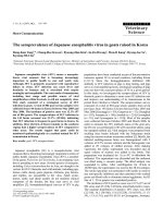
Since 1975, the Korea National Institute of Health has
annually checked JEV activity from vector mosquitoes
collected between July and September in nine provinces
of South Korea (Figure 1). Black-light traps were operated
* Correspondence:
1
WHO Japanese Encephalitis Regional Reference Laboratory for the Western
Pacific Region/Division of Arboviruses, National Institute of Health, Korea
Centers for Disease Control and Prevention, Seoul, Republic of Korea
Full list of author information is available at the end of the article
Yun et al. Virology Journal 2010, 7:127
/>Page 2 of 7
once a week in cattle sheds and the mosquitoes were
identified morphologically and categorized to the species
level. Only Culex tritaeniorhynchus mosquitoes (the
major JEV vector in Korea) were processed for virus iso-
lation, using suckling mice as described previously [18].
Seventeen strains from among the JEV strains isolated in
South Korea in 1983-2005 were initially characterized in
the present study (Table 1). Viral RNA was extracted
from the stocks of each virus using the QIAamp Viral
RNA Mini Kit (Qiagen, Valencia, CA, USA). The purified
RNA was used as the template for cDNA synthesis using
the SuperScript™ III first-strand synthesis system (Invit-
rogen, Carlsbad, CA, USA) with primer JE-2623AS (NS1
region, 5'-GCTTTGTGGACGATCTTCGC-3'), accord-
ing to the manufacturer's instructions. The synthesized
cDNA was then used for PCR amplification with
AccuPrime™ Pfx DNA polymerase (Invitrogen) and prim-
ers JE-723 S (prM/M region, 5'-CGGACCAGGCATTC-
CAA-3') and JE-2623AS. The primers were designed
according to the consensus sequences of three Korean
JEV strains (K94P05, K87P39, and KV1899). The ampli-
fied products (1.9 kb) were purified and sequenced using
the ABI PRISM BigDye Terminator Cycle Sequencing Kit
and an ABI 3730 × l sequencer (Applied Biosystems, Fos-
ter City, CA, USA) at Macrogen (Seoul, Korea). The
nucleotide sequences of the E genes (1,500 nt) were com-
pared with those of other JEV strains representing each
genotype and different geographic regions. A total of 86 E
gene sequences were initially collected and 29 strains rep-
resenting each country and genotype were finally selected
(Table 2). A multiple alignment was generated with the
ClustalX 2.0.11 program [20] and the percentage similari-
ties between the aligned sequences were calculated using
the MegAlign program implemented in the Lasergene
software (DNASTAR, Madison, WI, USA). Phylogenetic
analyses based on the E gene were performed with the
neighbor-joining (NJ) and maximum likelihood (ML)
methods using MEGA 4.0 [21] and TREE-PUZZLE 5.2
[22], respectively. The E gene sequence of the Murray
Valley encephalitis virus (MVEV) was used as the out-
group (GenBank accession no. NC_000943
). For the NJ
tree, the Tamura-Nei model was used to compute the
genetic distances, and the reliability of the tree was tested
by bootstrap analysis with 1,000 replications. For the ML
Table 1: Details of 22 strains of JEV from South Korea*
Strain Year Source Location Accession no.
K82P01 1982 Culex tritaeniorhynchus Youngkwang U34926
K83P34 1983 Culex tritaeniorhynchus IU FJ938231
K83P44 1983 Culex tritaeniorhynchus IU FJ938232
K84A071 1984 Culex tritaeniorhynchus IU FJ938224
K87A07 1987 Culex tritaeniorhynchus IU FJ938225
K87A071 1987 Culex tritaeniorhynchus IU FJ938226
K87P39 1987 Culex tritaeniorhynchus Wando U34927
K88A07 1988 Culex tritaeniorhynchus IU FJ938227
K88A071 1988 Culex tritaeniorhynchus IU FJ938228
K89A07 1989 Culex tritaeniorhynchus IU FJ938229
K91P55 1991 Culex tritaeniorhynchus Wando U34928
K93A07 1993 Culex tritaeniorhynchus IU FJ938230
K94A07 1994 Culex tritaeniorhynchus IU FJ938216
K94A071 1994 Culex tritaeniorhynchus IU FJ938217
K94P05 1994 Culex tritaeniorhynchus Wando U34929
K95A07 1995 Culex tritaeniorhynchus IU FJ938218
K96A07 1996 Culex tritaeniorhynchus IU FJ938219
KV1899 1999 Pig serum Gyeonggi AY316157
K01-GN 2001 Culex tritaeniorhynchus Gyeong-Nam FJ938220
K01-JB 2001 Culex tritaeniorhynchus Jeon-Buk FJ938221
K01-JN 2001 Culex tritaeniorhynchus Jeon-Nam FJ938222
K05-GS 2005 Culex tritaeniorhynchus Gunsan FJ938223
* Five isolates sequenced previously are indicated in boldface type.
IU: information unavailable.
Yun et al. Virology Journal 2010, 7:127
/>Page 3 of 7
tree, the HKY85 evolutionary model of nucleotide substi-
tution was used to build the tree based on the complete E
gene. The statistical significance of each internal branch
of the tree was indicated as a quartet puzzling (QP) value.
Other parameters for the ML tree are available upon
request. All the trees were produced with the MEGA 4.0
software.
Twenty-two Korean JEV strains showed minimal
sequence similarities (uncorrected p-distances) of 87.3%
and 96.2% at the nucleotide and amino acid sequence lev-
els, respectively (Figure 2). Except for K82P01 and
K91P55 strains, Korean JEV strains were divided into two
groups, genotypes 1 and 3. The nucleotide sequence
divergence within the genotypes were only 0.3-1.7%
(mean 1.1%, genotype 1) and 0.1-2.7% (mean 1.5%, geno-
type 3), respectively. The sequence divergence between
the two genotypes was 11.5%-12.7% (mean 12.0%).
K82P01 showed nucleotide divergences of 9.5%-9.8%
from the genotype 1 strains and 3.8%-4.5% from the gen-
otype 3 strains. K91P55 showed nucleotide divergences of
5.2%-5.9% from genotype 1 and 7.6-8.2% from genotype
3.
The E gene sequences of the Korean JEV strains showed
remarkable genetic stability. Five pairs of E gene
sequences from 10 Korean strains (K83P34 and K88A07,
K84A071 and K87A071, K93A07 and K96A07, K94A07
and K95A07, and K01JN and K01-JB) were identical,
despite differences in their geographic distributions and
the maximum five-year time span. This genetic stability
in JEV was also detected in strains from Taiwan, China,
Table 2: Details of 29 JEV strains compared with Korean strains
Strain Year Location Source Genotype Accession no.
Fu 1995 Australia Human serum 2 AF217620
P3 1949 China Human brain 3 AY243844
YN 1954 China Human brain 3 AY243838
SA 14 1954 China Mosquito 3 U14163
YN79-Bao83 1979 China Mosquito 1 DQ404128
YN86-B8639 1986 China Mosquito 1 DQ404133
SH-53 2001 China Mosquito 1 AY555757
SH03-124 2003 China Mosquito 1 DQ404100
SH04-3 2004 China Mosquito 3 DQ404105
SH17M-07 2007 China Mosquito 1 EU429297
GP78 1978 India Human brain 3 AF075723
JKT5441 1981 Indonesia Mosquito 2 U70406
JKT7003 1981 Indonesia Mosquito 4 U70408
JKT9092 1981 Indonesia Mosquito 4 U70409
Nakayama 1935 Japan Human brain 3 U70413
JaOH0566 1966 Japan Human brain 3 AY029207
JaOArS1186 1986 Japan Mosquito 3 AB028262
JaOArK5990 1990 Japan Unknown 3 AB028268
Ishikawa 1994 Japan Pig 1 AB051292
JaNAr0990 1990 Japan Mosquito 3 AY427797
JaNAr32-04 2004 Japan Mosquito 1 FJ185151
PhAn1242 1984 Philippines Pig serum 3 U70417
B1065 1983 Thailand Pig blood 2 U70388
ThCMAr6793 1993 Thailand Mosquito 1 D45363
H49778 1987 Sri Lanka Human brain 3 U70395
VN207 1986 Vietnam Human brain 3 AY376461
VN50 1989 Vietnam Human brain 3 AY376463
VN78 2002 Vietnam Mosquito 1 AY376467
Muar 1952 Singapore Human brain 5 [30]
Yun et al. Virology Journal 2010, 7:127
/>Page 4 of 7
and Japan [6,13,23]. When phylogenetic analyses were
performed, the branching patterns on both the NJ and
ML trees were similar, with slight differences in the reli-
ability indices. Thus, only ML tree is presented in this
study (Figure 3). Most Korean strains were divided into
genotypes 1 and 3. Ten Korean strains grouped in geno-
type 3, together with those isolated in China, Japan, India,
Philippines, Sri Lanka, and Vietnam between the 1930 s
and the early 1990 s. Another 10 Korean strains clustered
in genotype 1, together with strains isolated in China,
Japan, Thailand, and Vietnam between the late 1970 s and
the present day. Historically, the genotypic classification
of some JEV strains was discordant, depending on the
phylogenetic markers or tree construction method used,
including substitution models.
Two Korean JEV strains, K82P01 and K91P55, were
notably problematic in phylogenetic analyses. K82P01
was grouped in genotype 3 or unclassified based on the E
gene sequence, and K91P55 was classified in either geno-
type 1 or genotype 3 depending on the gene region used
for the phylogenetic analysis [7,10,16]. Moreover, in an
analysis of Flavivirus recombination, K82P01 and
K91P55 appeared to be putative recombinant strains
derived from genotype 1 and 3 strains [24]. When we
analyzed the two strains using the RDP3 program [25],
both were shown to be recombinant strains (data not
shown). Chuang and Chen recently provided experimen-
Figure 1 Locations of JEV vector surveillance in South Korea. Mos-
quitoes were caught in nine provinces, excluding Seoul, once a week
between July and September. The mosquito collection sites are indi-
cated as closed circles. Youngkwang and Wando are located in Jeon-
Nam Province. Gunsan is located in Jeon-Buk Province.
Figure 2 Nucleotide and amino acid sequence similarities among Korean JEV strains. The percentage similarities between the aligned nucle-
otide and deduced amino acid sequences were calculated (uncorrected p-distances) with the MegAlign program implemented in the Lasergene soft-
ware. The nucleotide similarities (%) are shown above the diagonal and the deduced amino acid identities (%) are shown below the diagonal.
Yun et al. Virology Journal 2010, 7:127
/>Page 5 of 7
Figure 3 Maximum likelihood tree of 51 JEV strains representing four different genotypes, including 22 Korean strains. The HKY85 evolu-
tionary model of nucleotide substitution was used to construct a ML tree for the complete E gene sequence. The tree was rooted with the E gene
sequence of the Murray Valley encephalitis virus (MVEV, accession no. NC_000943). Branch reliability is indicated with quartet puzzling (QP) values.
Branches showing QP reliability > 70% can be considered well supported [22]. The scale bar indicates the number of base substitutions per site. Korean
strains are indicated as closed circles and the JEV genotypes are as defined previously [8].
Yun et al. Virology Journal 2010, 7:127
/>Page 6 of 7
tal evidence that RNA recombination occurs in JEV [26].
This genotypic conflict may be confirmed by sequencing
the E gene again or, more effectively, the full genome.
However, in this study, we could not pursue this
research because the two strains were lost during long-
term storage. Therefore, we suggest that these two strains
are not used in future studies of JEV evolution. Our
results indicate that the genotypes of the Korean JEV
strains changed from genotype 3 to genotype 1 around
1993, with both genotypes isolated in 1994 (Figure 3).
Since then, only genotype 1 strains have been isolated in
South Korea. Before the present study, it was reported
that genotype 1 was introduced into Korea around 1991
(K91P55 strain) or 1994 (K94P05 strain) [14,15,27].
Interestingly, this genotype change was also reported in
Japan in 1991 [12,23], in China in 1979 [13], in Vietnam in
2001 [9], and in Thailand in 1991 [14]. Although several
explanations have been offered [5,7,9], we believe that
migrating water birds may be a major mediator of the
new genotypes in these regions. Consistent with this sug-
gestion, the cattle egret, black-crowned night heron, and
little egret (the major JEV reservoir) are migratory spe-
cies in at least the countries of Japan, Korea, and China
[28,29].
In summary, this study reports that at least two distinct
genotypes of JEV have circulated in South Korea. Geno-
type 3 strains were predominant in Korea before 1993,
when genotype 1 strain K93A07 was first isolated. The
two genotypes were detected simultaneously in 1994 but
since then, only genotype 1 has been isolated in South
Korea.
Competing interests
The authors declare that they have no competing interests.
Authors' contributions
SMY performed the experiments and contributed to the preparation of the
manuscript. JEC, SYK, JR, and WYC collected the specimens and contributed to
the data analysis. YRJ and MGH contributed to the data analysis and the prepa-
ration of the manuscript. YEJ designed the study, performed the experiments,
and prepared the manuscript. All authors have read and approved the final
manuscript.
Acknowledgements
The authors thank all the researchers who have been engaged in the JE Epi-
demic Forecast Program since 1975 for their devotion to duty. Thanks are also
due to Gi-Chang Bing at the Migratory Birds Center, National Park Research
Institute, for providing information about Korean water birds. This research was
undertaken with a grant from the National Institute of Health, Korea Centers for
Disease Control and Prevention.
Author Details
WHO Japanese Encephalitis Regional Reference Laboratory for the Western
Pacific Region/Division of Arboviruses, National Institute of Health, Korea
Centers for Disease Control and Prevention, Seoul, Republic of Korea
References
1. Vaughn DW, Hoke CH Jr: The epidemiology of Japanese encephalitis:
prospects for prevention. Epidemiol Rev 1992, 14:197-221.
2. Chambers TJ, Hahn CS, Galler R, Rice CM: Flavivirus genome
organization, expression, and replication. Annu Rev Microbiol 1990,
44:649-688.
3. Holland J, Spindler K, Horodyski F, Grabau E, Nichol S, VandePol S: Rapid
evolution of RNA genomes. Science 1982, 215:1577-1585.
4. Chen WR, Tesh RB, Rico-Hesse R: Genetic variation of Japanese
encephalitis virus in nature. J Gen Virol 1990, 71(Pt 12):2915-2922.
5. Chen WR, Rico-Hesse R, Tesh RB: A new genotype of Japanese
encephalitis virus from Indonesia. Am J Trop Med Hyg 1992, 47:61-69.
6. Jan LR, Yueh YY, Wu YC, Horng CB, Wang GR: Genetic variation of
Japanese encephalitis virus in Taiwan. Am J Trop Med Hyg 2000,
62:446-452.
7. Williams DT, Wang LF, Daniels PW, Mackenzie JS: Molecular
characterization of the first Australian isolate of Japanese encephalitis
virus, the FU strain. J Gen Virol 2000, 81:2471-2480.
8. Solomon T, Ni H, Beasley DW, Ekkelenkamp M, Cardosa MJ, Barrett AD:
Origin and evolution of Japanese encephalitis virus in southeast Asia. J
Virol 2003, 77:3091-3098.
9. Nga PT, del Carmen Parquet M, Cuong VD, Ma SP, Hasebe F, Inoue S,
Makino Y, Takagi M, Nam VS, Morita K: Shift in Japanese encephalitis
virus (JEV) genotype circulating in northern Vietnam: implications for
frequent introductions of JEV from Southeast Asia to East Asia. J Gen
Virol 2004, 85:1625-1631.
10. Uchil PD, Satchidanandam V: Phylogenetic analysis of Japanese
encephalitis virus: envelope gene based analysis reveals a fifth
genotype, geographic clustering, and multiple introductions of the
virus into the Indian subcontinent. Am J Trop Med Hyg 2001, 65:242-251.
11. Pyke AT, Williams DT, Nisbet DJ, van den Hurk AF, Taylor CT, Johansen CA,
Macdonald J, Hall RA, Simmons RJ, Mason RJ, Lee JM, Ritchie SA, Smith GA,
Mackenzie JS: The appearance of a second genotype of Japanese
encephalitis virus in the Australasian region. Am J Trop Med Hyg 2001,
65:747-753.
12. Ma SP, Yoshida Y, Makino Y, Tadano M, Ono T, Ogawa M: Short report: a
major genotype of Japanese encephalitis virus currently circulating in
Japan. Am J Trop Med Hyg 2003, 69:151-154.
13. Wang HY, Takasaki T, Fu SH, Sun XH, Zhang HL, Wang ZX, Hao ZY, Zhang
JK, Tang Q, Kotaki A, Tajima S, Liang XF, Yang WZ, Kurane I, Liang GD:
Molecular epidemiological analysis of Japanese encephalitis virus in
China. J Gen Virol 2007, 88:885-894.
14. Nitatpattana N, Dubot-Peres A, Gouilh MA, Souris M, Barbazan P, Yoksan S,
de Lamballerie X, Gonzalez JP: Change in Japanese encephalitis virus
distribution, Thailand. Emerg Infect Dis 2008, 14:1762-1765.
15. Nam JH, Chae SL, Won SY, Kim EJ, Yoon KS, Kim BI, Jeong YS, Cho HW:
Short report: genetic heterogeneity of Japanese encephalitis virus
assessed via analysis of the full-length genome sequence of a Korean
isolate. Am J Trop Med Hyg 2001, 65:388-392.
16. Yun SI, Kim SY, Choi WY, Nam JH, Ju YR, Park KY, Cho HW, Lee YM:
Molecular characterization of the full-length genome of the Japanese
encephalitis viral strain K87P39. Virus Res 2003, 96:129-140.
17. Yang DK, Kim BH, Kweon CH, Kwon JH, Lim SI, Han HR: Biophysical
characterization of Japanese encephalitis virus (KV1899) isolated from
pigs in Korea. J Vet Sci 2004, 5:125-130.
18. Chung YJ, Nam JH, Ban SJ, Cho HW: Antigenic and genetic analysis of
Japanese encephalitis viruses isolated from Korea. Am J Trop Med Hyg
1996, 55:91-97.
19. Nam JH, Chung YJ, Ban SJ, Kim EJ, Park YK, Cho HW: Envelope gene
sequence variation among Japanese encephalitis viruses isolated in
Korea. Acta Virol 1996, 40:303-309.
20. Thompson JD, Gibson TJ, Plewniak F, Jeanmougin F, Higgins DG: The
CLUSTAL_X windows interface: flexible strategies for multiple
sequence alignment aided by quality analysis tools. Nucleic Acids Res
1997, 25:4876-4882.
21. Tamura K, Dudley J, Nei M, Kumar S: MEGA4: Molecular Evolutionary
Genetics Analysis (MEGA) software version 4.0. Mol Biol Evol 2007,
24:1596-1599.
22. Schmidt HA, Strimmer K, Vingron M, von Haeseler A: TREE-PUZZLE:
maximum likelihood phylogenetic analysis using quartets and parallel
computing. Bioinformatics 2002, 18:502-504.
Received: 26 April 2010 Accepted: 14 June 2010
Published: 14 June 2010
This article is available from: 2010 Y un et al; lice nsee BioMed Central Ltd . This is an Open Access article distributed under the terms of the Creative Commons Attribution License ( ), which permits unrestricted use, distribution, and reproduction in any medium, provided the original work is properly cited.Virology Journal 2010, 7:127
Yun et al. Virology Journal 2010, 7:127
/>Page 7 of 7
23. Nerome R, Tajima S, Takasaki T, Yoshida T, Kotaki A, Lim CK, Ito M,
Sugiyama A, Yamauchi A, Yano T, Kameyama T, Morishita I, Kuwayama M,
Ogawa T, Sahara K, Ikegaya A: Molecular epidemiological analyses of
Japanese encephalitis virus isolates from swine in Japan from 2002 to
2004. J Gen Virol 2007, 88:2762-2768.
24. Twiddy SS, Holmes EC: The extent of homologous recombination in
members of the genus Flavivirus. J Gen Virol 2003, 84:429-440.
25. Martin D, Rybicki E: RDP: detection of recombination amongst aligned
sequences. Bioinformatics 2000, 16:562-563.
26. Chuang CK, Chen WJ: Experimental evidence that RNA recombination
occurs in the Japanese encephalitis virus. Virology 2009, 394:286-297.
27. Nitatpattana N, Apiwathnasorn C, Barbazan P, Leemingsawat S, Yoksan S,
Gonzalez JP: First isolation of Japanese encephalitis from Culex
quinquefasciatus in Thailand. Southeast Asian J Trop Med Public Health
2005, 36:875-878.
28. Yamashina Institute for Ornithology: Atlas of Japanese migratory birds
from 1961 to 1995. Japan, Bird Migration Research Center 2002.
29. Park JG, Seo JH: Guide for Korean wild birds (water birds). Korea,
Shingubook 2008.
30. Hasegawa H, Yoshida M, Fujita S, Kobayashi Y: Comparison of structural
proteins among antigenically different Japanese encephalitis virus
strains. Vaccine 1994, 12:841-844.
doi: 10.1186/1743-422X-7-127
Cite this article as: Yun et al., Molecular epidemiology of Japanese encepha-
litis virus circulating in South Korea, 1983-2005 Virology Journal 2010, 7:127