Báo cáo Y học: Molecular interaction of neutral trehalase with other enzymes of trehalose metabolism in the fission yeast Schizosaccharomyces pombe pdf
Bạn đang xem bản rút gọn của tài liệu. Xem và tải ngay bản đầy đủ của tài liệu tại đây (205.83 KB, 9 trang )
Molecular interaction of neutral trehalase with other
enzymes of trehalose metabolism in the fission yeast
Schizosaccharomyces pombe
Teresa Soto, Alejandro Franco, S. Padmanabhan, Jero Vicente-Soler, Jose Cansado and Mariano Gacto
Department of Genetics and Microbiology, Facultad de Biologı
´
a, University of Murcia, Spain
Trehalose metabolism is an essential component of the stress
response in yeast cells. In this work we show that the prod-
ucts of the principal genes involved in trehalose metabolism
in Schizosaccharomyces pombe, tps1
+
(coding for trehalose-
6-P synthase, Tps1p), ntp1
+
(encoding neutral trehalase,
Ntp1p) and tpp1
+
(that codes for trehalose-6-P phospha-
tase, Tpp1p), interact in vitro with each other and with
themselves to form protein complexes. Disruption of the
gene tps1
+
blocks the activation of the neutral trehalase
induced by heat shock but not by osmotic stress. We propose
that this association may reflect the Tps1p-dependent
requirement for thermal activation of trehalase. Data
reported here indicate that following a heat shock the
enzyme activity of trehalase is associated with Ntp1p dimers
or trimers but not with either Ntp1p monomers or with
complexes involving Tps1p. These results raise the possibility
that heat shock and osmotic stress activate trehalase differ-
entially by acting in the first case through an specific mech-
anism involving Tps1p–Ntp1p complexes. This study
provides the first evidence for the participation of the cata-
bolic enzyme trehalase in the structural framework of a
regulatory macromolecular complex containing trehalose-
6-P synthase in the fission yeast.
Keywords: neutral trehalase; stress; protein interaction.
Synthesis and degradation of the nonreducing disaccharide
trehalose is carried out by several enzymes that are widely
distributed and conserved among prokaryotes and eukary-
otes. One probable reason for this conservation is the ability
of trehalose to function as a general stress protectant in
living organisms. Recent studies have focused on the role of
trehalose in stabilizing cellular structures under conditions
like desiccation, osmotic or oxidative stresses, and mild heat
shock [1,2]. Studies in vitro have confirmed the exceptional
properties of trehalose in protecting biological membranes
or enzymes subjected to different types of extreme condi-
tions [3,4]. All these findings suggest that the trehalose
turnover must occur in a coordinated way for this sugar to
play its diverse functional roles during the life cycle.
Consequently, the study of the enzymes involved in
trehalose metabolism and the subtle regulation of their
activities at the molecular and cellular level have received a
great deal of attention in the last decade. The best known
picture for trehalose synthesis and mobilization in simple
eukaryotes has emerged from studies in the budding yeast
Saccharomyces cerevisiae, where trehalose synthesis is
basically a two-step process: trehalose 6-phosphate synthesis
by trehalose-6-P synthase (TPS1) from UDP-glucose and
glucose 6-P as substrates, and dephosphorylation of treha-
lose-6-P to trehalose by trehalose-6-P phosphatase (TPS2).
Studies based on two-hybrid analyses and on Western blot
analyses of complexes obtained by gel filtration fractiona-
tion concluded that TPS1 and TPS2 together with proteins
TSL1andTPS3(whichactasregulatorsofbothsynthase
and phosphatase activities) form part of a multimeric
protein complex of approximate molecular mass 800 kDa
called the trehalose synthase complex [5–7]. In this complex,
TPS1, TPS2, and TPS3 subunits interact with each other
and among themselves (as dimers or higher order oligo-
mers), whereas TSL1 interacts only with TPS1 and TPS2 [6].
Another component of trehalose metabolism is the hydro-
lysis of trehalose to glucose. This is catalyzed by the enzyme
neutral trehalase (NTH1), whose activity is regulated
by phosphorylation of the enzyme protein at serine residues
[8,9].
Comparatively much less is known about the regulation
of the trehalose synthesis in the evolutionarily distant yeast
Schizosaccharomyces pombe. In this fission yeast, the tps1
+
gene codes for trehalose-6-P synthase (Tps1p), that synthe-
sizes trehalose-6-P as occurs in S. cerevisiae [10]. However,
in contrast to the behaviour observed in S. cerevisiae, Dtps1
strains of S. pombe are able to grow on glucose or other
readily fermentable carbon sources, although disruption of
this gene does prevent spore germination [10]. Recently, we
characterized a second gene of the trehalose biosynthetic
pathway in S. pombe,namedtpp1
+
, which codes for
trehalose-6-P phosphatase (Tpp1p) and is responsible for
the synthesis of trehalose from trehalose-6-P [11]. In
S. pombe, trehalose degradation is due to the action of a
84-kDa neutral trehalase protein (Ntp1p), encoded by
ntp1
+
gene [12,13] activated by phosphorylation on
Correspondence to J. Cansado, Department of Genetics and
Microbiology, Facultad de Biologı
´
a, University of Murcia,
30071 Murcia, Spain. Fax: + 34 68 363963, Tel.: + 34 68 364953,
E-mail:
Abbreviations: EMM2, Edinburgh minimal medium; GST, glutathi-
one S-transferase; Ha6H, hemagglutinin antigen epitope and six
histidines; Ntp1p, neutral trehalase protein; Tps1p, trehalose-6-P
synthase protein; Tpp1p, trehalose-6-P phosphatase protein; TTC,
triphenyltetrazolium chloride
(Received 15 March 2002, revised 6 May 2002, accepted 27 June 2002)
Eur. J. Biochem. 269, 3847–3855 (2002) Ó FEBS 2002 doi:10.1046/j.1432-1033.2002.03082.x
supplementing cultures with glucose and a nitrogen source.
Activation of this enzyme protein appears to be mediated by
protein kinases Pka1p and Sck1p [14,15]. In addition,
mRNA levels of ntp1
+
rise when S. pombe cultures are
subjected to thermal, osmotic or oxidative stresses [12,16].
The increased expression of ntp1
+
is accompanied by a
rise in enzyme activity, which is also regulated by the
Pka1p/Sck1p pathway during osmotic and oxidative
stress [16,17]. In a previous work, we showed that mutants
of S. pombe disrupted in trehalose-6-P synthase function
were unable to increase neutral trehalase activity under heat
shock conditions or after the addition of glucose or
nitrogen source. However, these Dtps1 strains still respond
to osmotic stress by increasing trehalase levels [18]. Thus, in
S. pombe, trehalose-6-P synthase appears to be involved in
the regulation of the thermal- and nutrient-induced activa-
tion of neutral trehalase but not during activation on
osmotic stress. These observations raised the question of
whether trehalose-6-P synthase and neutral trehalase would
interact in vivo. In this paper we demonstrate that both
Tps1p and Ntp1p interact in vivo and are part of distinct
trehalose-6-P synthase/trehalase complexes. Moreover,
trehalose-6-P phosphatase (Tpp1p) is likely a member of
these complexes suggesting that regulation of trehalose
synthesis and breakdown may be integrated mechanisms.
These findings indicate a divergence in the molecular designs
controlling trehalose metabolism in S. pombe from those in
S. cerevisiae.
MATERIALS AND METHODS
Strains and culture media
The S. pombe strains employed in this study are listed in
Table 1. They were routinely grown with shaking at 28 °C
in YES [19] or EMM2 with or without thiamin (5 mgÆL
)1
)
[12]. Culture media were supplemented with adenine,
leucine, histidine or uracil (100 mgÆL
)1
, all obtained from
Sigma Chemical Co.) depending on the requirements for
each particular strain. For sporulation of diploids, MEL
medium was employed [19]. Solid media were made by the
addition of 2% (w/v) bacto-agar (Difco Laboratories).
Transformation of S. pombe strains was performed by the
lithium acetate method as described elsewhere [19]. Escher-
ichia coli DH5aF¢ was employed as a host to propagate
plasmids. It was grown at 37 °C in Luria–Bertani medium
plus 50 lgÆmL
)1
ampicillin.
Construction of Ntp1p-, Tps1p-, and Tpp1p-tagged
strains
A5¢ truncated version of ntp1
+
ORF was amplified by PCR
employing the 5¢ oligonucleotide NTP-1 (CCG
CTCGAG
TCGAATATCTGCCGGAAG, which hybridizes at posi-
tions 701–718 in the ntp1
+
ORF and contains an internal
XhoIsite),andthe3¢ oligonucleotide TAG-3 (CTAC
G
GCGGCCGCCATTTTTATGAATGGAAA, which
hybridizes at the 3¢ end of ntp1
+
ORF and incorporates a
NotI site placed immediately upstream of the TAA stop
codon). The restriction sites in both oligonucleotides are
underlined. PCR amplification employing the Expand high-
fidelity system (Roche Molecular Biochemicals) generated a
1.6 kb fragment that was cleaved with XhoIandNotIand
cloned into plasmids pIH-ura and pIH-LEU. These are
integrative plasmids derived from plasmid pDS472a [20],
without nmt promoter and ars1 sequences, and with ura4
+
or LEU2 as selectable markers, which allow the construc-
tion of vectors with Ha6H tag fusions at the C-terminus.
The resulting plasmids were digested at the unique SfiIsite
within the ntp1
+
coding region (at position 1665) and the
linear fragments transformed into haploid strains MM1 and
MM2, to target integration at the ntp1
+
locus. Uracil or
leucine prototrophs were selected for strains MM1 and
MM2, respectively. The identification of strains C3 and
C69, with one copy of Ntp1p–Ha6H expressed from the
genomic ntp1
+
promoter, was verified by Southern blot
analysis and immunoblot of whole-cell extracts with anti-
Ha Ig (see below).
To obtain Tps1p-tagged strains, a 5¢ truncated version of
tps1
+
ORF was amplified by PCR employing the 5¢
oligonucleotide TPSINT-1 (CCG
CTCGAGCCTAACGG
TGTGGAATAC, which hybridizes at positions 715–732 in
the tps1
+
ORF and shows an internal XhoIsite),andthe3¢
oligonucleotide TAF-3 (CTACG
GCGGCCGCCCGAGC
TAGAATTCATCGA, which hybridizes at the 3¢ end of
tps1
+
ORF and incorporates a NotI site placed immediately
upstream of the TAA stop codon). The 0.7 kb amplified
PCR fragment was cleaved with XhoIandNotI and cloned
into integrative plasmids pIH-ura and pIG-ura (fusion to a
GST tag at C-terminus). The resulting plasmids were
digested at the unique NcoI site within tps1
+
(at position
1297) and the linear fragments transformed into haploid
strain MM1, to target integration at the tps1
+
locus. Uracil
prototrophs were selected and the identification of strains
C4 and C5, with one copy of Tps1p–GST or Tps1p–Ha6H
Table 1. S. pombe strains used in this study.
Strain Genotype Source/Reference
MM1 h
+
ade6-M216 leu 1-32 ura4-D18 M. Yamamoto
MM2 h
–
ade6-M210 leu 1-32 ura4-D18 A. Dura
´
n
C3 h
+
ade6-M216 leu 1-32 ura4-D18 ntp1
+
:Ha6H (ura4
+
) This study
C69 h
–
ade6-M210 leu 1-32 ura4-D18 ntp1
+
:Ha6H (LEU2) This study
C4 h
)
ade6-M216 leu 1-32 ura4-D18 tps1
+
:GST (ura4
+
) This study
C5 h
)
ade6-M216 leu 1-32 ura4-D18 tps1
+
:Ha6H (ura4
+
) This study
C694 h
+
ade6-M216 leu 1-32 ura4-D18 ntp1
+
:Ha6H (LEU2) tps1
+
:GST (ura4
+
) This study
MMPI-3a h
+
ade6-M216 leu 1-32 ura4-D18 tpp1
+
:Ha6H (ura4
+
) [11]
MMPI-3b h
–
ade6-M210 leu 1-32 ura4-D18 tpp1
+
:Ha6H (ura4
+
) [11]
C33 h
–
ade6-M210 leu 1-32 ura4-D18 tpp1
+
:Ha6H (ura4
+
) ntp1
+
:Ha6H (ura4
+
) tps1
+
:Ha6H (ura4
+
) This study
3848 T. Soto et al. (Eur. J. Biochem. 269) Ó FEBS 2002
expressed from the genomic tps1
+
promoter, was verified
by Southern blot analysis and immunoblot of whole-cell
extracts with anti-GST or anti-Ha Ig, respectively.
The double-tagged strain C694 (Ntp1p–Ha6H, Tps1p–
GST) was constructed by mating strains C69 and C4, and
selecting diploids in EMM2 medium without supplements.
Sporulation was performed in MEL medium and the spores
purified by glusulase treatment [21] were allowed to
germinate in minimal medium plus adenine. Strains with
the double-tagged genotype were identified by Southern and
immunoblot analysis with anti-HA and anti-GST Ig.
Double-tagged strain C33 (Ntp1p–Ha6H, Tpp1p–Ha6H)
was constructed by mating strains C3 and MMPI-3b, and
selecting diploids in EMM2 medium with leucine. After
spore purification and germination, the double-tagged
strains were identified by Southern and immunoblot
analysis with anti-Ha Ig. The triple-tagged strain C335
(Ntp1p–Ha6H, Tps1p–Ha6H, Tpp1p–Ha6H) was obtained
after mating strains C33 and C5, and Southern and
immunoblot analysis of germinated spores, as described
above.
Expression of Tps1p–GST and Ntp1p–GST fusions
The tps1
+
ORF was amplified by PCR with oligonucleotides
TPS-5 (CCG
CTCGAGGAATCTTTGTTTTGCTGA,
which hybridizes at sequences upstream of the ATG start
codon in the tps1
+
ORF and shows an internal XhoIsite),
and TAF-3. The 1.5 kb product was cloned into the XhoI/
NotI sites of plasmid pDS472M, which is a derivative of
plasmid pDS472a with an attenuated version of the nmt1
promoter and LEU2 selectable marker, to create plasmid
pTGST, which expresses trehalose-6-P synthase (Tps1)
fused to GST at the C-terminus under the control of the
medium strength thiamin-repressible promoter. pTGST
and control plasmid pDS472M (unfused GST) were
transformed into strains C3, C5 and MMPI-3, and leucine
prototrophs selected in EMM2 medium plus adenine.
For Ntp1p–GST expression, ntp1
+
ORF was amplified
by PCR with oligonucleotides NTP-5 (CCG
CTCGAGG
CTATCATTCGTGAATAG, whichhybridizesatsequences
upstream of the ATG start codon in the ntp1
+
ORF and
shows an internal XhoI site), and TAG-3. The 1.5 kb product
was cloned as above into pDS472M to create plasmid
pNGST, containing an in-frame fusion wherethe 3¢ endofthe
ntp1
+
ORF is followed by the GST epitope, and whose
expression is under the control of the medium strength nmt1
thiamin-repressible promoter. Both pNGST and control
plasmid pDS472M were transformed into strains C3, C5 and
MMPI-3, and leucine prototrophs were selected.
Purification of Ha6H- and GST-tagged proteins
by affinity chromatography
Total cell homogenates were prepared under native condi-
tions employing chilled acid-washed glass beads and lysis
buffer (10% glycerol, 50 m
M
Tris/HCl pH 7.5, 150 m
M
NaCl, 0.1% Nonidet NP-40, plus an specific protease
inhibitor cocktail for fungal and yeast extracts obtained
from Sigma Chemical Co.). The lysate was removed and
cleared by centrifugation at 10 000 g for 30 min. Ha6H-
tagged proteins were purified by using Ni
2+
-nitrilotriacetic
acid-agarose beads (Qiagen Inc.) whereas GST and GST-
tagged proteins were precipitated using glutathione glutathi-
one–Sepharose beads (Amersham-Pharmacia) following the
procedures described by Shiozaki & Russell [22].
Immunoprecipitation of Ha6H- and GST-tagged proteins
For immunoprecipitation of Ha6H-tagged proteins, the
extracts were incubated for 12 h at 4 °C with monoclonal
mouse anti-Ha Ig (clone 12CA5, Roche Molecular Bio-
chemicals), and the immunocomplexes were adsorbed with
protein A–agarose (Roche Molecular Biochemicals) for 4 h
at 4 °C. The immunoprecipitation of GST-tagged proteins
was performed with a polyclonal sheep anti-GST Ig
(Amersham-Pharmacia) and protein G–agarose (Roche
Molecular Biochemicals). In all cases the suppliers’ recom-
mendations were followed in terms of incubation times and
washing of the complexes.
Detection of neutral trehalase activity in gel
Affinity purified Ntp1p-Ha and Tps1p–GST proteins were
mixed with loading buffer (100 m
M
Mes pH 6, 15%
glycerol and 0.01% bromophenol blue), and resolved at
4 °C in native 6% polyacrylamide gels (200 V for 8 h),
employing Tris/borate pH 7.5 as running buffer. The gels
were then washed with 100 m
M
Mes pH 6.0 for 10 min, and
incubated with 100 m
M
Mes pH 6 plus 200 m
M
trehalose
(Sigma Chemical Co.) for 2 h at 30 °C. After washing with
distilled water, active neutral trehalase proteins were
detected in situ by incubating the gels with 0.1% TTC in
0.5
M
sodium hydroxide at 80 °C. Color development was
stopped with a 7.5% acetic acid solution.
Gel filtration
A Superdex-200 column (Amersham-Pharmacia) equili-
brated with buffer A (10 m
M
Mes, pH 6.0, 150 m
M
NaCl)
was used for size-exclusion analysis in an AKTA HPLC
system (Amersham-Pharmacia). Lower salt concentrations
were not used in order to minimize nonspecific electrostatic
interactions with the column matrix. The column was
calibrated using vitamin B
12
(1.3 kDa), cytochrome c
(12.4 kDa), carbonic anhydrase (29 kDa), ovalbumin
(43 kDa), BSA (66 kDa), yeast alcohol dehydrogenase
(150 kDa), b-amylase (200 kDa), apoferritin (430 kDa)
and thyroglobulin (670 kDa) (all from Sigma Chemical
Co.) at the concentrations recommended by the manufac-
turer. One-hundred microliters (1 mg total protein) of the
supernatant from ultracentrifuged extract obtained from
triple-tagged strain C335 grown to mid-log phase, after
either a heat shock (40 °C, 1 h) or an osmotic shock (0.75
M
NaCl, 2 h) were applied to the column with buffer A plus a
yeast protease inhibitor cocktail (Sigma Chemical Co.). A
flow rate of 0.4 mLÆmin
)1
was used and the elution was
tracked by absorbance at 280, 235 and 220 nm. Blue
Dextran (2000 kDa; Sigma) and vitamin B
12
were used for
void volume (V
o
)andtotalbedvolume(V
t
) determinations,
respectively. Molecular masses were estimated from a
calibration curve (correlation coefficients ‡ 0.98) generated
by linear regressions of the elution volume for each protein
using
SIGMAPLOT
(Jandel Scientific). To determine the
fractions containing Ntp1p-, Tps1p-, and Tpp1p–Ha6H
tagged proteins, 100 lL of each fraction (0.4 mL) were
Ó FEBS 2002 Neutral trehalase complexes in fission yeast (Eur. J. Biochem. 269) 3849
trichloroacetic-acid-precipitated, washed with cold acetone,
air-dried, resuspended in SDS gel sample buffer, and
resolved in SDS/PAGE (10%) gels. Immunoreactive bands
were detected by Western blot analysis with anti-Ha Ig (see
below).
SDS/PAGE and Western blotting
Proteins were resolved in 8 or 10% SDS/PAGE gels as
previously described [11], transferred to nitrocellulose filters
(Amersham-Pharmacia), and incubated with mouse anti-
Ha or sheep anti-GST Ig. The immunoreactive bands were
revealed with HRP-conjugated secondary Ig [anti-(mouse
IgG) Ig or anti-(sheep IgG) Ig; Sigma Chemical Co.] and the
ECL system (Amersham-Pharmacia).
Enzyme assays and trehalase activation
Trehalase activity was assayed after cell breakage as
described previously [23]. Activation of trehalase by heat
treatment or osmotic shock was carried out as indicated
earlier [17]. Enzyme activity in eluates was expressed as
nmol glucose produced per min. All trehalase determina-
tions were repeated at least three times with consistent
results. Representative results are shown. Specific activity of
trehalase in slab gels was expressed as enzyme units per mg
protein. Protein determination was performed by absor-
bance measurement at 280 and 205 nm according to the
method described in previously [24].
RESULTS
Neutral trehalase and trehalose-6-
P
synthase
association
in vitro
In order to analyse possible interactions between treha-
lose-6-P synthase and neutral trehalase interaction
in vitro, we constructed strains C3 and C69, which
express a C-terminal Ha6H-tagged version of neutral
trehalase (Ntp1p, 84 kDa) under the control of the
genomicpromoter.Thesestrainsshowedthesamegrowth
behavior and neutral trehalase activation pattern as the
parental strains MM1 and MM2 for a variety of
conditions. Strain C3 was further transformed with
plasmid pTGST, which expresses trehalose-6-P synthase
(55 kDa) fused to a GST epitope (25 kDa) at its
C-terminus under the regulation of an attenuated version
of the thiamin-repressible promoter [20]. Several transfor-
mants were cultured in minimal medium plus thiamin,
and then transferred to the same medium without thiamin
to allow synthesis of the Tps1p–GST fusion protein. The
cells were collected at mid-log phase of growth, and
Tps1p–GST was affinity-purified employing glutathione
(glutathione)–Sepharose beads. As shown in Fig. 1 (lane 2),
significant levels of Tps1p–GST were detected employing
anti-GST Ig under these conditions. However, if Tps1p
interacts with Ntp1p in vitro, Ntp1p–Ha6H should be
detectable with anti-Ha Ig after Tps1p–GST purification.
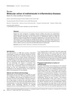
As shown in Fig. 1 (lane 6), this appears to be the case,
and a clear band of the size expected for Ntp1p–Ha6H
fusion was visible after hybridization with anti-Ha Ig.
Besides, the Ntp1p–Ha6H signal was absent when strain
C3 was transformed with a control plasmid that expresses
unfused GST (Fig. 1, lane 5). Taken together, these
results indicate that Ntp1p and Tps1p interact in vitro in
S. pombe, and that the nature of this association is
specific and independent of the presence of the GST
domain fused to Tps1p.
Ntp1p–Tps1p association takes place during
normal yeast growth and thermal shock, but might not
occur when the cells are stressed by an osmotic upshift [18].
To test this possibility, we performed the same experiment
described above by subjecting strain C3 plus plasmid
pTGST either to osmotic stress or to a thermal one. As
shown in Fig. 1, Ntp1p–Tps1p association was observed
not only in control, exponentially growing cells but also for
both thermal as well as osmotic stresses (lanes 7 and 8).
Thus, in S. pombe, Ntp1p–Tps1p interaction does not
appear to be transient but is stable, and is maintained even
under conditions where the presence of Tps1p is not needed
for neutral trehalase activation (osmotic shock). These
results, however, do not exclude by themselves the possibi-
lity that some Ntp1p might be in a free, nonassociated state
(see below).
We employed a medium-strength thiamin-repressible
promoter for Tps1p–GST expression in Ntp1p–Ha6H cells
to achieve low levels of Tps1p–GST synthesis. Although
Ntp1p–Ha6H was not detectable in the absence of the
Tps1p–GST fusion, it was conceivable that the described
Tps1p–Ntp1p interaction could be due, in part, to the
presence of nonphysiological levels of Tps1p–GST. In order
to clarify this point, we constructed the S. pombe double-
tagged strain C694. This strain expresses Ntp1p and Tps1p
fused at their C-terminus to Ha6H and GST epitopes,
respectively, and in both cases the synthesis is regulated by
their own genomic promoters. The Tps1p fusion protein is
active because strain C694 synthesizes trehalose at normal
level. This strain was grown in rich medium to mid-log
phase, and after obtaining the corresponding extracts,
Tps1p–GST and Ntp1p–Ha6H were immunoprecipitated
with anti-GST and anti-Ha Ig, respectively. As shown in
Fig. 2 (lane 2), the Ntp1p–Ha6H band was clearly detected
Fig. 1. Ntp1p–Tps1p association takes place in vivo in growing cells of
S. pombe, and in cells subjected to heat and osmotic stresses. Strain C3,
with a Ha6H epitope-tagged version of Ntp1p, was transformed with
plasmids pDS472a (unfused GST; lanes 1 and 5) or pTGST (Tps1p–
GST fusion; lanes 2, 3, 4, 6, 7 and 8). GST and Tps1p–GST fusions
were expressed using the medium-strength thiamin-regulated promoter
for 24 h. Yeast lysates prepared from exponentially growing cells
(lanes 2 and 6), or from either heat- (lanes 3 and 7) or osmotically
shocked cells (lanes 4 and 8), were adsorbed with glutathione–Sepha-
rose beads. After extensive washing in lysis buffer, the proteins bound
to the beads were analyzed by Western blot using anti-GST (lanes 1–4)
and anti-Ha Ig (lanes 5–8).
3850 T. Soto et al. (Eur. J. Biochem. 269) Ó FEBS 2002
with anti-Ha Ig in the complexes obtained from Tps1p–
GST immunoprecipitation, whereas Tps1p–GST was visi-
ble with anti-GST Ig after Ntp1p–Ha6H immunoprecipi-
tation (lane 5). In this strain, the existence of Ntp1p–Tps1p
interaction was evident not only in growing cells, but also
during heat or osmotic stress (not shown). These data
strongly suggest the existence of an in vivo association
between Ntp1p and Tps1p in S. pombe over a variety of
environmental conditions.
To ascertain the functional significance of this interac-
tion, we developed a gel assay for neutral trehalase
activity (see Materials and methods) and determined the
enzyme activity of Ntp1p bounded to Tps1p in exponen-
tially growing cells from double-tagged strain C694 and in
cultures subjected to heat or osmotic stress. As shown in
Fig. 2B, affinity-purified Ntp1p–Ha6H from growing cells
displayed a typical pattern of neutral trehalase activity,
with low level of enzyme activity in unstressed cells that
increases strongly upon stress. Notably, we were unable to
detect in situ any neutral trehalase activity associated to
native Tps1–GST protein purified with glutathione–
Sepharose beads in samples from either unstressed,
osmotic- or heat-shocked cells. A quantitative estimation
of neutral trehalase activity gave similar results (Fig. 2B,
lower panel). Because a significant fraction of Ntp1p
protein is bounded in vitro toTps1p(seeFigs1and2A),
these results demonstrate that in S. pombe the active form
of neutral trehalase exists in a free, non Tps1p-associated
state, independently of the environmental condition used
for stress.
We focussed then our attention on the likelihood that
Tps1p or Ntp1p undergo self-association. This has been
previously reported for the Tps1p homologue in
S. cerevisiae [6]. Using the same experimental procedure
used in Fig. 1, we observed that self-association does
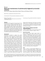
indeed take place for both proteins. As can be seen in
Fig. 3A (lane 4) and Fig. 3B (lane 4), both Ntp1p–
Ha6H and Tps1p–Ha6H were detected employing anti-
Ha Ig after Ntp1p–GST and Tps1p–GST purification,
respectively. These and other results (see below) support
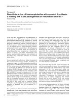
Fig. 2. Ntp1p and Tps1p coimmunoprecipitate and associate to form
complexes devoid of neutral trehalase activity. (A) The double-tagged
strain C694 (Ntp1p–Ha6H, Tps1p–GST) was grown to mid-log phase,
and Tps1p–GST and Ntp1p–Ha6H were immunoprecipitated from
the corresponding extracts with anti-Ha Ig (lanes 1 and 5) or anti-GST
Ig (lanes 2 and 4) Ig. After incubation with protein A–agarose
(anti-Ha immunoprecipitations) or protein G–agarose (anti-GST
immunoprecipitations) and extensive washing, the immunocomplexes
were resolved by SDS/PAGE and analyzed by Western blot using anti-
Ha (lanes 1, 2 and 3) or anti-GST Ig (lanes 3, 5 and 6). Lanes 3 and 6
correspond to negative controls without immunoprecipitation but
incubated with protein A–agarose (lane 3) or protein G–agarose (lane
6) to account for possible nonspecific protein binding to the matrix. (B)
Upper panel: Affinity-purified Ntp1p–Ha6H and Tps1p–GST were
prepared from exponentially growing cells of strain C694 prior (lanes 1
and 4) and after osmotic (lanes 2 and 5) or heat stress (lanes 3 and 6).
Samples were resolved by native PAGE and spots developed for
neutral trehalase activity. Lower panel: quantitative estimation of
enzyme activity (as trehalase units per mg protein) in each sample.
Fig. 3. Ntp1p and Tps1p self-interact in vivo. (A) Ntp1p–Ntp1p interaction. The Ntp1p–Ha6H epitope-tagged strain C3 was transformed with
plasmids pDS472a (unfused GST; lanes 1 and 3) or pNGST (Ntp1p–GST fusion; lanes 2 and 4). GST and Ntp1p–GST fusions were expressed
using the medium-strength thiamin-regulated promoter for 24 h. Yeast lysates were adsorbed with glutathione–Sepharose beads and after washing
in lysis buffer the proteins bound to the beads were analyzed by SDS/PAGE and Western blotting using anti-GST Ig (lanes 1 and 2) and anti-Ha Ig
(lanes 3 and 4). (B) Tps1p–Tps1p interaction. The Tps1p–Ha6H epitope-tagged strain C5 was transformed with plasmids pDS472a (unfused GST;
lanes 1 and 3) or pTGST (Tps1p–GST fusion; lanes 2 and 4). GST and Tps1p–GST fusions were expressed using the medium-strength thiamin-
regulated promoter for 24 h. Yeast lysates were processed as described above, and the proteins bound to glutathione beads were analyzed after
SDS/PAGE using anti-GST Ig (lanes 1 and 2) and anti-Ha Ig (lanes 3 and 4).
Ó FEBS 2002 Neutral trehalase complexes in fission yeast (Eur. J. Biochem. 269) 3851
the existence of a multiprotein complex involved in the
regulation of trehalose synthesis and breakdown in
S. pombe.
Trehalose- 6P phosphatase (Tpp1) is a member of the
Ntp1–Tps1 complex
Recently a third member of the trehalose metabolism
pathway in S. pombe,thetpp1
+
gene, which codes for
trehalose-6-P phosphatase, has been isolated and charac-
terized [11]. The tpp1
+
gene has considerable sequence
homology to S. cerevisiae TPS2, which encodes trehalose-
6-P phosphatase. S. cerevisiae Tps2 has been shown to
interact, among others, with Tps1, and form part of the
trehalose synthase complex in this yeast [6,7]. Based on these
precedents, we examined if Tpp1p interacts with both Tps1p
and Ntp1p in S. pombe. The strain MMPI-3a, that
expresses a Ha6H-tagged version of Tpp1p [11], was
transformed separately with plasmids pTGST and pNGST
(expressing Tps1p and Ntp1p fused to GST, respectively),
and the GST fusions were purified with glutathione–
Sepharose beads. In either case (Fig. 4A,B), the 100 kDa
Tpp1p–HA6H protein coprecipitated with the purified GST
fusion proteins, whereas it was absent in control experi-
ments (with plasmids expressing unfused GST). These
results clearly suggest that Tpp1p may also participate
in vivo to form Ntp1p–Tps1p complexes. However, as for
Tps1p–Ntp1p complexes (see Fig. 2B), trehalase activity
wasabsentinTpp1p–Ntp1passemblieswhenassayedongel
slabs (data not shown).
Analysis of the Ntp1p–Tps1p–Tpp1p complex
by HPLC-gel filtration
The results obtained thus far suggest the existence of
complexes in S. pombe formed by mutual interaction
among Ntp1p, Tps1p, and Tpp1p. To gain additional
information on their physical nature in terms of size and
composition, we constructed the triple-tagged strain C335
by mating double-tagged strain C33 (Ntp1p–Ha6H,
Tpp1p–Ha6H) and Tps1p–Ha6H tagged strain C5, which
affords simultaneous detection of Tps1p, Ntp1p and
Tpp1p by Western blot analysis using anti-Ha Ig. Protein
extracts obtained from exponentially growing cells of strain
C335 were fractionated in native solution conditions by gel
filtration employing a Superdex-200 HPLC column. The
fractions eluted from the column were subjected to SDS/
PAGE, blotted, and subsequently analyzed by Western
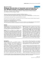
blot with anti-Ha Ig. As shown in Fig. 5A, the elution
profiles for Tps1p, Ntp1p and Tpp1p are quite intricate,
with Tps1p being detected at higher levels than either
Ntp1p or Tpp1p. Moreover, Tps1p was detected over a
wide range of elution volumes extending from that
expected for the monomeric protein (fractions 18–20) to
high molecular mass complexes close to 700–800 kDa
(fractions 4–5), as well as intermediate-sized complexes.
Neutral trehalase (Ntp1p) was also detected in virtually
all the fractions, but was most pronounced in the
700–800 kDa complex fractions and in those correspond-
ing to sizes between 80 kDa (free monomer) and 300 kDa.
Finally, Tpp1p showed an elution profile similar to Ntp1p
and, like Tps1p and Ntp1p, was also found in the 800 kDa
complex. These data indicate that, in addition to the
monomeric forms, at least two distinct complexes exist
containing significant amounts of all three proteins.
One corresponds to the high molecular mass complex
(700–800 kDa) and the other to a more diffusely spread
population corresponding to lower molecular mass range
(80–250 kDa, fractions 12–17). To clarify which of these
protein complexes exhibit neutral trehalase activity, we
prepared log-phase cultures from strain C335 and subjected
them to thermal (40 °C, 1 h) or osmotic (0.75
M
NaCl, 2 h)
stress. The corresponding cell extracts were then fraction-
ated by gel filtration under the conditions described above.
No significant differences in the elution profile of Ntp1p,
Tps1p and Tpp1p were found under these circumstances,
despite an increase in the overall signal (as the three
proteins behave as heat shock proteins). Surprisingly, when
the eluted fractions were assayed for neutral trehalase, no
enzyme activity was found associated with Ntp1p mono-
mers or in fractions corresponding to the high molecular
mass trehalose synthase–trehalase complex (fractions 4–5).
Instead, the heat-activated neutral trehalase was mainly
detected in complexes of about 250 kDa, whereas neutral
Fig. 4. Tpp1p associates with Ntp1p and Tps1p in vivo . (A) Tpp1p–Tps1p interaction. The Tpp1p–Ha6H epitope-tagged strain MMPI-3a was
transformed with plasmids pDS472a (unfused GST; lanes 1 and 3) or pTGST (Tps1p–GST fusion; lanes 2 and 4). GST and Tps1p–GST fusions
were expressed using the medium-strength thiamin-regulated promoter for 24 h. Yeast lysates were then adsorbed with glutathione–Sepharose
beads, and after extensive washing the proteins bound to the beads were analyzed by Western blot using anti-GST Ig (lanes 1 and 2) and anti-Ha Ig
(lanes 3 and 4). (B) Ntp1p–Tpp1p interaction. Strain MMPI-3a was transformed with plasmids pDS472a (unfused GST; lanes 1 and 3) or pNGST
(Ntp1p–GST fusion; lanes 2 and 4). GST and Tps1p–GST fusions were expressed using the medium-strength thiamin-regulated promoter for 24 h,
purified as described above, and analyzed using anti-GST (lanes 1 and 2) and anti-Ha Ig (lanes 3 and 4).
3852 T. Soto et al. (Eur. J. Biochem. 269) Ó FEBS 2002
trehalase activated by osmotic shock peaked mostly in
fractions corresponding to somewhat lower molecular
masses. These enzyme activities fit the expected elution
for trehalase trimers and dimers, respectively.
DISCUSSION
Two main lines of evidence lead us to propose that in the
fission yeast S. pombe neutral trehalase (Ntp1p) interacts
in vitro with other proteins involved in trehalose metabo-
lism. First, the activation of Ntp1p by heat shock or
nutritional stimuli only takes place in the presence of
trehalose-6-P synthase (Tps1p) [18]. Secondly, in contrast
to the neutral trehalase in S. cerevisiae [25], all attempts to
activate S. pombe Ntp1p in vitro have been unsuccessful to
date. Taken together, these results support the idea that in
S. pombe the regulation of Ntp1p activity under stress relies
on the existence of some kind of interaction between Ntp1p
and Tps1p, or other elements, that may be lost under the
conditions of the in vitro assay for activation. In the present
work, we have performed experiments including affinity
chromatography, immunoprecipitation, and gel filtration to
demonstrate that not only Tps1p, but also Tpp1p, interact
with Ntp1p and that macromolecular complexes, involving
trehalose synthase/phosphatase and trehalase, might indeed
exist in this yeast. The demonstration of interactions
between Ntp1p–Tps1p and Ntp1p–Tpp1p (Figs 1, 2 and 4)
is, to our knowledge, the first description of an association
between the hydrolytic enzyme trehalase and proteins
involved in synthesis of trehalose, thus revealing a novel
relationship in trehalose metabolism.
In E. coli, trehalases are monomeric enzymes [26].
However, in S. cerevisiae the functional cytoplasmic neutral
trehalase (NTH1) is probably a dimer, as deduced from gel
filtration experiments with active enzyme [25]. No evidence
has been reported demonstrating that NTH1 participates in
the formation of the well-characterized trehalose synthase
complex in the budding yeast [6,7]. In S. pombe, we find that
part of Ntp1p is apparently present as free monomeric
protein that does not display trehalase activity in vitro
(Fig. 5). The elution profile of trehalase activity in gel
filtration indicated that after heat or osmotic shock only
fractions corresponding to protein sizes between 170 and
300 kDa contain active enzyme. On the other hand, Ntp1p
eluted mainly in two peaks, either as part of a 700–800 kDa
complex, together with Tps1p and Tpp1p, or, as expected,
in the fractions containing trehalase activity, which again
showed the coexistence of Tps1p and Tpp1p (Fig. 5).
Because Ntp1p molecules also associate among themselves
(Fig. 2), these results alone would not allow us to establish
whether trehalase activity correlates with self-assembly of
Ntp1p homomultimers or with the formation of hetero-
meric complexes involving Tps1p and Tpp1p in addition to
Ntp1p. However, the observation that Tps1p–(Tpp1p)–
Ntp1p complexes lack trehalase activity (Fig. 2B) strongly
favors the first interpretation. Active trehalase might thus
arise from the specific self-assembly of a discrete number of
Ntp1p molecules to form small oligomers (probably dimers
or trimers, considering the molecular mass of each putative
subunit, 84 kDa). An intriguing fact is that there is a small
but reproducible shift in the elution behavior of trehalase
depending on the nature of the activation stress (Fig. 5B).
This shift might argue against the participation of a unique
set of interactions involving association of Ntp1p molecules
and could be taken to indicate a specific event occurring
during activation by hyperthermia that is absent in the
response elicited by osmotic stress. However, the elution
pattern of trehalase cannot be exclusively interpreted in
Fig. 5. Analysis of Tps1p–Tpp1p–Ntp1p complexes by gel filtration. (A) A high-speed supernatant of cell-free extracts from exponentially growing,
triple Ha6H-tagged strain C335, was size fractionated by gel filtration through a Superdex-200 column. The proteins from 100 lLofeachfraction
were concentrated by trichloroacetic acid precipitation, separated by SDS/PAGE, transferred to nitrocellulose and incubated with anti-Ha Ig. After
incubation with an HRP-conjugated secondary antibody [anti-(mouse IgG) Ig], the signals specific for Ha-tagged proteins were visualized with the
ECL system. V
0
indicates the void volume. (B) Elution profile of activated neutral trehalase in S. pombe. Exponentially growing cultures of strain
C335 were subjected to either heat shock (open circles) or osmotic stress (closed circles), and the correspondent protein extracts size fractionatedas
described for (A). Neutral trehalase activity (expressed as nmol glucose produced per min) was assayed in 250 lLofeachfraction.
Ó FEBS 2002 Neutral trehalase complexes in fission yeast (Eur. J. Biochem. 269) 3853
terms of changes in the complex composition. Because gel
filtration is responsive to Stokes radius, one should also
consider that the conformation has merely changed upon
activation depending on the triggering stimulus while the
composition is unaltered.
Tps1p is probably the most abundant protein related to
trehalose metabolism present in S. pombe (Fig. 5), although
we can not exclude that the differential relative expression of
Ntp1p, Tps1p and Tpp1p in eluted extracts might be an
experimental artifact arising from differences in the acces-
sibility of the antibody to the Ha tag. However, there is a
parallel in S. cerevisiae, where the Tps1p homologue (TPS1)
is also present at levels higher than those of the other
constituents of the trehalose synthase complex [7]. In
S. cerevisiae, trehalose is synthesized by the 800 kDa
trehalose synthase complex as well as by free TPS1 [7]. It
is unknown whether Tps1p activity in S. pombe is present in
the high molecular mass trehalose synthase–trehalase com-
plex or linked to forms of lower molecular mass as for
neutral trehalase, although accumulation of trehalose by
ntp1-deleted strains seems to indicate that Tps1p function
does not require association with Ntp1p. In any case, our
data suggest the existence of different types of Ntp1p
complexes in S. pombe that might be specifically activated
as a function of the external stimulus. If so, an attractive
explanation for the above results would be that S. pombe
harbors at least two forms of Ntp1p that are capable of
being activated, one as a Ntp1p–Ntp1p homodimer
( 170 kDa), that becomes activated during osmotic shock,
and the other as a Tps1p–(Tpp1p)–Ntp1p heterocomplex
( 250 kDa) activated during heat shock. Apparently,
trehalase requires association with Tps1p while being
activated by heat shock acquires enzymatic activity only
when detached from the heat-induced activation complex.
Other possibilities may exist, but some experimental data
are consistent with this hypothesis. For instance, in contrast
to heat shock, Ntp1p activation during osmotic shock is
independent of the presence of Tps1p [18]. Also, the
activation of neutral trehalase induced by heat shock is
largely a post-translational event that exhibits considerably
faster kinetics than the osmotically induced activation
[17,23]. In the latter case, the Pka/Sck1p-mediated activa-
tion of Ntp1p appears to occur at the level of the enzyme
synthesized de novo [17]. Finally, the Tps1p–Ntp1p associ-
ation can occur under any growth condition or stress
treatment (Fig. 1), which lends physiological significance to
this association. In this context it should be mentioned that
Tpp1p disruption mutants show heat-shock activation of
Ntp1p [11], which supports the view that Tpp1p, but not
Tps1p, may be dispensable for this activation of trehalase.
The advantages of a complex bearing enzymes separately
implicated in synthesis and breakdown of trehalose could
provide improved control efficiency of the respective
enzymatic activities as has been suggested in the case of
the synthesizing Tps1–Tps2 complex in S. cerevisiae [7]. On
the other hand, although this work has demonstrated the
existence of Tps1p–(Tpp1p)–Ntp1p complexes, one or more
of these may also interact with, as yet, unknown elements to
form the final complex. In this context, an exhaustive search
for homologues to Tps1p, Tpp1p and Ntp1p in the
databases of the S. pombe sequencing project (Sanger
Center, Cambridge, UK), revealed the existence of three
putative ORFs (SPAC3G6.09c, SPAC2F8.05 and
SPACUNK.16c) encoding proteins that show amino acid
identity with Tps1p and Tpp1p ranging from 34 to 42%,
and that may be good candidates for additional interac-
tions. More remarkably, the degree of identity of these ORF
products is also high with respect to TSL1 and TPS3, two
members of the trehalose synthase complex in S. cerevisiae
that regulate both trehalose synthase and phosphatase
activities. In particular, the protein sequence deduced from
ORF SPAC2F8.05 indicated 37% identity with TSL1,
whereas that of SPACUNK.16c showed a 38% of identity
with TPS3. Hence, as described for the TPS1–TPS2
complex in S. cerevisiae, it is tempting to speculate that in
S. pombe these ORFs code for proteins that somehow
regulate trehalose metabolism by interacting with some
members of the enzyme proteins described here. The
data presented in this report reveal a crucial difference
between the two organisms. Unlike previously reported for
S. cerevisiae [25], both the anabolic and catabolic enzymes
might be integrated in some instances into a regulatory
complex in S. pombe.
ACKNOWLEDGEMENTS
T. S. and A. F. contributed equally to this work. We thank Prof F. J.
Murillo for generous access to the HPLC equipment and F. Garro for
expert technical assistance. We are indebted to Profs. M. Yamamoto,
S. L. Forsburg and A. Duran for kindly providing plasmids and
strains. A. F. is a predoctoral fellow of PFPI from the University of
Murcia. This work was supported in part by grant BMC2001-0135
from MCYT, Spain.
REFERENCES
1. Eroglu, A., Russo, M.J., Bieganski, R., Fowler, A., Cheley, S.,
Bayley, H. & Toner, M. (2000) Intracellular trehalose improves
the survival of cryopreserved mammalian cells. Nat. Biotechnol.
18, 163–167.
2. Guo, N., Puhlev, I., Brown, D.R., Mansbridge, J. & Levine, F.
(2000) Trehalose expression confers desiccation tolerance on
human cells. Nat. Biotechnol. 18, 168–171.
3. Colaco, C., Sen, S., Thangavelu, M., Pinder, S. & Roser, B. (1992)
Extraordinary stability of enzymes dried in trehalose: simplified
molecular biology. Biotechnology 10, 1007–1011.
4. Singer, M.A. & Lindquist, S. (1998) Multiple effects of trehalose
on protein folding in vitro and in vivo. Mol. Cell 1, 639–648.
5. Vuorio, O.E., Kalkkinen, N. & Londesborough, J. (1993) Cloning
of two related genes encoding the 56-kDa and 123-kDa subunits of
trehalose synthase from the yeast Saccharomyces cerevisiae. Eur. J.
Biochem. 216, 849–861.
6. Reinders, A., Burckert, N., Hohmann, S., Thevelein, J.M., Boller,
T., Wiemken, A. & De Virgilio, C. (1997) Structural analysis of the
subunits of the trehalose-6-phosphate synthase/phosphatase
complex in Saccharomyces cerevisiae and thsir function during
heat shock. Mol. Microbiol. 24, 687–695.
7.Bell,W.,Sun,W.,Hohmann,S.,Wera,S.,Reinders,A.,De
Virgilio, C., Wiemken, A. & Thevelein, J.M. (1998) Composition
and functional analysis of the Saccharomyces cerevisiae trehalose
synthase complex. J. Biol. Chem. 273, 33311–33319.
8. Kopp, M., Mu
¨
ller, H. & Holzer, H. (1993) Molecular analysis of
the neutral trehalase gene from Saccharomyces cerevisiae. J. Biol.
Chem. 268, 4766–4774.
9. Wera, S., De Schrijver, E., Geyskens, I., Nwaka, S. & Thevelein,
J.M. (1999) Opposite roles of trehalase activity in heat-shock
recovery and heat-shock survival in Saccharomyces cerevisiae.
Biochem. J. 343, 621–626.
3854 T. Soto et al. (Eur. J. Biochem. 269) Ó FEBS 2002
10. Blazquez, M.A., Stucka, R., Feldmann, H. & Gancedo, C. (1994)
Trehalose-6-P synthase is dispensable for growth on glucose but
not for spore germination in Schizosaccharomyces pombe.
J. Bacteriol. 176, 3895–3902.
11. Franco, A., Soto, T., Vicente-Soler, J., Valero Guillen, P.,
Cansado, J. & Gacto, M. (2000) Characterization of tpp1+ as
encoding a main trehalose-6-P phosphatase in the fission yeast
Schizosaccharomyces pombe. J. Bacteriol. 182, 5880–5884.
12. Cansado,J.,Soto,T.,Fernandez,J.,Vicente-Soler,J.&Gacto,M.
(1998) Characterization of mutants devoid of neutral trehalase
activity in the fission yeast Schizosaccharomyces pombe: partial
protection from heat shock and high-salt stress. J. Bacteriol. 180,
1342–1345.
13. Soto, T., Fernandez, J., Dominguez, A., Vicente-Soler, J.,
Cansado, J. & Gacto, M. (1998) Analysis of the ntp1
+
gene,
encoding neutral trehalase in the fission yeast Schizosaccharomyces
pombe. Biochim. Biophys. Acta 1443, 225–229.
14. Soto,T.,Fernandez,J.,Cansado,J.,Vicente-Soler,J.&Gacto,M.
(1995) Glucose-induced, cyclic-AMP-independent signalling
pathway for activation of neutral trehalase in the fission
yeast Schizosaccharomyces pombe. Microbiology 141,
2665–2671.
15. Soto,T.,Fernandez,J.,Cansado,J.,Vicente-Soler,J.&Gacto,M.
(1997) Protein kinase Sck1 is involved in trehalase activation by
glucose and nitrogen source in the fission yeast Schizosaccharo-
myces pombe. Microbiology 143, 2457–2463.
16. Fernandez, J., Soto, T., Franco, A., Vicente-Soler, J., Cansado, J.
& Gacto, M. (1998) Enhancement of neutral trehalase activity by
oxidative stress in the fission yeast Schizosaccharomyces pombe.
Fungal Genet. Biol. 25, 79–86.
17. Fernandez,J.,Soto,T.,Vicente-Soler,J.,Cansado,J.&Gacto,M.
(1997) Osmo-stress-induced changes in neutral trehalase activity of
the fission yeast Schizosaccharomyces pombe. Biochim. Biophys.
Acta 1357, 41–48.
18. Cansado, J., Vicente-Soler, J., Soto, T., Fernandez, J. & Gacto, M.
(1998) Trehalose-6-P synthase is essential for trehalase activation
triggered by glucose, nitrogen source or heat shock, but not by
osmostress. Schizosaccharomyces pombe. Biochim. Biophys. Acta.
1381, 271–278.
19. Moreno, S., Klar, A. & Nurse, P. (1991) Molecular genetic ana-
lysis of the fission yeast Schizosaccharomyces pombe. Methods
Enzymol. 194, 795–823.
20. Forsburg, S.L. & Sherman, D.A. (1997) General purpose tagging
vectors for fission yeast. Gene 191, 191–195.
21. Kelly, T.J., Martin, G.S., Forsburg, S.L., Stephen, R.J., Russo, A.
& Nurse, P. (1993) The fission yeast cdc18+ gene product couples
S-phase to start and mitosis. Cell 74, 371–382.
22. Shiozaki, K. & Russell, P. (1997) Stress-activated protein kinase
pathwayincellcyclecontroloffissionyeast.Methods Enzymol.
283, 506–520.
23. Carrillo, D., Vicente-Soler, J. & Gacto, M. (1994) Cyclic AMP
signalling pathway and trehalase activation in the fission yeast
Schizosaccharomyces pombe. Microbiology 140, 1467–1472.
24. Stoscheck, C.M. (1990) Quantitation of protein. Methods Enzy-
mol. 182, 50–68.
25. Londesborough, J. & Varimo, K. (1984) Characterization of two
trehalases in baker’s yeast. Biochem. J. 219, 511–518.
26. Uhland, K., Mondigler, M., Spiess, C., Prinz, W. & Ehrmann, M.
(2000) Determinants of translocation and folding of TreF, a
trehalase of Escherichia coli. J. Biol. Chem. 275, 23439–23445.
Ó FEBS 2002 Neutral trehalase complexes in fission yeast (Eur. J. Biochem. 269) 3855