Báo cáo y học: "A VLP-based vaccine targeting domain III of the West Nile virus E protein protects from lethal infection in mice" pptx
Bạn đang xem bản rút gọn của tài liệu. Xem và tải ngay bản đầy đủ của tài liệu tại đây (722.51 KB, 9 trang )
Spohn et al. Virology Journal 2010, 7:146
/>Open Access
RESEARCH
© 2010 Spohn et al; licensee BioMed Central Ltd. This is an Open Access article distributed under the terms of the Creative Commons
Attribution License ( which permits unrestricted use, distribution, and reproduction in
any medium, provided the original work is properly cited.
Research
A VLP-based vaccine targeting domain III of the
West Nile virus E protein protects from lethal
infection in mice
Gunther Spohn*
1
, Gary T Jennings
1
, Byron EE Martina
2
, Iris Keller
1,4
, Markus Beck
1,5
, Paul Pumpens
3
,
Albert DME Osterhaus
2
and Martin F Bachmann
1
Abstract
Background: Since its first appearance in the USA in 1999, West Nile virus (WNV) has spread in the Western
hemisphere and continues to represent an important public health concern. In the absence of effective treatment,
there is a medical need for the development of a safe and efficient vaccine. Live attenuated WNV vaccines have shown
promise in preclinical and clinical studies but might carry inherent risks due to the possibility of reversion to more
virulent forms. Subunit vaccines based on the large envelope (E) glycoprotein of WNV have therefore been explored as
an alternative approach. Although these vaccines were shown to protect from disease in animal models, multiple
injections and/or strong adjuvants were required to reach efficacy, underscoring the need for more immunogenic, yet
safe DIII-based vaccines.
Results: We produced a conjugate vaccine against WNV consisting of recombinantly expressed domain III (DIII) of the
E glycoprotein chemically cross-linked to virus-like particles derived from the recently discovered bacteriophage
AP205. In contrast to isolated DIII protein, which required three administrations to induce detectable antibody titers in
mice, high titers of DIII-specific antibodies were induced after a single injection of the conjugate vaccine. These
antibodies were able to neutralize the virus in vitro and provided partial protection from a challenge with a lethal dose
of WNV. Three injections of the vaccine induced high titers of virus-neutralizing antibodies, and completely protected
mice from WNV infection.
Conclusions: The immunogenicity of DIII can be strongly enhanced by conjugation to virus-like particles of the
bacteriophage AP205. The superior immunogenicity of the conjugate vaccine with respect to other DIII-based subunit
vaccines, its anticipated favourable safety profile and low production costs highlight its potential as an efficacious and
cost-effective prophylaxis against WNV.
Background
West Nile virus (WNV) is a positive-stranded RNA flavi-
virus grouped within the Japanese encephalitis virus sero-
complex. Transmitted primarily between birds via Culex
mosquitoes, it occasionally infects humans, where it usu-
ally remains asymptomatic or causes a mild undifferenti-
ated febrile illness called West Nile fever. Under certain
conditions, mainly in immunocompromised or elderly
individuals, and in individuals deficient in expression of
the chemokine receptor CCR5, WNV infection can
develop into severe, potentially life-threatening encepha-
litis [1-4]. In 2002, WNV was responsible for the largest
outbreak of arthropod-borne encephalitis recorded in the
USA, accounting for 2946 diagnosed cases and 284
deaths [5]. Since then the virus has been spreading
throughout the USA, as well as Canada, Mexico and the
Caribbean basin [6]. Isolated clinical cases have also been
reported in recent years in Mediterranean countries, sug-
gesting emergence of the virus in Western Europe [7,8].
In the absence of an effective treatment, there is a medi-
cal need for the development of a safe and efficient pro-
phylactic vaccine against WNV.
* Correspondence:
1
Cytos Biotechnology, Wagistrasse 25, 8952 Schlieren, Switzerland
Full list of author information is available at the end of the article
Spohn et al. Virology Journal 2010, 7:146
/>Page 2 of 9
A chimeric virus incorporating the envelope proteins of
WNV into the infectious backbone of a yellow fever vac-
cine strain is currently being developed as a live-attenu-
ated vaccine [9-11]. While immunogenic in humans, such
a vaccine carries the inherent risk of reversion to a more
virulent form, requiring stringent monitoring of the pro-
duction process and careful safety assessment during
clinical development. Alternative vaccination strategies
are therefore focusing on recombinant subunit vaccines
based on the large envelope glycoprotein (E) of WNV.
The E protein is crucial for virus attachment and entry
into host cells and is also the major antigen eliciting neu-
tralizing antibody responses [12]. In particular a structur-
ally distinct domain of the E protein (DIII) has been
proposed as the receptor-binding domain [13]. Antibod-
ies recognizing epitopes in this domain have been shown
to neutralize the virus in vitro [14-19] and passive trans-
fer of DIII-specific antibodies has been shown to protect
mice from WNV challenge [19]. Subunit vaccines based
on recombinantly expressed DIII have been tested in ani-
mal models and have proven effective in protecting from
WNV infection [20-24]. However, multiple injections
and/or strong adjuvants were needed to induce neutraliz-
ing antibody responses, indicating that isolated DIII is
poorly immunogenic.
We have previously shown that by displaying antigens
in a repetitive and highly ordered fashion on the surface
of virus-like particles (VLPs) derived from the bacterio-
phage Qβ, specific B cells can be efficiently activated and
rapid and robust antibody responses can be induced [25-
28]. Here we describe the production of a conjugate vac-
cine based on recombinant DIII covalently linked to VLPs
derived from the recently discovered bacteriophage
AP205. A single injection of the conjugate vaccine was
sufficient to induce virus-neutralizing antibodies and
provide significant protection from WNV challenge,
demonstrating its superior immunogenicity over previ-
ously described DIII-based vaccines and highlighting its
potential as an efficient and safe prophylaxis against
WNV.
Results
Production of the AP205 VLP Carrier
The bacteriophage AP205 has first been isolated from
Acinetobacter spp. and belongs to the Leviviridae family,
a group of ssRNA phages which infects a wide range of
Gram-negative bacteria. Its positive-stranded RNA
genome comprises three large open reading frames,
which encode the maturation, coat and replicase proteins
[29]. The icosahedral T = 3 phage particles are 29 nm in
size and contain the genomic RNA in a densely packaged
fashion [30]. In analogy to the coat proteins of the related
phages Qβ [31] and MS2 [32], the AP205 coat protein
self-assembles into virus-like particles upon recombinant
expression in E. coli [33]. We purified AP205 VLPs from
lysates of E. coli overexpressing the coat protein by
sequential anion exchange and mixed-mode cation/anion
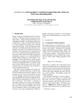
exchange chromatography. Figure 1 shows that AP205
VLPs purified in this way were 25-30 nm in size and
strongly resembled in their appearance the wild type par-
ticles [29]. Spectrophotometric analysis and agarose gel
electrophoresis indicated that AP205 VLPs contained
approximately 25-30 μg of host cell RNA per 100 μg of
coat protein (not shown).
Production of the DIII-C-AP205 Conjugate Vaccine
Domain III of the WNV E protein was engineered to
comprise a hexahistidine tag and a cysteine-containing
linker at its C-terminus (DIII-C). Upon expression in E.
coli the protein formed insoluble inclusion bodies. DIII-C
was solubilized in urea, purified under denaturing condi-
tions by affinity chromatography and refolded into its
native form by stepwise dialysis. A heterobifunctional
cross-linker was used to covalently conjugate DIII-C via
the introduced C-terminal cysteine residue to lysine resi-
dues on the surface of AP205 VLPs. The isolated vaccine
components and the conjugate vaccine (DIII-C-AP205)
were analysed by SDS-PAGE and Western Blot, as well as
size exclusion chromatography, with results shown in Fig-
ure 2. Recombinant DIII-C migrated as a distinct 13.5
kDa band in reducing, denaturing SDS-PAGE (Figure 2A,
left panel), consistent with its predicted molecular
weight. Size exclusion analysis of the protein showed a
single peak with an elution volume corresponding to an
Figure 1 Electron micrograph of purified AP205 VLPs. Purified
AP205 VLPs were adsorbed onto carbon-Formvar-coated grids, stained
with 2% phosphotungstic acid and subjected to transmission electron
microscopy.
Spohn et al. Virology Journal 2010, 7:146
/>Page 3 of 9
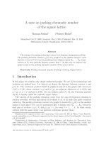
Figure 2 Production and characterization of the DIII-C-AP205 conjugate vaccine. A Derivatized AP205 (dAP205), DIII-C, and the dialysed conju-
gate vaccine (DIII-C-AP205) were analyzed by reducing, denaturing SDS-PAGE (left panel). Corresponding amounts of derivatized AP205, DIII-C, and of
the non-dialysed (nd) conjugate vaccine were also analysed by non-reducing, non-denaturing SDS-PAGE (right panel). For identification of the cou-
pling bands, proteins were separated on reducing, denaturing SDS-PAGE, blotted on nitrocellulose and detected with AP205- and His-tag- specific
antibodies (middle panels). Bands corresponding to AP205-crosslinked DIII-C are indicated by arrows. The 27 kDa band which is visible in the SDS-PAGE
and in the His-tag-specific Western Blot corresponds to dimeric DIII-C (*). B Size exclusion chromatography. A superdex 75 column was calibrated with
a molecular weight (MW) calibration kit and then loaded sequentially with the indicated proteins. AP205 VLPs and the conjugate vaccine DIII-C-AP205
elute in the void volume of the column (V
0
, 40 ml) while purified DIII-C elutes at 72.4 ml.
14
27
43
66
kDa
dAP205
DIII-C
DIII-C-AP205
α-His-tag
14
27
43
66
kDa
dAP205
DIII-C
DIII-C-AP205
α-AP205
14
27
43
66
kDa
dAP205
DIII-C
DIII-C-AP205
red.
denat.
SDS-PAGE
dAP205
DIII-C
DIII-C-AP205 (nd)
14
27
43
66
kDa
non-red.
non-denat.
SDS-PAGE
*
*
14
27
43
66
kDa
dAP205
DIII-C
DIII-C-AP205
α-His-tag
14
27
43
66
kDa
dAP205
DIII-C
DIII-C-AP205
α-AP205
14
27
43
66
kDa
dAP205
DIII-C
DIII-C-AP205
red.
denat.
SDS-PAGE
dAP205
DIII-C
DIII-C-AP205 (nd)
14
27
43
66
kDa
non-red.
non-denat.
SDS-PAGE
*
*
0
100
200
300
mAU
20 30 40 50 60 70 80 ml
400
0
50
100
150
mAU
20 30 40 50 60 70 80 ml
67
43 25
13
0
20
40
60
80
mAU
20 30 40 50 60 70 80 ml
0
100
200
300
mAU
20 30 40 50 60 70 80 ml
400
A
MW Calibration
AP205
DIII-C
DIII-C-AP205
V
0
(2000)
B
Spohn et al. Virology Journal 2010, 7:146
/>Page 4 of 9
apparent molecular weight of approximately 16 kDa,
indicating that DIII-C was indeed a folded monomer in
solution (Figure 2B). Analysis of the conjugate vaccine
DIII-C-AP205 showed the presence of several high
molecular weight bands in reducing, denaturing SDS-
PAGE, which reacted with both AP205- and His-tag spe-
cific antisera, demonstrating the successful crosslinking
of DIII-C to the AP205 VLPs (Figure 2A, middle panels).
Similar to AP205 VLPs, the DIII-C-AP205 conjugate vac-
cine eluted in the void volume of the size exclusion col-
umn, confirming its virus-like particle assembly state
(Figure 2B). In order to estimate the number of DIII-C
molecules displayed by each VLP in the conjugate vac-
cine, the corresponding amounts of DIII-C used in the
coupling reaction and of the non-dialysed DIII-C-AP205
conjugate vaccine were loaded side by side on a non-
reducing, non-denaturing SDS polyacrylamide gel (Fig-
ure 2A, right panel). Under these conditions, DIII-C still
migrates as a 13.5 kDa monomer while the conjugate vac-
cine migrates as a high molecular weight complex. Densi-
tometric analysis showed a 55% decrease in the amount
of free DIII-C after coupling to AP205. Assuming that the
decrease in free DIII-C is due to coupling to AP205 sub-
units and taking into account the molar ratio of VLP sub-
units and DIII-C molecules used in the coupling reaction
it could be calculated that an average 27% of AP205 sub-
units had been cross-linked to DIII-C molecules. As each
AP205 VLP is composed of 180 subunits, it can be esti-
mated that an average of 50 DIII-C molecules are dis-
played per vaccine particle.
Immunogenicity of DIII-C-AP205 in Mice
Groups of mice were immunized subcutaneously either
with the DIII-C-AP205 conjugate vaccine or a mixture of
the corresponding amounts of non-conjugated AP205
carrier and free DIII-C protein in the absence of any addi-
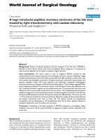
tional adjuvant. Figure 3 shows that one or two injections
of the non-conjugated AP205/DIII-C mixture did not
result in any detectable DIII-specific IgG antibodies, con-
firming the notion that isolated DIII is poorly immuno-
genic. A measurable IgG antibody response could be
induced only after a third injection (ELISA titer of 15,300
on day 42). In contrast, DIII-specific IgG titers were mea-
sured after only a single administration of the DIII-C-
AP205 conjugate vaccine (6,600 on day 14). A second vac-
cine injection boosted the specific antibodies to a titer of
106,900 (day 28), while a third injection did not lead to a
further increase. In the absence of additional injections,
antibody titers slowly declined with an approximate half
life of two months.
Vaccination with DIII-C-AP205 Induces Neutralizing
Antibodies and Protects from Lethal WNV Infection
Groups of mice were immunized with DIII-C-AP205
three times (days 0, 14, 28) either in the absence or in the
presence of Alum as adjuvant. One group was immunized
only once (day 28) in the presence of Alum. The neutral-
izing capacity of the induced antibodies was determined
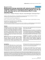
two weeks after the last injection (day 42). Figure 4A
shows that three injections of DIII-C-AP205 in the
absence or presence of adjuvant resulted in average neu-
tralizing titers of 360 and 760, respectively. Neutralizing
ability was also detected in sera from 6 of the 8 mice that
that received a single injection of vaccine. All vaccinated
mice and a group of control mice, that had been immu-
nized with the AP205 carrier alone, were then challenged
with a lethal dose of West Nile virus strain NY99. Figure
4B shows that all mice that had received three injections
of DIII-C-AP205 either in the presence or absence of
Alum survived the viral challenge. In contrast, all control
mice immunized with AP205 succumbed to the infection.
Interestingly, 5 out of 8 mice that had received a single
injection of DIII-C-AP205 in Alum also survived the
challenge with WNV. This survival rate is in the range of
the ones achieved after single doses of different live atten-
uated WNV vaccines in mice [9,34,35]. Vaccination with
DIII-C-AP205 resulted in a marked reduction of viremia
as measured by real time PCR in blood 3 and 7 days after
infection (Figures 4C and 4D, respectively). All mice that
had received three injections of DIII-C-AP205, as well as
6 out of 8 mice that had received a single injection had
cleared the virus by day 7; the 2 remaining mice of this
group had reduced viral titers as compared to the average
viral load of AP205-immunized animals at this time
point.
Discussion
Domain III of the envelope protein of WNV is a major
target of virus-neutralizing antibody responses and has
been identified as a promising candidate antigen for the
development of recombinant subunit vaccines [36]. In
this study we produced a highly immunogenic and effica-
cious WNV vaccine consisting of recombinant domain III
chemically cross-linked to virus-like particles of the bac-
teriophage AP205. The conjugate vaccine produced high
DIII-specific antibody titers in mice, which were able to
efficiently inhibit viral replication both in vitro and in
vivo. In contrast to other experimental vaccines based on
recombinant DIII and comprising different adjuvants
[20-23], a single injection in the presence of Alum was
sufficient to induce neutralizing antibody responses and
confer partial protection from WNV challenge (Figure 4).
Interestingly, the neutralizing titers observed (approxi-
mately 30-40) were in the range of those induced by a sin-
gle injection of live WNV vaccines such as a chimeric
attenuated flavivirus vaccine [9,35] or a recombinant
attenuated influenza strain expressing the WNV E pro-
tein [34]. Multiple injections either in the presence or
absence of Alum as adjuvant yielded sustained high titers
of DIII-specific antibodies, which efficiently neutralized
Spohn et al. Virology Journal 2010, 7:146
/>Page 5 of 9
the virus. The reason for the superior immunogenicity of
the DIII-C-AP205 conjugate vaccine most likely resides
in its virus-like connotations. It has been shown that
highly ordered and repetitive antigen arrays can cause an
efficient cross-linking of BCRs on specific B cells and
induce a rapid and sustained antibody response [37]. As
domain III is presented to the immune system in an ori-
ented and densely packaged fashion on the surface of the
AP205 VLP, it is likely that DIII-specific B cells are
promptly and efficiently activated to produce specific IgG
antibodies. The particulate nature of the VLP vaccine fur-
thermore ensures a preferential uptake by antigen-pre-
senting cells such as dendritic cells, and thereby an
efficient presentation of DIII- as well as AP205-derived
epitopes on MHC class II for the priming of specific T
H
cells. Activation of antigen-presenting cells is also
enhanced by bacterial RNA, which is spontaneously
packaged into the VLP carrier during the recombinant
expression and assembly process. Upon uptake by B cells
and APCs, the RNA is co-delivered with the AP205 parti-
cle to the endosomal compartment, where it can activate
TLR3 or TLR7/8 (for review see [38,39]).
In addition to its good immunogenicity, the DIII-C-
AP205 vaccine is also expected to be safe and well toler-
ated. In contrast to live vaccines based on attenuated
viruses, which inevitably carry the risk of genetic recom-
bination and mutation into a more virulent form, DIII-C-
AP205 is based on non-replicating virus-like particles
derived from a bacteriophage, which are unable to infect
mammalian cells. Moreover, both the VLP carrier and the
antigen components of the vaccine can be produced in
large amounts in bacterial expression systems and puri-
fied with relatively simple biochemical methods, suggest-
ing that large scale production of the conjugate vaccine
can be achieved in a cost-effective manner. A highly
immunogenic, yet safe and affordable WNV vaccine
would be attractive for veterinary prophylaxis and might
also be used in elderly or immunocompromised individu-
als in high-risk areas. The high immunogenicity of the
VLP vaccine might also offer the potential of inducing
cross-protection against related flaviviruses such as Japa-
nese encephalitis virus or dengue virus. That cross-pro-
tection may occur in principle has been shown by
immunization of mice with recombinant domain III of
the WNV E protein [21]. The increased immunogenicity
of the domain III by conjugation to the VLP carrier may
therefore be sufficient to confer cross-protective immu-
nity.
Conclusions
In the present study we show that the immunogenicity of
DIII of the WNV E protein can be strongly enhanced by
conjugation to virus-like particles of the bacteriophage
AP205. In contrast to other vaccination approaches based
on recombinant DIII, which require multiple injections
and/or strong adjuvants for the induction of neutralizing
antibodies, a single injection of the conjugate DIII-C-
AP205 vaccine in Alum was sufficient to induce a signifi-
Figure 3 Immunogenicity of DIII-C-AP205. Groups of female BALB/c mice (n = 4) were immunized subcutaneously three times (days 0, 14, and 28,
arrows) with either 50 μg of DIII-C-AP205 or a mixture of the corresponding amounts of free DIII-C protein (13.6 μg) and free AP205 VLPs (36.4 μg) in
the absence of adjuvants. DIII-C-specific IgG antibody titers were measured at the indicated time points. The dashed line indicates the detection limit.
Shown are group means ± SEM.
0 10 20 30 40
10
1
10
2
10
3
10
4
10
5
50 150 250 350
DIII-C + AP205
DIII-C-AP205
days
ELISA titer (OD50%)
Spohn et al. Virology Journal 2010, 7:146
/>Page 6 of 9
Figure 4 Immunization with DIII-C-AP205 protects from lethal WNV infection. A Induction of neutralizing antibodies. Groups of female C57BL/
6 mice (n = 8) were immunized subcutaneously on days 0, 14 and 28 with 50 μg DIII-C-AP205 either in the absence or presence of Alum as adjuvant
or with 50 μg AP205 VLPs in the absence of adjuvant. A fourth group was immunized once with 50 μg DIII-C-AP205 in Alum on day 28. Virus-neutral-
izing titers of individual sera were measured on day 42. The dashed line indicates the detection limit. Shown are individual titers and group means. B
Protection from WNV challenge. Two weeks after the last vaccine injection mice were challenged with a lethal dose of WNV (arrow). Statistical signif-
icance of differences in survival curves was calculated by log-rank test using GraphPad-Prism (***p < 0.001 vs. 3xAP205 control). p.i.= post infection C
and D Viral titers were determined 3 and 7 days post infection (p.i.) in blood of infected animals. Shown are individual titers and group means. The
Mann-Whitney test was used to assess statistical significance (**p < 0.01, ***p < 0.001 vs. 3xAP205 control).
day 3 p.i.
3x
A
P205
3x DIII-C-
A
P205
3x DI
I
I-C-AP205 + Alum
+ A
l
um1
x DIII-
C
-
A
P
2
05
10
0
10
1
10
2
10
3
10
4
10
5
10
6
***
***
***
virus titer
day 7 p.i.
3x
A
P205
3
x DIII-
C
-
A
P205
3x DIII-C-AP205 + Alum
1x DI
II
-C-AP205 + Alum
10
0
10
1
10
2
10
3
10
4
10
5
*** ***
**
virus titer
A
CD
B
3
x
AP20
5
3x DIII-C-AP205
3x DII
I
-C-
A
P2
0
5 +
Al
um
+ Al
um
1x
DI
II-
C-
AP20
5
10
1
10
2
10
3
10
4
neutralizing titer
0 5 10 15
0
20
40
60
80
100
***
***
3x AP205
3x DIII-C-AP205
3x DIII-C-AP205 + Alum
1x DIII-C-AP205 + Alum
days p.i.
% survival
Spohn et al. Virology Journal 2010, 7:146
/>Page 7 of 9
cant amount of virus-neutralizing antibodies in mice.
Three injections of the vaccine completely protected
mice from a lethal WNV challenge, even when given in
the absence of any adjuvant. The relatively low produc-
tion costs of the DIII-C-AP205 vaccine, its superior
immunogenicity with respect to other DIII-based
approaches and its anticipated good safety profile make it
an attractive candidate for WNV prophylaxis both in
humans and in veterinary applications.
Methods
Expression and Purification of AP205 Virus-like Particles
Cleared bacterial lysates containing the recombinantly
expressed coat protein of AP205 were dialysed against
AEX loading buffer (20 mM NaH
2
PO
4
pH 7.2) and loaded
on a Fractogel™ TMAE column (Merck). After removal of
host cell proteins by a salt wash with 333 mM NaCl, viral
capsids were eluted with 600 mM NaCl and dialyzed
against HAp loading buffer (5 mM NaH
2
PO
4
, 100 mM
NaCl, pH 6.8). Capsids were bound to a hydroxyapatite
column (Macro-prep ceramic hydroxyapatite type II, Bio-
rad) and eluted with 60 mM NaH
2
PO
4
, 220 mM NaCl, pH
6.8, resulting in depletion of bacterial LPS.
Expression and Purification of Recombinant Domain III of
the E Glycoprotein of WNV
A DNA fragment encoding domain III of the glycoprotein
E of WNV NY99 was amplified from plasmid pTRHis2A-
WNV-E [21] with the oligonucleotide pair WNV1/
WNV2 (5'-ATATATCATATGGAAAAATTGCAGTT-
GAAGG-3'; 5'-ATATATCTCGAGTTTGCCAATGCTG
C TTCCAG-3', NdeI and XhoI restriction sites are in
bold) and cloned into the expression vector pET42T [40].
The resulting plasmid encoded a fusion protein consist-
ing of domain III of the WNV E protein (corresponding
to amino acids 582-696 of the WNV polyprotein precur-
sor), a hexahistidine tag, and a short C-terminal, cysteine
containing linker (DIII-C). E. coli BL21 DE3 cells were
transformed with this plasmid, and protein expression
was induced in a logarithmic phase culture by addition of
isopropyl-β-D-thiogalactopyranoside to a final concen-
tration of 1 mM. After overnight growth bacteria were
harvested by centrifugation, resuspended in 50 mM
NaH
2
PO
4
, 150 mM NaCl, 10 mM MgCl
2
, 0.25% Triton X-
100, pH 7.2, and lysed by sonication. Nucleic acids were
digested by 1 h incubation at room temperature with
1500 U Benzonase (Sigma-Aldrich), and inclusion bodies
containing recombinant DIII-C were harvested by cen-
trifugation. After three washes with 100 mM Tris-Cl, 5
mM EDTA, 5 mM DTT, 2% Triton X-100, pH 7.0, inclu-
sion bodies were solubilized in 8 M urea, 100 mM Tris-
Cl, 100 mM DTT, pH 8.0, and loaded on a Ni-NTA col-
umn (Qiagen), which had been previously equilibrated
with 8 M Urea, 100 mM NaH
2
PO
4
, 10 mM Tris-Cl, 2 mM
β-mercaptoethanol, pH 8.0. Bound DIII-C was eluted
with 8 M urea, 100 mM NaH
2
PO
4
, 10 mM Tris, 2 mM β-
Mercaptoethanol pH 4.5, and dialysed against 2 M urea,
50 mM NaH
2
PO
4
, 0.5 M arginine, 0.5 mM oxidized gluta-
thione, 5 mM reduced glutathione, 10% glycerol, pH 8.5.
DIII-C was then refolded by stepwise dialysis against 50
mM NaH
2
PO
4
, 0.5 M arginine, 0.5 mM oxidized glutathi-
one, 5 mM reduced glutathione, 10% glycerol, pH 8.5, and
against 50 mM NaH
2
PO
4
, 10% glycerol, pH 8.5.
Chemical Cross-linking of Recombinant DIII-C to AP205
Virus-like Particles
AP205 VLPs (in PBS, pH 7.2) were first reacted for 1 h at
room temperature with a 2.5 fold molar excess of the het-
erobifunctional cross-linker succinimidyl-6-(β-maleimi-
dopropionamido)hexanoate (Pierce). Free cross-linker
was removed by dialysis against PBS, pH 7.2. Recombi-
nant DIII-C was incubated for 1 h at room temperature
with an equimolar amount of tri(2-carboxyethyl)phos-
phine-hydrochloride. Under these mildly reducing condi-
tions the cysteine residue contained in the linker is
reduced, while the internal disulfide bridge of DIII-C
remains intact. The reduced protein was then mixed with
the derivatized AP205 VLPs at a molar ratio of 1 DIII-C
monomer per 2 AP205 monomers and incubated over
night at 17°C to allow cross-linking. Free DIII-C was
removed by extensive dialysis against PBS pH 7.2 using
cellulose ester membranes with a cut-off of 100 kDa
(Spectrum Laboratories). The conjugate vaccine was ana-
lyzed by SDS-PAGE followed by Coomassie Blue staining
or by Western Blot using AP205- and His-tag- specific
antisera. The molecular masses of the DIII-C and AP205
monomers are similar; 13.5 kDa and 14.0 kDa, respec-
tively. The coupling product comprising one AP205
monomer covalently conjugated to one DIII-C monomer
co-migrates with the AP205 dimer band. Hence the cou-
pling efficiency could not simply be calculated by densi-
tometry of protein bands on a reducing SDS-PAGE
stained with Coomassie Blue. Instead the conjugate vac-
cine was loaded on a non-reducing non-denaturing SDS-
PAGE side by side with the corresponding amount of free
DIII-C, which had been used in the cross-linking reac-
tion. By comparing the intensities of the DIII-C mono-
mers before and after cross-linking to AP205 by
densitometry, the amount of DIII-C coupled to the
AP205 carrier could then be quantified.
Analysis of DIII-C, AP205 and DIII-C-AP205 by Size Exclusion
Chromatography
A superdex 75 column (GE Healthcare) was calibrated
with a mixture of Dextran Blue (~2000 kDa), BSA (67
kDa), Ovalbumin (43 kDa), Chymotrypsinogen (25 kDa),
and RNase A (14 kDa). AP205 VLPs, purified DIII-C pro-
Spohn et al. Virology Journal 2010, 7:146
/>Page 8 of 9
tein and the conjugate vaccine DIII-C-AP205 were then
sequentially analysed on the same column. The apparent
molecular weight of DIII-C was calculated from a stan-
dard curve obtained by plotting the logarithm of the
molecular weights of the protein standards against their
partition coefficients.
Immunogenicity of DIII-C-AP205
Female BALB/c mice (8 weeks of age) were purchased
from Charles River Laboratories. DIII-C-AP205 vaccine
or the mixture of the non-conjugated vaccine compo-
nents AP205 and recombinant DIII-C were diluted in
PBS to 200 μl and injected subcutaneously (100 μl on two
ventral sites) in the absence of additional adjuvants. Sera
from immunized mice were serially diluted in PBS con-
taining 0.05% Tween-20, 2% BSA, and applied to ELISA
plates (Nunc) that had been coated with 1 μg/ml recom-
binant DIII-C protein. Reactivity of serum antibodies
with the target protein was determined using a HRP-con-
jugated goat anti-mouse IgG secondary antibody (Jack-
son ImmunoResearch Laboratories) at a dilution of
1:1000 in PBS/0.05% Tween-20/2% BSA. After develop-
ment with 1,2-phenylenediamine dihydrochloride (0.4
mg/mL in 0.066 M Na
2
HPO
4
, 0.035 M citric acid, 0.01%
H
2
O
2
, pH 5.0) the optical density at 450 nm (OD
450 nm
)
was determined using an ELISA reader (Biorad). Titers
were expressed as the reciprocal of those serum dilutions
that lead to half-maximal OD
450 nm
(OD50%).
WNV Challenge
Female C57BL/6 mice (6 weeks of age) were purchased
from Harlan and allowed to acclimate to the facility for
one week before experiments were performed. Experi-
ments were approved by the animal ethics committee of
the Erasmus MC Rotterdam, The Netherlands. Mice were
immunized as indicated in the legend of Figure 4 and
challenged two weeks after the last immunization by an
intraperitoneal injection of a lethal dose of WNV-NY99
(1 × 10
6
TCID
50
). After the challenge, mice were main-
tained in isolation cages and observed daily for illness and
death for a period of 14 days. Blood was collected on days
3 and 7 after infection and viral titers were determined by
real-time PCR. The quantity of viral RNA was measured
with a one-step RT-PCR TaqMan protocol and ABI
PRISM 7500 detection instrument (EZ-kit, Applied Bio-
systems). The primers and probe used for WNV RNA
quantification were: forward primer 5'-TCACTGT-
CAACCCTTTTGTTTC-3'; reverse primer 5'-AAGG
GTGGTTCCAATTCAATC-3'; probe 5'-CCACGGCCA-
ACGCTAAGGTCC-3'. Serial dilutions of WNV stock
were used as standard, and results were expressed as
TCID
50
equivalents per gram of brain tissue. For determi-
nation of neutralizing antibody titers serial two-fold dilu-
tions of immune sera were incubated with 100 TCID
50
of
WNV strain NY99. Virus-neutralizing titers were
expressed as the reciprocal of the highest dilution that
still resulted in 100% suppression of the cytopathic effects
on Vero E6 cells [21].
Competing interests
ADMEO is a part-time employee (CSO) of Viroclinics B.V. (for details go to http:/
/www.erasmusmc.nl). The other authors declare that they have no competing
interests.
Authors' contributions
GS designed the experiments on vaccine production and immunogenicity
testing, performed experiments on vaccine analytics, coordinated the study
and drafted the manuscript. GTJ conceived and designed the project and
helped to draft the manuscript. BEEM designed and performed the WNV infec-
tion experiments, helped to coordinate the study and to draft the manuscript.
IK performed immunization and ELISA experiments. MB purified recombinant
DIII-C, and produced the conjugate vaccine. PP performed the electron micros-
copy experiments. ADMEO and MFB conceived and designed the project. All
authors read and approved the final manuscript.
Acknowledgements
Part of this work has been supported by a grant of the European Community
(contract LSHB-CT-2004-005246 "COMPUVAC").
The authors would like to thank Alexander Link for critical reading of the manu-
script.
Author Details
1
Cytos Biotechnology, Wagistrasse 25, 8952 Schlieren, Switzerland,
2
Erasmus
MC, Department of Virology, P.O. Box 2040, 3000 CA Rotterdam, The
Netherlands,
3
Latvian Biomedical Research and Study Centre, Ratsupites iela 1,
Riga, LV 1067, Latvia,
4
AO Foundation, Clavadelerstrasse 8, 7270 Davos Platz,
Switzerland and
5
ETH Zürich, Institute for Integrative Biology, Universitätstrasse
16, 8092 Zürich, Switzerland
References
1. Campbell GL, Marfin AA, Lanciotti RS, Gubler DJ: West Nile virus. Lancet
Infect Dis 2002, 2:519-529.
2. Hayes EB, Sejvar JJ, Zaki SR, Lanciotti RS, Bode AV, Campbell GL: Virology,
pathology, and clinical manifestations of West Nile virus disease.
Emerg Infect Dis 2005, 11:1174-1179.
3. Glass WG, McDermott DH, Lim JK, Lekhong S, Yu SF, Frank WA, Pape J,
Cheshier RC, Murphy PM: CCR5 deficiency increases risk of symptomatic
West Nile virus infection. J Exp Med 2006, 203:35-40.
4. Lim JK, Louie CY, Glaser C, Jean C, Johnson B, Johnson H, McDermott DH,
Murphy PM: Genetic deficiency of chemokine receptor CCR5 is a strong
risk factor for symptomatic West Nile virus infection: a meta-analysis of
4 cohorts in the US epidemic. J Infect Dis 2008, 197:262-265.
5. O'Leary DR, Marfin AA, Montgomery SP, Kipp AM, Lehman JA, Biggerstaff
BJ, Elko VL, Collins PD, Jones JE, Campbell GL: The epidemic of West Nile
virus in the United States, 2002. Vector Borne Zoonotic Dis 2004, 4:61-70.
6. Gubler DJ: The continuing spread of West Nile virus in the western
hemisphere. Clin Infect Dis 2007, 45:1039-1046.
7. Kaptoul D, Viladrich PF, Domingo C, Niubo J, Martinez-Yelamos S, De Ory
F, Tenorio A: West Nile virus in Spain: report of the first diagnosed case
(in Spain) in a human with aseptic meningitis. Scand J Infect Dis 2007,
39:70-71.
8. Rossini G, Cavrini F, Pierro A, Macini P, Finarelli A, Po C, Peroni G, Di Caro A,
Capobianchi M, Nicoletti L, et al.: First human case of West Nile virus
neuroinvasive infection in Italy, September 2008 - case report. Euro
Surveill 2008, 13:.
9. Arroyo J, Miller C, Catalan J, Myers GA, Ratterree MS, Trent DW, Monath TP:
ChimeriVax-West Nile virus live-attenuated vaccine: preclinical
evaluation of safety, immunogenicity, and efficacy. J Virol 2004,
78:12497-12507.
Received: 21 April 2010 Accepted: 6 July 2010
Published: 6 July 2010
This artic le is available fro m: http://www.v irologyj.com/co ntent/7/1/146© 2010 Spohn et al; licensee BioMed Central Ltd. This is an Open Access article distributed under the terms of the Creative Commons Attribution License ( which permits unrestricted use, distribution, and reproduction in any medium, provided the original work is properly cited.Virology Journal 2010, 7:146
Spohn et al. Virology Journal 2010, 7:146
/>Page 9 of 9
10. Monath TP, Liu J, Kanesa-Thasan N, Myers GA, Nichols R, Deary A,
McCarthy K, Johnson C, Ermak T, Shin S, et al.: A live, attenuated
recombinant West Nile virus vaccine. Proc Natl Acad Sci USA 2006,
103:6694-6699.
11. Guy B, Guirakhoo F, Barban V, Higgs S, Monath TP, Lang J: Preclinical and
clinical development of YFV 17D-based chimeric vaccines against
dengue, West Nile and Japanese encephalitis viruses. Vaccine
28:632-649.
12. Roehrig JT: Antigenic structure of flavivirus proteins. Adv Virus Res 2003,
59:141-175.
13. Lee JW, Chu JJ, Ng ML: Quantifying the specific binding between West
Nile virus envelope domain III protein and the cellular receptor
alphaVbeta3 integrin. J Biol Chem 2006, 281:1352-1360.
14. Beasley DW, Barrett AD: Identification of neutralizing epitopes within
structural domain III of the West Nile virus envelope protein. J Virol
2002, 76:13097-13100.
15. Volk DE, Beasley DW, Kallick DA, Holbrook MR, Barrett AD, Gorenstein DG:
Solution structure and antibody binding studies of the envelope
protein domain III from the New York strain of West Nile virus. J Biol
Chem 2004, 279:38755-38761.
16. Nybakken GE, Oliphant T, Johnson S, Burke S, Diamond MS, Fremont DH:
Structural basis of West Nile virus neutralization by a therapeutic
antibody. Nature 2005, 437:764-769.
17. Choi KS, Nah JJ, Ko YJ, Kim YJ, Joo YS: The DE loop of the domain III of the
envelope protein appears to be associated with West Nile virus
neutralization. Virus res 2007, 123:216-218.
18. Sanchez MD, Pierson TC, McAllister D, Hanna SL, Puffer BA, Valentine LE,
Murtadha MM, Hoxie JA, Doms RW: Characterization of neutralizing
antibodies to West Nile virus. Virology 2005, 336:70-82.
19. Oliphant T, Engle M, Nybakken GE, Doane C, Johnson S, Huang L, Gorlatov
S, Mehlhop E, Marri A, Chung KM, et al.: Development of a humanized
monoclonal antibody with therapeutic potential against West Nile
virus. Nat Medicine 2005, 11:522-530.
20. Wang T, Anderson JF, Magnarelli LA, Wong SJ, Koski RA, Fikrig E:
Immunization of mice against West Nile virus with recombinant
envelope protein. J Immunol 2001, 167:5273-5277.
21. Martina BE, Koraka P, van den Doel P, van Amerongen G, Rimmelzwaan
GF, Osterhaus AD: Immunization with West Nile virus envelope domain
III protects mice against lethal infection with homologous and
heterologous virus. Vaccine 2008, 26:153-157.
22. Ledizet M, Kar K, Foellmer HG, Wang T, Bushmich SL, Anderson JF, Fikrig E,
Koski RA: A recombinant envelope protein vaccine against West Nile
virus. Vaccine 2005, 23:3915-3924.
23. Chu JH, Chiang CC, Ng ML: Immunization of flavivirus West Nile
recombinant envelope domain III protein induced specific immune
response and protection against West Nile virus infection. J Immunol
2007, 178:2699-2705.
24. McDonald WF, Huleatt JW, Foellmer HG, Hewitt D, Tang J, Desai P, Price A,
Jacobs A, Takahashi VN, Huang Y, et al.: A West Nile virus recombinant
protein vaccine that coactivates innate and adaptive immunity. J Infect
Dis 2007, 195:1607-1617.
25. Spohn G, Guler R, Johansen P, Keller I, Jacobs M, Beck M, Rohner F, Bauer
M, Dietmeier K, Kundig TM, et al.: A virus-like particle-based vaccine
selectively targeting soluble TNF-alpha protects from arthritis without
inducing reactivation of latent tuberculosis. J Immunol 2007,
178:7450-7457.
26. Spohn G, Keller I, Beck M, Grest P, Jennings GT, Bachmann MF: Active
immunization with IL-1 displayed on virus-like particles protects from
autoimmune arthritis. Eur J Immunol 2008, 38:877-887.
27. Spohn G, Schwarz K, Maurer P, Illges H, Rajasekaran N, Choi Y, Jennings GT,
Bachmann MF: Protection against osteoporosis by active immunization
with TRANCE/RANKL displayed on virus-like particles. J Immunol 2005,
175:6211-6218.
28. Rohn TA, Jennings GT, Hernandez M, Grest P, Beck M, Zou Y, Kopf M,
Bachmann MF: Vaccination against IL-17 suppresses autoimmune
arthritis and encephalomyelitis. Eur J Immunol 2006, 36:2857-2867.
29. Klovins J, Overbeek GP, van den Worm SH, Ackermann HW, van Duin J:
Nucleotide sequence of a ssRNA phage from Acinetobacter: kinship to
coliphages. J Gen Virol 2002, 83:1523-1533.
30. van den Worm SH, Koning RI, Warmenhoven HJ, Koerten HK, van Duin J:
Cryo electron microscopy reconstructions of the Leviviridae unveil the
densest icosahedral RNA packing possible. J Mol Biol 2006, 363:858-865.
31. Kozlovska TM, Cielens I, Dreilinna D, Dislers A, Baumanis V, Ose V,
Pumpens P: Recombinant RNA phage Q beta capsid particles
synthesized and self-assembled in Escherichia coli. Gene 1993,
137:133-137.
32. Peabody DS: Translational repression by bacteriophage MS2 coat
protein expressed from a plasmid. A system for genetic analysis of a
protein-RNA interaction. J Biol Chem 1990, 265:5684-5689.
33. Tissot AC, Renhofa R, Schmitz N, Cielens I, Meijerink E, Ose V, Jennings GT,
Saudan P, Pumpens P, Bachmann MF: Versatile Virus-Like Particle Carrier
for Epitope Based Vaccines. PLOS One 2010, 5(3):e9809.
34. Martina BEE, van den Doel P, Koraka P, van Amerongen G, Spohn G,
Haagmans BL, Fouchier RAM, Osterhaus ADME, Rimmelzwaan GF: A
recombinant influenza A expressing domain III of West Nile virus
induces protective immune responses against influenza and West Nile
virus. PLOS One in press.
35. Huang CY, Silengo SJ, Whiteman MC, Kinney RM: Chimeric dengue 2
PDK-53/West Nile NY99 viruses retain the phenotypic attenuation
markers of the candidate PDK-53 vaccine virus and protect mice
against lethal challenge with West Nile virus. J Virol 2005, 79:7300-7310.
36. Diamond MS, Pierson TC, Fremont DH: The structural immunology of
antibody protection against West Nile virus. Immunol rev 2008,
225:212-225.
37. Bachmann MF, Rohrer UH, Kundig TM, Burki K, Hengartner H, Zinkernagel
RM: The influence of antigen organization on B cell responsiveness.
Science 1993, 262:1448-1451.
38. Spohn G, Bachmann MF: Exploiting viral properties for the rational
design of modern vaccines. Expert Rev Vaccines 2008, 7:43-54.
39. Jennings GT, Bachmann MF: The coming of age of virus-like particle
vaccines. Biol Chem 2008, 389:521-536.
40. Bachmann MF, Spohn G: WO2007/039552 A1: Interleukin-1 conjugates
and uses thereof. 2007.
doi: 10.1186/1743-422X-7-146
Cite this article as: Spohn et al., A VLP-based vaccine targeting domain III of
the West Nile virus E protein protects from lethal infection in mice Virology
Journal 2010, 7:146