Báo cáo y học: " Course of seasonal influenza A/Brisbane/59/07 H1N1 infection in the ferret" pot
Bạn đang xem bản rút gọn của tài liệu. Xem và tải ngay bản đầy đủ của tài liệu tại đây (738.39 KB, 5 trang )
McBrayer et al. Virology Journal 2010, 7:149
/>Open Access
SHORT REPORT
© 2010 McBrayer et al; licensee BioMed Central Ltd. This is an Open Access article distributed under the terms of the Creative Commons
Attribution License ( which permits unrestricted use, distribution, and reproduction in
any medium, provided the original work is properly cited.
Short report
Course of seasonal influenza A/Brisbane/59/07
H1N1 infection in the ferret
Alexis McBrayer
1
, Jeremy V Camp
1
, Ron Tapp
1
, Vladimir Yamshchikov
1
, Sheila Grimes
1
, Diana L Noah
1
,
Colleen B Jonsson
1,2
and Carl E Bruder*
1,3
Abstract
Every year, influenza viruses infect approximately 5-20% of the population in the United States leading to over 200,000
hospitalizations and 36,000 deaths from flu-related complications. In this study, we characterized the immune and
pathological progression of a seasonal strain of H1N1 influenza virus, A/Brisbane/59/2007 in a ferret model. The
immune response of the animals showed a dose-dependent increase with increased virus challenge, as indicated by
the presence of virus specific IgG, IgM, and neutralizing antibodies. Animals infected with higher doses of virus also
experienced increasing severity of clinical symptoms and fever at 2 days post-infection (DPI). Interestingly, weight loss
was more pronounced in animals infected with lower doses of virus compared to those infected with a higher dose;
these results were consistent with viral titers of swabs collected from the nares, but not the throat. Analyzed specimens
included nasal and throat swabs from 1, 3, 5, and 7 DPI as well as tissue samples from caudal lung and nasal turbinates.
Viral titers of the swab samples in all groups were higher on 1 and 3 DPI and returned to baseline levels by 7 DPI.
Analysis of nasal turbinates indicated presence of virus at 3 DPI in all infected groups, whereas virus was only detected
in the lungs of animals in the two highest dose groups. Histological analysis of the lungs showed a range of pathology,
such as chronic inflammation and bronchial epithelial hypertrophy. The results provided here offer important
endpoints for preclinical testing of the efficacy of new antiviral compounds and experimental vaccines.
Findings
Every year, influenza virus infects 5-20% of the US popu-
lation with numerous deaths attributed to primary influ-
enza infection or secondary bacterial pneumonia [1]. The
rapid evolution of new influenza virus strains and drug
resistant variants demands constant development of
treatments as well as reliable animal models allowing for
testing of these remedies [2,3]. Although a number of ani-
mal models are used for influenza research, ferrets are
ideal because they can be readily infected with human
isolates of influenza virus (in contrast to mice) and
exhibit symptoms similar to humans, such as fever,
coughing, sneezing, runny nose, lethargy [4-10], and
make a full recovery in 7-10 days [11,12]. Humans and
ferrets also share a similar distribution of α-2,6 and α-2,3
linked sialic acid residues, which serve as the receptor for
influenza attachment to airway epithelial cells, enabling
influenza to use the same cell entry mechanism [5,13,14].
Furthermore, ferrets are large enough to easily monitor
aspects of disease progression and yield enough materials
for immunological and virological analysis, [6,15-17].
Prior to clinical trials, safety and efficacy need to be dem-
onstrated in two animal models, one non-rodent, making
the ferret ideal.
We examined progression of A/Brisbane/59/2007 in
ferrets using a full series of endpoints; clinical symptoms,
gross and microscopic pathology, virology, and immunol-
ogy. A/Brisbane/59/07 was obtained from the Centers for
Disease Control and Prevention and propagated for 2
days at 34°C in 10-day embryonated hen's eggs [18]. Cas-
trated and de-scented Fitch ferrets (6-8 months of age,
800-1800 grams; Triple F Farms, Sayre, PA) were assigned
to one of 6 treatment groups (Table 1) by a weight-
matched computer-generated randomization procedure.
Five groups were challenged intranasally with increasing
doses of A/Brisbane/59/2007, and controls received PBS.
Changes in body temperature, body weight, and onset of
clinical symptoms were monitored for 7 days after chal-
lenge to measure disease progression and severity. Ana-
* Correspondence:
1
Southern Research Institute, 2000 9th Ave South, Birmingham, AL 35205, USA
Full list of author information is available at the end of the article
McBrayer et al. Virology Journal 2010, 7:149
/>Page 2 of 5
lyzed specimens included blood sera, and excreta
samples from nasal and throat swabs from 1, 3, 5, and 7
DPI and tissues from 3 and 7 DPI. Animal studies were
approved by Southern Research Institutional Animal
Care and Use Committee and met the recommended ani-
mal care guidelines.
Animals in groups infected with higher doses of influ-
enza experienced greater severity in clinical symptoms
compared to those in lower dose groups or control ani-
mals (Table 1). Groups infected with influenza demon-
strated significant weight loss at 2 through 7 DPI
compared to the control group. Animals also exhibited
elevated body temperature on 2 DPI. Flu-like symptoms,
such as sneezing, and nasal and ocular discharge were
seen. Most animals fully recovered by 7 DPI; however,
some animals relapsed with a recurrence of clear or
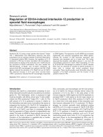
serous nasal discharge. Histological analysis of lungs
showed a range of pathology, such as bronchiolar epithe-
lial hypertrophy and inflammation. Macroscopic lung
lesions consisted of dark/mottled discoloration observed
in animals in all dose groups on 3 and 7 DPI. In animals
euthanized on 3 and 7 DPI, microscopic lesions consis-
tent with influenza infection were observed in all chal-
lenge groups, but not controls. Microscopic lesions in
lungs of influenza challenge dose groups consisted of
acute inflammation of the alveolus, bronchiole, and bron-
chiole lumen; chronic inflammation of the alveolus, bron-
chus, peribronchiolar interstitium and perivascular
interstitium; chronic-active inflammation of the alveolus;
hemosiderin pigmentation of the perivascular intersti-
tium; type II pneumocyte hyperplasia; bronchiolar hyper-
trophy; syncytia of the alveolus and bronchiole; and
regeneration of the bronchiole. Although the incidence
and severity of lesions was variable among dose groups,
these parameters tended to be the greatest in animals
infected with higher doses of virus. Excluding chronic
inflammation of the perivascular interstitium and bron-
chiolar hypertrophy, which ranged from minimal to mild
in severity, lesions noted were minimal in severity (Figure
1).
Viral load in swabs and tissues was analyzed by titration
to determine the TCID
50
. Briefly, MDCK cells (ATCC,
clone CCl-34) were grown in DMEM (4.5 g/L glucose,
10% FBS, 1% penicillin/streptomycin, 2 mM L-glutamine,
0.25 M HEPES (all from Gibco)) and seeded at a density
of 30,000 cells per well in 96-well plates then incubated at
37°C overnight. For infection, UltraMDCK media
(Lonza) (2 μg/mL Trypsin, 1% penicillin/streptomycin,
1% L-glutamine, and 2.5% HEPES) was used. Cells were
inoculated with 10-fold serially diluted samples from
swabs or tissue homogenates in quadruplicate format.
Plates were incubated 3 days at 37°C, 5% CO
2
and satu-
rated humidity, after which cytopathic effect (CPE) was
observed microscopically. The viability was determined
using a cell viability assay for the nasal and throat swabs
as well as for the nasal turbinates (Cell Titer Aqueous
One Reagent, Promega). The lungs were analyzed using A
cell based ELISA, since this method proved to be less sen-
Table 1: Study design and outline of clinical symptoms
Dose Group Challenge Material Infectious dose* Symptoms
1PBS None
2 A/Brisbane/59/2007
10
3.8
Discharge, Nose, Serous
Discharge, Nose Purulent
Discharge, Eye, Clear
3 A/Brisbane/59/2007
10
4.8
Discharge, Nose, Serous
4 A/Brisbane/59/2007
10
5.8
Discharge, Nose, Clear
Discharge, Eye, Clear
5 A/Brisbane/59/2007
10
6.8
Discharge, Nose, Serous
Discharge, Eye, Clear
Sneezing
6 A/Brisbane/59/2007
10
7.8
Discharge, Nose, Clear
Discharge, Nose, Serous
Discharge, Eye, Clear
Sneezing
* Infectious dose is measured as 50% egg infectious dose per mL (EID
50
/mL)
McBrayer et al. Virology Journal 2010, 7:149
/>Page 3 of 5
sitive to cell toxicity. Briefly, cell plates washed twice with
PBS (300 uL/well) and fixed (80% v/v Acetone, 50 μL/
well). After three repeats of PBS rinses followed by 10
min RT incubations, mouse-anti-nucleoprotein and
mouse anti-matrix protein antibodies (200 ng/mL, 50 μL/
well, ATCC) were added. The plates were then incubated
for 1 h at RT and washed three times with 300 μL/well of
PBS + 0.05% Tween-20 (PBST), after which 200 ng/mL of
HRP-conjugated horse anti-mouse IgG (H + L chains)
was added to (50 uL per well) and incubated for 1 h at RT.
Finally, the plates were developed using TMB 2-compo-
nent microwell peroxidase substrate kit (KPL). The reac-
tion was stopped using 1 M H
3
PO
4
, and plates were
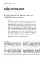
measured at an absorbance of 450 nm. Analysis of nasal
turbinates collected on 3 DPI showed similar titers
regardless of viral dose administered at challenge. Two
animals from group 6 and one animal from group 5
showed presence of virus in the lungs (Figure 2A). Results
showed dose-dependent infection in throat swabs for 1
and 3 DPI. Dose dependence is also seen for the nasal
swabs on 1 DPI (Spearman-Rho non-parametric testing,
r
s
> 0.85, Figure 2B and 2C). By 7 DPI, all groups returned
to baseline levels, indicating that the animals cleared the
infection (Figure 2B and 2C).
Immunological parameters were evaluated using virus
specific ferret IgG and IgM ELISA on sera collected on 3
and 7 DPI. Briefly, plates were coated with 1:200 dilution
of stock virus in PBS overnight at 4°C, blocked with 2%
donor goat serum (Sigma Aldrich) in PBS/0.05% v/v
Tween-20 for one hour. Ferret serum was then added and
2-fold serially diluted and incubated at 4°C overnight.
Anti-ferret IgG or IgM-HRP (1:10,000) (Rockland Immu-
nochemicals) was then added and after a one hour incu-
bation at 37°C, TMB substrate was added, the reaction
was stopped using 1 M H
3
PO
4
, and read at absorbance of
450 nm. At 7 DPI, influenza-specific IgM and IgG anti-
bodies increased relative to viral dose administered at
challenge (Spearman-Rho non-parametric testing, r
s
>
0.94, Figure 3A and 3B). No change between pre-immune
and post-immune sera collected at 3 DPI was detected.
Neutralization titer analysis was performed to detect
influenza-specific neutralizing antibodies in serum. Only
sera collected on 7 DPI was evaluated as ELISA results
suggested that no neutralizing antibodies were present on
3 DPI. As expected, no neutralizing antibodies were
detected in sera from control animals. Only 2 of 4 ani-
mals from group 2 and 1 of 4 animals from group 3 had
detectable neutralizing antibodies; however neutralizing
antibodies were seen in all animals in groups 4, 5 and 6
which were challenged with higher doses of virus (Figure
3C). Hematological analyses were also performed on
blood samples collected immediately prior to euthanasia.
Results showed an increase in the number of lympho-
cytes, neutrophils, and the total number of white blood
cells in infected groups compared to control (Figure
3D,E,F). There was only a slight increase in the number of
basophils and eosinophils in groups 4 and 5 compared to
controls.
To conclude, ferrets infected with A/Brisbane/59/2007
H1N1 displayed mild clinical symptoms, with weight loss,
Figure 2 TCID
50
Virus Titration Analysis. Blue dots indicate the titer
of individual animals, the red line indicates the average for the animals
tested in each group and day. For the caudal lung and nasal turbinates
(A), four animals per group were analyzed at 3 DPI. In (B), analysis of
throat and nasal swabs isolated at 1, 3, 5, and 7 DPI is shown. Eight sam-
ples per group were analyzed on 1 and 3 DPI, and four samples per
group were analyzed on days 5 and 7 due to the euthanasia of 50% of
the animals on 3 DPI.
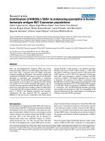
Figure 1 Clinical Pathology of A/Brisbane/59/2007 infected fer-
rets. (A) Control lung tissue; (B) Lung from ferret challenged with 10
3.8
EID
50
/ml with chronic inflammation in the bronchial glands; (C). Lung
from ferret challenged with 10
4.8
EID
50
/ml with bronchiolar epithelial
hypertrophy (white arrow) and, neutrophils and macrophages within
alveoli and airways (black arrow); (D). Lung of ferret challenged with
10
6.8
EID
50
/ml with a syncytium within an alveolus (see blue arrow). Im-
ages were taken at 400x magnification
E
B
CD
A
McBrayer et al. Virology Journal 2010, 7:149
/>Page 4 of 5
sneezing, nasal and ocular discharge as well as histo-
pathological lesions consistent with influenza infection.
Histopathology of the lungs indicated a localized immune
response. Virus titers exhibit dose dependence, with
higher titers early in the course of infection for the higher
doses. Lower doses suggest a delay of virus replication in
the samples tested. Homogenized nasal turbinates
showed a relatively even distribution over time points. In
contrast to a recently published study investigating the
pathological effects of a single dose of A/Brisbane/59/
2007 [19], we detected replicating virus in the lungs,
which indicates that this influenza strain is capable of
inducing infection in tissues of the lower respiratory
tract. High correlation is seen between viral dose at chal-
lenge and the immune response detected by virus specific
IgG and IgM ELISA, the neutralization index, and to the
viral titers of the throat swabs. To conclude, we describe
development of a ferret model for analysis of a seasonal
influenza strain. The results provide key endpoints for
preclinical testing of the efficacy of new antiviral com-
pounds and experiential vaccines.
Abbreviations
CPE: cytopathic effect; DPI: days post-infection; PBS: phosphate buffered saline;
TCID
50
: tissue culture infectious dose 50%.
Competing interests
The authors declare that they have no competing interests.
Authors' contributions
AM: immunological analysis, manuscript preparation, JVC: virological and
immunological analysis, manuscript preparation, RT: virological and immuno-
logical analysis, manuscript preparation, VY: immunological analysis, SG: clinical
pathology analysis, DN: virus preparation, CBJ: participated in design of study,
review of findings and manuscript preparation, CEB: participated in design,
direction of the study, data analysis and manuscript preparation
All authors have read and approved the final manuscript.
Acknowledgements
We would like to give special thanks to Nichole Tower for her editorial assis-
tance. We also thank the technical staff at Southern Research Institute for
excellent assistance on the in-life portion of this study. The study was funded
thorough the contract N01-AI-30063 from the NIH.
Author Details
1
Southern Research Institute, 2000 9th Ave South, Birmingham, AL 35205, USA,
2
Center for Predictive Medicine For Biodefense and Emerging Infectious
Disease, University of Louisville, KY 40292, USA and
3
Department of
Microbiology, Tumor and Cell Biology, Karolinska Institutet, Nobels väg 16, SE-
171 77 Stockholm, Sweden
References
1. Beigel JH: Influenza. Crit Care Med 2008, 36:2660-2666.
2. Lackenby A, Thompson CI, Democratis J: The potential impact of
neuraminidase inhibitor resistant influenza. Curr Opin Infect Dis 2008,
21:626-638.
3. Matsuoka Y, Lamirande EW, Subbarao K: The mouse model for influenza.
Curr Protoc Microbiol 2009, Chapter 15(Unit 15G 13):.
4. Moorman JP: Viral characteristics of influenza. South Med J 2003,
96:758-761.
5. van der Laan JW, Herberts C, Lambkin-Williams R, Boyers A, Mann AJ,
Oxford J: Animal models in influenza vaccine testing. Expert Rev
Vaccines 2008, 7:783-793.
6. Matsuoka Y, Lamirande EW, Subbarao K: The ferret model for influenza.
Curr Protoc Microbiol 2009, Chapter 15(Unit 15G 12):.
7. Kirkeby S, Martel CJ, Aasted B: Infection with human H1N1 influenza
virus affects the expression of sialic acids of metaplastic mucous cells
in the ferret airways. Virus Res 2009, 144:225-232.
8. Maher JA, DeStefano J: The ferret: an animal model to study influenza
virus. Lab Anim (NY) 2004, 33:50-53.
9. Reuman PD, Keely S, Schiff GM: Assessment of signs of influenza illness
in the ferret model. J Virol Methods 1989, 24:27-34.
10. Yen HL, Lipatov AS, Ilyushina NA, Govorkova EA, Franks J, Yilmaz N,
Douglas A, Hay A, Krauss S, Rehg JE, et al.: Inefficient transmission of
H5N1 influenza viruses in a ferret contact model. J Virol 2007,
81:6890-6898.
11. Herlocher ML, Truscon R, Elias S, Yen HL, Roberts NA, Ohmit SE, Monto AS:
Influenza viruses resistant to the antiviral drug oseltamivir:
transmission studies in ferrets. J Infect Dis 2004, 190:1627-1630.
12. Svitek N, Rudd PA, Obojes K, Pillet S, von Messling V: Severe seasonal
influenza in ferrets correlates with reduced interferon and increased IL-
6 induction. Virology 2008, 376:53-59.
13. Leigh MW, Connor RJ, Kelm S, Baum LG, Paulson JC: Receptor specificity
of influenza virus influences severity of illness in ferrets. Vaccine 1995,
13:1468-1473.
14. Nicholls JM, Bourne AJ, Chen H, Guan Y, Peiris JS: Sialic acid receptor
detection in the human respiratory tract: evidence for widespread
distribution of potential binding sites for human and avian influenza
viruses. Respir Res 2007, 8:73.
15. Boltz DA, Rehg JE, McClaren J, Webster RG, Govorkova EA: Oseltamivir
prophylactic regimens prevent H5N1 influenza morbidity and
mortality in a ferret model. J Infect Dis 2008, 197:1315-1323.
16. Lambkin R, Oxford JS, Bossuyt S, Mann A, Metcalfe IC, Herzog C, Viret JF,
Gluck R: Strong local and systemic protective immunity induced in the
ferret model by an intranasal virosome-formulated influenza subunit
vaccine. Vaccine 2004, 22:4390-4396.
Received: 25 June 2010 Accepted: 9 July 2010
Published: 9 July 2010
This artic le is available fro m: http://www.v irologyj.com/co ntent/7/1/149© 2010 McBrayer et al; licensee BioMed Central Ltd. This is an Open Access article distributed under the terms of the Creative Commons Attribution License ( which permits unrestricted use, distribution, and reproduction in any medium, provided the original work is properly cited.Virology Journal 2010, 7:149
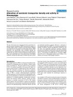
Figure 3 Humoral and Cellular Immunity. (A and B) ELISA data show
an increase in influenza specific IgM and IgG at 7 DPI compared to
mock-infected control animals. These data show that there is a dose-
dependent increase in antibody response. Bars indicate the average
difference per group between log
2
-transformed end-point dilutions
from pre-infection serum and post-infection serum. (C) Neutralization
titer analysis was performed in order to detect the presence of influen-
za-specific neutralizing antibodies in the serum. The presence of neu-
tralizing antibodies was measured only on 7 DPI. No neutralizing
antibodies were detected in the sera from control animals. Only 2 out
of 4 animals from group 2 and 1 out of 4 animals from group 3 had de-
tectable neutralizing antibodies, while all animals in groups 4, 5, and 6
had detectable neutralizing antibodies. (D, E, and F) The number of
neutrophils and lymphocytes, as well as the total number of white
blood cells, increased in animals infected with A/Brisbane/59/07 com-
pared to mock infected control animals. Y axis indicates number of
cells as 10
3
cells per mm
3
.
0
1
2
3
4
5
6
No. of cells
0
1
2
3
4
5
6
Endpoint Titer
B
E
0
1
2
3
4
5
6
7
Neutralization Index
0
2
4
6
8
10
12
14
No. of cells
C
F
0
2
4
6
8
Endpoint Titer
A
D
0
1
2
3
4
5
6
7
No. of cells
McBrayer et al. Virology Journal 2010, 7:149
/>Page 5 of 5
17. Middleton D, Rockman S, Pearse M, Barr I, Lowther S, Klippel J, Ryan D,
Brown L: Evaluation of vaccines for H5N1 influenza virus in ferrets
reveals the potential for protective single-shot immunization. J Virol
2009, 83:7770-7778.
18. Heyward JT, Klimas RA, Stapp MD, Obijeski JF: The rapid concentration
and purification of influenza virus from allantoic fluid. Arch Virol 1977,
55:107-119.
19. Rowe T, Leon AJ, Crevar CJ, Carter DM, Xu L, Ran L, Fang Y, Cameron CM,
Cameron MJ, Banner D, et al.: Modeling host responses in ferrets during
A/California/07/2009 influenza infection. Virology 401:257-265.
doi: 10.1186/1743-422X-7-149
Cite this article as: McBrayer et al., Course of seasonal influenza A/Brisbane/
59/07 H1N1 infection in the ferret Virology Journal 2010, 7:149