Báo cáo Y học: Two 1 : 1 binding modes for distamycin in the minor groove of d(GGCCAATTGG) docx
Bạn đang xem bản rút gọn của tài liệu. Xem và tải ngay bản đầy đủ của tài liệu tại đây (457.85 KB, 10 trang )
Two 1 : 1 binding modes for distamycin in the minor groove
of d(GGCCAATTGG)
Koen Uytterhoeven
1
, Jiri Sponer
2
and Luc Van Meervelt
1
1
Biomolecular Architecture, Department of Chemistry, Katholieke Universiteit Leuven, Belgium;
2
Institute of Biophysics,
Academy of Sciences of the Czech Republic, and National Center for Biomolecular Research, Brno, Czech Republic
Single-crystal X-ray structure determinations of the complex
between the minor-groove binder distamycin and
d(GGCCAATTGG) reveal two 1 : 1 binding modes which
differ in the orientation of the drug molecule in the minor
groove. The two crystals were grown from different cry-
stallization conditions and found to diffract to 2.38 and
1.85 A
˚
, respectively. The structures were refined to comple-
tion using SHELXL-93, resulting in a residual R factor of
20.30% for the 2.38-A
˚
resolution structure (including 46
water molecules) and 19.74% for the 1.85-A
˚
resolution
structure (including 74 water molecules). In both orienta-
tions, bifurcated hydrogen bonds are formed between the
amide nitrogen atoms of the drug and AT base pairs. With a
binding site of at least five base pairs, close contacts between
the terminal distamycin atoms and guanine amino groups
are inevitable. The detailed nature of several of these inter-
actions was further investigated by ab initio quantum
chemical methods.
Keywords: distamycin; drug–DNA complex; minor groove
binder; quantum chemical calculations; X-ray structure.
Distamycin A (Fig. 1) is a member of a family of naturally
occuring oligopeptides showing antiviral and antibiotic
properties. Like other minor-groove binder drugs, distamy-
cin binds noncovalently in the minor groove of DNA with a
binding preference for stretches of AT-rich sequences [1],
thereby preventing DNA and RNA synthesis by inhibition
of the corresponding polymerase reaction. The crystal
structure determination of a 1 : 1 distamycin–d(CGCAAA
TTTGCG) complex (12-dista) at 2.2 A
˚
resolution shows
that the drug covers five of the six AT base pairs [2]. The
amide nitrogen atoms of the drug form hydrogen bonds to
N3(A) and/or O2(T) atoms in the minor groove. The
complex is further stabilized by van der Waals’ and
electrostatic interactions.
The selectivity for AT-rich sequences of minor-groove
binders was first thought to have sterical reasons: the bulky
NH
2
group at the floor of the minor groove of CG-
containing regions can prevent binding of these drugs [3].
More recently, factors such as minor-groove width influen-
cing the extent of van der Waals’ interactions [4] and
electrostatic interactions between the positively charged
drug and the more negatively charged minor groove in the
case of AT sequences [5] were added.
Solution NMR studies have also discovered side-by-
side binding of two distamycin molecules in the minor
groove of d(CGCAATTGCG) [6]. More structural
information about this 2 : 1 binding mode was first
provided by the crystal structure of d(ICICICIC)–dista-
mycin [7] and later by side-by-side complexes of dista-
mycin with natural targets d(ICITACIC), d(ICATATIC)
and d(GTATATAC) [8,9]. Owing to the overlap of about
75%, the two staggered antiparallel distamycin molecules
span almost eight base pairs and are kept together by
dipole–dipole interactions between stacking pyrrole rings
and amide bonds. Each drug hydrogen-bonds with the
bases of only one DNA strand and stacks with the sugar
rings.
We have previously reported the structure determin-
ation at 1.9-A
˚
resolution of the complex of the shor-
ter minor-groove binder 4¢,6-diamidino-2-phenylindole
(DAPI) with d(GGCCAATTGG) (10-DAPI), revealing
a novel off-centered binding with a hydrogen bond
between the drug and a CG base pair [10]. In an attempt
to use similar crystal engineering techniques to improve
the resolution of 1 : 1 distamycin–DNA complexes
(currently 2.2 A
˚
for 12-dista and 2.0 A
˚
for the dista-
mycin–d(CGCGAATTC
+
GCG) complex where C
+
¼
5-methylcytidine (NDB entry code GDLB41), we have
cocrystallized distamycin with the decamer d(GGCCAA
TTGG). Intensity measurements for two crystals obtained
from different crystallization conditions were carried out
to 2.38 and 1.85 A
˚
resolution. Whereas for one crystal the
distamycin orientation and binding site is the same as in
12-dista, the orientation of the drug is inverted in the
other crystal. Both orientations show interactions between
thedrugandguanineNH
2
groups. For the inverted
orientation, the DNA–distamycin interaction is also
characterized by ab initio methods.
Correspondence to L. Van Meervelt, Biomolecular Architecture,
Department of Chemistry, Katholieke Universiteit Leuven,
Celestijnenlaan 200F, B-3001 Leuven (Heverlee), Belgium.
Fax: + 32 16 327990, Tel.: + 32 16 327609,
E-mail:
Abbreviations: 12-dista, crystal structure of the
d(CGCAAATTTGCG) complex (2); 10-DAPI, crystal structure of
the d(GGCCAATTGG)–DAPI complex (10); MPD, 2-methyl-2,
4-pentanediol; DAPI, 4¢,6-diamidino-2-phenylindole; HF, Hartree-
Fock; MP2, Moeller–Plesset perturbational theory.
Note: a web page is available at
/>(Received 20 December 2001, revised 10 April 2002,
accepted 23 April 2002)
Eur. J. Biochem. 269, 2868–2877 (2002) Ó FEBS 2002 doi:10.1046/j.1432-1033.2002.02952.x
EXPERIMENTAL PROCEDURES
Crystallization and data collection
The DNA decamer d(GGCCAATTGG) was purchased
from Oswel DNA service (University of Southampton, UK),
distamycin from Serva Biochemica (Heidelberg, Germany).
Crystals were grown at 16 °C using the sitting drop method
from two different conditions containing 54.4/33.25 m
M
sodium cacodylate buffer (pH 6.0), 35.0/105.0 m
M
MgCl
2
,
70 m
M
NaCl, 8.8% 2-methyl-2,4-pentanediol (MPD),
10.5 m
M
spermine, 0.25/0.42 m
M
ssDNA and 0.125/
0.21 m
M
distamycin against a 50/35% MPD stock solution.
From a bar-shaped crystal of dimensions 0.4 · 0.1 ·
0.05 mm from condition 1, intensity data were collected at
100 K on a MAR345 imaging plate detector at beamline
X11inanEMBLHamburg(k ¼ 0.9116 A
˚
) over a 105 ° u
range with increments of 1.5 ° using cryocooling techniques
with a crystal-to-detector distance of 350 mm.
A well-diffracting crystal of dimensions 0.2 · 0.1 ·
0.05 mm from condition 2 was mounted for data collection
at 100 K using a similar protocol at beamline BW7b in an
EMBL Hamburg (150 ° u range, crystal-to-detector dis-
tance 250 mm, k ¼ 0.8423 A
˚
).
Data were processed using the
DENZO
/scalepack [11]
suite of programs. Data collection statistics for both crystals
are given in Table 1. The final resolution limit of the
diffraction pattern was 2.38 A
˚
for crystal 1 and 1.85 A
˚
for
crystal 2.
Structure solution and refinement
Unit cell parameters and space group indicated isomorph-
ism with the d(GGCCAATTGG)–DAPI structure, which
was used as a starting model (NDB entry code DD0002,
except DAPI and solvent molecules) for further refinement
on F
2
using SHELXL-93 [12]. The nucleotides of strand 1
are labeled G1–G10 in the 5¢fi3¢ direction and G11–G20
on strand 2, and the drug is labeled D. After determination
of the weighting factor and positional adjustment of parts of
the DNA structure, water molecules were added, but not in
the minor-groove region. At this stage of the refinement, the
distamycin molecule was located in the (F
o
) F
c
) Fourier
difference map. In subsequent refinement cycles, more water
molecules were gradually added. During the conjugate-
gradient refinement, no torsion angle or hydrogen-bond
restraints were applied. The 1,2 and 1,3 distances used as
dictionary values for distamycin were based on netropsin
[13], except for the formamide end which was based on
fragments retrieved from the Cambridge Structural
Database [14].
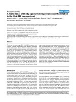
Fig. 1. Structure and numbering scheme of distamycin. Hydrogen
atoms attached to pyrrole and alkyl groups are not shown.
Table 1. Data collection and refinement statistics of the d(GGCCAATTGG)–distamycin complexes. NA, Not available.
Crystal 1 Crystal 2
Data collection statistics
Space group P2
1
2
1
2
1
P2
1
2
1
2
1
Unit cell (A
˚
)a¼ 26.011, b ¼ 40.861, c ¼ 53.164 a ¼ 25.289, b ¼ 36.439, c ¼ 53.047
Total no. of measured reflections 27524 43223
No. of independent reflections 2538 4223
Resolution (A
˚
) 2.38 1.85
Multiplicity 5.5 4.3
v
2
1.035 1.142
R
symm
(%) 4.1 (100.0–2.38 A
˚
) 4.9 (100.0–1.85 A
˚
)
25.1 (2.42–2.38 A
˚
) 19.5 (1.92–1.85 A
˚
)
Completeness (%) 92.7 (100.0–2.38 A
˚
) 93.0 (100.0–1.85 A
˚
)
93.4 (2.42–2.38 A
˚
) 96.9 (1.92–1.85 A
˚
)
Mean I/r(I) 22.6 21.1
Reflections with I >3 r(I) (%) 80.3 (100.0–2.38 A
˚
) 80.3 (100.0–1.85 A
˚
)
46.3 (2.42–2.38 A
˚
) 55.3 (1.92–1.85 A
˚
)
Refinement statistics
Resolution range (A
˚
) 100.0–2.38 100.0–1.85
R value/R
free
value (%) 20.30/NA 19.74/27.80
No. of nonsolvent atoms 445 445
No. of water molecules 46 74
Average B values of DNA (A
˚
2
) 66.6 28.2
Average B values of distamycin (A
˚
2
) 79.1 30.8
Average B values of water molecules (A
˚
2
) 73.0 43.0
Rmsd of bond lengths (A
˚
) 0.018 0.019
Rmsd of bond angles (°) 3.20 2.70
Ó FEBS 2002 Two 1 : 1 binding modes for distamycin (Eur. J. Biochem. 269) 2869
For crystal 1, the R value converged to 20.30% after
addition of 46 water molecules (R
free
was not used to avoid
further reduction of the number of data per parameter at
this resolution). For crystal 2, the first maps already
indicated an inverted orientation (hereafter called orienta-
tion B) of the drug molecule with respect to crystal 1
(orientation A). Therefore refinement for crystal 2 was
monitored using R
free
calculated for a reference set of 10%
of the reflections. Addition of 29 water molecules and
distamycin led to the following R values: R ¼ 23.88%,
R
free
¼ 32.31% for orientation A, and R ¼ 22.79%,
R
free
¼ 30.75% for orientation B. Final R values for crystal
2wereR ¼ 19.74%, R
free
¼ 28.01% for orientation B. An
independent refinement with orientation A resulted in
R ¼ 20.30%, R
free
¼ 29.84%. As a consequence and in
agreement with the electron density maps, orientation B was
retained for crystal 2.
Figure 2 shows the final (2F
o
) F
c
) electron-density maps
in the minor-groove region for both crystals. Refinement
statistics are presented in Table 1.
The helical parameters in accordance with the Tsukuba
Workshop guidelines [15], and torsion angles were calcula-
tedwiththe3
DNA
program [16].
Quantum chemical calculations
Ab initio quantum chemical calculations were used to
investigate intrinsic molecular interactions of a number of
close intergroup contacts observed in the crystal. Special
attention was given to contacts involving the guanine amino
groups. Appropriate fragments of the drug and several
proximal bases have been taken from the crystal structure.
Intermolecular positions of the interacting species were
frozen based on the crystal data using a set of constraints
involving three (in some cases more) appropriate non-
hydrogen atoms on each monomer. The rest of the structure
including all the hydrogen positions was relaxed using
gradient optimization. This procedure has been extensively
used in the past to investigate local contacts seen in DNA
crystal structures and allows full relaxation of the electronic
structure and hydrogen positions while keeping the systems
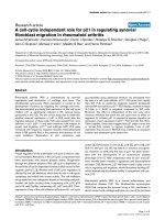
Fig. 2. Final (F
o
) F
c
) electron-density maps in the minor groove of the crystal structure of the d(GGCCAATTGG)–distamycin complex in which the
drug has been omitted from the final refined model. (A) Crystal 1 in orientation A; (B) crystal 2 in orientation B. The refined distamycin position is
superimposed on the difference density for reference, contouring at 1 r (yellow) and 2 r level (green). The figure was prepared with
BOBSCRIPT
[34]
and
RASTER
3
D
[35].
2870 K. Uytterhoeven et al.(Eur. J. Biochem. 269) Ó FEBS 2002
studied in the experimental geometry [10,17–20]. The
optimizations were carried out within the Hartree–Fock
(HF) approximation with the standard polarized 6–31G*
basis set. Although this level of calculations underestimates
the flexibility of amino groups, it nevertheless is sufficient to
reveal when the amino group is activated by molecular
interactions towards a partial sp
3
hybridization [21–23].
Interaction energy calculations between drug fragments
and nucleobases were carried out assuming the quantum
chemical-optimized geometries with inclusion of electron
correlation effects using the second-order Moeller-Plesset
perturbational theory (MP2) with the 6–31G basis set
augmented by diffuse d-polarization functions to all sec-
ond-row elements [exponents of 0.25, designated as
6–31G*(0.25)] to properly account for the dispersion
attraction [22,23]. The calculations were corrected for the
basis set superimposition error using the standard counter-
poise procedure. The quantum chemical procedure used in
this study allows reliable semiquantitative characterization
of the nature and intrinsic Ôin vacuoÕ strength of the observed
intermolecular contacts. All quantum chemical calculations
were carried out using the
GAUSSIAN
94 program suite [24].
RESULTS AND DISCUSSION
Both complexes (Fig. 3) have a conformation that closely
resembles the native decamer structure [25] and its DAPI
complex [10]: a central octamer d(CCAATTGG) consisting
of normal Watson–Crick base pairs is at both ends flanked
by two overhanging guanines forming triplets in the crystal
packing. All three drug–decamer complexes differ from the
native 1.15-A
˚
resolution structure in the phosphate confor-
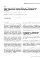
Fig. 3. Stereoscopic representation of the
distamycin–d(GGCCAATTGG) complexes.
(A) Crystal 1 in orientation A; (B) crystal 2 in
orientation B. The DNA is drawn with open
bonds, and the distamycin with solid bonds.
Nitrogen, oxygen and phosphor atoms are
grey, and carbon atoms are white. The figure
was prepared using B
OBSCRIPT
[34].
Ó FEBS 2002 Two 1 : 1 binding modes for distamycin (Eur. J. Biochem. 269) 2871
mation of residue C13, which correlates with the difference
in resolution as described previously [10]. As expected for
B-DNA, the sugar puckering modes for the central octamer
duplex are situated in the normal C3¢-exo to O4¢-endo
range, the southern part (C2¢-endo) of the pseudorotation
cycle. However, for the overhanging guanines, C3 and C13,
the puckering modes are closer to those for A-DNA, which
is a consequence of triplet formation.
DNA–distamycin interactions
Distamycin binds in the minor groove of the central
octamer. The expected binding site of at least five base
pairs makes interactions with GC base pairs inevitable.
Both structures not only differ in resolution, but also in the
orientation of the distamycin molecule (Fig. 4). In the lower
resolution structure of crystal 1, the orientation (orienta-
tion A) is similar to that described for 12-dista [2]. The
higher-resolution structure of crystal 2 shows an inverted
orientation of distamycin (orientation B).
Position of distamycin in the lower-resolution structure
(orientation A, Fig. 4A)
Distamycin binds to five base pairs covering the sequence
d(AATTG). The positively charged amidinium end group is
orientated toward the A5.T18 base pair with atom N9(D)
lying deep in the minor groove interacting with N3(A5)
(2.70 A
˚
) and the partial negative O4¢ atoms O4¢(A5)
(3.29 A
˚
)andO4¢(A6) (2.80 A
˚
). A close contact with the
amino group of G19 is avoided (N9(D)…N2(G19) 3.86 A
˚
).
The nearest neighbor of N8(D) is O4¢(3.41 A
˚
). Bifurcated
hydrogen bonds are formed between the amide nitrogen
atoms simultaneously to both N3(A6) and O2(T18) for
N7(D), O2(T7) and O2(T17) for N5(D), and N3(A15) and
N2(G9) for N1(D). N3(D) only has one close contact to
N3(A16) and is 3.59 A
˚
away from O2(T8). At the other end
of distamycin, the formamide O1(D) is oriented away from
the groove. The nearest neighbor of O1(D) is a sugar
O4¢(A15) atom at 3.50 A
˚
.
The three pyrrole rings are rotated to each other to
follow closely the curvature of the groove. Rings A and B
make an angle of 22.9 °, rings B and C 28.4 ° and ring C
makes an angle of 35.9 ° with the terminal amidinium
group. The drug–DNA complex is further stabilized by
van der Waals’ interactions mainly between carbon atoms
of the three pyrrole rings and the sugar–phosphate
backbone. No water molecules are in direct contact with
the drug molecule.
Position of distamycin in the higher-resolution
structure (orientation B, Fig. 4B)
It was clear from the density maps of the 1.85-A
˚
resolution
structure that the drug has to be rotated over 180 ° with
respect to orientation A. As a consequence, the amidinium
group is now orientated toward the G9–C12 base pair and
distamycin now interacts with six base pairs.
The N9(D) atom of the amidinium group is in close
contact with amino group N2 of guanine G9 (3.16 A
˚
)and
hydrogen-bonded with N3(A15) (2.85 A
˚
). This N9(D) atom
is also in contact with O2(C14) by two intermediate water
molecules.
The O1(D) atom of the formamide group now points
toward the C4–G19 base pair with a very close contact with
N2(G19)ofonly2.57A
˚
. Atom O1(D) is further in contact
with O2(C4) (3.39 A
˚
), O4¢(A5) (2.94 A
˚
)andtwowater
molecules (2.77 and 2.95 A
˚
). In the same formamide group,
atom N1(D) interacts with N3(A5) (3.12 A
˚
)andO4¢(A6)
(3.03 A
˚
).
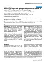
Fig. 4. Schematic view of the distamycin interactions in the minor groove of d(GGCCAATTGG)–distamycin structures. (A) Crystal 1; (B) crystal 2.
2872 K. Uytterhoeven et al.(Eur. J. Biochem. 269) Ó FEBS 2002
The nitrogen atoms of the amide linkers between the
pyrrole rings form bifurcated hydrogen bonds with the
thymine or adenine bases. The central N5(D) atom of
the drug is hydrogen-bonded to the two O2 atoms of the
thymine bases of the central AT steps. Nitrogen atoms
N3(D) and N7(D) bridge N3(adenine) and O2(thymine)
atoms of opposite strands. Furthermore, several stabilizing
van der Waals’ contacts between the O4¢ atoms of the DNA
sugars and distamycin atoms are observed.
Rings A and B make an angle of 8.6 °,ringsBandC
21.1 °, and ring C makes an angle of 15.7 ° with the terminal
amidinium group.
Quantum chemical analysis of the base–distamycin
contacts
For both orientations, the crystal structure reveals a number
of close intermolecular contacts including interactions
between the guanine amino groups and the terminal
distamycin atoms. Such amino-group interactions are often
assumed to be repulsive steric clashes. To clarify the nature
of the selected interactions, we used quantum chemical
calculations carried out at the MP2/6–31*(0.25)//HF/
6–31G* approximation (see Experimental procedures). As
the interactions were most surprising for the 1.85-A
˚
resolution structure, the calculations were only performed
for orientation B. Further, such calculations are usually
only performed for systems of resolution 2 A
˚
and better, as
outcomes of such calculations may be spoiled by inaccurate
positioning of the interacting groups.
The contact area of the distamycin amidinium end was
investigated by evaluating the hydrogen positions and
interaction energy terms for the interactions between the
terminal drug segment and the bases of base pair A15–T8
and base G9 (Table 2, Fig. 5). The drug binding to A15 is
very strong, with substantial electrostatic and dispersion
components indicating a strong hydrogen bond between
N9(D) and N3(A15). The intrinsic strength of this interac-
tion is halfway between CG Watson–Crick and AT
Watson–Crick base pairs [22,23]. Furthermore the drug
also recognizes T8 by a long-range electrostatic interaction
between N7(D) and O2(T8). The long-range nature of this
contact can be deduced considering both the geometry and
the negligible correlation component to the interaction
energy. The interaction with G9 is an energetically neutral
van der Waals’ contact with an attractive correlation
(dispersion) component and thus a repulsive HF compo-
nent. This means no active recognition. The guanine amino
group is certainly influenced by this contact. It has a
substantial pyramidal character: both hydrogen atoms
deviate 0.39 and 0.49 A
˚
from the best plane through the
base atoms. The amino-group nonplanarity is modestly
enhanced over isolated guanine optimized by the same
method (hydrogen deviations 0.10 and 0.41 A
˚
, respectively).
Note also that, because of increased propeller twisting of the
GC base pair, the cytosine base (its O2 atom) not involved
in the calculation helps to lead the guanine amino-group
hydrogen atoms away from the drug. A search in the
Cambridge Structural Database [14] shows that most amino
groups in guanine fragments are almost planar. Note,
however, that in vacuo the amino group is substantially
nonplanar [21–23], and this electronic structure feature may
reoccur in DNA when intermolecular interactions profit
from the nonplanarity [10,17–19,21–23]. This has been
observed previously for the DAPI–DNA complex [10] and
is seen to a lesser extent here also.
The interaction between the neutral formamide part of
the drug at the other end and the proximal base pairs
A5–T18 and C4–G19 is less favourable than the amidi-
nium–DNA interactions because of the neutral character of
this end (Table 2, Fig. 5). The composition of the interac-
tion energy shows that the drug interacts with the AT pair
more in a van der Waals’ than in a hydrogen-bonding
manner. (Hydrogen-bonded systems are always dominated
by a strong HF component, which could be obtained as the
difference between the total interaction energy and the
correlation component in Table 2.) The contact with the CG
base pair is weakly repulsive, but not enough to prevent
binding. The contact with C4 is indeed not favorable
because of the proximity of the two oxygen atoms O1(D)
and O2(C4) (3.39 A
˚
). Despite the very short distance
between O1(D) and N2(G19), the energy components show
that this contact is not a strong hydrogen bond at all. This
is also clear from the amino-group hydrogen positions,
pointing away from O1(D). To obtain an upper limit of the
possible attraction that can be achieved by this part of the
drug and a CG base pair, we alleviated all constraints during
the optimization. A completely unconstrained gas phase
optimization of the formamide part and base pair C4–G19
illustrates that not much of the hydrogen-bonding potential
is used. In the most optimal binding, the interaction energy
between G19 and the formamide fragment would be
)35.1 kJÆmol
)1
(E
cor
¼ )4.2 kJÆmol
)1
). However, this
optimal geometry is not possible because of the interactions
of the rest of the distamycin with the oligonucleotide. We
also carried out some additional optimizations with differ-
ent constraints (not shown). These confirmed that in the
refined crystal geometry the drug is rather too tightly
packed against the G19 base. The interaction can be
improved by locally relaxing the drug geometry, but no
hydrogen bond can be obtained close to the experimental
geometry. It should also be noted that the CG–formamide
end interaction might be supported by two crystallographic
water molecules (O116 and O126). Although several
Table 2. Total interaction energy and correlation component for the
interaction between terminal distamycin fragments and bases in close
contact with distamycin evaluated at the MP2/6–31G*(0.25)//HF/
6–31G* approximation and utilizing constraints taken from the crystal.
The electron correlation component of the interaction energy includes
the dispersion attraction and corrections to electrostatic and polar-
ization terms.
Base
Interaction
energy
(kJÆmol
)1
)
Correlation
component
(kJÆmol
)1
)
Amidinium end
A15 )81.1 )23.4
T8 )48.5 )1.3
G9 2.5 )19.6
Formamide end
A5 )19.6 )18.8
T18 )12.5 )1.3
C4 10.9 )5.4
G19 0.0 )14.6
Ó FEBS 2002 Two 1 : 1 binding modes for distamycin (Eur. J. Biochem. 269) 2873
optimization attempts (not shown) did not result in a
converged structure, the intermediate structures suggest that
the water molecules are actively involved.
Influence of the binding of distamycin on the DNA
conformation
One of the most important effects of drug binding on the
DNA conformation is the widening of the minor groove as
illustrated in Fig. 6. The width of the groove is measured by
taking the shortest H5¢–H4¢ distances between the two
DNA chains. In the native structure, the minor-groove
width is symmetric. Both distamycin and DAPI have a
similar asymmetric opening effect: the widening is more
pronounced at the 3¢ end of the first strand. Where DAPI
opens the groove over a distance of three to four base pairs,
distamycin has an effect on at least five base pairs. The
opening of the groove by distamycin (by 4A
˚
)ismore
pronounced than with DAPI (1.5 A
˚
). In the 2 : 1 side-by-
side complexes, the minor groove expands to 7.7 A
˚
in
order to accommodate two distamycin molecules.
Distamycin makes more close contacts with the atoms in
the minor groove in orientation A than in orientation B
(Table 3). For both orientations, the number of contacts
with both strands is almost equal, which was less empha-
sized for 12-dista.
The complexation of distamycin has no major effect on
interbase parameters (buckle, propellor twist, and opening)
Fig. 6. Comparison of the minor-groove width based on H4¢–H5¢ dis-
tances for the native decamer (blue) and its complex with distamycin
(black for the 2.38 A
˚
and red for the 1.85 A
˚
resolution structure) and
DAPI (green).
Fig. 5. Optimized geometry based on HF/6–31G* calculations of the interaction between (A) the amidinium end and bases A15, T8 and G9, and (B) the
formamide end and bases C4 and G19. Intermolecular geometry frozen according to the crystal data. Drug fragment atoms are yellow.
2874 K. Uytterhoeven et al.(Eur. J. Biochem. 269) Ó FEBS 2002
and cartesian neighboring base parameters (tilt, roll, twist,
shift slide, and rise). However, some small adaptions such as
the increased propeller twist of G14–G9 in orientation B are
observed, and are necessary to optimize the complexation.
Also the base stacking patterns are very similar to those
observed for the native decamer.
CONCLUSIONS
Both current crystal structures describe the interaction of
distamycin in the minor groove of the central CAATTG
sequence in a 1 : 1 binding mode. The present tight crystal
packing due to the triplet formation is not compatible with a
2 : 1 drug binding mode, which requires a much broader
minor groove. The opposite drug orientations in the minor
groove are despite the pseudo-twofold symmetry of the
palindromic sequence distinguishable because of the different
triplet formation at both ends of the oligonucleotide. The
length of the minor-groove binder makes contacts with the
G-NH
2
group at both ends of the drug inevitable. For the
novel orientation B, analyis of the absolute interaction
energies obtained by quantum chemical methods shows that
the presence of both G-NH
2
does not destabilize the
distamycin binding to an extent that it prevents complexa-
tion.
The amidinium end of the drug does not recognize G9
actively, but this region optimizes its conformation with
respect to the available space. The amidinium fragment ÔsitsÕ
on the G base; as a consequence, the G-NH
2
group becomes
pyramidal and the propeller twist of base pair G9–C14
increases by 5 ° compared with the native decamer, helping
the pyramidalization. Thus the DNA structure adapts to
host the drug molecule, including a modest amino-group
pyramidalization.
The contacts of the atoms O1(D) and N1(D) at the other
side of the drug are more complicated. The interaction
energy with A5 of )19.6 kJÆmol
)1
is close to those of a
water dimer. Combined with the N1(D)…N3(A5) distance
of 3.12 A
˚
, this could possibly lead to the conclusion that a
good hydrogen bond is formed. However, the composition
of the interaction energy (the dominating dispersion
component) and the N1(D)-H…N3(A5) angle (137 °)do
not support this view. The short contact between atoms
O1(D) and N2(G19) (2.57 A
˚
) again cannot be classified as
a strong hydrogen bond. Optimal hydrogen bonding in this
region as located by an unconstrained gas phase optimi-
zation is not possible here because of the other DNA–
distamycin interactions. It is helpful to check the electron
density again in this region: both O1(D) and C1(D) are not
in reasonable (2F
o
– F
c
) electron density, whereas the rest
of the drug molecule fits these maps nicely (Fig. 2B). Also
the temperature factors of these two atoms are much
higher than those of the other distamycin atoms (Fig. 7).
Most probably, this end of the drug has more than one
conformation, the average of which is observed. This
illustrates the use of quantum chemical calculations in
further analysis of crystallographic results, a combination
Table 3. Close contacts of atoms in the minor groove of d(GGCCAATTGG) or d(CGCAAATTTGCG) and distamycin atoms. Distances less than
3.6 A
˚
are considered close contacts.
d(GGCCAATTGG) + distamycin (orientation A)
C4 G19 C4¢,O4¢,C5¢ 3
A5 C1¢,O4¢, N3, C4 T18 C4¢,C5¢, O2, O4 8
A6 C1¢,C4¢,O4¢,C5¢, N3 T17 C1¢,C4¢,O4¢,O2 9
T7O3¢,C4¢,O4¢,C5¢, O2 A16 C4¢,O4¢,N3 8
T8O3¢,C4¢,C5¢, O2 A15 C1¢,O4¢, C2, N3, C4 9
G9 C4¢,O4¢,C5¢, O1P, N2, N3 C14 6
d(GGCCAATTGG) + distamycin (orientation B)
C4 O2 G19 C4¢,O4¢,C5¢,N2 5
A5 C1¢,O4¢, N3 T18 C4¢,O4¢,C5¢,O2 7
A6 C4¢,O4¢, N3 T17 C4¢,O4¢,O2 6
T7C4¢,O4¢,C5¢, O2 A16 C1¢,C4¢,O4¢, C2, N3 9
T8C4¢,O4¢,C5¢, O2 A15 C2, N3 6
G9 O4¢, N2, N3 C14 3
d(CGCAAATTTGCG) + distamycin (12-dista)
A4 C2, N3 T21 C4¢,O4¢,C5¢,O2 6
A5 C2, N3 T20 C4¢,O4¢,C5¢,O2 6
A6 C1¢,C4¢,O4¢,C5¢, C2, N3, C4 T19 7
T7C4¢ A18 N3 2
T8 O2 A17 1
T9C4¢,O4¢,C5¢ A16 3
Fig. 7. Temperature factors for distamycin in orientation B (crystal 2).
Red are high (B 55 A
˚
2
) and white are low (B 20A
˚
2
) temperature
factors. The figure was prepared using
BOBSCRIPT
[34].
Ó FEBS 2002 Two 1 : 1 binding modes for distamycin (Eur. J. Biochem. 269) 2875
that is so far unique in the field of biological macromol-
ecules. We plan to investigate this binding mode by an
extensive explicit-solvent molecular dynamics simulation in
the near future.
Competition experiments demonstrate that distamycin is
capable of replacing netropsin in its 1 : 1 and 2 : 1
complexes with DNA [26]. Compared with netropsin, both
distamycin orientations indeed bind better in the minor
groove [27,28].
Different 1 : 1 binding modes and orientations have been
reported for several minor-groove binders such as netropsin
[27,28] and Hoechst 33258 [29–32]. In the case of two
orientations fitting the electron density equally well, one can
conclude that both orientations in the minor groove are
energetically very similar, or that the resolution of the
crystal structure determination is not high enough. We have
shown that, for DAPI and distamycin, crystal engineering
techniques may overcome the problem of interpreting
electron-density maps. In our case in which both orienta-
tions occur in different crystals, one can also conclude that
the two distamycin orientations in the d(GGCCAATTGG)
minor groove are energetically equivalent. However, as the
1 : 1 association of distamycin to AT-rich sequences is
extremely fast [33], why is there no disorder in our
structures? Here the charged character of dystamycin may
play an important role. The three orthogonal twofold screw
axes present in the space group P2
1
2
1
2
1
in general prevent
similar parts of a molecule being in each others neighbor-
hood. Adaption of this packing scheme by the drug without
orientational disorder means that the distance between the
positive distamycin ends is always maximal, a situation that
is electrostatically more favourable.
ACKNOWLEDGEMENTS
This work was partly supported by the European Community–Access
to Research Infrastructure Action of the Improving Human Potential
Programme to the EMBL Hamburg Outstation, contract number:
HPRI-CT-1999-00017, and by the Fund for Scientific Research
(Flanders). We thank the staff of the EMBL Hamburg Outstation
for assistance. J. S. was supported by grant LN00A016 (National
Center for Biomolecular Research), MSMT CR. All quantum chemical
calculations were carried out at the Supercomputer Center, Brno.
REFERENCES
1. Zimmer, C. & Wahnert, U. (1986) Nonintercalating DNA-bind-
ing ligands: specificity of the interaction and their use as tools in
biophysical, biochemical and biological investigations of the
genetic material. Prog. Biophys. Mol. Biol. 47, 31–112.
2. Coll, M., Frederick, C.A., Wang, A.H J. & Rich, A. (1987) A
bifurcated hydrogen-bonded conformation in the d(AT) base pair
of the DNA dodecamer d(CGCAAATTTGCG) and its complex
with distamycin. Proc. Natl Acad. Sci. USA 84, 8385–8389.
3. Wartell, R.M., Larson, J.E. & Wells, R.D. (1974) Netropsin-
specific probe for A-T regions of duplex deoxyribonucleic acid.
J. Biol. Chem. 249, 6719–6731.
4. Neidle, S. (1992) Minor-groove width and accessibility in B-DNA
drug and protein complexes. FEBS Lett. 298, 97–99.
5. Pullman, B. (1983) Electrostatics of polymorphic DNA. J. Biomol.
Struct. Dyn. 1, 773–794.
6.Pelton,J.G.&Wemmer,D.E.(1989)Bindingmodesof dis-
tamycin A with d(CGCAAATTTGCG)
2
determined by two-
dimensional NMR. J. Am. Chem. Soc. 112, 1393–1399.
7. Chen, X., Ramakrishnam, B., Rao, S.T. & Sundaralingam,
M. (1994) Binding of two distamycin A molecules in the minor
groove of an alternating B-DNA duplex. Nat. Struct. Biol. 1,
169–175.
8. Chen, X., Ramakrishnan, B. & Sundaralingam, M. (1997) Crystal
structures of the side-by-side binding of distamycin to AT-con-
taining DNA octamers d(ICITACIC) and d(ICATATIC). J. Mol.
Biol. 267, 1157–1170.
9. Mitra, S.N., Wahl, M.C. & Sundaralingam, M. (1999) Structure
of the side-by-side binding of distamycin to d(GTATATAC)
2
.
Acta Crystallogr. D55, 602–609.
10. Vlieghe, D., Sponer, J. & Van Meervelt, L. (1999) Crystal structure
of d(GGCCAATTGG) complexed with DAPI reveals novel
binding mode. Biochemistry 38, 16443–16451.
11. Otwinowski, Z. & Minor, W. (1997) Processing of X-ray diffrac-
tion data collected in oscillation mode. Methods Enzymol. 276,
307–326.
12. Sheldrick, G.M. & Schneider, T.R. (1997) SHELXL: high-
resolution refinement. Methods Enzymol. 277, 319–343.
13.Berman,H.,Neidle,S.,Zimmer,C.&Thrum,H.(1979)
Netropsin, a DNA-binding oligopeptide structural and binding
studies. Biochim. Biophys. Acta 561, 124–131.
14. Allen, F.H. & Kennard, O. (1993) 3D search and research using
the Cambridge Structural Database. Chem. Design Automation
News 8, 1 & 31–37.
15. Olson, W.K., Bansal, M., Burley, S.K., Dickerson, R.E., Gerstein,
M., Harvey, S.C., Heinemann, U., Lu, X J., Neidle, S., Shakked,
Z., Sklenar, H., Suzuki, M., Tung, C S., Westhof, E., Wolberger,
C. & Berman, H.M. (2001) A standard reference frame for the
description of nucleic acid base-pair geometry. J. Mol. Biol. 313,
229–237.
16. Lu, X J., Shakked, Z. & Olson, W.K. (2000) A-DNA con-
formational motifs in ligand-bound double helices. J. Mol. Biol.
300, 819–840.
17. Sponer, J. & Hobza, P. (1994) Bifurcated hydrogen bonds in DNA
crystal structures. An ab initio quantum chemical study. J. Am.
Chem. Soc. 116, 709–714.
18. Sponer, J., Florian, J., Leszczynski, J. & Hobza, P. (1996) Non-
planar DNA base pairs. J. Biomol. Struct. Dyn. 13, 827–833.
19. Luisi, B., Orozco, M., Sponer, J., Luque, F.J. & Shakked, Z.
(1998) On the potential role of the amino nitrogen atom as a
hydrogen acceptor in macromolecules. J. Mol. Biol. 279, 1123–
1136.
20. Sponer, J., Florian, J., Ng, H K., Sponer, J.E. & Spackova, N.
(2000) Local conformational variations observed in B-DNA
crystals do not improve base stacking. Computational analysis of
base stacking in d(CATGGGCCCATG)
2
B«Aintermediate
crystal structure. Nucleic Acids Res. 24, 4893–4902.
21. Sponer, J. & Hobza, P. (1994) Nonplanar geometries of DNA
bases. Second order Moller–Plesset study. J. Phys. Chem. 98,
3161–3164.
22. Hobza, P. & Sponer, J. (1999) Structure, energetics, and dynamics
of the nucleic acid base pairs: nonempirical ab initio calculations.
Chem. Rev. 99, 3247–3276.
23. Sponer, J., Leszczynski, J. & Hobza, P. (2001) Electronic prop-
erties, hydrogen bonding, stacking and cation-binding of DNA
and RNA bases. Biopolymers 61, 3–21.
24. Frisch, M.J., Trucks, G.W., Schlegel, H.B., Gill, P.M.W.,
Johnson, B.G., Robb, M.A., Cheeseman, J.R., Keith, T.,
Petersson, G.A., Montgomery, J.A. et al. (1995) GAUSSIAN.
Gaussian, Inc, Pittsburgh, PA.
25. Vlieghe, D., Van Meervelt, L., Dautant, A., Gallois, B., Precigoux,
G. & Kennard, O. (1996) Parallel and antiparallel (G.GC)
2
triple
helix fragments in a crystal structure. Science 273, 1702–1705.
26. Lah, J. & Vesnaver, G. (2000) Binding of distamycin A and
netropsin to the 12mer DNA duplexes containing mixed AT.GC
2876 K. Uytterhoeven et al.(Eur. J. Biochem. 269) Ó FEBS 2002
sequences with at most five or three successive AT base pairs.
Biochemistry 39, 9317–9326.
27. Coll, M., Aymami, J., Van der Marel, G.A., van Boom, J.H.,
Rich, A. & Wang, A.H J. (1989) Molecular structure of the
netropsin–d(CGCGATATCGCG) complex: DNA conformation
in an alternating AT segment. Biochemistry 28, 310–320.
28. Goodsell, D.S., Kopka, M.L. & Dickerson, R.E. (1995) Refine-
ment of netropsin bound to DNA: bias and feedback in electron
density map interpretation. Biochemistry 34, 4983–4993.
29. Quintana, J.R., Lipanov, A.A. & Dickerson, R.E. (1991) Low-
temperature crystallographic analysis of the binding of Hoechst
33258 to the double-helical DNA dodecamer C-G-C-G-A-A-T-T-
C-G-C-G. Biochemistry 30, 10294–10306.
30. Pjura, P., Grzeskowiak, K. & Dickerson, R.E. (1987) Binding of
Hoechst 33258 to the minor groove of B-DNA. J. Mol. Biol. 197,
257–271.
31. Teng, M.K., Frederick, C.A., Usman, N. & Wang, A.H J. (1988)
The molecular structure of the complex of Hoechst 33258 and the
DNA dodecamer d(CGCGAATTCGCG). Nucleic Acids Res. 16,
2671–2690.
32. Carrondo, M., Coll, M., Aymami, J., Wang, A.H J., Van der
Marel, G.A., van Boom, J.H. & Rich, A. (1989) Binding of
Hoechst dye to d(CGCGATATCGCG) and its influence on the
conformation of the DNA fragment. Biochemistry 28, 7849–7859.
33. Baliga, R. & Crothers, D.M. (2000) On the kinetics of distamycin
binding to its target sites on duplex DNA. Proc. Natl Acad. Sci.
USA 97, 7814–7818.
34. Esnouf, R.M. (1999) Further additions to Molscript version 1.4
including reading and contouring electron-density maps. Acta
Cryst. D55, 938–940.
35. Merritt, E.A. & Murphy, M.E.P. (1994) RASTER3D version 2.
Acta Crystallogr. D50, 869–873.
Ó FEBS 2002 Two 1 : 1 binding modes for distamycin (Eur. J. Biochem. 269) 2877