Báo cáo y học: " Hepatitis B virus genotypes and precore and core mutants in UAE patients" ppt
Bạn đang xem bản rút gọn của tài liệu. Xem và tải ngay bản đầy đủ của tài liệu tại đây (725.36 KB, 8 trang )
Alfaresi et al. Virology Journal 2010, 7:160
/>Open Access
RESEARCH
© 2010 Alfaresi et al; licensee BioMed Central Ltd. This is an Open Access article distributed under the terms of the Creative Commons
Attribution License ( which permits unrestricted use, distribution, and reproduction in
any medium, provided the original work is properly cited.
Research
Hepatitis B virus genotypes and precore and core
mutants in UAE patients
Mubarak Alfaresi*, Abida Elkoush, Hajer Alshehhi, Azza Alzaabi and Adeel Islam
Abstract
Background: Knowledge of the HBV genotype with which a patient is infected is crucial information for a physician to
have when planning clinical treatment for that patient. Previous studies have suggested that there are possible
differences in the pathogenicity and therapeutic response of different HBV genotypes. However, the prevalence of the
various HBV genotypes and Precore and Core mutations is unknown in the UAE. Therefore, we sought to determine the
prevalence of the different HBV genotypes in the UAE population.
Methodology/Principal Findings: A total of 88 HBsAg-positive patients were included in the study.
A method for genotyping and subtyping HBV by partial HBsAg gene sequencing using primers that are
complementary to all known genotypes was used. Precore and core region of these viruses were also sequenced in 88
patients.
HBV genotype D was the most prevalent (79.5%) genotype identified in our study population, followed by genotypes
A (18.2%) and C (2.3%). The following subtypes were isolated: ayw2 (80.7%), adw2 (14.8%), and adw (2.3%). The HBV-
DNA viral load was higher in HBeAg-positive patients than it was in patients who were HBeAg-negative. Precore
mutants were found in 51 (58.0%) of 88 patients. Mutations in the basal core promotor were found in 22 (25.3%) of 88
patients.
Conclusion/Significance: HBV infection is a major health problem in the UAE, and while genotypes B and C are the
most prevalent HBV genotypes in the Asian population, our study reveals that genotype D is the predominant
genotype that is present in the UAE. More patients were HBeAg-negative than were HBeAg-positive in our study
sample, which could be due to the duration of infection of the included patients. Additionally, the viral loads of the
HBeAg-positive patients were higher those of the HBeAg-negative patients. Analysis of nucleotide 1858 showed
presence of thymine in all patients with genotypes C, and D and in a few patients with genotypes A. This nucleotide
was closely related to the presence of precore mutants. Mutations in the basal core promoter were found in 22 of 88
(25.3%) samples. These mutations were more frequent in patients infected with genotype A (37.5%) and not found in
patients infected with genotype C.
Background
Hepatitis B Virus (HBV) is a well-known agent of acute
and chronic hepatitis, liver cirrhosis and hepatocellular
carcinoma. Around 400 million people worldwide carry
the virus of which more than 250 million reside in
Asia[1].
The course of the disease can vary from a subclinical
self-limited illness to chronic active hepatitis, which can
either lead to death after many years or to fulminant hep-
atitis[2]. The chronic carrier state occurs in 5 to 10% of
individuals who are infected as adults and in 85 to 90% of
those who are infected during infancy[3]. The outcome of
infection depends upon many factors, such as the host
immune status, their age at the time of infection, and the
degree of viral replication that occurs. Another factor
that has been postulated to affect the outcome of infec-
tion is the genetic variability of the virus, which influ-
ences its expression of viral antigens[4]. However, the
impact that the natural genetic variability of the virus has
* Correspondence:
1
Department of Pathology &Laboratory Medicine, Zayed Military Hospital, Abu
Dhabi, UAE
Full list of author information is available at the end of the article
Alfaresi et al. Virology Journal 2010, 7:160
/>Page 2 of 8
on infected patients' clinical course has only recently
become a topic of research.
HBV was formerly classified into four different sub-
types that were afterward subdivided according to the
antigenic determinants of HBsAg in adw (adw2 and
adw4), ayw (ayw1, ayw2, ayw3, and ayw4), adr (adrq
adrq), and ayr. Subtype a is common to the majority of
viruses and is related to a neutralizing epitope. Diver-
gence of the complete genome in a same subtype is ca.
8%, similar to the one found between different sub-
types[5].
Genotypically, HBV is divided into eight groups, A-H.
These groups were identified based on an intergroup
divergence of 8%[5] or 4% in the gene S sequence[6]. Gen-
otype A is pandemic and is most prevalent in Northern
Europe, North America, and Central Africa. Isolates of
genotypes B and C have been observed in Southeast Asia
and the Far East. Genotype D is distributed worldwide
and is most prevalent in the Mediterranean region. Gen-
otypes E and F are prevalent in West Africa and in the
Amerindian population, respectively[7,8]. Recently, geno-
type G was identified in the USA and France[9]. Geno-
type H was also recently found in Central America[10].
The genotypes and subtypes are useful clinical and epide-
miological markers[11,12] because it is well known that
genotypes vary geographically and correlate strongly with
ethnicity[4,7].
In the natural course of chronic HBV infection, the loss
of HBeAg expression and the appearance of antibodies
directed against it (Anti-HBe) are usually accompanied
by cessation of viral replication. However such a serology
profile may also be seen in individuals who harbor pre-
core (PC) and basal core promoter (BCP) mutants where
replicative infection continues. The frequent genomic
mutation that leads to HBeAg negativity is the mutation
of the nucleotide (nt) 1896 from G to A (G-A). This muta-
tion converts codon 28 of the precore sequence to a ter-
mination codon (TGGTTAG) and thus prevents HBeAg
from being expressed[13]. PC variants are more common
among patients with genotype D (65 to 75 percent) than
genotype A (9 to 18 percent)[14,15].
A second group of mutations affect the basal core pro-
moter region and result in a transcriptional reduction of
precore but not pregenomic and core mRNA[16]. These
HBeAg suppressive strains contain mutations of nt1762
from A to T (A1762T) and nt 1764 from G to A (G1764
A) in the BCP region and are the predominant quasispe-
cies in chronic hepatitis patients[17-20]. These mutations
may be found in isolation or in conjunction with PC
mutations. Occurrence of these mutations result in
increase in viral load[16,17,21,22]. These changes were
initially thought to be related to a "HBeAg-negative phe-
notype" but recent studies showed that they may also be
found in some HBeAg-positive patients, especially those
with chronic hepatitis[18,23].
In the present study, we sought to determine the preva-
lence of the various HBV genotypes in the UAE as this
information was previously unknown. We determined
the HBV genotype, subtype, viral load, and HBeAg anti-
body status and examined those and other clinical char-
acteristics (including age and gender) of the patients
included in our study to determine if there was a correla-
tion between the molecular characteristics of the HBV
virus with which a patient was infected and their clinical
characteristics. We also verified the frequency of precore
and BCP mutations in the UAE patients.
Methods
Patients
A total of 88 consecutive serum samples from HBsAg-
positive patients between the period of January 2008 till
December 2009 were evaluated in this study. Most of
samples collection was done with no relation of symp-
toms appearance, but as a routine check up or for follow
up. These samples were derived from 74 males and 14
females with a mean age of 35.33 ± 11.5 years (range: 18
to 70 years). All of these patients were UAE citizens.
Serum samples were stored at -20°C and thawed immedi-
ately before use. This work has been approved by the
Zayed military hospital. No written consent was needed
for this work since no additional sample was taken for the
study. The samples were evaluated for the presence of
several serological markers of HBV infection (including
HBeAg, anti-HBeAg, and HBsAg) using the bioMérieux
ELISA kit according to the manufacturer's instructions.
Detection of HBV-DNA by PCR (polymerase chain reaction)
The extraction and amplification of HBV-DNA was car-
ried out by nested PCR using the methods described by
Kaneko et al.[24].
Analysis of HBV sequences from different genotypes
We used selected primers that have been described previ-
ously[25] and that corresponded to conserved regions of
the various HBV genotypes that flank heterogeneous
intervening regions to distinguish between the HBV gen-
otypes. The region selected for amplification also
included the amino acid loop corresponding to the a, d/y,
and w/r allelic subtypic determinants as well as mutations
that have been shown to be related to the HBIg antibody,
the anti-HBs monoclonal antibody, and vaccine resis-
tance. The following primers were selected: 1) FHBS1, 5'-
GAG TCT AGA CTC GTG GTG GAC TTC-3'; 2)
FHBS2, 5'-CGT GGT GGA CTT CTC TCA ATT TTC-3';
3) RHBS1, 5'-AAA TKG CAC TAG TAA ACT GAG
CCA-3'; and 4) RHBS2, 5'-GCC ARG AGA AAC GGR
CTG AGG CCC-3'. The positions in the HBV genome
Alfaresi et al. Virology Journal 2010, 7:160
/>Page 3 of 8
(strain HBVADW; GenBank accession number V00866)
to which the primers corresponded were as follows: 1)
HBS1F (positions 244 to 267), 2) HBS2F (positions 255 to
278), 3) HBS2R (positions 648 to 671), and 4) HBS1R
(positions 668 to 691). Serum samples were treated as
described above and subjected to two rounds of amplifi-
cation sequentially with outer (FHBS1 and RHBS1) and
inner (FHBS2 and RHBS2) primers. The amplification
conditions for the two rounds of the nested PCR were as
follows: initial denaturation at 94°C for 20 s, followed by
30 cycles of amplification at 94°C for 20 s, 56°C for 20 s,
and 72°C for 30 s, followed by a final extension step at
72°C for 1 min in a PTC-200 Thermocycler (MJ Research,
Watertown, Mass.).
Detection of BCP and precore mutants
For the detection of BCP and precore mutants, HBV-
DNA-positive samples were amplified by using the prim-
ers described by Takahashi et al.[19].
Sequencing reaction
PCR products were subjected to cycle sequencing reac-
tions as described previously[26] using the second round
primers and the ABI Prism BigDye Terminator Cycle
Sequencing Ready Reaction Kit (Applied Biosystems,
Foster City, Calif.). After purification, the samples were
denatured and loaded onto a 5% Long Ranger 6 M urea
gel (Long Ranger Gel Solution; FMC) and sequenced
using an automated ABI Prism 377 DNA Sequencer
(Applied Biosystems).
Sequence analysis
Genotyping, BCP, and precore mutant analysis were car-
ried out by sequence comparison with known sequences
from different HBV genotypes that have been previously
described and were aligned as described above. The
Geneious program (Biomatters, Inc.) was used for geno-
typing as well as for phylogenetic and molecular evolu-
tionary analyses. The DNA sequences obtained from the
PCR analyses were aligned using Geneious and then ana-
lyzed using the neighbor-joining method via a distance
matrix that was calculated using the Kimura two-parame-
ter model[24]. Woolly monkey hepatitis B virus (Gen-
Bank accession number AF046996
) was used as an
outgroup.
HBV DNA Quantification
All samples were submitted to HBV DNA quantification
using the commercial TaqMan Amplicor HBV assay
(Roche Diagnostics), which has a lower limit of detection
of 12 IU/L.
Statistical analysis
For statistical analysis, we used the PASW Statistics soft-
ware package, version 18.0. Either the χ
2
test with the
Yates correction or Fischer's exact test was used to ana-
lyze quantitative data and to compare proportions. All
calculated P-values were two-tailed and all P-values <
0.05 were considered to be statistically significant.
GenBank accession numbers
Sequences from the S gene that were acquired during this
study were deposited in the GenBank under numbers
GU594063
-GU594150.
Results
The baseline characteristics of the study population as
well as the frequencies with which the various HBV geno-
types and subtypes were observed are shown in Table 1.
All 88 patients (100%) were citizens of the UAE. In this
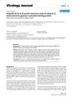
group [as was shown in phylogenetic analysis (Fig. 1)],
genotype D was the most prevalent (79.5%) followed by
genotype A (18.2%) and genotype C (2.3%). The following
subtypes were detected: ayw2 (80.7%), adw2 (14.8%), and
adw (2.3%). Two patients infected with HBV genotype C
could not be subtyped. Within genotype A, subtypes adw
and adw2 were detected, while within genotype D, only
subtype ayw2 was detected.
The prevalence of each of the HBV genotypes that were
isolated from the included patients was also assessed with
respect to patient age (Table 2). There was no trend
observed in the distribution of genotypes among the vari-
ous age groups (P = 0.674). However, genotype A and C
Table 1: Baseline characteristics and HBV genotype
frequencies of the 88 HBV-infected patients included in
this study
HBeAg-positive
patients
N = 88 (%)
Gender M/F 74/14(84/16)
Mean age Years 35.33 ± 11.5
Age range Years 18-70
Genotype A 16 (18.2)
C2 (2.3)
D 70 (79.5)
Subtype ayw2 71 (80.7)
adw2 13 (14.8)
adw 2 (2.3)
HBeAg status Positive 5 (5.7)
Inactive carrier ALT < 65 U/L 71 (80.7)
Chronic hepatitis ALT ≥65 U/L 17 (19.3)
Alfaresi et al. Virology Journal 2010, 7:160
/>Page 4 of 8
Figure 1 Neighbor-joining tree of a specific HBV-DNA nucleotide fragment (S gene) from viral isolates obtained from the study population.
Woolly monkey Hepatitis B virus (GenBank accession number AF046996
) was used as an outgroup.
Alfaresi et al. Virology Journal 2010, 7:160
/>Page 5 of 8
were not observed in individuals aged between 10 and 20
years of age, whereas genotype D was observed in all age
groups. Males comprised a larger proportion of our study
population than females, and because our study popula-
tion was comprised of 88 consecutive HBV-infected
patients, males appeared to be more frequently infected
with HBV than females.
We compared the mean alanine aminotransferase
(ALT) levels of patients infected with each of the three
genotypes that were detected. The highest mean ALT
level (51.30 ± 18.32 U/L) was found in the individuals
infected with HBV genotype D (Fig. 2). However, there
was no significant difference in ALT level observed with
respect to HBV genotype (P = 0.27).
In our study, 83 (94.3%) of patients were HBeAg-nega-
tive and 5 (5.7%) were HBeAg-positive (Table 3). We did
not observe any statically significant differences between
HBeAg-negative and HBeAg-positive patients with
regard to gender, age, or HBV genotype. The mean HBV-
DNA level was significantly higher among HBeAg-posi-
tive patients (3.02 × 10
7
IU/mL) than it was in HBeAg-
negative patients (3205964 IU/mL; P = 0.004). Addition-
ally, HBeAg-positive patients were more likely to have
chronic hepatitis than HBeAg-negative patients (P =
0.048).
In 88 patients, we also analyzed the BCP and precore
region of HBV. The frequency of mutants in these regions
among the different genotypes, as well as the results of
the nt 1858 analysis, are shown in Table 4. Precore
mutants were more frequent in patients infected with
genotype C and D virus (P < 0.0001).
Analysis of nt 1858 showed the presence of thymine in
all patients with genotypes C and D and 56.5% in patients
with genotype A. This nucleotide was closely related to
the presence of precore mutants (P = 0.002).
Mutations in the basal core promoter were found in 22
of 88 (25.3%) samples. These mutations were more fre-
quent in patients infected with genotype A (37.5%), less
frequent among genotype D-infected patients (23.2%)
and not found in patients infected with genotype C, but
no statistical significant difference was found.
Discussion
HBV infection is an important global health problem that
places a continuously increasing burden on developing
countries like the UAE. About 400 million people world-
wide are chronic carriers of the virus[1]. In addition to
the serological classification of HBV isolates into nine
subtypes on the basis of HBsAg determinants[27], a
genetic classification based on the comparison of com-
plete genomes has defined eight genotypes of HBV (A to
H).
Differences in the distribution and clinical characteris-
tics of the eight HBV genotypes have been studied exten-
sively around the world. Better responses to treatment
have been reported for genotypes A and B than genotypes
D and C[28-30]. On the other hand, progression to
chronic hepatitis or to more severe diseases, such as
hepatocellular carcinoma, has been shown to occur most
frequently in patients infected with genotypes A and
C[28,31-33]. Data regarding genotype F that has been
reported thus far have been scant, but in one study, death
related to liver disease was observed more frequently in
patients infected with genotype F than in those infected
with genotypes A or D[29].
The 88 samples that were genotyped indicated that
genotype D had the highest prevalence in our population,
followed by genotype A. Genotypes E and F were not iso-
lated from any of our patients, indicating that these geno-
Table 2: Distribution of HBV genotypes by patient age
Age group (years) Genotype A (N) Genotype C (N) Genotype D (N)
1 to 20 0 0 5
21 to 40 12 1 43
41 to 60 3 1 21
> 60101
Total 16 2 70
Figure 2 Mean ALT values(U/L) of patients infected with the three
HBV genotypes that were identified in this patient population.
Alfaresi et al. Virology Journal 2010, 7:160
/>Page 6 of 8
types are not present in this region. Interestingly, the
initial studies on HBV genotype distribution in other
parts of Asia found that genotypes B and C were the most
prevalent genotypes in this region. However, almost all of
these studies were performed in Japan and China, which
are geographically distant from the UAE. Later studies
revealed that seven of the eight HBV genotypes are pres-
ent in Asia[34]. For instance, the predominant HBV gen-
otypes in India have been shown to be genotypes A and
D[35], while the predominant HBV genotype in Afghani-
stan was found to be genotype D[36]. The epidemiologi-
cal data about the prevalence of the seven HBV genotypes
that have been observed in Asia have revealed the pre-
dominance of genotype D in this region.
Genotypes E, G, and H were not found in our study
population. Genotype E has only been isolated in certain
regions of Africa[37] and in one Haitian child who was
infected with this HBV genotype in Belgium[38]. Geno-
type G was found to be present in about 10% of patients
in France and United States in a previous epidemiological
study[9].
In this study, the most important predictor of an ele-
vated ALT level and a high HBV-DNA level was HBeAg
status, as HBeAg-positive patients were more likely to
have higher ALT levels and HBV-DNA levels than
HBeAg-negative patients.
Among the HBeAg-positive patients, we found that
40% were inactive carriers, compared to 60% of patients
with chronic hepatitis B (Table 3). Among 88 patients
included in this study, all of whom were HBsAg-positive
and HBV-DNA positive, 5 (5.7%) were HBeAg-positive
and 83 (94.3%) were HBeAg-negative. Therefore, in our
population, a high percentage of HBV appear to be
HBeAg-negative. In other studies that have been per-
formed in different patient populations, considerable dif-
ferences between the percentages of HBeAg-positive and
HBeAg-negative patients were also observed. These pre-
vious authors encountered a higher prevalence of
HBeAg-negative patients than HBeAg-positive patients
in their studies, with HBeAg negativity rates varying from
52.5% to 63.3%[39-41]. The majority of the HBV-positive
patients in our study had probably been infected for a
long time and had therefore likely developed mutations in
the pre-core region. Therefore, a number of included
patients were probably HBeAg-negative but anti-HBeAg-
positive.
Precore mutants had an intermediate frequency in our
population (58%). Such mutants were found in all
patients infected with genotype C, with high frequency in
patients infected with genotypes D, and at a very low fre-
quency in genotype A-infected patients. The occurrence
of this mutation is dependent upon the nucleotide (cyto-
sine or thymine) at position 1858, which forms a base pair
with nt 1896 in the pregenomic RNA loop at the ε
encapsidation sign. A thymine at position 1858 is particu-
larly common in genotype D viruses. The presence of a
cytosine at position 1858 precludes the G-to-A mutation
at nt 1896, since this would destabilize the stem-loop
structure of the RNA encapsidation signal[13]. Interac-
tions of this encapsidation signal with the viral DNA
polymerase is an essential step in the viral replication
cycle, and it has been hypothesized that the increased
strength of the guanine-to-cytosine base pairing found in
pre-core mutants would implicate in a stronger ε encapsi-
Table 3: Baseline characteristics and genotype distribution frequencies of the included HBV-infected patients, stratified
by HBeAg status (n = 88)
Characteristics HBeAg-positive HBeAg-negative P-value
(n = 5) (n = 83)
Mean age Years 39 ± 10.2 35 ± 11.6 0.377
Gender M 3 (60%) 71 (85.5%)
F 2 (40%) 12 (14.5%) 0.178
Genotypes A 1 (20%) 15 (18%) 0.13
C0 2 (2.4%)
D 4 (80%) 66 (79.6%)
Inactive carrier ALT < 65 U/L 2 (40%) 69 (83%) 0.048
Chronic hepatitis ALT ≥65 U/L 3 (60%) 14 (17%)
HBV-DNA viral load (IU/mL)
3.02 × 10
7
3205964 0.004
Alfaresi et al. Virology Journal 2010, 7:160
/>Page 7 of 8
dation sign, allowing a more efficient replication of these
strains. Genotype A usually shows a cytosine at this posi-
tion[42]. Our results from nt 1858 analysis corroborate
with these data, since we also have found the presence of
cytosine only in genotypes A, which showed a low fre-
quency of precore mutants.
The frequency of this mutation varies widely around
the world. Castro et al.[43] studied Brazilian patients and
found the precore stop codon mutation in only 24% of
them a result similar to ours. These authors also demon-
strated the higher frequency of the stop codon mutation
at nt 1896 in the isolates that had T1858. On the other
hand, the prevalence of this mutation in China was
86%[44]. This discrepancy may reflect differences in gen-
otype distributions in each studied population, since gen-
otype D viruses are common in the UAE populations,
whereas in China genotypes B and C are more common.
The BCP mutation was found in 25.3% of our patients
and was present in A and D genotypes. BCP mutants fre-
quency ranged from 23.2% in genotype D to 37.5% in gen-
otype A. On the other hand, other authors have reported
that the presence of these mutants is not related to the
presence of precore mutants[43]. This point should be
further analyzed in our population.
Conclusion
HBV genotype influences the severity of liver disease that
patients experience as well as their response to interferon
and antiviral therapy. It is also thought to influence the
emergence of resistant strains. Therefore, patients
infected with certain genotypes of HBV that are known to
be resistant to common treatment regimens can be coun-
seled to seek alternative therapeutic options to spare
them the cost and burden of treatment. The knowledge of
the prevalence of HBV genotypes in a certain region is
thus of immense importance to allow the proper and
effective management of HBV patients in that region. As
we have reported for the first time, HBV genotype D
appears to be the predominant genotype in the UAE. In
this study, the viral loads of HBeAg-positive patients were
higher than those of HBeAg-negative individuals. There-
fore, it should be considered worthwhile for clinicians to
adopt better strategies to prevent and cure HBV infec-
tion. Precore mutants are more common among geno-
type C and D-infected patients, whereas BCP mutants
were present in A and D genotypes. These types of stud-
ies are thus important both for epidemiological reasons
and because they can help promote efforts to develop
effective treatments for HBV.
Competing interests
The authors declare that they have no competing interests.
Authors' contributions
MS: conceived and designed the experiments, participated in experiments,
analyzed data, wrote the paper, AE: participated in experiments, HA; partici-
pated in experiments, AA: participated in experiments, AI: contributed to data
analysis.
All authors have read and approved the final manuscript.
Author Details
Department of Pathology &Laboratory Medicine, Zayed Military Hospital, Abu
Dhabi, UAE
References
1. Lee WM: Hepatitis B virus infection. N Engl J Med 1997, 337:1733-1745.
2. Mahoney FJ: Update on diagnosis, management, and prevention of
hepatitis B virus infection. Clin Microbiol Rev 1999, 12:351-366.
3. Gust I, Crowe S: The Global importance of viral hepatitis. Clin Trop Med
Commun Dis 1986, 1:11.
4. Lau JY, Wright TL: Molecular virology and pathogenesis of hepatitis B.
Lancet 1993, 342:1335-1340.
5. Okamoto H, Tsuda F, Sakugawa H, Sastrosoewignjo RI, Imai M, Miyakawa
Y, Mayumi M: Typing hepatitis B virus by homology in nucleotide
sequence: comparison of surface antigen subtypes. J Gen Virol 1988,
69(Pt 10):2575-2583.
6. Norder H, Hammas B, Lofdahl S, Courouce AM, Magnius LO: Comparison
of the amino acid sequences of nine different serotypes of hepatitis B
surface antigen and genomic classification of the corresponding
hepatitis B virus strains. J Gen Virol 1992, 73(Pt 5):1201-1208.
7. Norder H, Courouce AM, Coursaget P, Echevarria JM, Lee SD, Mushahwar
IK, Robertson BH, Locarnini S, Magnius LO: Genetic diversity of hepatitis
Received: 23 April 2010 Accepted: 15 July 2010
Published: 15 July 2010
This artic le is available fro m: http://www.v irologyj.com/co ntent/7/1/160© 2010 Alfaresi et al; licensee BioMed Central Ltd. This is an Open Access article distributed under the terms of the Creative Commons Attribution License ( which permits unrestricted use, distribution, and reproduction in any medium, provided the original work is properly cited.Virology Journal 2010, 7:160
Table 4: HBV genotypes and their relationship to viral features
HBV BCP and precore region mutations(no. [%])
genotype B CP nt 1858 Precore region
WT Mutant C T WT Mutant
A 10(62.5) 6(37.5) 7(43.8) 9(56.3) 15(93.8) 1(6.3)
C 2(100) 0 0 2(100) 0 2(100)
D 53(76.8) 16(25.3) 0 70(100) 22(31.4) 48(68.6)
Total 65(74.7) 22(25.3) 7(8) 81(92) 37(42) 51(58)
Alfaresi et al. Virology Journal 2010, 7:160
/>Page 8 of 8
B virus strains derived worldwide: genotypes, subgenotypes, and
HBsAg subtypes. Intervirology 2004, 47:289-309.
8. Magnius LO, Norder H: Subtypes, genotypes and molecular
epidemiology of the hepatitis B virus as reflected by sequence
variability of the S-gene. Intervirology 1995, 38:24-34.
9. Stuyver L, De Gendt S, Van Geyt C, Zoulim F, Fried M, Schinazi RF, Rossau R:
A new genotype of hepatitis B virus: complete genome and
phylogenetic relatedness. J Gen Virol 2000, 81:67-74.
10. Arauz-Ruiz P, Norder H, Robertson BH, Magnius LO: Genotype H: a new
Amerindian genotype of hepatitis B virus revealed in Central America.
J Gen Virol 2002, 83:2059-2073.
11. Kao JH: Hepatitis B viral genotypes: clinical relevance and molecular
characteristics. J Gastroenterol Hepatol 2002, 17:643-650.
12. Sakai T, Shiraki K, Inoue H, Okano H, Deguchi M, Sugimoto K, Ohmori S,
Murata K, Fujioka H, Takase K, et al.: HBV subtype as a marker of the
clinical course of chronic HBV infection in Japanese patients. J Med
Virol 2002, 68:175-181.
13. Lok AS, Akarca U, Greene S: Mutations in the pre-core region of hepatitis
B virus serve to enhance the stability of the secondary structure of the
pre-genome encapsidation signal. Proc Natl Acad Sci USA 1994,
91:4077-4081.
14. Rodriguez-Frias F, Buti M, Jardi R, Cotrina M, Viladomiu L, Esteban R,
Guardia J: Hepatitis B virus infection: precore mutants and its relation to
viral genotypes and core mutations. Hepatology 1995, 22:1641-1647.
15. Grandjacques C, Pradat P, Stuyver L, Chevallier M, Chevallier P, Pichoud C,
Maisonnas M, Trepo C, Zoulim F: Rapid detection of genotypes and
mutations in the pre-core promoter and the pre-core region of
hepatitis B virus genome: correlation with viral persistence and disease
severity. J Hepatol 2000, 33:430-439.
16. Hunt CM, McGill JM, Allen MI, Condreay LD: Clinical relevance of
hepatitis B viral mutations. Hepatology 2000, 31:1037-1044.
17. Buckwold VE, Xu Z, Yen TS, Ou JH: Effects of a frequent double-
nucleotide basal core promoter mutation and its putative single-
nucleotide precursor mutations on hepatitis B virus gene expression
and replication. J Gen Virol 1997, 78(Pt 8):2055-2065.
18. Okamoto H, Tsuda F, Akahane Y, Sugai Y, Yoshiba M, Moriyama K, Tanaka
T, Miyakawa Y, Mayumi M: Hepatitis B virus with mutations in the core
promoter for an e antigen-negative phenotype in carriers with
antibody to e antigen. J Virol 1994, 68:8102-8110.
19. Takahashi K, Aoyama K, Ohno N, Iwata K, Akahane Y, Baba K, Yoshizawa H,
Mishiro S: The precore/core promoter mutant (T1762A1764) of
hepatitis B virus: clinical significance and an easy method for
detection. J Gen Virol 1995, 76(Pt 12):3159-3164.
20. Kurosaki M, Enomoto N, Asahina Y, Sakuma I, Ikeda T, Tozuka S, Izumi N,
Marumo F, Sato C: Mutations in the core promoter region of hepatitis B
virus in patients with chronic hepatitis B. J Med Virol 1996, 49:115-123.
21. Buckwold VE, Xu Z, Chen M, Yen TS, Ou JH: Effects of a naturally
occurring mutation in the hepatitis B virus basal core promoter on
precore gene expression and viral replication. J Virol 1996,
70:5845-5851.
22. Moriyama K, Okamoto H, Tsuda F, Mayumi M: Reduced precore
transcription and enhanced core-pregenome transcription of hepatitis
B virus DNA after replacement of the precore-core promoter with
sequences associated with e antigen-seronegative persistent
infections. Virology 1996, 226:269-280.
23. Kidd-Ljunggren K, Oberg M, Kidd AH: Hepatitis B virus X gene 1751 to
1764 mutations: implications for HBeAg status and disease. J Gen Virol
1997, 78(Pt 6):1469-1478.
24. Kimura M: A simple method for estimating evolutionary rates of base
substitutions through comparative studies of nucleotide sequences. J
Mol Evol 1980, 16:111-120.
25. Sitnik R, Pinho JR, Bertolini DA, Bernardini AP, Da Silva LC, Carrilho FJ:
Hepatitis B virus genotypes and precore and core mutants in Brazilian
patients. J Clin Microbiol 2004, 42:2455-2460.
26. Sanger F, Nicklen S, Coulson AR: DNA sequencing with chain-
terminating inhibitors. Proc Natl Acad Sci USA 1977, 74:5463-5467.
27. Courouce-Pauty AM, Lemaire JM, Roux JF: New hepatitis B surface
antigen subtypes inside the ad category. Vox Sang 1978, 35:304-308.
28. Orito E, Mizokami M, Sakugawa H, Michitaka K, Ishikawa K, Ichida T,
Okanoue T, Yotsuyanagi H, Iino S: A case-control study for clinical and
molecular biological differences between hepatitis B viruses of
genotypes B and C. Japan HBV Genotype Research Group. Hepatology
2001, 33:218-223.
29. Sanchez-Tapias JM, Costa J, Mas M, Bruguera M, Rodes J: Influence of
hepatitis B virus genotype on the long-term outcome of chronic
hepatitis B in western patients. Gastroenterology 2003, 125:2.
30. Zhang X, Zoulim F, Habersetzer F, Xiong S, Trepo C: Analysis of hepatitis B
virus genotypes and pre-core region variability during interferon
treatment of HBe antigen negative chronic hepatitis B. J Med Virol 1996,
48:8-16.
31. Ding X, Mizokami M, Yao G, Xu B, Orito E, Ueda R, Nakanishi M: Hepatitis B
virus genotype distribution among chronic hepatitis B virus carriers in
Shanghai, China. Intervirology 2001, 44:43-47.
32. Mayerat C, Mantegani A, Frei PC: Does hepatitis B virus (HBV) genotype
influence the clinical outcome of HBV infection? J Viral Hepat 1999,
6:299-304.
33. Orito E, Ichida T, Sakugawa H, Sata M, Horiike N, Hino K, Okita K, Okanoue
T, Iino S, Tanaka E, et al.: Geographic distribution of hepatitis B virus
(HBV) genotype in patients with chronic HBV infection in Japan.
Hepatology 2001, 34:590-594.
34. Toan NL, Song le H, Kremsner PG, Duy DN, Binh VQ, Koeberlein B, Kaiser S,
Kandolf R, Torresi J, Bock CT: Impact of the hepatitis B virus genotype
and genotype mixtures on the course of liver disease in Vietnam.
Hepatology 2006, 43:1375-1384.
35. Thakur V, Guptan RC, Kazim SN, Malhotra V, Sarin SK: Profile, spectrum
and significance of HBV genotypes in chronic liver disease patients in
the Indian subcontinent. J Gastroenterol Hepatol 2002, 17:165-170.
36. Amini-Bavil-Olyaee S, Alavian SM, Adeli A, Sarrami-Forooshani R, Sabahi F,
Sabouri E, Tavangar HR, Azizi M, Mahboudi F: Hepatitis B virus
genotyping, core promoter, and precore/core mutations among
Afghan patients infected with hepatitis B: a preliminary report. J Med
Virol 2006, 78:358-364.
37. Mizokami M, Nakano T, Orito E, Tanaka Y, Sakugawa H, Mukaide M,
Robertson BH: Hepatitis B virus genotype assignment using restriction
fragment length polymorphism patterns. FEBS Lett 1999, 450:66-71.
38. Liu HF, Sokal E, Goubau P: Wide variety of genotypes and geographic
origins of hepatitis B virus in Belgian children. J Pediatr Gastroenterol
Nutr 2001, 32:274-277.
39. Tsugeno H, Yamada G, Kinoshita M, Shimomura H, Iwasaki Y, Tsuji T:
Quantitative analysis of wild-type and precore mutant hepatitis B virus
in carriers. Hepatol Res 2002, 23:48-54.
40. Yoo B, Park J, Kim HJ, Lee DH, Chan YJ, Park SM: Precore and core
Promotor Mutations of Hepatitis B Virus and Hepatitis B e Antigen-
Negative Chronic Hepatitis B in Korea. J Hepatology 2003, 38:6.
41. Lim CK, Tan JT, Khoo JB, Ravichandran A, Low HM, Chan YC, Ton SH:
Correlations of HBV genotypes, mutations affecting HBeAg expression
and HBeAg/anti-HBe status in HBV carriers. Int J Med Sci 2006, 3:14-20.
42. Li JS, Tong SP, Wen YM, Vitvitski L, Zhang Q, Trepo C: Hepatitis B virus
genotype A rarely circulates as an HBe-minus mutant: possible
contribution of a single nucleotide in the precore region. J Virol 1993,
67:5402-5410.
43. De Castro L, Niel C, Gomes SA: Low frequency of mutations in the core
promoter and precore regions of hepatitis B virus in anti-HBe positive
Brazilian carriers. BMC Microbiol 2001, 1:10.
44. Chan HL, Hussain M, Lok AS: Different hepatitis B virus genotypes are
associated with different mutations in the core promoter and precore
regions during hepatitis B e antigen seroconversion. Hepatology 1999,
29:976-984.
doi: 10.1186/1743-422X-7-160
Cite this article as: Alfaresi et al., Hepatitis B virus genotypes and precore
and core mutants in UAE patients Virology Journal 2010, 7:160