Báo cáo y học: " Oligomerization of Uukuniemi virus nucleocapsid protein" pptx
Bạn đang xem bản rút gọn của tài liệu. Xem và tải ngay bản đầy đủ của tài liệu tại đây (3.75 MB, 13 trang )
RESEARC H Open Access
Oligomerization of Uukuniemi virus nucleocapsid
protein
Anna Katz
1*†
, Alexander N Freiberg
2†
, Vera Backström
1,3,5
, Axel R Schulz
2
, Angelo Mateos
2
, Liisa Holm
3
,
Ralf F Pettersson
4
, Antti Vaheri
1
, Ramon Flick
2,6
, Alexander Plyusnin
1
Abstract
Background: Uukuniemi virus (UUKV) belongs to the Phlebovirus genus in the family Bunyaviridae. As a non-
pathogenic virus for humans UUKV has served as a safe model bunyavirus in a number of studies addressing
fundamental questions such as organization and regulation of viral genes, genom e replication, structure and
assembly. The present study is focused on the oligomerization of the UUKV nucleocapsid (N) protein, which plays
an important role in several steps of virus replication. The aim was to locate the domains involved in the N protein
oligomerization and study the process in detail.
Results: A set of experiments concentrating on the N- and C-termini of the protein was performed, first by
completely or partially deleting putative N-N-interaction domains and then by introducing point mutations of
amino acid residu es. Mutagenesis strategy was based on the computer modeling of secondary and tertiary
structure of the N protein. The N protein mutants were studied in chemical cross-linking, immunofluorescence,
mammalian two-hybrid, minigenome, and virus-like particle-forming assays. The data showed that the
oligomerization ability of UUKV-N protein depends on the presence of intact a-helices on both termini of the N
protein molecule and that a specific structure in the N-terminal region plays a crucial role in the N-N interaction(s).
This structure is formed by two a-helices, rich in amino acid residues with aromatic (W7, F10, W19, F27, F31) or
long aliphatic (I14, I24) side chains. Furthermore, some of the N-terminal mutations (e.g. I14A, I24A, F31A) affected
the N protein functionality both in mammalian two-hybrid and minigenome assays.
Conclusions: UUKV-N protein has ability to form ol igomers in chemical cross-linking and mammalian two-hybrid
assays. In mutational analysis, some of the introduced single-point mutations abolished the N protein functionality
both in mammalian two-hybrid and minigenome assays, suggesting that especially the N-terminal region of the
UUKV-N protein is essential for the N-N interaction.
Background
Uukuniemi virus (UUKV) belongs to the Phlebovirus
genus in the family Bunyaviridae. Some members of the
family are important human pathogens, e.g. Crimean-
Congo hemorrhagic fever virus, hantaviruses, and Rift
Valley fever virus (RVFV) [1]. UUKV was first isolated
from ticks in Uuku niemi, Finland, in 1959 [2], and a s a
non-pathogenic virus for humans [3], UUKV has served
as a safe model bunyavirus in a number of studies
addressing fundamental questio ns, e.g. organizati on and
regulation of viral genes, structure an d assembly [4-7].
Like other Buny aviridae,UUKVisanenvelopedvirus
with a tripartite RNA genome of negative polarity. The
large (L) segment encodes the RNA-dependent RNA
polymerase (L protein), and the medium (M) segment
encodes two glycoproteins, G
N
and G
C
.Thesmall(S)
segment encodes the nucleocapsid (N) protein and, in
positive sense orientation, the non -structural protein [1].
N protein play s a cen tral role in the replication, tran-
scription and assembly of RNA viruses. In negative-
strand RNA viruses (NSRV), including bunyaviruses,
both the v RNA and cRNA are encapsidated by the N
protein into a ribonucleopr otein (RNP) complex, which
serves as template for transcription and r eplication of
* Correspondence:
† Contributed equally
1
Department of Virology, Infection Biology Research Program, Haartman
Institute, P.O. Box 21, 00014, University of Helsinki, Helsinki, Finland
Full list of author information is available at the end of the article
Katz et al. Virology Journal 2010, 7:187
/>© 2010 Katz et al; licensee BioMed Central Ltd. This is an Open Access article distributed under the terms of the Creative Commons
Attribution License ( which permits unrestricted use, distribution, and reproduction in
any medium, provided the original work is properly cited.
the viral genome [8]. In the course of RNA encapsida-
tion, the N protein of NSRV forms oligomers.
Among the NSRV, this oligomerization abili ty has
been demonstrated for several viruses, for example Mar-
burg virus (Filoviridae)[9],Sendaivirus(Paramyxoviri-
dae) [10], and influenza A virus (Orthomyxoviridae)
[11]. In addition, N protein 3D-structures for four
viruses were solved recently - rabies and vesicular sto-
matitis viruses ( Rhabdoviridae) [12,13], Borna disease
virus (Bornaviridae) [14], and influenza A virus [15] -
revealing the oligomerization domains in detail.
The ability of N protein to oli gomerize has also been
shown for bunyaviruses in different genera: Bunyamwera
virus (BUNV) (Orthobunyavirus) [16], hantaviruses
(Hantavirus) [17,18], tomato spotted wilt virus (Tospo-
virus)[19],andRVFV(Phlebovirus) [ 20]. Throughout
the five genera, the sizes of bunyaviral N proteins differ
from 25 to 30 kDa (orthobunya-, phlebo-, and tospo-
viruses) to double the size, 48 to 54 kDa (hanta- and
nairoviruses). The mode of N protein oligomerizati on
seems to differ between the genera as well. BUNV-N
protein was shown to form dimers, trimers and higher
multimers [16,21], and Tula hantavirus N protein to
form oligomers t hrough trimer formation, where the N-
terminal coiled-coils are involv ed [22-25]. These coiled-
coiled domain structures have a lso been solved for two
hantaviruses, Sin Nombre virus and Andes hantavirus
[26,27]. A head-to-head and tail-to-tail fashion of oligo-
merization was suggested for both BUNV and Tula
hantavirus N proteins. For RVFV-N protein, dimer for-
mation was suggested, and the N-N interacting domain
was mapped to the first 71 N-terminal residues [20].
Further details of the oligomerization process remain
largely unknown.
In the present study, we focused on the oligomeriza-
tion of the UUKV-N protein. Our first experiments
using the mammalian two-hybrid (M2H) system and
chemical cross-linking showed that the UUKV-N pro-
tein molecules can interact with ea ch other. The aim
was then to locate domains involved in the N protein
oligomerization and to study this process in more detail.
Results
Analysis of UUKV-N protein in the chemical cross-linking
and M2H assays
To study the N p rotein oligomerization, COS-7 cells
were transfected with pcDNA-UUKV-N constructs and
lysates were treated with the chemical cross-linker, BS
3
(Fig. 1). In the absence of BS
3
, the monomeric form of
the N protein (~25 kDa) was predominant in immuno-
blotting (Fig. 1, lane 1). In the presence of BS
3
,the
intensity of monomeric band decreased and the dimeric
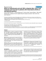
Figure 1 Cross-linking of the UUKV-N protein. N protein containing COS-7 cell lysates were treated with 0.1 or 0.5 mM of cross-linking
reagent (BS
3
), and resolved by 10% SDS-PAGE under denaturing conditions. An immunoblot shows that omitting BS
3
, the majority of the full-
length UUKV-N protein migrated as 25 kDa monomeric form (lane 1). Addition of BS
3
decreased the amount of the monomeric form, while the
amount of dimeric and multimeric forms were simultaneously increasing (lanes 2 and 3). In mock-transfected samples no N protein was
detected (lanes 4 to 6).
Katz et al. Virology Journal 2010, 7:187
/>Page 2 of 13
form (~50 kDa) was formed. In the l ysates treated with
0.5mMofBS
3
also a band of ~75 kDa appeared, sug-
gesting the presence of trimers (Fig. 1, lane 3). MOCK-
transfected COS-7 cells served as control (Fig. 1, lanes 4
to 6). A band just above the N-dimer as w ell as the
bands that move slower than the N-trimer probably pre-
sented unrelated cross-reacting cellular proteins.
These results were in agreement with those obtained
using the M2H assay: UUKV-N protein was expressed
as a fusion with the DNA-BD and DNA-AD domains,
resulting in high luciferase reporter signal. Same M2H
vectors without N protein inserts served as negative
controls, leading t o very lo w signals ( data not shown).
Thus, both assays clearly demonstrated the N protein
ability to form dimers and higher oligomers.
Secondary and tertiary structure predictions for UUKV-N
protein
For the secondary stucture, J pred, Psipred and Predict-
Protein programs predicted the UUKV-N (254 aa resi-
dues) to be an a-helix-rich protein containing 13 to 15
a-helices with one to thr ee short b-strands. Only minor
differences in the length and location of the a-helices
were observed between these models. In our working
model we adopted 13 a-helices constantly predicted by
all used programs (Fig. 2). For the N-terminal region
the predictions showed either one l ong a-helix (aa
residues 5-33), or two shorter a-helices (aa residues 7-
19/5-17 and 21-33). In the C-terminal region, two a-
helices (aa residues 220/221-229/230 and 239/241-252/
253) were predicted (see a1, a2, a12 and a13 in Fig. 2).
Next, the secondary structure predictions for UUKV-
N were compared to N proteins of four other phlebo-
viruses (RVFV, SFSV, TOSV and PTV). Predictions by
the Jpred and PsiPred programs showed that the overall
secondary structure is well conserved among phlebo-
viruses (data not shown). For the N-terminus, predicted
a-helices were of the same length (a1: 11-15 residues;
a2: 12-13 residues) and located at simi lar positions (Fig.
3A). The same was true for the last C-terminal a-
helices: a13 and a12 were predicted to be respectively,
11-14 and 10-11 aa residues long , wit h a shor t b-strand
located between them. Shor ter a-helices were predicted
within the central part of the molecule, with less uni-
form pattern than in the well-conserved N- and C-term-
inal parts. This analysis suggested that conserved a-
helices perform an important, fundamental function
shared by all analyzed phleboviral N proteins.
Tertiary structure of the UUKV-N protein was pre-
dicted using Robetta server’s ab initio modelin g. Alto-
gether, ten models were obtained, one of them i s shown
in Fig. 3B. All showed the same overall pattern of fold-
ing and, i n agreement with the secondary structure pre-
dictions, contained 13-16 a-helices. Similarly to the
Figure 2 Secondary structure predictions for UUKV-N protein (Jpred, PsiPred and PredictProtein servers) and mutagenesis strategy.To
study the oligomerization ability of UUKV-N protein, five deletion mutants were designed: ΔN19, ΔN34, ΔC38, ΔC17, and ΔC10.
Katz et al. Virology Journal 2010, 7:187
/>Page 3 of 13
PsiPred prediction, all Robetta models showed two a-
helices separated by 2-3 a a residues at the N-terminal
part of the molecule. At the C-terminal region, two anti-
parallel b-strands were predicted (Fig. 3B).
These models of UUKV-N protein were used to define
our mutagenesis strategy. Since earlier studies on hanta -
viruses, RVFV, and BUNV showed that the very term-
inal regions of the N protein are especially important
for the oligomerization [16,20,23,25], we focused on
these regions.
Analysis of the N-N interactions in M2H and minigenome
assays
First, pr edicted a-helices were gradually removed from
the N- and C-termini of the N protein molecule gener-
ating f ive deletion mutants: ΔN19, ΔN34, ΔC38, ΔC17,
and ΔC10 (Fig. 2). In mutant ΔN19, the first a-helix,
and in mutant ΔN34, the two first a-helices were
deleted. Similar strategy was implemented for the C-ter-
minus; in the mutant ΔC10, half of the last a-helix was
removed, in mutant ΔC17 the entire last a-helix, and in
mutant ΔC38 the last two a-helices were deleted (Fig.
2). These mutants were tested in the M2H s ystem, and
further studied using the minigenome system, immuno -
fluorescence and cross-linking assays.
In the M2H system the full-length N protein showed
strong N-N interaction ability (Fig. 4A). This ability
decreased in the mutant ΔN19, where the remaining
activity was only 25 ± 3%, and totally vanished in the
mutant ΔN34 (Fig. 4A). This effect was repeatedly seen
with DNA-AD-fused truncated proteins, even if the y
reacted with the full-length DNA-BD-N fusion protein.
Two constructs, ΔN19-DNA-BD and ΔN34-DNA-BD,
showed artificially high signals in the luciferase reporter
assay, perhaps due to misfolding of the N fusion protein.
All three C-terminal mutants, ΔC10 (Fig. 4A), ΔC17 and
ΔC38 (data not shown) were completely non-func tional,
even if introduced in only one interacting partner
(DNA-AD). To exclude the possibility that the lack of
reactivity in the M2H assay was due to inefficient
Figure 3 Design for the U UKV-N protein mutations based on multiple alignments and 2D- and 3D-structure predictions. (A) Jpred
alignment shows the N- and C-termini of N proteins among five members in the genus Phlebovirus. a-helix-forming aa residues are shadowed.
Point mutations for the UUKV-N protein were targeted to the aromatic and hydrophobic aa residues. (B) Robetta server’s ab initio model for the
UUKV-N protein: the first two predicted a-helices in N-terminus are shown in blue, and the C-terminus is shown in green. (C) The N-terminal
part of the Robetta model (Fig. 3B) showing the residues presumably involved in the oligomerization (except Y33 facing outside of the a-
helices).
Katz et al. Virology Journal 2010, 7:187
/>Page 4 of 13
protein production, expression levels of truncated N
proteins in COS-7 cells were confirmed by immunoblot-
ting. Even though there was variation in th e expression
levels, it could not explain the differences in the M2H
results (Fig. 4B).
Deletion mutants were further tested in the UUKV
minigenome system. The N protein molecules suppo-
sedly interact with each other and also with viral gen-
ome segments and replication intermediates and form
RNPs. Interrupting t he ability to form N-oligomers
should interfere with m inigenome transcription and
replication. All five N- and C-terminal deletions com-
pletely abolished the function of the N protein, result-
ing in a negative CAT signal (Fig. 5). Mutants ΔN19
and ΔC10 were negative in CAT assays; this suggested
that both terminal moieties are needed for the oligo-
merization process. Expression of the UUKV-N
mutants was verified by immunofluorescence assay
(IFA) (Fig. 6) and by immunoblotting (data not
shown). Thus the results obtained with the minige-
nome system were in agreement with those of the
M2H assay: both N- and C-terminal a-helices are
essential for the N-N interactions and even small dele-
tions completely destroyed the protein function (Fig.
4A and 5). Furthermore, the results of the cross-link-
ing assay confirm ed that all N- and C- terminal dele-
tions severely affected the ability of N protein to
oligomerize ( data not shown).
Figure 4 N-N interaction of truncated UUKV-N protein constructs in the M2H assay and verification of the protein expression. (A)
Truncation ΔN19 affected the N-N interaction, ΔN34 and C-terminal truncation ΔC10 destroyed the oligomerization ability completely. Deleted
regions are shown in white. Numbers are averages of normalized luciferase activity values (%), where the wt N-N interaction was set as 100%. ±
standard deviations are calculated for the mean values. (B) N protein expression of the M2H constructs was verified in immunoblotting. MAbs
were used to detect the N protein fusions with DNA-AD (lanes 1 to 4), and DNA-BD (lanes 5 to 8) constructs, which migrated as 44-46 kDa
bands.
Katz et al. Virology Journal 2010, 7:187
/>Page 5 of 13
Analysis of the intracellular localization and distribution
of wt and truncated UUKV-N protein using
immunofluorescence assay
Next, we examined where the wt and truncated UUKV-
N protein localizes in transfected cells and whether
there are differences in the staining pattern (Fig. 6).
Wild type (wt) N protein localized in the cytoplasm and
formed larger aggregates (Fig. 6), resembling UUKV-
infected cells (Fig. 6), as also presented earlier [28]. Of
the N-terminal truncations, ΔN19didnothaveamajor
effect on the appearance of the stained protein aggre-
gates. It resembled the pattern of wt N protein, although
some diffuse staining was observed. However, truncation
ΔN34 differed from the wt N protein remarkably: the
protein was dispersed throughout the cytoplasm as a
diffuse net, with on ly a few microgra nular aggregates
observed (Fig. 6). In all C-terminal truncations, both
intracellular localization and the staining pattern of the
N protein were strongly affected. These truncated N
proteins were dispersed as a diffuse pattern in the cyto-
plasm, with some small microgranular aggregates similar
to mutant ΔN34. Truncated proteins were also observed
in the nuclei, the effect was most pronounced with the
longest truncation, ΔC38 (Fig. 6). Most importantly, no
protein aggregates, characteristic of the wt N protein,
were seen.
Bioinformatic analysis and mutagenesis strategy of point
mutations
Experiments with truncated UUKV-N proteins directed
us to focus on the N- and C-termini of the molecule,
using site-directed mutagenesis to define aa residues
involved in the N-N interactions. Sequence alignment
and secondary structure predictions revealed very few
conserved aa residues within the last C-terminal a-helix.
To check whether this helix is directly involved in the
N-N interaction(s) two mutations were introduced:
R251, conserved in all phleboviruses (except K in
RVFV), was replaced with alanine, and the double
mutant QQ244,245AA, was designed to evaluate the
possible role of the polar side chains in the N-N interac-
tion. These mutants did not differ from the wt N pro-
tein in M2H, minigenome and VLP assays (Table 1, Fig.
7, lanes 12 and 13), and also in cross-linking and IFA
(data not shown). It seems that the last C-terminal a-
helix is not directly involved in the oligomerization but
is indispensable for maintaining a proper folding of the
whole molecule and hence its functional competence.
We therefore concentrated on the N-terminal part of
the protein.
Most models indicated t hat the N-terminal region (aa
1-33), forms two a-helices separated by a short turn
(Fig. 3C). This region is rich with aromatic and hydro-
phobic residues. In Robetta 3D-model of UUKV-N pro-
tein (Fig. 3B and 3C), it was observed that W7, F10, I14,
W19, I24, F27 and F31 are facing the same side of a a-
helical projection of the molecule. This structure with
two parallel a-helices resemble s the N-terminal coiled-
coil structure of the hantaviral N protein that was
shown to be important for the N-N interactions
[23,25,29]. The mo del for UUKV-N protein suggests
that the shared h ydrophobic space between first two a-
helices is not exposed to the solvent. After a conforma-
tional change opening the structure, the listed aa resi-
dues could interact with their partners in t he other N-
monomer. To evaluate the contribution of these resi-
dues to the N-N interaction, and the overall functi onal-
ity of the N protein molecule, eight point mutants were
generated: W7A, F10A, I14A, W19A, I24A, F27A, F31A,
and Y33A (Fig. 3A, arrowheads). Tyrosine at position 33
was facing outside of the a-helices, and therefore its
replacement with alanine was expected to have no effect
on the oligomerization.
Analysis of the N-terminal point mutations in M2H and
minigenome assays
Eight alanine substitutions were introduced to the N
protein fused with the DNA-BD and DNA-AD for M2H
assay and pcDNA-UUKV-N expression plasmids. In
M2Hassay,fourmutants,F10A,I14A,I24A,andF31A,
Figure 5 Comparative CAT analysis on N protein deletion
mutants. BHK-21 cells were transfected with UUKV minigenome
plasmids: (UUKV M-CAT), viral polymerase expression (pCMV-UUKV-L)
and wt or mutant pcDNA-UUKV-N. Cells were analyzed for CAT
activity at 48 h post-transfection. The N- and C-terminal deletion
mutants (lanes 3 to 7) were compared with wt N protein (lane 2),
showing that all the deletion mutants were non-functional in the
minigenome system. In the negative control (lane 1) pCMV-UUKV-L
was omitted.
Katz et al. Virology Journal 2010, 7:187
/>Page 6 of 13
showed a reduced N-N interaction ability. Four other
mutations, W7A, W19A, F27A, and Y33A (our negative
control), acted as the wt N protein (Table 1). In the
minigenome system, five m utations (W7A, I14A, I24A,
F27A, and F31A) completely abolished CAT expression,
mutations F10A and W19A had a moderate impact,
whereas mutation Y33A did not affect the functionality
of the protein (Fig. 7, upper panel, and Table 1).
Expression of all mutants was verified by I FA (Fig. 6)
and immunoblotting (data not shown).
Analysis of the point mutations in the virus-like particle
(VLP) system
To furt her test point mutations o n the N protein func-
tional competence, we used recently developed infec-
tious VLP system for UUKV [30], in which cells
Figure 6 Immunofl uorescence analysis on the UUKV-N protein mutants at 24 h post-transfection. The panel of UUKV-N deletion and
point mutants shows that the intracellular localization and the oligomerization ability of some mutants (e.g. ΔN34 and ΔC17) was altered
compared to the wt N protein and UUKV infected BHK-21 cells.
Katz et al. Virology Journal 2010, 7:187
/>Page 7 of 13
transfected with expression plasmids encoding for
UUKV-G
N
/G
C
, N protein, an d viral po lymerase,
together with the UUKV minigenome, generate VLPs
containing the minigenome. UUK-VLPs are released
into the cell supernatant and able to infect new cells. It
was assumed that if the N protein functionality is
affected, it would inhibit, or even abolish, both packa-
ging and transfer ability of t he minigenome. No CAT
activity was detected in the negative control of VLP-
infected cells omitting UUKV-L and UUKV-G
N
/G
C
,
respectively (Fig. 7, lower panel, lanes 1 a nd 2). The
positive control containing UUKV-G
N
/G
C
showed
strong CAT activit y (Fig. 7, lower panel, lane 3). Six N-
terminal point mutants (W7A, I14A, W19A, I24A,
F27A, and F31A) showed reduced CAT signal indicating
the affected N protein ability to oligomerize and/or
encapsidate minigenome RNA (Fig. 7 and Table 1).
Three of these mutants, I14A, I24A, and F31A,
showed reduced functional competence in the minige-
nome assay and affected N-N interaction ability in the
M2H assay. In addition, two mutants, W7A and F27A,
showed either reduced or totally inhibited functional
Table 1 Summary of the results: UUKV-N protein point
mutants in the M2 H, minigenome, and
immunofluorescence assays
UUKV-N protein
point mutants
M2H*
% of interaction
Minigenome
†
VLP
†
IFA
‡
Wt N protein 100 +++ +++ +++ +++
W7A > 100 (3) +++ - - +++
F10A 34 ± 4 (2) + + + +++
I14A 60 ± 18 (3) ++ - - +
W19A > 100 (3) +++ +/++ - +
I24A 56 ± 12 (2) ++ - - +
F27A > 100 (3) +++ - - ++
F31A 23 ± 5 (2) + - - ++
Y33A > 100 (2) +++ +++ +++ +++
QQ244,245AA > 100 (2) +++ +++ +++ +++
R251A 96 ± 11 (2) +++ +++ +++ +++
* Full-length N-N protein interaction (100%, +++) was compared to the N-N
interaction of point mutants, range from not affected (+++) to reduced (++)
and to substantially reduce d interaction (+). Each test was performed in
triplicates, number of repetitions for each test is given in parentheses.
†
In minigenome and VLP systems the CAT expression was either non-affected
(+++), reduced (++), subst antially reduced (+), or completely inhibited (-).
‡
Point mutants forming aggregates resembling full-length N protein (+++),
showing microgranular (++) or diffuse (+) fluorescence pattern.
Figure 7 UUKV-N point mutants analyzed by comparative CAT analysis and VLP transfer of UUKV M-CAT minigenome. Upper panel : In
comparative CAT analysis, BHK-21 cells were transfected with UUKV M-CAT, pCMV-UUKV-L and wt or mutant pcDNA-UUKV-N plasmids, and the
glycoprotein expression plasmid pCMV-UUKV-G
N
/G
C
was co-transfected for VLP transfer of the minigenome. Cells were analyzed for CAT activity,
and VLP-containing supernatant was used to infect new cells pre-transfected with pCMV-UUKV-L and wt UUKV-N protein. Lower panel: UUKV-N
point mutants analyzed by CAT expression and VLP transfer of UUKV M-CAT minigenome. Negative controls omit either pCMV-UUKV-L (lane 1)
or include pCMV-UUKV-L but omit pCMV-UUKV-G
N
/G
C
(lane 2; negative control for VLP transfer).
Katz et al. Virology Journal 2010, 7:187
/>Page 8 of 13
ability in the minigenome assay and in the M2H assay
gave artificially high signals. This discrepancy could
indicate on a possible involvement of these residues in
RNA-binding. Our control (Y33A) as well as two C-
terminal mutants (R251A and QQ244,245AA) acted
similarly in the VLP-assay compared to the wt N pro-
tein,i.e.wereabletoencapsidate, package and transfer
a functional minigenome. To summarize, the results of
the VLP assay were in agreement with the other two
tests described above. They also logically showed that
the demand for the funct ional competence of the
involved components, including the N protein, is higher
in this more integral system thus fewer alterations are
tolerated.
Immunofluorescence microscopy of UUKV-N protein
mutants
All eight N-terminal and two C-terminal point muta-
tions were also tested in IFA. Six mutants, including our
negative control and two C-terminal mutants (W7A,
F10A, F27A, Y33A, QQ244,245AA, and R251A) behaved
as the wt N protein. They formed aggregates located
mostly in the perinuclear region (a typical mut ant from
this grou p, Y33A, is shown on Fig 6). In sharp c ontrast,
four other mutants (I14A, W19A, I24A, and F31A)
showed a diffuse to microgranular pattern of staining
(Fig.6anddatanotshown).Theseresultssuggested
that at least some of the mutations, which inflicted the
N-N interactions, also affected the intrac ellular localiza-
tion of the N protein.
Results of different assays are summarized in Table 1.
For six mutants the correlation was good. These muta-
tions included those that affected the N protein func-
tionality ( I14A, I24A and F31A), and also the ones
inflicting no detectable damage (Y33A, QQ244,245AA,
R251A). The results for other mutations were ambigu-
ous. Mutants W7A and F27A were particularly interest-
ing: a lthough they were capable to interact in the M2H
assay and their IFA-staining pattern was the same as of
the wt N protein, these mutations were not functional
in the minigenome assay.
Discussion
Results presented in this paper show that the molecules
of UUKV-N protein molecules can interact with each
other. Thus, in this respect, the UUKV-N protein
resembles the nucleocapsid proteins of other NSRV
[9-14,16-19].
Our experiments revealed that the oligomerization
ability of UUKV-N protein depends on the presence o f
intact a-helices on both termini of the molecule. More-
over, the point mutagenesis data in comb ination with
the computer modeling, suggested that a specific struc-
ture in the N-terminal region plays a crucial role in the
N-N interaction(s). This structure (Fig. 3C) is formed by
two a-helices, rich in aa residues with aromatic (W7,
F10, W19, F27, F31, Y33) or long aliphatic (I14, I24)
side chains. Seven of these residues are predicted to face
thesamesideofthea-helical structure and presumably
form an interacting surface during the oligomerization
process. The side chain of Y33 is oriented differently
(Fig. 3C) and therefore this residue served as a useful
control. Replacement of any of the seven above-men-
tioned aa residues w ith alanines significantly reduced
the N protein functional ity in at least one of three func-
tional assays: M2H, minigenome or VLP assays and, as
expected, the mutation Y33A did not affect the N pro-
tein functionality (Table 1). Note that the M2H assay is
best suited for the direct evaluation of the N-N interact-
ing ability, whereas the minigenome and VLP assays are
more complex and thus more demanding in terms of
functional competence of the N protein. It should be
capable not only to oligomerize but also to interact with
the RNA temp late, the cytoplasmic tail of G
N
protein
and, perhaps with other components of a viral transcrip-
tion-replication-pack aging machinery such as the L pro-
tein. This could explain the results observed with
mutants W7A and F27A: although they were capable to
interact in the M2H assay and their IFA-staining pattern
was the same as that of the wt N protein, these muta-
tions were not functional in the minigenome assay sug-
gesting that these aa residues might be involved in other
functions, for example, RNA binding. Th ree of these
seven mutants (I14A, I24A, and F31A) showed also a
changed pattern of the intracellular distribution of N
proteinseenintheIFA.Thisdiffuseormicrogranular
pattern in IFA staining most probably reflected a
reduced oligomerization ability of the N protein mole-
cule (Fig. 6).
Our data correlated well with the earlier observations
made for another phlebovirus, RVFV. Le May et al. [20]
mapped the N-N interacting domain to the first 71 N-
terminal residues of RVFV-N protein and showed the
particular importance of Y4 and F11 (corresponding to
W7 and I14 of UUKV-N) for the oligomerization. Sec-
ondary structure predictions suggest a universal mode
of folding for phleboviral N proteins, thus the oligomeri-
zation mechanism might be similar in all members of
the Phlebovirus genus, and could also share some
important features with other bunyaviral N proteins.
Indeed, the very recent publication on structure of
RVFV-N protein [31], suggest that due to high sequence
identity, all phlebovirus N proteins have the same fold,
which may also exist throughout the Bunyaviridae
family.
Similarly to phleboviral N proteins, the N proteins of
BUNV (genus Orthobunyavirus) and hantaviruses (genus
Hantavirus) oligomerize in a head-to-head and tail-to-
Katz et al. Virology Journal 2010, 7:187
/>Page 9 of 13
tail fashion, and in all three genera the N-terminus of
the N protein plays an imp ortant role [16,21,22,25]. One
would expe ct to see some differences between the gen-
era as well. Indeed, in hanta viruses the C-terminal part
of the N protein plays crucial role in the oligomerization
thus even point mutations introduced to this region can
totally destroy the functionality of the molecule [25]. In
mapping of the BUNV-N protein [21] the N-N i nteract-
ing residue s were located to the N-te rminal region, the
middle r egion and the C -terminus of the N protein. In
contrast, in RVFV-N protein the C-terminal region was
not found essential for the N-N interaction [20] and our
data on UUKV showed that point mutations supposedly
destroying proper folding of the last C-terminal a-helix
did not affect the N protein functionality (Fig. 7 and
Table 1). Although deletion of the C-terminal a-helices
resulted in loss of activity in the minigenome and M2H
assays, this could be caused by overall misfold ing of the
truncated protein.
In this paper, 3D-model of UUKV-N protein predicted
using an ab initio approach has been proven useful to
direct experiments to analyze N-N interactions. In the
absence of X-ray crystallography or NMR structures of
UUKV-N protein, our 3D-model might be helpful also
in studying other functions of the molecule, such as
RNA binding that is highly relevant to the N protein oli-
gomerization, since both processes are coupled. Studies
on RNA-binding properties of UUKV-N protein go
beyond the frame of this project. Our preliminary data
on mapping the N protein RNA-binding domain con-
firmed that predictions drawn from the model are rea-
sonably accurate [Katz et al., MS in preparation].
Details of the UUKV-N protein oligo merization
remain largely unknown. As a working hypothesis one
could consider two alternative modes of interaction
between specific structures formed by the N-terminal a-
helices: (1) The interaction occurs between intact struc-
tures; in this case two interacting surfaces that are
formed by aromatic and long aliphatic side chains are
coming into close proximity and form a shared hydro-
phobic space, and (2). The interaction occurs after a
conformational change that opens the structure and
forms a new interacting surface mainly, or even exclu-
sively, from the same side chains. In the above men-
tioned work, Raymond and co-authors [31] showed the
importance of the hydropho bic residues - both in main-
taining the structural stability and as sites for the N-N
interaction, as we suggest in our study.
Conclusions
Our results show t hat UUKV-N protein has ability to
form oligomers in chemical cross-linking and mamma-
lian two-hybrid assays. This oligomerization ability
depends on the presence of intact a-helices on both
termini of the molecule. Moreover, a set of N protein
mutations were analyzed in minigenome and mamma-
lian two-hybrid assays; this data in combination with the
computer modeling suggested that a specific structure in
the N-terminal region plays a crucial role in the N-N
interactions.
Methods
Viruses and cells
The origin and the preparation of the UUKV prototype
strain S23 have been described earlier [32]. All cell lines
were from the ATCC: BHK-21 cells were grown in
Glasgow minimal essential medium (GMEM; Invitro-
gen), COS-7 cells in Dulbecco’s modified Eagle medium
(DMEM), HeLa cells in minimum essential medium
(MEM), and Sf9 insect cells in SF-900 II SF medium
(Invitrogen). The media were supplemented w ith 10%
fetal bovine serum, 2 mM L-glutamine, 100 IU of peni-
cillin/ml, and 100 μg streptomycin/ml and maintained
at 37°C in a 5%-CO
2
atmosphere.
Antibodies and antisera
UUKV-N protein was detected with earlier described
[28], or commercial (ProSci Inc.) rabbit polyclonal anti-
bodies, and mouse monoclonal antibodies (MAbs) (R. F.
Pettersson, unpublished data) against UUKV-N protein.
The UUKV-N fusion proteins used in the M2H assay
were detected using MAbs raised against GAL4 DNA-
binding (DNA-BD) and/or VP16 DNA activation (DNA-
AD) domains (Santa Cruz Biotechnology).
Plasmids
UUKV-N mutants were derived from plasmid pGEM-
3N [33] containing complete UUKV-N protein cDNA.
The UUKV- N ORF was ampl ified using Pfu DNA poly-
merase (Fermentas), digested with the restriction endo-
nucleases HindIII and XbaI, and cloned into pcDNA3.1
(+) (Invitrogen), resulting in the construct pcDNA-
UUKV-N encoding the full-length, wild type (wt) N pro-
tein. N- and C-terminal truncations were introduced by
oligonucleotide-directed muta genesis u sing primers car-
rying HindIII/XbaI restriction sites. These PCR produc ts
were also cloned into the plasmids used in the M2H
assay: pM1, containing DNA-BD and/or pVP16 contain-
ing DNA-AD domain (BD Biosciences Clontech). Ala-
nine substitutions were introduced into the plasmids
with site-directed mutagenesis kit (Stratagene) according
to the manufacturer’s instructions. The accuracy cloning
was verified by restriction analysis and sequencing.
Structural analysis of UUKV-N protein and sequence
alignments
The secondary structure of U UKV-N protein was pre-
dicted using servers Jpred dee.
Katz et al. Virology Journal 2010, 7:187
/>Page 10 of 13
ac.uk/~www-jpred/[34], PsiPred .
uk/psipred/[35], and PredictProtein dict-
protein.org/[36]. Tertiary structure of the N protein was
predicted using Robetta server’s ab initio modeling
and phlebovirus N
protein sequences were aligned using ClustalW program
/>Chemical cross-linking
COS-7 cells were grown to 70-80% confluence and
transfected with pcDNA-UUKV-N constructs and
FuGene6 reagent (Roche Applied Science) according to
the manufacturer’s instructions. After 24 h cells were
washed and scraped to PBS, pH 7.4, and freeze-thawed
three times. Cell lysates were cross-linked using 0.1 and
0.5 mM bis[sulfosuccinimidyl] suberate (BS
3
)(Thermo
Fisher Scientific) for 30 min at room temperature (RT).
After quenching the reaction with 50 mM Tris-HCl, pH
7.5, proteins were separated on 10% SDS-PAGE. Pro-
teins were detected by immunoblotting with a rabbit
antiserum against UUKV-N protein [28], followed by
incubation with horseradish peroxidase (HRP)-conju-
gated swine anti-rabbit IgG antibody (Dako) and visua-
lized using the enhanced chemiluminescence (ECL)
method.
Mammalian two-hybrid (M2H) assay
HeLa cells were grown to 70-80% confluence and trans-
fected with 0.5 μg of pM-UUKV-N and pVP-UUKV-N
constructs expressing the full-length or mutated N pro-
tein, 0.5 μg of the reporter-e ncoded plasmid pG5luc
expressing the firefly luciferase (FL, Promega), and 0.01
μg of pRL-SV40 expressing Renilla luciferase (RL, Pro-
mega) to normalize t he results. Each mutant was tested
in triplicate (2 μl FuGene6 reagent for each reaction),
and all experiments w ere performed at least t wice. The
reporter gene activities were determined 24 h post-
transfection with the Dual-Luciferase Reporter Assay
System (Promega). To balance inherent variations in the
M2H assay, the FL-values were normalized using the
RL-values: Normalized value of experiment A = [(RL-
value f rom N-N interaction/R L-value of experiment A)
× (FL-value of experiment A)]. The formula for percent
interaction = (normalized value of experiment A/nor-
malized value of N-N interaction) × 100.
For immunoblotting, COS-7 cells were transfected
with M2H c onstructs using the FuGene6 reagent,
washed at 24 h, and lysed with the Passive Lysis Buffer
(Dual-Luciferase Reporter Assay System) for 15 min on
ice. Cell lysates were clarified and proteins were sepa-
rated on 10% SDS-PAGE under reducing conditions,
and transfered to nitrocellulose membrane. Proteins
were detected by incubation with MAbs recognizing
DNA-BD and DNA-AD domains, followed by
incubation with HRP-conjugated rabbit anti-mouse IgG
antibodies (Dako), and visualized using the ECL system.
Immunofluorescence assay (IFA)
BHK-21 cells were grown on coverslips to 70-80% con-
fluence, and either transfected with 0.5 μgofwtor
mutant pcDNA-UUKV-N constructs with 2 μlof
FuGene6 reagent, or infected with UUKV with unde-
fined multiplicity of infection (estimate from 1 to 5) for
1 h, after which t he medium was replaced. At 24 h
post-transfection and infection, cells were washed and
fixed with 3% paraformaldehyde (15 min) and permeabi-
lized with 0.1% Triton X-100 (30 min), both in PBS.
After washing, the coverslips were incubated with two
UUKV-N MAbs (30 min), followed by FI TC-conjugated
rabbit anti-mouse IgG antibodies (Dako) (30 min), all
incubations at RT. Images were collected with Zeiss
Axioplan 2 microscope.
RNA Pol I-driven UUKV minigenome system
BHK -21 cells were grown to 80% confluence and trans-
fected with UUKV M-CAT, pCMV-UUKV-L [5], and wt
or mutated pcDNA-UUKV-N, using 4 μl o f LipofectA-
MINE™2000 transfection reagent (Invitrogen) foll owing
the manufacturer’s instructions. Cells were analyzed for
reporter gene chloramphenicol acetyltransferase (CAT)
activity 48 h post-transfection. The CAT assay was per-
formed as described earlier [5], and according to the
manufacturer’s instructions (FAST CAT Kit; Invitrogen).
Bri efly, cells were harvested in PBS, resus pended in 250
mM Tris-HCl (pH 7.4) and lysed by three f reeze-thaw
cycles. Clarified cell lysates were mixed with 9 mM
acetyl coenzyme A (Sigma Aldrich), Component A (Fast
CAT Kit) and 250 mM Tris-HCl (pH 7.4). After 2 to
4 h at 37°C, samples were prepared for thin-layer chro-
matography and the reaction products were visualized
by UV illumination.
Virus-like particle (VLP) system for UUKV
VLP infection was performed as described earlier [30]:
Briefly, supernatant of cells transfected with the above-
mentioned minigenome plasmids and pCMV-UUKV-
G
N
/G
C
expressing the UUKV M segment was transfered
to BHK-21 cells. These cells were transfected with
pCMV-UUKV-L and wt pcDNA-UUKV-N 24 h prior
VLP passage to maintain replication and transcription of
the transfered minigenome, thereby allowing expression
of the reporter gene. After 1 h incubation the inoculum
was replaced by fresh medium and ce lls were analyzed
for CAT activity 48 h post-infection. The CAT assay
was performed as described above. For immunoblo tting,
cells were lysed in t he M-PER® R eagent (Pierce), and
incubated 20 min on ice. Cell lysates were separated by
10% SDS-PAGE and UU KV-N proteins were detect ed
Katz et al. Virology Journal 2010, 7:187
/>Page 11 of 13
by immunoblotting with a rabbit polyclonal anti-UUKV-
N antibody (ProSci), followed by incubation with HRP-
labeled goat anti-rabbit IgG antibody (Sigma).
Acknowledgements
This work was supported by the Helsinki Biomedical Graduate School and
Sigrid Jusélius Foundation, Helsinki.
Author details
1
Department of Virology, Infection Biology Research Program, Haartman
Institute, P.O. Box 21, 00014, University of Helsinki, Helsinki, Finland.
2
Department of Pathology, University of Texas Medical Branch, 301
University Boulevard, Galveston, TX 77555-0609, USA.
3
Structural Genomics
Group, Institute of Biotechnology P.O. Box 56, 00014 University of Helsinki,
Helsinki, Finland.
4
Ludwig Institute for Cancer Research, Stockholm Branch,
Karolinska Institute, P.O. Box 240, 17177 Stockholm, Sweden.
5
Software Point,
Valkjärventie 1, 02130 Espoo, Finland.
6
BioProtection Systems Corporation,
2901 South Loop Drive, Suite 3360, Bldg. 3, Ames, IA 50010-8646, USA.
Authors’ contributions
AK and AP designed the study, AV, LH, RP and RF were involved in the
study design. AK prepared recombinant plasmids and introduced all
truncations and point mutations. AK, VB and LH performed bioinformatic
analysis. AK performed chemical cross-linking, M2H assay, IFA, and
immunoblotting for the M2H constructs; AF, ARS and AM performed the
minigenome and VLP assays including the immunoblotting. AK, AF, VB, RF
and AP analyzed the data. AK, AF and AP wrote the draft of the manuscript.
All authors read and approved the final manuscript.
Competing interests
The authors declare that they have no competing interests.
Received: 16 June 2010 Accepted: 10 August 2010
Published: 10 August 2010
References
1. Nichol ST, Beaty BJ, Elliott RM, Goldbach R, Plyusnin A, Schmaljohn CS,
Tesh RB: Family Bunyaviridae. Virus Taxonomy: Eighth Report of the
International Committee on Taxonomy of Viruses London, UK: Elsevier
Academic PressFauquet CM, Mayo MA, Maniloff J, Desselberger U, Ball LA
2005, 695-716.
2. Oker-Blom N, Salminen A, Brummer-Korvenkontio M, Kääriäinen L,
Weckström P: Isolation of some viruses other than typical tick-borne
encephalitis viruses from Ixodes ricinus ticks in Finland. Ann Med exp Biol
Fenn 1964, 42:109-112.
3. Saikku P, Brummer-Korvenkontio M: Arboviruses in Finland. II. Isolation
and characterization of Uukuniemi virus, a virus associated with ticks
and birds. Am J Trop Med Hyg 1973, 22(3):390-399.
4. Andersson AM, Pettersson RF: Targeting of a short peptide derived from
the cytoplasmic tail of the G1 membrane glycoprotein of Uukuniemi
virus (Bunyaviridae) to the Golgi complex. J Virol 1998, 72(12):9585-9596.
5. Flick R, Pettersson RF: Reverse genetics system for Uukuniemi virus
(Bunyaviridae): RNA polymerase I-catalyzed expression of chimeric viral
RNAs. J Virol 2001, 75(4):1643-1655.
6. Flick K, Katz A, Överby A, Feldmann H, Pettersson RF, Flick R: Functional
analysis of the noncoding regions of the Uukuniemi virus (Bunyaviridae)
RNA segments. J Virol 2004, 78(21):11726-11738.
7. Överby AK, Pettersson RF, Grünewald K, Huiskonen JT: Insights into
bunyavirus architecture from electron cryotomography of Uukuniemi
virus. Proc Natl Acad Sci USA 2008, 105(7):2375-2379.
8. Kaukinen P, Vaheri A, Plyusnin A: Hantavirus nucleocapsid protein: a
multifunctional molecule with both housekeeping and ambassadorial
duties. Arch Virol 2005, 150(9):1693-1713.
9. Becker S, Rinne C, Hofsäss U, Klenk HD, Mühlberger E: Interactions of
Marburg virus nucleocapsid proteins. Virology 1998, 249(2):406-417.
10. Myers TM, Pieters A, Moyer SA: A highly conserved region of the Sendai
virus nucleocapsid protein contributes to the NP-NP binding domain.
Virology 1997, 229(2):322-335.
11. Ortega J, Martín-Benito J, Zurcher T, Valpuesta JM, Carrascosa JL, Ortín J:
Ultrastructural and functional analyses of recombinant influenza virus
ribonucleoproteins suggest dimerization of nucleoprotein during virus
amplification. J Virol 2000, 74(1):156-163.
12. Albertini AA, Wernimont AK, Muziol T, Ravelli RB, Clapier CR, Schoehn G,
Weissenhorn W, Ruigrok RW: Crystal structure of the rabies virus
nucleoprotein-RNA complex. Science 2006, 313(5785):360-363.
13. Green TJ, Zhang X, Wertz GW, Luo M: Structure of the vesicular stomatitis
virus nucleoprotein-RNA complex. Science 2006, 313(5785):357-360.
14. Rudolph MG, Kraus I, Dickmanns A, Eickmann M, Garten W, Ficner R: Crystal
structure of the Borna disease virus nucleoprotein. Structure (Camb) 2003,
11(10):1219-1226.
15. Ye Q, Krug RM, Tao YJ: The mechanism by which influenza A virus
nucleoprotein forms oligomers and binds RNA. Nature 2006,
444(7122):1078-1082.
16. Leonard VH, Kohl A, Osborne JC, McLees A, Elliott RM: Homotypic
interaction of Bunyamwera virus nucleocapsid protein. J Virol 2005,
79(20):13166-13172.
17. Alfadhli A, Love Z, Arvidson B, Seeds J, Willey J, Barklis E: Hantavirus
nucleocapsid protein oligomerization. J Virol 2001, 75(4):2019-2023.
18. Kaukinen P, Koistinen V, Vapalahti O, Vaheri A, Plyusnin A: Interaction
between molecules of hantavirus nucleocapsid protein. J Gen Virol 2001,
82(Pt 8):1845-1853.
19. Snippe M, Borst JW, Goldbach R, Kormelink R: The use of fluorescence
microscopy to visualise homotypic interactions of tomato spotted wilt
virus nucleocapsid protein in living cells. J Virol Methods 2005,
125(1):15-22.
20. Le May N, Gauliard N, Billecocq A, Bouloy M: The N terminus of Rift Valley
fever virus nucleoprotein is essential for dimerization. J Virol 2005,
79(18):11974-11980.
21. Eifan SA, Elliott RM: Mutational analysis of the Bunyamwera
orthobunyavirus nucleocapsid protein gene. J Virol 2009,
83(21):11307-11317.
22. Alminaite A, Halttunen V, Kumar V, Vaheri A, Holm L, Plyusnin A:
Oligomerization of hantavirus nucleocapsid protein: analysis of the N-
terminal coiled-coil domain. J Virol 2006, 80(18):9073-9081.
23. Alminaite A, Backström V, Vaheri A, Plyusnin A: Oligomerization of
hantaviral nucleocapsid protein: charged residues in the N-terminal
coiled-coil domain contribute to intermolecular interactions. J Gen Virol
2008, 89(Pt 9):2167-2174.
24. Kaukinen P, Vaheri A, Plyusnin A: Mapping of the regions involved in
homotypic interactions of Tula hantavirus N protein. J Virol 2003,
77(20):10910-10916.
25. Kaukinen P, Kumar V, Tulimäki K, Engelhardt P, Vaheri A, Plyusnin A:
Oligomerization of Hantavirus N protein: C-terminal alpha-helices
interact to form a shared hydrophobic space. J Virol 2004,
78(24):13669-13677.
26. Boudko SP, Kuhn RJ, Rossmann MG: The coiled-coil domain structure of
the Sin Nombre virus nucleocapsid protein. J Mol Biol 2007,
366(5):1538-1544.
27. Wang Y, Boudreaux DM, Estrada DF, Egan CW, St Jeor SC, De Guzman RN:
NMR structure of the N-terminal coiled coil domain of the Andes
hantavirus nucleocapsid protein. J Biol Chem 2008, 283(42)
:28297-28304.
28. Kuismanen E, Hedman K, Saraste J, Pettersson RF: Uukuniemi virus
maturation: accumulation of virus particles and viral antigens in the
Golgi complex. Mol Cell Biol 1982, 2(11):1444-1458.
29. Alfadhli A, Steel E, Finlay L, Bachinger HP, Barklis E: Hantavirus
nucleocapsid protein coiled-coil domains. J Biol Chem 2002,
277(30):27103-27108.
30. Överby AK, Popov V, Neve EP, Pettersson RF: Generation and analysis of
infectious virus-like particles of Uukuniemi virus (Bunyaviridae): a useful
system for studying bunyaviral packaging and budding. J Virol 2006,
80(21):10428-10435.
31. Raymond DD, Piper ME, Gerrard SR, Smith JL: Structure of the Rift Valley
fever virus nucleocapsid protein reveals another architecture for RNA
encapsidation. Proc Natl Acad Sci USA 2010, 107(26):11769-11774.
32. Pettersson R, Kääriäinen L: The ribonucleic acids of Uukuniemi virus, a
noncubical tick-borne arbovirus. Virology 1973, 56(2):608-619.
33. Simons JF, Persson R, Pettersson RF: Association of the nonstructural
protein NSs of Uukuniemi virus with the 40 S ribosomal subunit. J Virol
1992, 66(7):4233-4241.
Katz et al. Virology Journal 2010, 7:187
/>Page 12 of 13
34. Cole C, Barber JD, Barton GJ: The Jpred 3 secondary structure prediction
server. Nucleic Acids Res 2008, , 36 Web Server: W197-201.
35. Bryson K, McGuffin LJ, Marsden RL, Ward JJ, Sodhi JS, Jones DT: Protein
structure prediction servers at University College London. Nucleic Acids
Res 2005, , 33 Web Server: W36-8.
36. Rost B, Yachdav G, Liu J: The PredictProtein server. Nucleic Acids Res 2004, ,
32 Web Server: W321-6.
37. Bonneau R, Strauss CE, Rohl CA, Chivian D, Bradley P, Malmström L,
Robertson T, Baker D: De novo prediction of three-dimensional structures
for major protein families. J Mol Biol 2002, 322(1) :65-78.
38. Chivian D, Kim DE, Malmström L, Schonbrun J, Rohl CA, Baker D: Prediction
of CASP6 structures using automated Robetta protocols. Proteins 2005,
61(Suppl 7):157-166.
doi:10.1186/1743-422X-7-187
Cite this article as: Katz et al.: Oligomerization of Uukuniemi virus
nucleocapsid protein. Virology Journal 2010 7:187.
Submit your next manuscript to BioMed Central
and take full advantage of:
• Convenient online submission
• Thorough peer review
• No space constraints or color figure charges
• Immediate publication on acceptance
• Inclusion in PubMed, CAS, Scopus and Google Scholar
• Research which is freely available for redistribution
Submit your manuscript at
www.biomedcentral.com/submit
Katz et al. Virology Journal 2010, 7:187
/>Page 13 of 13