Báo cáo y học: " Molecular characterization of Chikungunya virus isolates from clinical samples and adult Aedes albopictus mosquitoes emerged from larvae from Kerala, South India" pot
Bạn đang xem bản rút gọn của tài liệu. Xem và tải ngay bản đầy đủ của tài liệu tại đây (2.63 MB, 8 trang )
SHORT REPOR T Open Access
Molecular characterization of Chikungunya virus
isolates from clinical samples and adult Aedes
albopictus mosquitoes emerged from larvae
from Kerala, South India
Kudukkil P Niyas
1
, Rachy Abraham
1
, Ramakrishnan Nair Unnikrishnan
2
, Thomas Mathew
2,3
, Sajith Nair
1
,
Anoop Manakkadan
1
, Aneesh Issac
1
, Easwaran Sreekumar
1*
Abstract
Chikungunya virus (CHIKV), an arthritogenic alphavirus, is transmitted to humans by infected Aedes (Ae.) aegypti and
Ae.albopictus mosquitoes. In the study, reverse-transcription PCR (RT PCR) and virus isolation detected CHIKV in
patient samples and also in adult Ae.albopictus mosquitoes that was derived from larvae collected during a chikun-
gunya (CHIK) outbreak in Kerala in 2009. The CHIKV strains involved in the outbreak were the East, Central and
South African (ECSA) genotype that had the E1 A226V mutation. The viral strains from the mosquitoes and CHIK
patients from the same area showed a close relationship based on phylogenetic analysis. Genetic characterization
by partial sequencing of non-structural protein 2 (nsP2; 378 bp), envelope E1 (505 bp) and E2 (428 bp) identified
one critical mutation in the E2 protein coding region of these CHIKV strains. This novel, non-conservative mutation,
L210Q, consistently present in both human and mosquito-derived samples studied, was within the region of the
E2 protein (amino acids E2 200-220) that determines mosquito cell infectivity in many alpha viruses. Our results
show the involvement of Ae. albopictus in this outbreak in Kerala and appearance of CHIKV with novel genetic
changes. Detection of virus in adult mosquitoes, emerged in the laboratory from larvae, also points to the possibi-
lity of transovarial transmission (TOT) of mutant CHIKV strains in mosquitoes.
Findings
Chikungunya virus (CHIKV) is an alphavirus of the
Togaviridae family and is an important re-emerging
pathogen. It has been responsible for major fever epi-
demics in many parts of the world [ 1,2]. The disease,
chikungunya (CHIK), is characterized by high fever,
headache, myalg ia, severe and prolonged arthralgia, and
erythematous skin rashes [1]. In general, it is considered
as a self-limiting illness. However, recent outbreaks of
CHIK exhibited unusual severity, neurological complica-
tions and suspected mortality [3-6]. The disease is trans-
mitted by the bite of Aedes ( Ae.) aegypt i and Ae.
albopictus mosquitoes. Studies have shown that Ae.
albopictus facilitates rapid transmission of the new
strains of CHIKV that had adaptive mutations in the
viral genome [7,8].
CHIK epidemic has caused considerable morbidity in
recent years in India [9,10]. Kerala, in South India, was
one among the worst affected states [11-14] . Abundance
of Ae.albopictus in many parts of the state was impli-
cated for the rapid spread of the infection [11]. Recent
studies carried out in CHIKV from Kerala [11,12,14]
have revealed novel genetic changes in the virus isolates
from 2006-2008 outbreaks. Reports on virus isolation
from mosquito vectors from the region are currently
not available. The aim of the present work was to look
for novel genetic changes in the isolates from 2009 by
sequence analysis of selected genomic regions, and also
to look for CHIKV in Ae. albopictus mosquitoes
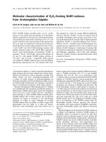
ThestudywasdoneduringafeveroutbreakinMay-
September 2009 in Kozhikkode district of northern Ker-
ala (Figure 1). All the patients included in the study had
* Correspondence:
1
Molecular Virology Laboratory, Rajiv Gandhi Centre for Biotechnology
(RGCB), Thycaud P.O., Thiruvananthapuram-695014, Kerala, India
Full list of author information is available at the end of the article
Niyas et al. Virology Journal 2010, 7:189
/>© 2010 Niyas et al; licensee BioMed Central Ltd. This is an Open Access article distributed under the terms of the Creative Commons
Attribution License ( which permits unrestr icted use, distribution, an d reproduction in
any medium, provided the original work is properly cited.
classical symptoms of CHIK [15]. Samples were
obtained from the outpatient department of three Pri-
mary Health Centres (Olavanna, Beypore and Chaliyum)
in the district. 2-5ml of whole blood was collected from
patients who were clinically diagnosed with CHIK and
had a history of fever of 1-5 days duration. Samples
were transported to the laboratory in wet-ice; serum
was separated and stored in aliquots at -80°C. Standard
ethical and bio-safety guidelines were followed, and
informed consent was obtained from all the patients
prior to blood withdrawal.
For virus detection in mosquitoes, households of
CHIK patients, whose serum samples were confirmed in
the laboratory by RT-PCR, were subsequent ly visited
and larval sampling was done. Stagnant water collected
in discarded articles such as coconut shells, broken
earthern-wares, plastic bottles and damaged drains were
searched for Ae. albopictus larvae. Third and fourth
instar larvae and pupae were phenotypically identified
in situ using standard keys and these were collected and
transferred to containers with fresh water. Four house-
holds each in Olavanna and Chaliyum, and three house-
holds in Beypore were surveyed. Larvae and pupae
collected from each location were made into a single
poo l. In the laboratory, these three pools were indepen-
dently reared in bowls with water, kept in mosquito
cag es at an ambient temperature of 25-30°C and a rela-
tive humidity 60-70%. The newly emerged adult mosqui-
toes were collected and frozen at -20°C for 30 minutes.
Whole-mosquito tissue extracts were prepared by
Figure 1 Map of Kerala showing the location of sample collection areas.
Niyas et al. Virology Journal 2010, 7:189
/>Page 2 of 8
homogenizing pools of adult mosquitoes [each pool with
30 individual mosquitoes (both males and females)
representing a single location]. Frozen mosquitoes w ere
homogenized in 700 μl of Dulbecos Modified Eagle’s
Medium (DMEM) using a micropestle. These were then
clarified by centrifugation at 800 × g at 4°C and steri-
lized b y filtering through 0.2 μMmembranefilter
(Millex GV, Millipore) and used for RNA isolation.
RNA isolation from the 70 patient serum samples and
the three extracts fr om mosquito samples were carried
out using QIAamp Viral RNA Mini kit (Qiagen, GmBH,
Hilden) exactly as per the kit protocol. Single-step RT
PCR was done using 10 μl of the isolated RNA from all
the samples using Fidelitaq RT-PCR kit (USB, Cleveland,
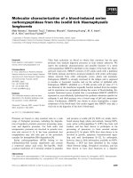
Ohio), as previously described [14]. PCR primers (Table
1; Figure 2) for CHIKV detection PCR were designed
based on earlier reports [16] and on the conserved
genomic regions of local strains of CHIKV [14]. The
conditions for RT PCR were: a reverse transcription
step at 50°C for 45 min; followed by 35 cycles of ther-
mal cycling, which included denaturation at 95°C for 1
min, annealing at 55°C for 1 min, and an extension at
68°C for 2 min. Extreme care was taken to avoid PCR-
contamination, by carrying out the pre-and post amplifi-
cation steps in laboratories located in separate buildings
andalsobyincludinganon-templatecontrolinall
amplifications.
For nucleotide sequencing and phylogenetic analysis,
15 clinical samples and all the three mosquito-derived
samples were used. Only clinical samples that gave a
high intensity amplicon in the primary detection PCR
were selected to ensure that sufficient DNA would be
available for sequencing reactions. Five clinical samples
each from Olavanna, Beypore and Chaliyum were used,
making a total of 15 samples. Selected regions of the
CHIKV genome (nucleotide position, with respect to
S27 reference sequence AF369024: nsP2 3134-3636; E2
8832-9332; E1 10246-10539; Table 1; Figure 2) were
amplified by RT PCR as described above using new sets
of primers (Table1; Figure 2). These specific regions
were chosen as they showed nucleotide variability and
novel mutations in our previous study with the l ocal
strains of CHIKV [14], making them suitable for phylo-
genetic analysis. Purified PCR products were directly
subjected to automated DNA seque ncing as per manu-
facturer’s directions in an ABI-Prism 3730 Genetic ana-
lyzer (PE Applied Biosystems, Foster City, CA). The
sequences were aligned with corresponding CHIKV
sequences obtained from NCBI GenBank using Clustal
W program of MEGA3.1 [17] software, with Kimura-2
distance correction. To get representation from different
gene segments in the evolution of the CHIKV strains,
the partial sequences of nsP2, E2 and E1 genes were
arranged in tandem to obtain a 1311 bp sequence (Fig-
ure 3a), which was then used for phylogenetic analysis.
The phylogeny was reconstructed by Neighbor-Joining
method with 10,000 bootstrap replications using the
MEGA 3.1 program. 100 μL of the mosquito extracts or
patient serum samples were u sed for CHIKV isolation
in confluent monolayer of Vero cells cultured in 75cm
2
flasks, as per standardized protocols [14]. T he titration
of CHIKV in the infected cultures was done by plaque
assayusingacarboxymethyl-celluloseoverlaymethod
[18] on Vero cells.
CHIKV RNA was detected in 49 out of the 70 patient
samples (70%) and in adult mosquitoes derived from lar-
vae from Chaliyum and Olavanna by RT PCR (Figure 4).
All the three mosquito derived samples were positive for
CHIKV, as indicated by cytopathic effects and RT PCR
(Figure 4), in the 3
rd
passage of virus isolation in Vero
Table 1 Details of the primers used for PCR amplification in the study
Primer Name Sequence (5’!3’); location with respect to S27 sequence
(GenBank Accession AF369024)
Target T
a
(°C)
Amplicon size Reference
RT PCR for CHIKV detection in patient and adult mosquitoes derived from larvae
E1 F tacccatttatgtggggc (10246-10263) 52 294bp [16]
E1 R gcctttgtacaccacgatt (10539-10521) E1
NSP2F tgccatgggaataatagagactccg (1682-1699)
ChR6 gcgagtcaaccgtacgtgcag (2390-2370) nsP2 55 709bp This study
ChF27 gtcccctaagagacacattg (11486-11505)
ChR28 tacgtccctgtgggttcggagaat (11798-11780) 3’NTR 52 313bp [14]
RT PCR of partial sequences CHIKV genes for sequencing and phylogenetic analysis
E1Fseq1 gctccgcgtcctttacc (10389-10405)
E1Rseq1 atggcgacgcccccaaagtc (10943-10924) E1 55 555bp This study
ChF21 gggacacttcatcctggc (8832-8849) [14]
ChR22 acatttgccagcggaaac (9332-9315) E2 55 501bp
ChF8 cctatcctcgaaacagcg (3134-3151) [14]
ChR9 gtgactctcttagtaggc (3636-3619) nsP2 45 503bp
Niyas et al. Virology Journal 2010, 7:189
/>Page 3 of 8
cell monolayer cultures. In plaque assays, the culture
supernatants from these infected cells had a virus titre
of 2.0 × 10
11
,3.3×10
10
,1.4×10
10
plaque forming
units (pfu) ml
-1
for samples from Olavanna, Chaliyum
and Beypore, respectively.
Analysis of the partial nucleotide sequenc es of nsP2
(378 bp; position 3246-3623), E1 (position 10427-10931)
and E2 (position 8893- 9320) revealed a few random
nucleotide changes in the CHKV isolates studied (Addi-
tional File 1) with respect to the corresponding
sequences of the previous isolates from Kerala [14]. The
nucleotide change T3297C observed in the 2007 & 2008
Kerala isolates, causi ng an L539 S mutation in the nsP2
protein, was absent in CHIKV strains of the present
outbreak. A novel substitution (T3296C) was consis-
tently observed in a few strains from patients
(RGCB711, RGCB730, and RGCB755) and in all the
three isolates from mosquito samples. However, this was
a synonymous substitution. The E1 sequence of all the
strains had the C10670T substitution resulting in the
A226V mutation identified in the re cent isolates of
CHIKV [3,14]. Another new substitution (E1 G10864A)
detected consistently in all the mosquito-derived strains
and two of the clinical isolates (RGCB711 & RGCB755)
can result in an amino acid change of V291I. Two
nucleotide substitutions (A9114G and T9170A) were
observed in the E2 coding region of all the strains stu-
died from the outbreak. The latter substitution resulted
in an am ino acid change L210Q in the pre dicted
sequence of amino acids of the E2 pro tein. Phylogene tic
analysis revealed that the strains involved in the out-
break were closely related to the East-Central South
African genotype of the CHIKV (Figure 3b). The gene
sequences of CHIKV obtained from mosquito and
patient samples formed a close cluster, distinct from the
strains isolated previously from Kerala [14], rest of India
and other parts of the world. This show a common
genetic origin of the virus strains from patients and
mosquitoes in this outbreak.
Apart from these genetic changes, an interesting
observationinthestudywas the detection of CHIKV
from adult mosquitoes derived from larval samples.
Considering that these mosquitoes were freshly
emerged in the laboratory from the larvae collected
from areas encountering a CHIK outbreak and did not
have a blood-meal, the possibility of acquiring the
virus through transovarial transmission (TOT) can be
thought of. Even though TOT has been proven in fla-
viviruses [19-23], the occurrence of this phenomenon
in alphaviruses is still inconclusive [24-27]. Studies
using a Réunion Island isolate of the CHIKV from
2006 outbreak [Strain 06.21; GenBank: AM258992]
could not demonstrate vertical transmission in the
mosquito vector [25]. The mosquito infectivity of
alphaviruses is modulated by mutations in specific viral
proteins [28-31]. Amino acid residues 200-220 of the
E2 protein determine the cellular receptor tropism and
mid-gut infectivity in Ae. aegypti mosquitoes [28,30].
An E2 I211T mutation was found to strongly enhance
Ae.albopictus infectivity of CHIKV strains with the E1
A226V change [31]. Both the mutations were present
in the isolates in this study and also in the recent
Indian isolates [10,12,14] (Figure 5). Interestingly, the
novel mutation in E2 (L210Q) that was detected exclu-
sively in these 2009 CHIKV strains was adjacent to the
E2-211 position. This substitution of the aliphatic
amino acid leucine with glutamine, an amino acid with
polar side chains, can have critical effects on local
Figure 2 Location of primers in the CHIKV genome. Positions are numbered with respect to S27 sequence (GenBank Accession AF369024).
Niyas et al. Virology Journal 2010, 7:189
/>Page 4 of 8
protein structure. One of the predicted effects of such
amino acid changes is the exposure of buried protein
surfaces. Possibly, this may alter the interaction of E2
with other proteins, particularly with cellular receptors,
and may change the tissue tropism. However, more
studies are required to understand the effects of the
L210Q mutation.
The results from this study, along with the previous
observations [11,12,14], indicate a constant genomic
evolution of the CHIKV strains circulating in Kerala.
The availability of large numbers of Ae.albopictus vector
mosquitoes [11] and an immunologically naïve human
population unexposed to CHIK in different parts of the
state might facilitate recurrent infections and viral
Figure 3 Phylogenetic analysis of the CHIKV partial nsP2, E2 and E1 coding region nucleotide sequences. a) Tandem arrangement of the
sequences used for the analysis. Numbers indicate the position with respect to the sequence of S27 strain (AF369024). b) Neighbor-Joining Tree
of corresponding sequences of CHIKV strains derived from human clinical samples constructed with 10,000 bootstrap replications. The human
and mosquito sequences obtained from the study are marked ‘black triangle’ and ‘black diamond’, respectively. GenBank accession numbers and
strain names are indicated. Scale bar represents the number of substitutions/site. Sequences of recent Kerala isolates are indicated by ‘!’.
Niyas et al. Virology Journal 2010, 7:189
/>Page 5 of 8
evolution. Emergence of newer strains with altered viru-
lence and transmission potential is a possible out come
of the long term viral persistence in the community.
Further entomological and viro logica l studies with these
new CHIKV strains would help to understand the chan-
ging epidemiology of this re-emerging virus.
Figure 4 RTPCRbaseddetectionofCHIKVRNAinadult
mosquitoes derived from larvae. WE-mosquito whole extract; P1,
P2, P3-RNA from viral passage 1, 2 & 3 in Vero cells; M-molecular
weight marker.
Figure 5 Alignment of predicted amino acid sequences of the partial E2 protein of CHIKV strains. The newly identifie d L210Q mutation
in Kerala strains is indicated. The CHIKV strain from Réunion island, which was previously used in vertical transmission studies [25], is marked as
‘** ‘.
Niyas et al. Virology Journal 2010, 7:189
/>Page 6 of 8
Additional material
Additional file 1: Clustal W alignment of the partial nucleotide
sequences of Chikungunya virus nsP2, E2 and E1 protein coding
region.
Acknowledgements
The authors are thankful to the medical staff of the primary health centres
(PHC) in Olavanna, Beypore and Chaliyum for the help extended for the
patient sample collection. The financial assistance by Department of
Biotechnology, Government of India as intramural funding and the
encouragement and support by the Director, RGCB, are gratefully
acknowledged.
Author details
1
Molecular Virology Laboratory, Rajiv Gandhi Centre for Biotechnology
(RGCB), Thycaud P.O., Thiruvananthapuram-695014, Kerala, India.
2
State
Disease Control and Monitoring Cell (SDCMC), National Rural Health Mission
(NRHM), Government of Kerala, Thiruvananthapuram-695014, Kerala, India.
3
Department of Community Medicine, Medical College, Thiruvananthapuram,
Kerala, India.
Authors’ contributions
KPN, SN, AM, and AI obtained patient samples, carried out RT PCR and
sequencing studies. RA did the virus isolation. TM made the administrative
arrangements for obtaining samples from the hospitals, and was involved in
identifying CHIK patients and collecting blood samples. RNU did the
collection, identification and rearing of mosquito larvae. ES conceived the
study and drafted the manuscript. All authors read and approved the final
manuscript.
Competing interests
The authors declare that they have no competing interests.
Received: 18 March 2010 Accepted: 13 August 2010
Published: 13 August 2010
References
1. Powers AM, Logue CH: Changing patterns of chikungunya virus:
re-emergence of a zoonotic arbovirus. J Gen Virol 2007, 88:2363-2377.
2. Staples JE, Breiman RF, Powers AM: Chikungunya fever: an
epidemiological review of a re-emerging infectious disease. Clin Infect Dis
2009, 49:942-948.
3. Schuffenecker I, Iteman I, Michault A, Murri S, Frangeul L, Vaney MC,
Lavenir R, Pardigon N, Reynes JM, Pettinelli F, et al: Genome
microevolution of chikungunya viruses causing the Indian Ocean
outbreak. PLoS Med 2006, 3:e263.
4. Rezza G, Nicoletti L, Angelini R, Romi R, Finarelli AC, Panning M, Cordioli P,
Fortuna C, Boros S, Magurano F, et al: Infection with chikungunya virus in
Italy: an outbreak in a temperate region. Lancet 2007, 370:1840-1846.
5. Chandak NH, Kashyap RS, Kabra D, Karandikar P, Saha SS, Morey SH,
Purohit HJ, Taori GM, Daginawala HF: Neurological complications of
Chikungunya virus infection. Neurol India 2009, 57:177-180.
6. Das T, Jaffar-Bandjee MC, Hoarau JJ, Krejbich Trotot P, Denizot M, Lee-Pat-
Yuen G, Sahoo R, Guiraud P, Ramful D, Robin S, et al: Chikungunya fever:
CNS infection and pathologies of a re-emerging arbovirus. Prog Neurobiol
2009, 91:121-9.
7. Tsetsarkin KA, Vanlandingham DL, McGee CE, Higgs S: A single mutation in
chikungunya virus affects vector specificity and epidemic potential. PLoS
Pathog 2007, 3:e201.
8. Vazeille M, Moutailler S, Coudrier D, Rousseaux C, Khun H, Huerre M,
Thiria J, Dehecq JS, Fontenille D, Schuffenecker I, et al: Two Chikungunya
isolates from the outbreak of La Reunion (Indian Ocean) exhibit
different patterns of infection in the mosquito, Aedes albopictus. PLoS
One 2007, 2:e1168.
9. Yergolkar PN, Tandale BV, Arankalle VA, Sathe PS, Sudeep AB, Gandhe SS,
Gokhle MD, Jacob GP, Hundekar SL, Mishra AC: Chikungunya outbreaks
caused by African genotype, India. Emerg Infect Dis 2006, 12:1580-1583.
10. Arankalle VA, Shrivastava S, Cherian S, Gunjikar RS, Walimbe AM, Jadhav SM,
Sudeep AB, Mishra AC: Genetic divergence of Chikungunya viruses in
India (1963-2006) with special reference to the 2005-2006 explosive
epidemic. J Gen Virol 2007, 88:1967-76.
11. Kumar NP, Joseph R, Kamaraj T, Jambulingam P: A226V mutation in virus
during the 2007 chikungunya outbreak in Kerala, India. J Gen Virol 2008,
89:1945-1948.
12. Santhosh SR, Dash PK, Parida MM, Khan M, Tiwari M, Lakshmana Rao PV:
Comparative full genome analysis revealed E1: A226V shift in 2007
Indian Chikungunya virus isolates. Virus Res
2008, 135:36-41.
13. Kannan M, Rajendran R, Sunish IP, Balasubramaniam R, Arunachalam N,
Paramsivan R, Tewari SC, Samuel PP, Tyagi BK: A study on chikungunya
outbreak during 2007 in Kerala, south India. Indian J Med Res 2009,
129:311-315.
14. Sreekumar E, Issac A, Nair S, Hariharan R, Janki MB, Arathy DS, Regu R,
Mathew T, Anoop M, Niyas KP, Pillai MR: Genetic characterization of 2006-
2008 isolates of Chikungunya virus from Kerala, South India, by whole
genome sequence analysis. Virus Genes 2010, 40:14-27.
15. WHO/SEARO (2008) Chikungunya Fever, a re-emerging disease in Asia.
WHO South East Asia Regional Office. [ />Section10/Section2246.htm].
16. Hasebe F, Parquet MC, Pandey BD, Mathenge EG, Morita K,
Balasubramaniam V, Saat Z, Yusop A, Sinniah M, Natkunam S, Igarashi A:
Combined detection and genotyping of Chikungunya virus by a specific
reverse transcription-polymerase chain reaction. J Med Virol 2002,
67:370-374.
17. Kumar S, Tamura K, Nei M: MEGA3: Integrated software for Molecular
Evolutionary Genetics Analysis and sequence alignment. Brief Bioinform
2004, 5:150-163.
18. Escobar-Herrera J, Medina-Ramirez FJ, Gutierrez-Escolano AL: A
carboxymethyl-cellulose plaque assay for feline calicivirus. J Virol Methods
2007, 146:393-396.
19. Rosen L, Shroyer DA, Tesh RB, Freier JE, Lien JC: Transovarial transmission
of dengue viruses by mosquitoes Aedes albopictus and Aedes aegypti.
Am J Trop Med Hyg 1983, 32:1108-1119.
20. Gunther J, Martinez-Munoz JP, Perez-Ishiwara DG, Salas-Benito J: Evidence
of vertical transmission of dengue virus in two endemic localities in the
state of Oaxaca, Mexico. Intervirology 2007, 50:347-352.
21. Mishra AC, Mourya DT: Transovarial transmission of West Nile virus in
Culex vishnui mosquito. Indian J Med Res 2001, 114:212-214.
22. Rosen L, Shroyer DA, Lien JC: Transovarial transmission of Japanese
encephalitis virus by Culex tritaeniorhynchus mosquitoes. Am J Trop Med
Hyg 1980, 29:711-712.
23. Aitken TH, Tesh RB, Beaty BJ, Rosen L: Transovarial transmission of yellow
fever virus by mosquitoes (Aedes aegypti). Am J Trop Med Hyg 1979,
28:119-121.
24. Mourya DT: Absence of transovarial transmission of Chikungunya virus in
Aedes aegypti &Ae. albopictus mosquitoes. Indian J Med Res 1987,
85:593-595.
25. Vazeille M, Mousson L, Failloux AB: Failure to demonstrate experimental
vertical transmission of the epidemic strain of Chikungunya virus in
Aedes albopictus from La Reunion Island, Indian Ocean. Mem Inst
Oswaldo Cruz 2009, 104:632-635.
26. Zytoon EM, el-Belbasi HI, Matsumura T: Transovarial transmission of
chikungunya virus by Aedes albopictus mosquitoes ingesting
microfilariae of Dirofilaria immitis under laboratory conditions. Microbiol
Immunol 1993, 37:419-421.
27. Thavara U, Tawatsin A, Pengsakul T, Bhakdeenuan P, Chanama S,
Anantapreecha S, Molito C, Chompoosri J, Thammapalo S,
Sawanpanyalert P, Siriyasatien P: Outbreak of chikungunya fever in
Thailand and virus detection in field population of vector mosquitoes,
Aedes aegypti (L.) and Aedes albopictus Skuse (Diptera: Culicidae).
Southeast Asian J Trop Med Public Health 2009, 40:951-962.
28. Myles KM, Pierro DJ, Olson KE: Deletions in the putative cell receptor-
binding domain of Sindbis virus strain MRE16 E2 glycoprotein reduce
midgut infectivity in Aedes aegypti. J Virol 2003, 77:8872-8881.
Niyas et al. Virology Journal 2010, 7:189
/>Page 7 of 8
29. Pierro DJ, Powers EL, Olson KE: Genetic determinants of Sindbis virus
strain TR339 affecting midgut infection in the mosquito Aedes aegypti. J
Gen Virol 2007, 88:1545-1554.
30. Pierro DJ, Powers EL, Olson KE: Genetic determinants of Sindbis virus
mosquito infection are associated with a highly conserved alphavirus
and flavivirus envelope sequence. J Virol 2008, 82:2966-2974.
31. Tsetsarkin KA, McGee CE, Volk SM, Vanlandingham DL, Weaver SC, Higgs S:
Epistatic roles of E2 glycoprotein mutations in adaption of chikungunya
virus to Aedes albopictus and Ae. aegypti mosquitoes. PLoS One 2009, 4:
e6835.
doi:10.1186/1743-422X-7-189
Cite this article as: Niyas et al.: Molecular characterization of
Chikungunya virus isolates from clinical samples and adult Aedes
albopictus mosquitoes emerged from larvae from Kerala, South India.
Virology Journal 2010 7:189.
Submit your next manuscript to BioMed Central
and take full advantage of:
• Convenient online submission
• Thorough peer review
• No space constraints or color figure charges
• Immediate publication on acceptance
• Inclusion in PubMed, CAS, Scopus and Google Scholar
• Research which is freely available for redistribution
Submit your manuscript at
www.biomedcentral.com/submit
Niyas et al. Virology Journal 2010, 7:189
/>Page 8 of 8