Báo cáo y học: "Molecular epidemiology of Hepatitis C virus genotypes in Khyber Pakhtoonkhaw of Pakistan" pptx
Bạn đang xem bản rút gọn của tài liệu. Xem và tải ngay bản đầy đủ của tài liệu tại đây (667.51 KB, 7 trang )
RESEARC H Open Access
Molecular epidemiology of Hepatitis C virus
genotypes in Khyber Pakhtoonkhaw of Pakistan
Amjad Ali
1
, Habib Ahmed
1*
, Muhammad Idrees
2
Abstract
Six major Hepatitis C virus (HCV) genotypes and hundreds of subtypes have been identified globally. All these gen-
otypes are generally studied for epidemiology, their vaccine development and clinical management. This article
comments the frequency distribution of various HCV genotypes circulate in different areas/districts of Khyber Pakh-
toonkhaw Province of Pakistan. Sum of 415 HCV RNA PCR positive sera samples were tested by a molecular geno-
typing assay. Data analysis revealed that out of these 415 HCV RNA positive patients 243 were males and 172 were
females. Distribution breakup of the patients was 135, 58, 51, 51, 36, 32, 6, 7and 9 patients come from the districts
of Abbottabad, Mardan, Pehawar, Swat, Haripure, Swabi and Dera Ismail Khan, respectively . Out of the tested sam-
ples, genotype specific PCR fragments were observed in 299 (74.82%) patient serum samples. The distribution of
genotypes of the typeable samples was as fallows: 3 patients (0.72%) each were infected with genotype 1a and
genotype 1b; 240 patients (80.26%) of genotype 3a; 25 patients (6.00%) genotype 3b; and 28 patients (6.73%) were
observed as with mixed genotypic infection. Sums of 116 serum samples (27.88%) were still found untypeable by
the used molecular genotyping system.
In conclusion, HCV genotypes 1a, 1b, 3a and 3b are distributed in various parts of KPK among which the genotype
3a is the most frequent genotype.
Background
Hepat itis C virus (HCV) infection is accountable for the
second most common cause of viral hepatitis and is one
of the most important Flaviviridae infections with sig-
nificant clinical problems all over the globe in humans
[1]. At least six major HCV genotypes and hundreds of
subtypes have been identified worldwide so far [2] . Dis-
similar HCV genotypes are related to epidemiological
studies, response rates to anti-viral treatment, vaccine
development and clinical management of the infection
[3]. HCV genotype is the strongest foret elling factor for
sustained virological response since patients with differ-
ent HCV genotypes act in response differently to alpha
interferon therapy [4,5]. Solid evidence has been estab-
lished that HCV genotype-2 and genotype-3 infected
patents are more likely to have a sustained virological
response (SVR) to anti-vi ral therapy than patients
infected with ge notype-1 HCV infections [6]. The
reported rates of SVR to interferon plus ribavirin
combination therapy are 65% and 30%, in patients
infected with HCV-2/3 and HCV-1 genotypes respec-
tively [7,8]. As the patient genotype has a vital role in
treatment outcome therefore, should be done before
starting standard interferon therapy.
ThreeHCVgenotypessuchasHCV-1,HCV-2,and
HCV-3 have worldwide distribution and their relative
prevalence varies from one geograp hic area to another.
HCV-1a and 1b subtypes are the most p revailing geno-
types circulating in the United States of America and
Europe [4,9-11]. In Japan the most common circulating
HCV subtype is 1b [12]. HCV-2a and 2b subtypes are
mostly common in North America, Europe, and Japan
and subtype 2c is found commonly in northern Italy
[9-12]. HCV-4 is the most prevalent genotype circulat-
ing in North Africa and the Middle East [13,14]. HCV-5
and HCV-6 genotypes are establish only in South Africa
and Hong Kong, respectively [15,16].
A small number of studies are available from Pakistan
on the d istribution of different hepatitis C virus geno-
types only from the provinces of Punjab and Sindh
[13,17-19]. No such study on the frequency distribution
of various HCV genotypes and their modes of infectivity
* Correspondence:
1
Deparment of Genetics, Hazara University, Garden Campus Mansehra
Khyber Pakhtoonkhaw, Pakistan
Full list of author information is available at the end of the article
Ali et al. Virology Journal 2010, 7:203
/>© 2010 Ali et al; licensee BioMed Central Ltd. This is an Open Acce ss article distributed under the terms of the Creative Commons
Attribution License (http://creativecom mons.org/licenses/by/2.0), which permits unrestricted use, distribution, and reproduction in
any medium , provided the original work is properly cited.
for different genotypes is available from Khyber Pakh-
toonkhaw (KPK) of Pakistan. Therefore, this study was
initiated to find out the molecular epidemio logy of var-
ious HCV genotypes and subtypes present in KPK
region of Pakistan and further to find out linked risk
factors for its transmission.
Methods
Sampling
For the determination of HCV genotyping serum sam-
ples were collected along with specifically designed data
sheets from patients admitted/attending various tertiary
collection centers situated in different districts/parts of
KPK, Pakistan. A written informed consent was taken
from each patient. A printed data sheet was also filled
for each patient contained demographic characteristic,
possible mode of transmission, area/district, and esti-
mated time of infection along with complete address
and contact numbers of the patients.
HCV RNA qualitative and quantitative PCRs
HCV RNA was detected qualitatively using reverse tran-
scriptase (RT) PCR as described before [17]. Briefly,
total RNA was isolated from 150 μlpatient’sserasam-
ples using Gentra RNA isolation kit (Puregene, Minnea-
polis, MN 55441 USA) according to the kit protocol.
Complimentary DNA (cDNA) of HCV 5’ NCR was
synthesized using 100 units of Moloney murine leuke-
mia virus (MMLV) reverse transcriptase enzyme (RTEs)
(Invitrogen, Corp., California USA) with 5 pM of outer
antisense primer. Two rounds of PCR amplifications
were done (first round PCR and Nested PCR) with two
unites of Taq DNA polymerase enzyme (Invitrogen,
Corp., California USA) in a volume of 2o μl reaction
mix. The nested PCR products were run on 2% agarose
gel contained ethidium bromide as DNA stain. The sp e-
cific HCV PCR bands were visualized under UV
transilluminator.
HCV RNA was quantified in all qualitative PCR pos i-
tive sera using SmartCycler II Real-time PCR (Cepheid,
Sunnyvale, Calif. USA) utilizing HCV RN A quantifica-
tion kits (Sacace Biotechnologies, Italy). The SmartCy-
cler II system is a PCR system by which amplification
and detection were accomplished concurrently with
TaqMan technology (Applied Biosystems, Foster City,
Calif) using fluorescent probes to detect amplification
after each replicating cycle. The lower and upper detec-
tion limits of the used assay were 5.0 × 10
2
and 5.0 ×
10
8
IU/mL, respectively. Specimens yielding values
above the upper limit were diluted 100-fold, retested
and the obtained values were multiplied by this dilution
factor to get the actual HCV RNA concentration in
international units per mL.
HCV Genotyping
For all the samples HCV genotyping was carried out
using molecular HCV genotyping method previously
published by Idrees [20]. Briefly, 10 μl (about 50 ng) of
the extracted RNA was reverse transcribed to cDNA
using 100 U of M-MLV RTEs at 37°C for 50 minutes.
The RTEs were killed at 96°C for 5 minutes. Two μlof
cDNA was used for the amplification of 470-bp region
from HCV 5’NCR+Core region in first roun d PCR. Each
first round PCR sample was subjected to two second-
rounds nested PCR amplifications first with mix-A pri-
mers and the second with mix-B primers in a reaction
volume of 20 μl. Mix-A had genotype-specific primers
for 1a, 1b, 1c, 3a, 3c and 4 genotypes and mix-B con-
tained genotype-specific primers for 2a, 2c, 3b, 5a, and
6a genotypes. The second round PCR products were
electrophoresed on a 2% agarose gel to separate type-
specific PCR fragment. The gel w as stained with
ethidium bromide and was observed under UV transillu-
minator. A 100-bp DNA ladder (Invitrogen, Corp., Cali-
fornia, USA) was run in each gel as DNA size marker
and the HCV genotype for each sample was determined
by identifying the HCV genotype-specific PCR band.
The gel photograph was taken using gel documen tation
system (Geldoc System, Eppendorf Inc, Germany).
Statistical analysis
SPSS version 10.0 for windows was used for the analysis
of data and summary statistics. The results for all vari-
ables were set in the form of rates (%). Fisher’s exact
and Chi Square tests were applied to find out the posi-
tive association among the categorical variables. The
data was obtainable as mean values or number of
patients. P-value less than 0.05 was considered as
significant.
Results
Patients demographic
Results regarding the demographic distribution of HCV
patients geno typed are summarized in figure 1. The fig-
ure also show enrolment and disposition criteria of the
patients. The results revealed that out of total 663 anti-
HCV positive sera that were received from different dis-
tricts of KPK province, 523 were found positive by HCV
qualitative PCR where as 140 sera samples were found
negative by PCR and were thus excluded from further
evaluation. Viral load was determined on all the 523
HCV RNA positive samples. In 108 samples the viral
load was less than 500 IU/ml that is sensitivity of the
genotyp ing assay, therefore, these low titer sera samples
were excluded from the study for further genotyping
analysis as were below the sensitivity limits of the geno-
typing assay. The selected 415 sera samples with
Ali et al. Virology Journal 2010, 7:203
/>Page 2 of 7
moderate to high viral load (500 -5.0 × 10
8
IU/mL or
above were tested by type-specific genotyping assay. The
genotyped sera samples revealed that 136 belonged to
Abbottabad region, 58 to Bannu, 80 to Kohat, 51 to
Mardan, 36 to P eshawar, 32 to Swat, 6 to Haripur, 7 to
Swabi and 9 patients came from D.I. khan. As all these
serum samples that were included in the current study
were tested HCV-RNA positive with enough viral load
and could thus be genotyped by the utilized genotype-
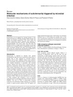
specific PCR assay. Figure-2 shows a typical agarose gel
showing different HCV genotype-specific bands (HCV-
1a & HCV-3a).
Pattern of HCV genotypes in the study population
The distribution of HCV genotypes in the population
analyzed is given in the table 1. The data shows that out
of 415 tested sera samples, type-specific PCR fragments
were seen in 299 (72.04%) whereas 116 (27.95%) s era
samples were found untypable in th e current study. The
pattern of HCV genotypes of the typeable samples seen
in the current study were in the order of: 240 (57.83%)
were genotype 3a, 25 (6.02%) were with genotype 3b, 3
(0.72%)were1aand3(0.72%)were1b.whereassum28
(6.73%) sera samples were infected with mixed genotype.
Frequency distribution of HCV genotypes in different
districts of KPK
Frequency distribution of different HCV genotypes were
recorded from individuals belonged to various districts
of KPK is shown in table 2. Among the determined gen-
otypes 136 patients were from Abbottabad. Among the
genotyped samples from Abbottabad, 83 (61.02%)
belonged to genotype 3a, 5 (3.67%) were genotype 3b,
11 (8.08%) patients were infected by mixed genotype
and 37 (27.02%) patients were observed as of unknown
genotype. From Bannu HCV positive cases were 58,
among these 32 (5 5.17%) were genotype 3a, 5 (8.62%)
3b, 3 (5.17%) were of dual genotype i.e 3a/1b. Sum 18
(31.03%) patients were of unknown genotype from Dis-
trictBannu.Among9patientsfromD.I.Khan,6
(66.66%) belonged to 3a genotype, 3 (33.33%) patients
were observed as untypeable. Total 80 patients were
positive to the co rresponding virus from region of
Koh at, 1 (1.25%) patient each from 1a and 1b genotype,
39 (48.75%) were of 3a genotype, 2 (2.5%) 3b genotype,
mixed genotype was 6 (7.5%). Patients having unknown
HCV genotype were 31 (38.75%). Among 51 Mardans
patients 1a was 1 (1.96%) and 1b were 2 (3.92%), 3a
were 28 (54.90%), 3b were 8 (1 5.68%), 7 (13.72) were
dual genotypes and 5 (9.80) were untypeable genotypes.
Figure 1 Study disposition. For study enrollments the patients were required to have chronic HCV with positive anti-HCV ELISA. The patients
were also required to have detectable HCV RNA by qualitative RT-PCR and viral load >500 IU/ml and belonged to Khyber Pakhtoonkhaw
province of Pakistan.
Ali et al. Virology Journal 2010, 7:203
/>Page 3 of 7
Of the 36 isolates from Peshawar city, 26 (72.22%) were
3a, 3 (8 .33%) were 3b, 7 (19.44%) were untypeable and
none was observed as with dual genotype. Of the 32
positive sera samples isolated from district Swat, 1a gen-
otype one (3.125%) was of 1a genotype, 21 (65.625%) 3a,
2 (6.25%) 3b, mixed genotype 1 (3.125%), 7 (19.44%)
were of untypeable genotype. From Haripur subtype 3a
were present in 5 (83.33%) patients and 1 (16.66%) sam-
ple was found untypeable. All the 7 patients’ sera col-
lected from Swabi were found with untypeable
genotypes.
Occurrence of HCV with mixed genotypes
Table 3 shows the prevalence of HCV m ixed-genotype
infections determined during the current study in differ-
ent populations across KPK of Pakistan. Total 28 HCV
isolates were found having two genotypes. Of these, 11
belonged to Abbot tabad region, 3 to Bannu district, 6 to
Kohat district, 7 to Mardan district and only one to
Swat district. Fourteen of the HCV infection with mixed
genotypes had HCV genotypes 3a and 3b followed by
3a + 1b that were 10 (35.71%), 3a + 1a in 3 (10.71%)
and 3a + 2b in 1 (3.57%).
Potential risk factors associated with the transmission of
various genotypes
Various possible risk factors observed in the current
study responsible for infection transmission with each
HCV genotypes are given in table 4. Over all the prob-
able modes of spread observed were: 58.1% due to mul-
tiple uses of needles especially syringes, 16.7% due to
surgeries (both major and minor), 3.3% due to blood
and blood products infectivity and in 23.1% patients the
mode of spread was not known and therefore were
sporadic. The foremost mode of contamination in
patients with HCV genotype 3a and 3b was multiple use
and re-use of needles/syringes that was 70% and 60%
respectively. All the ge notype 1a and about 75% 1b
infected patients got their infection during surgeries.
Sixty percent of the patients having dual infections were
sporadic where the route of infectivity was unknown to
them. Majority (58.1%) of untypable patients were
infected due to contaminated needles and syringes fol-
lowed by surgeries and dental procedures.
Discussion
Khyber Pakhtoonkhaw (KPK) previously known as the
North-West Frontier Province (NWP) is situated in the
North-western of Pakistan and is one of the four pro-
vinces of Pakistan. It borders Gilgit-Baldistan to the
north-east, Aghanistan to the north-west, the Federal
Administrativ e Tribal Areas (FATA) to the west and
south, Azad Jammu & Kash mir to the east, Balochistan
to the south and Punjab and the Islamabad Capital
Figure 2 Typical agarose gel electrophoresis patterns of PCR products from two different HCV genotypes. Lanes 1 and 7 showing 50-bp
DNA size ladder maker; Lanes 2-3 showed HCV-1a specific bands (210-bp); Lane 4-5 showed HCV-3a specific band (258-bp), Lane 6 showed
Positive Control (258-bp HCV-3a genotype-specific band) and Lane 8 showed Negative Control (No band).
Table 1 Frequency distribution of HCV genotypes and
subtypes in the studied population (N = 415)
HCV Genotype HCV Subtype No. of Isolates Percentage
11a
1b
3
3
0.72
0.72
33a
3b
240
25
57.83
6.02
Mixed 28 6.73
Undetermined 116 27.95
Total 415 100
Ali et al. Virology Journal 2010, 7:203
/>Page 4 of 7
Territory to the south-east. KPK is the third most popu-
lous province of the country. The main ethnic group in
the province is Pakhtuns, followed by a number of smal-
ler ethnic groups most notably, the Hindkowans; there-
fore, in the current study we tried to determine the
pattern of HCV genotype in this specific ethnic group-
Pakhtuns. A recently published genotype-specific PCR-
based method [20] with increased sensitivity and specifi-
city was employed for HCV genotypes determination.
The data presented here corresponds to the preceding
studies, in which genotypes, sub-types and/or serotypes
were determined [9,21-23]. Analysis of the data sho wed
that genotype 3a is the predominant genotype circulat-
ing in patients with chronic hepatitis C. These findings
verified results of the earlier studies from Pakistan
[17-19] which have concluded that genotype 3a is the
most prevalent HCV genotype in Pakistan. Similarly in
India, the predominant HCV genotype is 3a [24,25].
Our finding regarding distribution of the genotype
seems to be similar to the genotype pattern reported
from other Far Asian country such as Nepal [26] but
differentfromthoseinSouthAsiancountriessuchas
Japan [27], Thailand [28] and Vietnam where genotype
1 is the major HCV genotype circulating in their
populations.
Our study led to several important findings. The first
finding is incidence of HCV genotypes that confirms the
findings of another stu dy from this country [23]. The
second important finding of the study was the isolation
of 27% isolates that were undetermined as no genotype-
specific PCR products were seen for these samples. All
these 116 sera samples with indetermined genotypes
were HCV-RNA positive by qualitative PCR and were
with sufficient viral titer therefore might be genoty ped
by the utilized genotype-specific PCR assay. A recent
study from other parts of Pakistan showed only 6%
HCV infected sera samples with untypable genotypes by
this molecular biology-based system [23]. The high rate
of untypable results seen in the current study may be
due to the reason that m ajority (more than eighty per-
cent) of our untypable patients had received standard
interferon plus ribavirin treatment in the past and were
either non-responders or were relapsed thereafter. Why
the previously treated patients are difficult to genotype
with higher sensitivity using this molecular based geno-
typing assay is not known to us.
We were unable to isolate even a single HCV-4 geno-
type from any infected patient that is believed to be
absent from Pakistan, and is the most prevalent HCV
genotype in Middle East [13]. None of the patients of
the current study was found infected by genotype 5a
and 6a. The two genotypes are reported from South
Africa and Hong Kong, respectively [15,16] and may be
absent or very rare in this part of the world.
Table 2 Prevalence of HCV of comprise genotypes in different geographical regions of KPK of Pakistan
Geno-type Sub-
type
Isolated
from
Abbottabad
Isolated
from
Bannu
Isolated
from
Kohat
Isolated
from
Mardan
Isolated from
Peshawar
Isolated
from
Swat
Isolated
from Hari
pur
Isolated
from
Swabi
Isolated
from
D.I.
khan
P
value
1 1a 0 0 1 (1.25) 1 (1.96%) 0 1
(3.125%)
000NS
1b 0 0 1 (1.25%) 2 (3.92%) 0 0 0 0 0 NS
3 3a 83 (61.02%) 32 (55.17%) 39
(48.75%)
28
(54.90%)
26(2.22%) 21
(65.625%)
5 (3.33%) 0 6 (6.66%) <0.05
3b 5 (3.67%) 5 (8.62%) 2 (2.5%) 8
(15.68%)
3 (8.33%) 2 (6.25%) 0 0 0 < 0.05
Mixed 11(8.08%) 3 (5.17%) 6 (7.5%) 7
(13.72%)
01
(3.125%)
000NS
Undetermined 37 (27.20%) 18 (31.03%) 31
(38.75%)
5 (9.80%) 7 (19.44%) 7
(21.375%)
1 (6.66%) 7 (100%) 3 (3.33%) > 0.05
Total 136 58 80 51 36 32 6 7 9
Table 3 Prevalence of HCV mixed genotypes in KPK, Pakistan
Mixed
genotype
From
Abbottabad
From
Bannu
From
DIK
From
Kohat
From
Mardan
From
Peshawar
From
Swat
From
Haripur
From
Swabi
N
3a+3b 8 0 0 4 2 0 0 0 0 14
3a+1a 2 0 0 0 0 0 1 0 0 3
3a+1b 0 3 0 2 5 0 0 0 0 10
3a+2b 1 0 0 0 0 0 0 0 0 1
Total 11 3 0 6 7 0 1 0 0 28
Ali et al. Virology Journal 2010, 7:203
/>Page 5 of 7
The distribution of HCV genotypes for this population
was examined district wise in order to establish a base
line for regional differences in HCV pattern in KPK. No
regional difference with respect to HCV genotype distri-
bution in all districts was observed where the most pre-
valent genotype is 3a. However, a difference was
observed in district Swabi where all the isolates were
found untypeable. All these isolates had high titer of
HCV RNA and could thus be genotyped however;
majority of these patients had a history of interferon
treatment.
In the current study s um 28 isolates of HCV patients
had two genotypes at a time in their blood. Majority of
these (39%) were the residents of district Abbottabad
region where blood transfusion is common in thalassae-
mic patients. More than half of our patients with dual
infection had HCV genotypes 3a and 3b. Like other stu-
dies, the prevalence of HCV mixed-genotype infections
was high in thalassaemic patients who had received mul-
tiple blood transfusions. The overall rate of HCV mixed-
genotype infections was 6.7%, which is the same as
reported recently by Idrees and Riazuddin [23] from
other provinces of the country.
It has been recognized in the current study that differ-
ent HCV genotypes might be associated with different
transmission routes. For example genotype 3a appears
to be prevalent among injection drug users and dual
infection among thalassaemic patients who had received
blood transfusion several times in life. It is believed that
HCV-3a was introduced into North America and the
United Kingdom with the widespread use of heroin in
the 1960s [29]. For more than 58% of our patients the
probable modes of transmission observed were multiple
uses and re-uses of needles/syringes. In 16.7% patients it
was due to surgeries (both major and minor), 3.3% due
to blood and blood products contamination and in
23.1% patients the mode of cont amination was not
known and was sporadic. The dominant mode of con-
tamination in patients with HCV genotype 3a and 3b
was multiple and re-use of needles/syringes that was
70% and 60% respectively. All the genotype 1a and 75%
1b infect ed patients got their infection during surgeries.
Sixty percent of the patients having dual infections were
sporadic where t he route of contamination was
unknown to them. Majority (58.1%) of untypable
patients were infected by contaminated needles and syr-
inges followed by surgeries and d ental procedures. In
Pakistan HCV-3a is the most widespread genotype as
been also observed in the current study. It is believed
that this genotype is spread by medical practitioners like
doctors, vaccination teams and other medical persons
used non-disposable syringes for injections attended a
number of patients in the past. Mass vaccination in the
recent past in which un-sterilized syringes were used
might have enhanced the infection rate in this country
[23]. This type of practice is still common in the coun-
tryside especially in KPK province which needs effective
check for minimizing the spread of HCV infection and
the transmission of other communicable diseases.
Theonlylimitationofthisstudyisthedetectionof
large number (27%) of samples with untypable geno-
types. All these samples were HCV-RNA positive, had
sufficient viral titer and therefore might be genotyped
by sequencing method to designate the exact genotype,
however, we were unable to sequence these samples due
to lack of sequencing facility in our campus.
Conclusion
We conclude that (i) HCV genotypes 1a, 1b, 3a and 3b
are distributed in various parts of KPK (ii) genotype 3a
is the most frequent genotype circulating in KPK (iii)
Major mode of HCV transmission is multiple uses and
re-uses of needles/syringes.
Abbreviations
HCV: hepatitis C virus; M-MLV: Molony-murine leukemia virus; NWFP: North
West frontier province; KPK: Khyber Pakhtoonkhaw; ABI: Applied Biosyst em
Inc.; RT-PCR: reverse transcriptase polymerase chain reaction; cDNA:
complimentary DNA.
Author details
1
Deparment of Genetics, Hazara University, Garden Campus Mansehra
Khyber Pakhtoonkhaw, Pakistan.
2
Division of Molecular Virology, National
Table 4 Potential routes of transmission of various HCV genotypes
HCV Possible routes of transmission
HCV
subtypes(N)
Re use of needles syringes (%) Surgery, dentil operation (%) Blood Transfusion (%) Unclassified
(%)
1a (3) 0 3 (100) 0 0
1b (3) 0 2 (75) 0 1 (25)
3a (240) 168 (70) 33 (13.8) 6 (2.5) 33 (13.8)
3b (25) 15 (60) 7 (28) 0 3 (12)
Mixed (28) 5 (17.9) 4 (14.3) 02 (7.1) 17 (60.7)
Undetermined (116) 53 (45.7) 21 (18.1) 0 42 (36.2)
Total (415) 241 (58.1) 70 (16.7) 08 (1.9) 96 (23.13)
Ali et al. Virology Journal 2010, 7:203
/>Page 6 of 7
Centre of Excellence in Molecular Biology, 87-West Canal Bank Road Thokar
Niaz Baig Lahore-53700, University of the Punjab Lahore, Pakistan.
Authors’ contributions
HA conceived the study, participated in its design and coordination and
gave a critical view of manuscript writing. AA collected epidemiological
data, performed genotype analysis and analyzed the data statistically. MI
helped AA in molecular genotyping assays and gave a critical view of
manuscript writing and participated in data analysis. All the authors read
and approved the final manuscript.
Competing interests
The authors declare that they have no competing interests.
Received: 2 August 2010 Accepted: 26 August 2010
Published: 26 August 2010
References
1. Leiveven J, Pegasys RBV: Improves Fibrosis in Responders, relapsers &
Nonresponders with Advanced Fibrosis. 55th Annual Meeting of the
American Association for the Study of Liver Disease: 2004 October 29–
November 2 Boston, MA, USA.
2. Zein NN, Persing DH: Hepatitis C Genotypes: current trends and future
implications. Mayo Clin Proc 1996, 71:458-462.
3. Liew M, Erali M, Page S, Hillyard D, Wittwer C: Hepatitis C Genotyping by
Denaturing High-Performance Liquid Chromatography. J Clin Microbiol
2004, 42(1):158-163.
4. Zein NN, Rakela J, Krawitt EL, Reddy KR, Tominaga T, Persing DH: Hepatitis
C virus genotypes in the United States: epidemiology, pathogenicity,
and response to interferon therapy. Ann Intern Med 1996, 125:634-639.
5. Trepo C: Seminar on hepatitis C. European Commission Public Health Unit
1994.
6. Dusheiko G, Schmilovitz H, Brown D, McOmish F, Yap PL, Simmonds P:
Hepatitis C virus genotypes: an investigation of type-specific differences
in geographic origin and disease. Hepatology 1996, 19:13-18.
7. McHutchison JG, Gordon SC, Schiff ER, Shiffman ML, Lee WM, Rustgi VK,
Goodman ZD: Interferon alfa-2b alone or in combination with ribavirin
as initial treatment for chronic hepatitis C. Hepatitis Interventional
Therapy Group. N Engl J Med 1998, 339:1485-1492.
8. Poynard T, Marcellin P, Lee SS, Niederau C, Minuk GS, Ideo G, Bain V,
Heathcote J, Zeuzem S, Trepo C, Albrecht J: Randomized trial of interferon
alpha2b plus ribavirin for 48 weeks or for 24 weeks versus interferon
alpha2b plus placebo for 48 weeks for treatment of chronic infection
with hepatitis C virus. International Hepatitis Interventional Therapy
Group (IHIT). Lancet 1998, 352:1426-1432.
9. McOmish F, Yap PIL, Dow BC, Follett EAC, Seed C, Keller AJ, Cobain TJ,
Krusius T, Kolho E, Naukkarinen R, Lin C, Lai C, Leong S, Medgyesi GA,
He’jjas M, Kiyokawa H, Fukada K, Cuypers T, Saeed AA, Al-Rasheed AM,
Lin M, Simmonds P: Geographic distribution of hepatitis C virus
genotypes in blood donors: an international collaborative survey. J Clin
Microbiol 1994, 32:884-92.
10. Dusheiko G, Main J, Thomas H: Ribavirin treatment for patients with
chronic hepatitis C: results of a placebo-controlled study. J Hepatol 1994,
25(5):591-8.
11. Nousbaum JB, Pol S, Nalpas B, Landais P, Berthelot P, Brechot C, the
Collaborative Study Group: Hepatitis C virus type 1b (II) infection in
France and Italy. Ann Intern Med 1995, 122:161-168.
12. Takada NS, Takase S, Takada A, Date T: Differences in the hepatitis C virus
genotypes in different countries. J Hepatol 1993, 17:277-283.
13. Abdulkarim AS, Zein NN, Germer JJ, Kolbert CP, Kabbani L, Krajnik KL,
Hola A, Agha MN, Tourogman M, Persing DH: Hepatitis C virus genotypes
and hepatitis G virus in hemodialysis patients from Syria: identification
of two novel hepatitis C virus subtypes. Am J Trop Med Hyg 1998,
59:571-576.
14. Chamberlain RW, Adams N, Saeed AA, Simmonds P, Elliot RM: Complete
nucleotide sequence of a type 4 hepatitis C virus variant, the
predominantgenotype in the Middle East. J Gen Virol 1997, 78:1341-1347.
15. Simmonds P, Holmes EC, Cha TA, Chan SW, McOmish F, Irvine B, Beall E,
Yap PL, Kolberg J, Urdea MS: Classification of hepatitis C virus into six
major genotypes and a series of subtypes by phylogenetic analysis of
the NS-5 region. J Gen Virol 1993, 74:2391-9.
16. Cha TA, Kolberg J, Irvine B, Stempien M, Beall E, Yano M, Choo QL,
Houghton M, Kuo G, Han JH, Urdea MS: Use of a signature nucleotide
sequence of hepatitis C virus for detection of viral RNA in human serum
and plasma. J Clin Microbiol 1992, 29:2528-2534.
17. Idrees M: Common genotypes of hepatitis C virus present in Pakistan.
Pak J Med Res 2001, 40:(2): 46-49.
18. Shah HA, Jafri WS, Malik I, Prescott L, Simmonds P: Hepatitis C virus (HCV)
genotypes and chronic liver disease in Pakistan. J Gastroenterol Hepatol
1997, 12:758-761.
19. Idrees M: Detection of Six Serotypes of HCV in anti-HCV Positive Patients
and rate of ALT/AST abnormalities. Pak J Microbiol 2001, 2:61-65.
20. Idrees M: Development of an improved HCV Genotyping Assay for the
Detection of Common Genotypes and subtypes in Pakistan. J Virol Meth
2008, 150(1):50-56.
21. Jarvis LM, Ludlam CA, Ellender JA, Nemes L, Field SP, Song E,
Chuansumrit A, Preston FE, Simmonds P: Investigation of the relative
infectivity and pathogenicity of different hepatitis C virus genotypes in
hemophiliacs. Blood 1996, 87:3007-11.
22. Pa’r A, Gervain J, Go’gl A: Hepatitis C virus infection: pathogenesis,
diagnosis and treatment. Scand J Gastroenterol Suppl 1998, 228:107-14.
23. Idrees M, Riazuddin S: Frequency Distribution of Hepatitis C Virus
Genotypes in Different Geographical Regions of Pakistan and their
Possible Routes of Transmission. BMC Infectious Diseases 2008, 8:69.
24. Chowdhury A, Santra A, Chaudhuri S, Dhali GK, Chaudhuri S, Maity SG,
Naik TN, Bhattacharya SK, Mazumder DN: Hepatitis C virus infection in the
general population: a community-based study in west Bengal, India.
Hepatology 2003, 37(4):802-9.
25. Singh B, Verma M, Verma K: Markers for transfusion-associated hepatitis
in north Indian blood donors: prevalence and trends. Jpn J Infect Dis
2004, 57(2):49-51.
26. Tokita H, Shrestha SM, Okamoto H, Sakamoto M, Horikita M, Iizuka H,
Shrestha S, Miyakawa Y, Mayumi M: Hepatitis C virus variants from Nepal
with novel genotypes and their classification into the third major group.
J Gen Virol 1994, 75:931-936.
27. Shinji T, Kyaw Y, Gokan K, Tanaka Y, Ochi K: Analysis of HCV genotypes
from blood donors shows three new HCV types 6 subgroups exist in
Myanmar. Acta Med Okayama 2004, 58(3):135-42.
28. Tokita H, Okamoto H, Luengrojanakul P, Vareesangthip K, Chainuvati T,
Iizuka H, Tsuda F, Miyakawa Y, Mayumi M: Hepatitis C virus variants from
Thailand classifiable into five novel genotypes in the sixth (6b), seventh
(7c, 7d) and ninth (9b, 9c) major genetic groups. J Gen Virol 1995,
76:2329-2335.
29. Pawlotsky JM, Dussaix E, Simmonds P, Prescott L, Pellet C, Lau-rent-Puig P,
Labonne C, Remire J, Darthuy F, Duval J, Buffet C, Etienne JP, Dhumeaux D:
Hepatitis C virus (HCV) genotype determination: genotyping versus
serotyping. Hepatology 1995, 22:359A.
doi:10.1186/1743-422X-7-203
Cite this article as: Ali et al.: Molecular epidemiology of Hepatitis C virus
genotypes in Khyber Pakhtoonkhaw of Pakistan. Virology Journal 2010
7:203.
Submit your next manuscript to BioMed Central
and take full advantage of:
• Convenient online submission
• Thorough peer review
• No space constraints or color figure charges
• Immediate publication on acceptance
• Inclusion in PubMed, CAS, Scopus and Google Scholar
• Research which is freely available for redistribution
Submit your manuscript at
www.biomedcentral.com/submit
Ali et al. Virology Journal 2010, 7:203
/>Page 7 of 7