Báo cáo khoa học: " Reverse genetic characterization of the natural genomic deletion in SARS-Coronavirus strain Frankfurt-1 open reading frame 7b reveals an attenuating function of the 7b protein in-vitro and in-vivo" docx
Bạn đang xem bản rút gọn của tài liệu. Xem và tải ngay bản đầy đủ của tài liệu tại đây (2.09 MB, 17 trang )
BioMed Central
Page 1 of 17
(page number not for citation purposes)
Virology Journal
Open Access
Research
Reverse genetic characterization of the natural genomic deletion in
SARS-Coronavirus strain Frankfurt-1 open reading frame 7b
reveals an attenuating function of the 7b protein in-vitro and in-vivo
Susanne Pfefferle
1
, Verena Krähling
2
, Vanessa Ditt
3
, Klaus Grywna
1
,
Elke Mühlberger
2,4,5
and Christian Drosten*
1,3
Address:
1
Clinical Virology Group, Bernhard Nocht Institute for Tropical Medicine, Hamburg, Germany,
2
Department of Virology, Philipps
University Marburg, Germany,
3
Institute of Virology, University of Bonn Medical Centre, Bonn, Germany,
4
National Infectious Diseases
Laboratories Institute, Boston, USA and
5
Department of Microbiology, Boston University School of Medicine, Boston, USA
Email: Susanne Pfefferle - ; Verena Krähling - ; Vanessa Ditt - ;
Klaus Grywna - ; Elke Mühlberger - ; Christian Drosten* -
* Corresponding author
Abstract
During the outbreak of SARS in 2002/3, a prototype virus was isolated from a patient in Frankfurt/
Germany (strain Frankfurt-1). As opposed to all other SARS-Coronavirus strains, Frankfurt-1 has
a 45-nucleotide deletion in the transmembrane domain of its ORF 7b protein. When over-
expressed in HEK 293 cells, the full-length protein but not the variant with the deletion caused
interferon beta induction and cleavage of procaspase 3. To study the role of ORF 7b in the context
of virus replication, we cloned a full genome cDNA copy of Frankfurt-1 in a bacterial artificial
chromosome downstream of a T7 RNA polymerase promoter. Transfection of capped RNA
transcribed from this construct yielded infectious virus that was indistinguishable from the original
virus isolate. The presumed Frankfurt-1 ancestor with an intact ORF 7b was reconstructed. In
CaCo-2 and HUH7 cells, but not in Vero cells, the variant carrying the ORF 7b deletion had a
replicative advantage against the parental virus (4- and 6-fold increase of virus RNA in supernatant,
respectively). This effect was neither associated with changes in the induction or secretion of type
I interferon, nor with altered induction of apoptosis in cell culture. However, pretreatment of cells
with interferon beta caused the deleted virus to replicate to higher titers than the parental strain
(3.4-fold in Vero cells, 7.9-fold in CaCo-2 cells).
In Syrian Golden Hamsters inoculated intranasally with 10e4 plaque forming units of either virus,
mean titers of infectious virus and viral RNA in the lungs after 24 h were increased 23- and 94.8-
fold, respectively, with the deleted virus. This difference could explain earlier observations of
enhanced virulence of Frankfurt-1 in Hamsters as compared to other SARS-Coronavirus reference
strains and identifies the SARS-CoV 7b protein as an attenuating factor with the SARS-Coronavirus
genome. Because attenuation was focused on the early phase of infection in-vivo, ORF 7b might have
contributed to the delayed accumulation of virus in patients that was suggested to have limited the
spread of the SARS epidemic.
Published: 24 August 2009
Virology Journal 2009, 6:131 doi:10.1186/1743-422X-6-131
Received: 30 July 2009
Accepted: 24 August 2009
This article is available from: />© 2009 Pfefferle et al; licensee BioMed Central Ltd.
This is an Open Access article distributed under the terms of the Creative Commons Attribution License ( />),
which permits unrestricted use, distribution, and reproduction in any medium, provided the original work is properly cited.
Virology Journal 2009, 6:131 />Page 2 of 17
(page number not for citation purposes)
Introduction
The severe acute respiratory syndrome (SARS) emerged in
the end of 2002 in China and caused an international epi-
demic [1]. Its causative agent, a hitherto unknown Coro-
navirus (CoV) is thought to have been circulating in an
animal reservoir before it crossed species barriers into
humans [2-7]. Bats have been implicated as the original
reservoir of all CoV, and the large range of relevant human
and animal CoV has been suggested to be resulting from
host switching events [8-16].
In the context of viral host switching, it is interesting that
several SARS-CoV proteins encoded on subgenomic (sg)
RNAs seem to be dispensable for virus replication in cul-
tured cells of primate or rodent origin, as well as in rodent
models [17-19]. Because these ORFs are not shared
between different CoV groups, they are referred to as
group-specific ORFs [20]. Proteins encoded by group-spe-
cific ORFs have been shown to influence pathogenesis,
virus replication, or host immune response [17,20-24].
During the human SARS epidemic, SARS-CoV has rapidly
acquired deletions in several of its group-specific ORFs
[7,25-27]. The original functions of associated proteins
might exemplify mechanisms through which highly path-
ogenic zoonotic viruses such as the SARS-CoV can persist
in their reservoirs without causing disease.
The characterization of virus proteins can be unreliable if
only the protein of interest is studied on its own. The
study of proteins in the whole virus context reflects virus-
host interactions more realistically, and takes into account
intraviral protein interactions. Such experiments can be
done using reverse genetics techniques which for most
plus-strand viruses rely on cloned cDNA copies of the
whole RNA genome that can be mutagenized in-vitro [28-
30]. Different approaches have been followed to imple-
ment CoV reverse genetics. A great challenge in this regard
is the huge size of the CoV genome, making cloning pro-
cedures difficult because plasmid-based cDNA constructs
are instable in E. coli. In-vitro ligation of subgenomic
cDNA fragments without the assembly of full-length plas-
mids has been successfully used to establish CoV reverse
genetics [31-33]. As an alternative, full-length cDNA cop-
ies have been reconstructed and kept in vaccinia virus
[34,35]. A third approach is based on bacterial artificial
chromosomes (BAC) for keeping full-length CoV cDNA
stable, owing to a low copy number of BAC DNA per E.
coli cell [36-39]. The first two systems use T7 RNA
polymerase promoter-driven in-vitro transcription of
capped, infectious RNA that is transfected into cells. The
latter uses a CMV promoter and relies on the transfection
of full-length cDNA into cells, which is then transcribed in
the nucleus into infectious RNA. In this study we have
implemented a modified approach to CoV reverse genet-
ics by cloning the entire SARS-CoV genome downstream
of a T7 RNA polymerase promotor in a BAC. Using the lin-
earized BAC construct as a template for in vitro transcrip-
tion, this system combines plasmid-based handling of
cDNA constructs with direct delivery of genome-like RNA
into the cytoplasm.
The novel system was used to characterize a 45 nucleotide
in-frame deletion in ORF 7b that is present in the primary
isolate of SARS-CoV prototype strain Frankfurt-1 [20].
This specific deletion is not present in any other of > 150
SARS-CoV ORF 7b sequences in GenBank, and in none of
the SARS-like bat CoV. However, deletions of the whole
ORF 7b and beyond have been acquired by SARS-CoV
during the SARS epidemic in humans [25-27].
The ORF 7b protein is a 44 amino acid protein that is tran-
scribed by a noncanonical leaky scanning mechanism
from the second ORF encoded on subgenomic RNA 7
[20,40]. The protein is a type III integral transmembrane
protein located in the Golgi compartment [41]. It has
been shown previously that the protein is a structural vir-
ion component, that it is dispensable for replication in
various cell cultures, and that it induces apoptosis in cul-
tured cells if overexpressed [18,40]. The pro-apoptotic
effect seems to be limited to late stages of the apoptotic
cascade [18]. Qualitatively the same effect was confirmed
in studies on a recombinant virus, containing a combined
deletion of ORF 7a and ORF 7b [18]. However, it is
unclear to what extent either the ORF 7a or ORF 7b pro-
teins, respectively, contribute to the effect. It is also
unclear to what degree the ORF 7b protein alone influ-
ences virus replication in-vivo. This is relevant for the
Frankfurt-1 virus because it has been used as a model virus
in several studies on pathogenesis and antiviral drug
research (e.g [42-45]). Finally it is unclear whether the
Frankfurt-1 ORF 7b deletion has been acquired during cell
culture, or whether it may have been present already in
the patient and may have undergone transmission.
In this study, primary clinical samples from the Frankfurt
index patient and a secondary case who acquired her
infection from him were re-analyzed. Frankfurt-1 viruses
with and without the deletion were then reconstructed by
reverse genetics. Effects of the deletion on interferon
induction and response, on induction of apoptosis, and
on in-vivo replication in Syrian Golden hamsters were
determined.
Results
Origin of the ORF 7b deletion
The Frankfurt-1 SARS-CoV cell culture isolate contained a
45 nt in-frame deletion within a predicted transmem-
brane region. A back-translated BLAST search on the
nucleotide database (tBLASTn) showed that this deletion
was not present in any of > 150 SARS-CoV ORF 7b
Virology Journal 2009, 6:131 />Page 3 of 17
(page number not for citation purposes)
sequences in GenBank (except in an independent
sequence of the Frankfurt strain), and in none of 8 SARS-
like bat-CoV sequenced in the ORF 7b region (Figure 1).
To determine whether the deletion originated from the
infected patient or was generated in cell culture, RT-PCR
was used to screen for the deletion in several sequential
samples from the Frankfurt index patient of whom the
Frankfurt-1 isolate had been taken. As shown in Figure 1,
all patient samples yielded DNA bands of higher molecu-
lar weight than those from the cell culture isolate, indicat-
ing absence of the deletion in the patient. Of note, clinical
samples from the wife of the index patient, who got
infected by her husband in later course, did not contain
the deletion either (Figure 1). To exclude that a minor
background of the virus population in patient samples
might have carried the deletion already prior to virus iso-
lation, a second PCR was done with a primer bridging the
deleted region (i.e., it bound up- and downstream of the
deletion and would only amplify deletion-containing
viruses). The deleted virus could not be detected in any
patient sample. It was therefore assumed that the virus
had acquired the deletion during isolation in cell culture.
Expression of ORF 7b but not ORF 7b with the 45 nt
deletion induces apoptosis and the type I interferon
response
Several SARS-CoV accessory gene products have been
shown to be involved in the induction of apoptosis,
including the 7a and 7b proteins [18,46,47]. To analyze
whether the deletion in ORF 7b had any influence on its
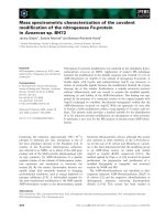
Amino acid variability in ORF 7b and RT-PCR analysis of ORF 7b in clinical samples versus cell culture isolateFigure 1
Amino acid variability in ORF 7b and RT-PCR analysis of ORF 7b in clinical samples versus cell culture isolate.
(A) ORF 7b amino acid alignment of all SARS- and SARS-like CoV available in GenBank (sequences yielding identical alignments
in the region of interest were deleted). The transmembrane domain [41] is shaded in black/grey. The left column shows Gen-
Bank accession numbers of representative genomes for each unique amino acid sequence, along with the starting nucleotide
positions of ORF 7b in each GenBank entry. The right hand column shows strain designations and their sources (human, civet,
bat). Only one sequence derived from the Frankfurt-1 strain (AB257344
) shows a 45 nucleotide in-frame deletion in the pre-
dicted transmembrane domain (TMD). The drawing below the alignment panel represents the ORF 7b in recombinant virus
r7bΔTMD. (B) Amplification of a 403 bp fragment of ORF 7b by RT-PCR in clinical samples taken after the initial isolation of
strain Frankfurt-1 from the Frankfurt index patient (bronchoalveolar lavage sample (BAL) [lane 2], sputum sample [lane 3] stool
sample [lane 4]), as well as a sputum sample from the wife of the index patient (wife, lane 5) [2]. Lane 7 shows the correspond-
ing amplification product in the original sputum sample that yielded the Frankfurt-1 isolate. Lane 8 depicts the PCR product of
the virus isolate derived from this sample.
Virology Journal 2009, 6:131 />Page 4 of 17
(page number not for citation purposes)
ability to induce apoptosis, Vero E6 cells were transfected
with expression plasmids encoding ORF 7a, ORF 7b or
ORF 7b del containing a deletion exactly corresponding to
that in Frankfurt-1. Control cells were infected with
Sendai virus (SeV) or left untreated. Forty-eight hours
later, lysates were analyzed for procaspase 3 cleavage by
Western blot using an antibody that detects cleaved and
non-cleaved forms. As shown in Figure 2A, cleavage of
caspase 3 was observed in cells expressing ORF 7a and
ORF 7b. Interestingly, expression of ORF 7b del did not
cause caspase 3 cleavage.
To examine the effect of the ORF 7b deletion on the type
I IFN response, reporter gene assays were performed. Cells
were transfected with plasmids encoding ORF 7a, ORF 7b
or ORF 7b del, respectively. All cells were co-transfected
with the pHISG-54 reporter plasmid containing the firefly
luciferase gene under the control of the ISRE region of the
human IFN-stimulated gene 54. Expression plasmid pRL-
SV40 encoding Renilla luciferase was co-transfected to
normalize for interferon-independent stimulation of tran-
scription. Twenty-four hours later, the cells were infected
with SeV to induce IFN-mediated reporter gene expres-
sion. Cells were lyzed 24 h post infection and subjected to
reporter gene assays. As shown in Figure 2B, expression of
both ORF 7a and ORF 7b but not ORF 7b del induced
IFN-dependent reporter gene expression. In those cultures
superinfected with SeV, none of the plasmids reduced the
SeV-associated, IFN-dependent reporter gene expression.
The Ebolavirus VP35, a known antagonist of interferon
induction, clearly showed reduction of reporter gene
expression if used in the same system (Figure 2B) [48,49].
These distinct findings prompted us to elucidate 7b pro-
tein functions in the natural virus context. To be able to
measure even marginal phenotypical differences we
decided to reconstruct both genotypes while establishing
a novel reverse genetic system.
Construction of a full-length infectious cDNA clone
In order to clone subgenomic portions of the SARS-CoV
genome, seven PCR fragments covering the whole
genome were generated with primers described by Yount
et al. [32]. Fragments were initially cloned in high copy
number plasmid vectors, or, if refractory to cloning, in
low copy plasmids as shown in Figure 3. Except some
marker mutations (see below), the sequence of cDNA
inserts in the seven resulting subclones was corrected to
match that of the cell culture-derived virus by plasmid-
based inverse PCR and fragment-extension PCR. For con-
struction of the variant with an intact ORF 7b, the 45 nt
deletion was filled in by oligonucleotide extension PCR
on subclone pF (Figure 3). Corrected subclones were
assembled in a stepwise procedure into four BAC clones
containing about a quarter of the SARS-CoV genome each,
which where then joined into a full length BAC cDNA
clone (refer to Figure 3 and the Materials and Methods
section for more details on the construction). BACs con-
taining both versions of subclone F were assembled. Both
BACs were sequenced, confirming presence of all marker
mutations and absence of any further mutations (refer to
Influence on apoptosis and type I interferon induction by overexpression of ORF 7a, ORF 7b, and ORF 7b with the Frankfurt-1-specific deletionFigure 2
Influence on apoptosis and type I interferon induc-
tion by overexpression of ORF 7a, ORF 7b, and ORF
7b with the Frankfurt-1-specific deletion. (A) Cleavage
of procaspase 3 analyzed by Western blot on cell lysates 48 h
after transfection with indicated plasmids or infection with
Sendai virus (20 hemagglutinating units). (B) Interferon beta
promoter-specific reporter gene expression (y-axis), shown
as the factor of induction as compared to the mock-trans-
fected, non-superinfected control (see below). The assay was
done by transfection of HEK 293 cells with plasmids express-
ing either Ebolavirus VP35, ORF 7a, ORF 7b, or ORF 7b with
a deletion corresponding to the ORF 7b deletion in Frank-
furt-1 (x-axis), as well as reporter constructs for the inter-
feron beta promoter (Firefly luciferase) and the SV40
promoter (Renilla luciferase). 24 h post transfection, cells
were either superinfected with SeV (20 hemagglutinating
units) or left uninfected. Interferon-specific reporter gene
expression was determined 24 h after superinfection (black
bars) or mock infection (grey bars). The experiment was
done in triplicate and standard deviations are shown. To
determine interferon-specific expression, the Firefly lumines-
cence signal was divided by the Renilla luciferase signal.
Virology Journal 2009, 6:131 />Page 5 of 17
(page number not for citation purposes)
GenBank accession number FJ429166). One whole BAC
was digested with Bgl I, which was present at seven posi-
tions on the BAC construct. As shown in Figure 4A, frag-
ments of the expected sizes were obtained.
The linearized BAC cDNA and a PCR product containing
the nucleocapsid gene were in-vitro transcribed and co-
transfected in BHK cells. Because BHK cells did not sup-
port SARS-CoV replication, supernatants were transferred
Assembly of a full-length SARS-CoV cDNA clone in a BAC (refer to Materials and Methods section for a detailed description of construction steps)Figure 3
Assembly of a full-length SARS-CoV cDNA clone in a BAC (refer to Materials and Methods section for a
detailed description of construction steps). (A) Arrows symbolize positions of PCR fragments on the SARS-CoV
genome. These were cloned in subgenomic plasmids. (B) Subgenomic plasmids pA1 pF. Plasmids are either based on pSMART
(identified by an "S" symbol within the respective clones) or on pCR2.1 (no symbol). Squares on each plasmid symbolize the
approximate positions of erroneous mutations from initial cloning corrected by fragment-extension technique before assembly
to higher-order clones. Small extension-PCR symbols above clones pB and pF indicate mutations introduced into plasmids to
facilitate subsequent construction steps (deletion of an Mlu I-site in pB) or to fill in the 45 nt deletion in ORF 7b in pF. (C)
Assembly of quarter clones. Circles represent plasmids, ovals represent BACs. Bold grey arrows symbolize essential BAC-
encoded genes reconstituted during BAC ligation, in order to achieve high cloning efficiency. Restriction digestion steps are
symbolized by thin arrows. The utilized restriction enzymes are identified next to the arrows. PCR primer symbols (small
arrows) next to plasmids indicate that these plasmids were first amplified with primers introducing restriction sites (identified
next to primer symbols) before the resulting products were double-digested as indicated. The large horizontal arrows below
plasmids pA1 and pA2 indicate that these fragments were joined by overlap-extension PCR with primers eliminating a Bgl I
restriction site as symbolized by a square on both of the parental plasmids. In each construction, fragment ends shown in close
proximity were first ligated in-vitro. The ligation products were then purified, ligated at sites drawn in greater distance, and
transformed in E. coli.
Virology Journal 2009, 6:131 />Page 6 of 17
(page number not for citation purposes)
to Vero cells susceptible for SARS-CoV infection. Virus
progeny was identified by immunofluorescence analysis
with anti-SARS-CoV patient serum after 24 h (Figure 4B),
as well as by plaque assays after 48 h (Figure 4B). Electron
microscopy showed intracellular structures compatible
with sites of virion assembly as well as mature virus parti-
cles (Figure 4C).
The recombinant virus containing the full-length ORF 7b
gene was named rSCV. The virus containing the deletion
in ORF 7b was termed r7bΔTMD. Both viruses were
amplified on Vero cells and stored for further experi-
ments. To confirm the purity of virus preparations, two
different RT-PCR assays were done. The first assay utilized
primers spanning the deletion in ORF 7b, as shown in Fig-
ure 5A. Both preparations yielded singular PCR products
whose molecular weight was lower for r7bΔTMD than for
rSCV. The molecular weight difference corresponded to
the size of the ORF 7b deletion. For confirmation, a sec-
ond RT-PCR assay was done with a primer hybridizing
with the deleted portion of ORF 7b that was missing in
r7bΔTMD. A singular band was obtained for rSCV but not
for r7bΔTMD (Figure 5A). Identity of all PCR products
was confirmed by sequencing.
The 7b protein is expressed in cells during SARS-CoV
infection
Since an appropriate antibody directed against ORF 7b
was not available when we started these studies, a
DDDDK (flag-) tag sequence was introduced in the infec-
tious clone prSCV by overlap-extension PCR at the C-ter-
minus of ORF 7b. As shown in Figure 6A, a protein band
corresponding to the predicted molecular weight of the 7b
protein (5.3 kDa) was specifically detected in rSCV7bflag-
infected cells using an anti-flag antibody. Also, immun-
ofluorescence analyses revealed a dotted perinuclear pat-
tern in rSCV7bflag-infected cells stained with an anti-flag
antibody, whereas rSCV-infected cells incubated with the
same antibody did not show fluorescence (Figure 6B).
Expression of the nucleocapsid (N) protein was con-
firmed with a human serum directed mainly against N
with both viruses (Figure 6B).
It was concluded that the ORF 7b protein of the recom-
binant viruses was expressed in infected cells, and that its
principal properties are not affected by a C-terminal flag-
tag epitope. These findings, including the pattern of fluo-
rescence when expressing ORF 7b, were consistent with
earlier reports by Pekosz et al [18,40].
The deletion in ORF 7b enhances growth of virus in cell
culture
Growth properties of rSCV and r7bΔTMD on different cell
lines were compared. Plaque morphology was deter-
mined for both viruses, with no discernible differences
(Figure 5B). Because plaque assay could only show cells
that die from virus infection, the same experiment was
repeated and read out by immunofocus assay, using
serum of a human SARS survivor. There was no difference
in immunofocus morphology (Figure 5B).
Growth curves in three different cell cultures were deter-
mined next. Virus RNA was measured in supernatant by
real-time RT-PCR. A multiplicity of infection (MOI) of
0.001 was used for both recombinant viruses in Vero and
CaCo-2 cells. For HuH7 cell, an MOI of 0.01 was used,
due to their lower susceptibility to SARS-CoV infection. In
Vero cells, very similar increases in RNA concentration
were observed with both viruses during 48 hours (Figure
5C). In CaCo-2 and HuH7 cells, respectively, r7bΔTMD
accumulated about 4- and 6-fold more RNA than rSCV. It
was concluded that the deleted virus had a slight but
reproducible growth advantage in the latter cell lines. In
the absence of mechanisms of adaptive immunity, repli-
cation of RNA viruses is controlled by production of and
response to type-I interferons, as well as apoptosis of
Recovery of recombinant virusFigure 4
Recovery of recombinant virus. (A) Digestion of full-
length BAC cDNA clone prSCV with the restriction enzyme
Bgl I. The BAC construct had seven Bgl I restriction sites at
positions 4454, 8783, 12146, 19000, 24124, 31719, and
36168, resulting in 7 digestion fragments as annotated in the
gel picture: 7595 bp (infectious clone Fragment F as identified
in Figure 3A with appending BAC fragment [digestion frag-
ment 1]); 6854 bp (Fragment D, [2]); 5124 bp (Fragement E,
[3]); 4972 bp (Fragment A with appending BAC fragment,
[4]); 4449 bp (BAC fragment, [5]); 3362 bp (Fragment C, [6]);
4330 bp (Fragment B, [7]) (B) Analysis of supernatants taken
from BHK cells 24 h after transfection with in-vitro transcripts
from the BAC cDNA clone. Supernatant was diluted as indi-
cated and plated on Vero cells. The top panel shows the
results of indirect immunofluorescence analysis using a
human polyclonal antiserum. The bottom panel shows the
results of plaque assays on the same Vero cells. (C) Electron
micrograph of Vero cells infected as described above. (D)
Detail from (C).
Virology Journal 2009, 6:131 />Page 7 of 17
(page number not for citation purposes)
infected cells. Taking into account our findings in overex-
pression experiments, central elements of these systems
were therefore examined in cells infected with both virus
variants.
ORF 7b is not involved in the ablation of interferon
induction observed during SARS-CoV infection
Because Vero cells as well as HuH-7 cells are deficient in
interferon induction [50], HEK 293-lp cells were used to
analyze interferon beta mRNA transcription. These cells
have been shown to be capable of inducing and secreting
interferon, and they are susceptible to SARS-CoV infection
[50]. HEK 293-lp cells were seeded in six-well plates and
infected with rSCV or r7bΔTMD at an MOI of 5. As shown
in Figure 7A, infection with the control virus NDV ele-
vated the transcription level of interferon beta mRNA by a
factor of 100. rSCV did not induce interferon beta mRNA
transcription, confirming earlier findings [50]. Induction
of transcription was not observed with r7bΔTMD either,
indicating that the ORF 7b protein is not involved in the
Comparison of recombinant viruses rSCV and r7bΔTMDFigure 5
Comparison of recombinant viruses rSCV and
r7bΔTMD. (A) RT-PCRs on supernatants of Vero cells
spanning the region of the ORF 7b deletion (RT-PCR 1) or
targeting the sequence deleted in ORF7bΔTMD (RT-PCR 2).
rSCV is the full-length ORF 7b virus; r7bΔTMD is the virus
with the Frankfurt-1-specific deletion in ORF 7b as shown in
Figure 1. (B) Plaque assay using crystal violet stain and
immunofocus assay using a polyclonal protein patient serum
reacting predominantly against the N protein (anti-N). (C)
Relative Log RNA concentration (copies per mL) in viral
supernatants after growth in cell lines as indicated. The zero
value on the y-axis represents the starting RNA concentra-
tions after virus absorption (1 h) and change of medium in
each culture. Other data for each culture were normalized
by subtraction of the logarithmic starting concentration. Each
datum point shows the mean value of three independent
experiments.
Expression of ORF 7bFigure 6
Expression of ORF 7b. (A) Detection of ORF 7b-flag
expression with an anti-flag antibody by Western blot analy-
sis. The 10 kD band is non-specifically detected in all samples.
(B) Vero cells were infected with the flag-tagged recombinant
virus rSCV7bflag or with the recombinant virus rSCV and
subjected to IFA at 24 h p.i. IFA was done with anti-flag anti-
body (left panel, anti-flag) or a convalescent patient serum
reacting predominantly against the SARS-CoV nucleocapsid
protein (right panel, anti N).
Virology Journal 2009, 6:131 />Page 8 of 17
(page number not for citation purposes)
ablation of interferon induction conferred during SARS-
CoV replication. Essentially the same results were
obtained with CaCo-2 cells (Figure 7B).
ORF 7b does not interfere with interferon alpha
production
HEK 293-lp cells were used to study release of interferon
alpha in the supernatants of infected cells. It has been
reported by Spiegel et al. that interferon alpha expression
is induced in SARS-CoV-infected 293-lp cells to a certain
level [50]. Exactly the same cells were obtained from F.
Weber, University of Freiburg, and interferon alpha tran-
scription after infection with SARS-CoV was qualitatively
confirmed by RT-PCR in our laboratory (not shown). The
level of interferon alpha was then determined by EIA in
supernatant of 293-lp cells, 48 h after infection of both
viruses at an MOI of 5. As shown in Figure 7A, infection
with the control virus NDV elevated the interferon alpha
level in supernatant by a factor of 3, while neither rSCV
nor r7bΔTMD caused detectable secretion.
Virus with the deletion in ORF7b has a slight replicative
advantage in cells pretreated with interferon beta
To study the effects of interferon on replication of both
viruses, Vero cells were pre-treated with increasing con-
centrations of interferon beta in order to induce an antivi-
ral state. Cells were infected with either rSCV or r7bΔTMD
at an MOI of 0.001. As shown in Figure 7B, r7bΔTMD rep-
licated to marginally higher virus concentrations than
rSCV in presence of interferon (up to 3.4 fold increase).
Since in our hands CaCo-2 cells were more resistant to
interferon beta pre-treatment than Vero cells, experiments
were repeated with higher concentrations of interferon
using CaCo-2 cells. More efficient replication (up to 7.9-
fold increase) was again observed for r7bΔTMD (Figure
7B).
The deletion in ORF 7b does not alter the capability of
virus to induce apoptosis in cell culture
Programmed, caspase-mediated death of infected cells is
an efficient way of controlling virus replication. Several
SARS-CoV accessory gene products have been implicated
in the induction of apoptosis, including the ORF 7a and
ORF 7b proteins as confirmed in this study (Figure 2).
Activation of apoptosis was therefore compared in cells
infected with either rSCV or r7bΔTMD. Vero cells were
infected at an MOI of 5 of either virus and assayed by
Western blot for activation of caspase 3, the central ele-
ment of the apoptosis induction cascade. As opposed to
the clear effect seen in overexpression experiments (Figure
2), both viruses induced partial cleavage of procaspase 3
at 60 hours post infection, and complete cleavage after 72
hours (Figure 8). To confirm these results we analyzed
cleavage of poly-ADP ribose polymerase type 1 (PARP-1),
a downstream effect of caspase-3 activation [51]. As
shown in Figure 8, Western blot showed little differences
in processing of PARP-1 in Vero cells with both viruses. It
was concluded that the deletion-dependent ablation of
the pro-apaptotic effect of ORF 7b as observed in overex-
pression experiments was irrelevant in the context of full
virus replication in cell culture.
The deletion in ORF 7b confers a significant replicative
advantage in Syrian golden hamsters
Deletions in and around the sgRNA 7 region occurred dur-
ing the 2003 epidemic and were transmitted in the com-
munity [25-27]. In order to elucidate whether the ORF 7b
deletion might influence replication in-vivo, both viruses
were tested in hamsters. Syrian Golden hamsters have
been shown to be an acceptable rodent model for SARS-
CoV replication and pathogenicity [52,53]. Four groups of
three hamsters were infected via the intranasal route with
10
4
PFU of either rSCV or r7bΔTMD, and sacrificed on day
1 or 3, respectively. Whole lungs were minced and tested
for infectious virus and viral RNA. The deleted virus
yielded 95-fold more infectious particles and 23-fold
more RNA copies in the lungs on day 1 (Figure 9 and
Table 1). Differences decreased but remained qualitatively
equivalent by day 3 (16-fold and 1.8-fold more infectious
virus and RNA, respectively). The differences in RNA con-
centrations were borderline significant on day 1 (Table 1).
T-tests did not identify further significant differences
between our small groups of animals, and we did not
want to use more animals for these experiments. In one of
three animals sacrificed on day 1 post infection, rSCV
failed to replicate entirely (Figure 9).
The replication advantage for r7bΔTMD was in concord-
ance with findings in CaCo-2 and HuH-7 cell cultures
(Figure 5).
Discussion
In the present study we have characterized a naturally-
acquired deletion in the ORF 7b of the primary SARS-CoV
Frankfurt-1 isolate by reverse genetics. In contrast to other
plus strand RNA viruses it has taken rather long to com-
plete the first coronavirus reverse genetics systems
[28,30,31,34,37,54]. It has been difficult to clone com-
plete CoV genomes due to their large sizes and toxicity or
lability of constructs in E. coli [31,34,37]. This has been
circumvented by Baric et al. by the use of subgenomic
plasmids that are ligated in-vitro to full genomic cDNA,
prior to transcription and electroporation [32]. We tried
this approach initially, but we failed to generate sufficient
amounts of full-length cDNA for in-vitro transcription.
Thiel et al. have described an approach to generating full-
length cDNA by stepwise assembly of an entire coronavi-
rus genome in a pox virus backbone [34]. As we had not
worked with pox viruses before, this technique appeared
rather difficult to establish. As a third alternative,
Virology Journal 2009, 6:131 />Page 9 of 17
(page number not for citation purposes)
Interferon induction, production and sensitivityFigure 7
Interferon induction, production and sensitivity. (A) Left panel, interferon beta mRNA as quantified by real-time RT-
PCR in 293-lp cells infected with rSCV or r7bΔTMD at an MOI of 5. Medium from mock-infected cells or cells infected at the
same MOI with NDV served as controls. One PCR unit (y-axis) represents ten times the minimum concentration of interferon
beta RNA detectable by the assay. (A) Right panel, interferon alpha secreted in supernatant of the same cells, as measured by
EIA. The IFN standard exemplifies the sensitivity and linear range of the assay. (B) Viral RNA concentrations measured by real-
time RT-PCR after two days of infection in cells pre-treated with increasing concentrations of interferon beta (x-axis). The left
panel shows the results of triplicate experiments on Vero cells, the right panel shows results of duplicate experiments on
CaCo-2 cells. For each graph the zero value indicates the Log RNA concentration achieved without interferon, to which the
rest of the data were normalized. Viruses and cells used in each experiment are stated in the panels.
Virology Journal 2009, 6:131 />Page 10 of 17
(page number not for citation purposes)
Enjuanes and coworkers have presented an approach
based on cloning of the entire genome in BAC and trans-
fecting the BAC-contained viral cDNA under the control
of a CMV promoter [37]. The use of BAC DNA provides
the remarkable benefit of being able to handle full length
genomic DNA in one plasmid backbone, using standard
DNA cloning techniques. As demonstrated in several stud-
ies of that group [24,36,37,55-57], BAC manipulations
are rather fast and straightforward, while providing little
opportunity for de-novo mutations resulting from DNA
manipulation steps. In our strategy we used a bacteri-
ophage T7-derived RNA polymerase promoter instead of
the CMV promoter because we wanted to provide a
genome that most closely resembled that of the virus,
using cytoplasmic sites for replication and circumventing
transcription and possible splicing in the nucleus
[37,38,56]. A T7 promoter has not been used before with
a plasmid-contained CoV cDNA genome; it was conceiva-
ble that leaky transcription might enhance underlying tox-
icity of CoV genomes in E. coli. Our study shows that the
SARS-CoV genome is stable in BAC despite the T7 pro-
moter. Interestingly, Enjuanes and colleagues have made
BAC-based full length clones for different CoV and
reported that their SARS-CoV BAC clone was more stable
than, e.g., the one they developed for TGEV [36]. The
SARS-CoV genome may thus be more stable in E. coli than
that of other CoVs. It remains to be seen whether com-
bined T7/BAC infectious cDNA clones can also be con-
structed for other CoVs.
The 45 nucleotide in-frame deletion in the transmem-
brane domain of ORF 7b is a paramount feature of the
Frankfurt-1 strain. This strain has been employed as a pro-
totypic SARS-CoV in several studies on pathogenesis and
antiviral therapy (e.g., [42-45]). By analysis of primary
clinical samples from the patients treated in 2003 for
SARS in Frankfurt, we could show that the mutation has
been selected during initial isolation in cell culture, and
that it did not stem from the Frankfurt index patient [2].
Initial characterizations of the protein by overexpression
experiments suggested reduced induction of interferon
and apoptosis in association with the deletion, which led
us to reconstruct the corresponding viruses with and with-
out the deletion by reverse genetics. In concordance with
earlier findings, type I interferon was neither induced nor
produced by either SARS-CoV variant in our study [50,58-
61]. It is assumed that CoV either encode a range of pro-
teins interacting with interferon sensing, or shield their
RNA from immune recognition through the formation of
double membrane vesicle-based replication compart-
ments [60,62-64]. Our experiments suggest that ORF 7b is
not necessary for SARS-CoV counteraction against the
induction of the interferon beta promoter. It also seems
unlikely that ORF 7b contributes to the interference of
SARS-CoV with secretion of interferon alpha [62]. How-
ever, the deleted virus showed slightly decreased sensitiv-
ity to pretreatment of cells with interferon. This effect was
remarkable since earlier studies only determined opposite
(= evasive) effects on the interferon response for CoV
accessory proteins. These include interference with the
interferon signalling cascade in the case of SARS-CoV pro-
tein 6, or prevention of activation of interferon-sensitive
Induction of apoptosis by recombinant coronaviruses rSCV and r7bΔTMDFigure 8
Induction of apoptosis by recombinant coronaviruses
rSCV and r7bΔTMD. Vero FM cells were infected with
rSCV or r7bΔTMD at an MOI of 5. Cleavage of caspase 3 and
PARP-1 at 60 and 72 hours post infection was analyzed by
Western Blot analysis.
In-vivo effect of the ORF7b deletionFigure 9
In-vivo effect of the ORF7b deletion. Golden Syrian
hamsters were infected with 10
4
PFU of rSCV and r7bΔTMD
(x-axis). Heat inactivated rSCV served as mock control. For
each point of time post infection, three animals per virus var-
iant were sacrificed (animals 1, 2, 3 as identified on the x-
axis). Lungs were taken in total. Viral titers were determined
by plaque assay and viral RNA was quantified by real-time
RT-PCR. Light grey bars represent log copies of viral RNA,
dark grey bars represent PFU per g lung tissue. The arrow
indicates one animal with failure of virus replication.
Virology Journal 2009, 6:131 />Page 11 of 17
(page number not for citation purposes)
genes for mouse hepatitis virus nucleocapsid protein
[61,65]. Here we observed an ORF 7b-dependent exten-
sion of the replication-attenuating effect of interferon.
However, the additional extent of attenuation on top of
the effect of interferon beta was of the same size as that
observed in untreated cell cultures (compare Figure 5 and
Figure 7) and did hardly increase with increasing inter-
feron concentrations. This suggests an additive rather than
a synergistic effect of ORF 7b and interferon on the atten-
uation of virus replication. In spite of the high relevance
of the interferon response for controlling SARS-CoV repli-
cation, we should therefore assume that ORF 7b plays no
role in the context of the type I interferon system [62,66].
Apoptosis of target cells can limit virus infection in-vitro
and in-vivo. Our initial overexpression experiments
pointed to a strong pro-apoptotic effect of intact ORF 7b,
which was in concordance with a study by Schaecher et al.
who found that sgRNA 7-derived proteins activated cas-
pase 3 if overexpressed [40]. Complementary to their
study, however, our experiments did not confirm any sim-
ilar effect specifically for the ORF 7b protein in the full
virus context. Schaecher et al. studied a recombinant virus
with a double deletion of both ORF 7a and 7b, and this
virus induced apoptosis clearly less efficiently than the
parent full-length virus [18,19]. The most likely explana-
tion for the difference between both viruses is that the
pro-apoptotic effect of gene 7 proteins observed by Schae-
cher et al. was contributed by ORF 7a rather than ORF 7b.
Even though the ORF 7b deletion in Frankfurt-1 was not
affecting interferon and apoptosis systems, the virus with
a deletion seems to have been selected during isolation in
cell culture and shows a replicative advantage in two of
three cell lines. This is remarkable because SARS-CoV var-
iants with deletions in the ORF 7 (and also ORF 8) gene
region have been transmitted and maintained in humans
in the late phases of the 2003 epidemic [25-27]. It has
never been formally addressed whether these viruses
might have undergone particularly efficient transmission.
We therefore determined whether the ORF 7b deletion in
Frankfurt-1 conferred a replicative advantage in-vivo, using
Syrian Golden Hamsters as a model of human SARS-CoV
infection [52,53]. Interestingly, the enhancing effect of
the ORF 7b deletion was even more pronounced in ham-
sters than in cell culture. Hamsters infected with the
deleted variant had significantly more virus RNA and a 95-
fold increase of infectious virus titers in their lungs after
24 h. The rate of successful infections was 6/6 with the
deleted virus and 5/6 with the full virus. In concordance
with these observations, Roberts et al. have described ca.
10-fold more efficient replication of Frankfurt-1 in ham-
sters as compared to Urbani and HKU-39849 [52,53].
Mortality in Hamsters was only observed with Frankfurt-1
(3 of 20 animals) but not Urbani and HKU-39849
[52,53]. It was suggested that an amino acid exchange
(L1148F) in the S2-domain of the spike protein of Frank-
furt-1 against both Urbani and HKU-39849 might explain
the difference. However, a replicative difference in extent
similar to that reported by Roberts et al. was observed in
our study between two variants of Frankfurt-1 that dif-
fered only by the ORF 7b deletion. As the deletion is not
present in Urbani or any other prototype strain, this iden-
tifies the 7b protein as a potential attenuating factor
within the genome of SARS-CoV.
We have seen in this study that the attenuating effect of
ORF 7b was focused on the early phase of infection in-
vivo. Because it has been suggested that delayed accumula-
tion of high virus concentrations in infected patients has
limited the spread of SARS-CoV in the population, it is
tempting to speculate that the occurrence of viruses with
deletions in the ORF7/8 region in the late phase of the
2003 epidemic might have added to the efficiency of virus
transmission in humans [67-69]. It will be interesting in
the future to investigate the exact mechanism of ORF 7b-
dependent attenuation, and to determine whether this
might contribute to the maintenance of virus in its natural
reservoir.
Materials and methods
Cells and viruses
The original Vero cells on which Frankfurt-1 was primarily
isolated (hereafter termed Vero FM, obtained from Jin-
Table 1: Virus replication levels in hamster lungs
Virus replication (mean* of N animals) T-test*
rSCV
(Virus titer, RNA concentration, N animals)
r7bΔTMD
(Virus titer, RNA concentration, N animals)
p
Day 1 p.i. 1.04 × 10
7
(9.00 × 10
6
- 1.2 × 10
7
) PFU/g 9.86 × 10
8
(2.4 × 10
7
- 1.8 × 10
8
) PFU/g 0.15
6.65 × 10
7
(1.84 × 10
7
- 2.4 × 10
8
) copies/g 1.53 × 10
9
(9.98 × 10
8
- 2.2 × 10
9
) copies/g 0.052
n = 2 n = 3
Day 3 p.i. 9.86 × 10
6
(8.00 × 10
5
- 1.20 × 10
8
) PFU/g 1.63 × 10
8
(1.00 × 10
8
- 3.6 × 10
8
) PFU/g 0.13
4.27 × 10
8
(1.68 × 10
8
- 2.1 × 10
9)
copies/g 7.70E × 10
8
(7.50 × 10
8
- 8.10 × 10
8
) copies/g 0.5
n = 3 n = 3
* Means were determined upon logarithmic data and calculated back into linear values. Two-tailed t-tests were done on logarithmic values
Virology Journal 2009, 6:131 />Page 12 of 17
(page number not for citation purposes)
drich Cinatl, Universtiy of Frankfurt), human hepatoma
cells HuH7 (ATCC CCL-185), human colonic cancer cells
CaCo-2 (ATCC HTB-37) and human embryonic kidney
cells HEK 293-low passage (hereafter termed 293-lp,
obtained from Friedemann Weber, University of Freiburg
[50]) were maintained and grown in Dulbecco's modified
Eagle medium (DMEM) containing 10% foetal calf serum
(FCS, PAA, Pasching, Austria), 1 mM glutamine (PAA), 1
mM sodium pyruvate (PAA), 1% non-essential amino
acids (PAA), 100 U/ml penicillin (PAA), and 100 μg/ml
streptomycin (PAA). All experiments with 293-lp cells
were performed between cell passage 42 and 48.
The SARS-CoV Frankfurt-1 isolate [2,20,70] was titrated
on Vero FM cells. Sindbis virus (SV) derived from infec-
tious cDNA clone pTOTO [28] was obtained from Beate
Kümmerer, BNI, Hamburg and titrated on Vero FM cells.
Sendai virus (SeV) strain Cantell was obtained from Chris-
topher Basler, Mount Sinai School of Medicine, New York,
propagated in 11-day-old embryonated chicken eggs, and
titrated by standard hemagglutination test. Newcastle dis-
ease virus (NDV) strain PPMV-1/pigeon/Germany/R151
was obtained from the virus collection of the Friedrich
Löffler Institute, Riems, Germany, and titrated on Vero FM
cells.
Virus quantification by cell culture and RT-PCR
Plaque assays were done with Avicel overlays (RC581,
FMC BioPolymer, Belgium) as described elsewhere [71].
Immunofocus assay used the same overlay and was other-
wise performed as described previously [72]. Viral RNA
quantification using in-vitro transcribed RNA standards
was done as described previously [2].
General cloning and mutagenesis techniques
Standard cloning techniques were used. All gel purifica-
tions were done with the QIAEX II kit (Qiagen, Hilden,
Germany). DNA constructs were electroporated into E.
cloni (Lucigen, Middleton, USA) or Stbl 3 E. coli cells (Inv-
itrogen, Karlsruhe, Germany). Prior to digestion with
methylation-sensitive endonucleases plasmids were trans-
formed in Sure cells (Stratagene, La Jolla, USA). BAC prep-
arations were done with the Nucleobond
®
AX-kit
(Macherey Nagel, Germany) as instructed. Plasmid-based
inverse PCR was performed with QuikChange XXL kit
(Stratagene, USA). PCR mutagenesis by overlap-extension
PCR used Phusion
®
DNA polymerase and around 50 ng of
input plasmid DNA. SARS-CoV coding sequence within
constructs was fully sequenced after every mutagenic step.
Cloning of subgenomic plasmids
Total RNA was extracted from infected Vero cells with the
Qiagen RNeasy kit. Using primers described by Yount et
al. [32], cDNA fragments spanning the SARS-CoV genome
were generated by RT-PCR using Superscript III reverse
transcriptase and Expand High Fidelity DNA polymerase
mixture. These primers inserted Bgl I restriction sites at
fragment borders and a T7 promoter in front of the 5'end
of the genome [32]. In addition to the strategy described
by Yount et al., a Not I restriction site was introduced
downstream of the genomic poly-A tail. Figure 3 gives an
overview of cloned fragments. Fragment A was cloned in
two parts (A1 and A2, Figure 3), using primer Afwd 5'-
TACTAATACGACTCACTATAGATATTAGGTTTTTACC TA
CCCAGG-3' and A1rev 5'-aatgccagtatgacctgagccaatatc-3'
and A2fwd 5'-GATATTGGCTCAGGTCATACTGGCATT-3'
and Arev 5'-ACACCATAGTCAACGATGCC-3'. After correc-
tion of errors both inserts were amplified from plasmids
and used as templates in an overlap-extension PCR. A nat-
urally existing Bgl I restriction site at genome position
1572 was thereby deleted. The extension product was sub-
cloned in pSMART, resulting in clone pA. PCR products B,
D, and E were cloned in pCR2.1 (Invitrogen). Fragments
C and F were cloned in pSMART Low Copy Kanamycin
vectors (Lucigen) after instability was observed in pCR2.1.
A 45 nt deletion present in the Frankfurt-1 virus isolate (nt
27654 to 27699 in Genbank Accession No AY310120
),
was filled in by overlap-extension PCR. A region including
restriction sites BamH I (genome position 26045) and
Not I (following the 3'end of genome) was amplified
from subclone pF in two halves using appropriate outer
primers and overlap-extension primers 5'-TTTCTGCTAT-
TCCTTGTTTT AATAATGCTTATTAT ATTTTGGTTT-
TCACTCGAAATCCAGGATCTAGAAG-3' and 5'-
ATTATTAAAACAAGGAATAGCAGAAAGGC TAA AAAGC
ACAAATAGAAGTCAATTAAAGTGAGCTCATTC-3'.
The fragments were overlap-extended, digested with
BamH I and Not I, and cloned back into the correspond-
ing restriction sites in clone pF. All clones were verified by
sequencing. Using the same technique, a DDDDK (flag-)
tag sequence was introduced at the C-terminus of ORF 7b,
with overlap-extension primers 5'-GATTACAAGGATGAC-
GACGATAAGTAAACGAACATGAAACTTCTC-3' and 5'-
CTTATCGTCGTCATCCTTGTAATCGACTTTGGTACAAG-
GTTCT-3'.
Assembly of full length BAC cDNA clone
BAC vector pBeloBAC11 was obtained from NEB, Boston,
USA. The Nco I site at position 890 was oblated by primer
extension mutagenesis, resulting in pBelodNco. The Not
I-Not I multiple cloning site fragment was removed from
pBelodNco and replaced by an oligonucleotide adapter
containing Nsi I, BsaH I, Sph I and Not I restriction sites
in sequence, resulting in pBeloAd4. Fragment A was
amplified from plasmid pA with primers 5'-
AGTAATGGGCCC
TAAGTACTAATACGACTCACTATAGA-
TATTAGG-3' and 5'-ACACCATAGTCAACGATGCC-3',
thereby introducing a PspOM I site upstream of the T7-
promotor (Figure 3). The fragment was digested with
Virology Journal 2009, 6:131 />Page 13 of 17
(page number not for citation purposes)
PspOM I and Bgl I and ligated to the long EcoR I Not I
fragment of pBeloAd4 (pBeloAd4A, Figure 1). The 5'-most
3,062 nt were amplified from plasmid pB using primers
5'-GCCTATATGCATGGATGTTAGAG-3' and 5'-ATGAAT-
GCGGCCGC
TACACTCAACACGTGTGGCACGC-3',
thereby introducing a Not I site immediately downstream
of the Mlu I site at position 7453. The PCR product was
digested with Bgl I and Not I, gel purified, and ligated to
the dephosphorylated short Not I EcoR I fragment of
pBeloAd4 (pBeloAd4B1, Figure 3). pBeloAd4A and
pBeloAd4B were gel purified and ligated, resulting in
quarter clone pAB, (Figure 3).
The 3'-most 5,536 nt were amplified from plasmid B3 in
two parts, using primers 5'-TAGACTACGCCGGCG
-
TAGCCTTAGGTTTAAAAACAATTGCCACTC-3' and 5'-
TACACTCAACACGTGTGGCACGATTGCGCT-3' (5'-part);
and primers 5'-AGCGCAATCGTGCCACACGTGTTGAGT-
GTA-3'and 5'-TGAACCGCCACGCTGGCTAAACC-3' (3'-
part), respectively. Both products were overlap-extended,
resulting in a PCR product with a depleted Mlu I site at
position 7453. The product also contained a Not I site
upstream of the Bsu36 I site at position 6544, introduced
by a primer 5'-overhang. The product was Not I and Bgl I
digested and ligated to the long EcoR I Not I fragment of
pBeloAd4 (pBeloAd4B2, Figure 3).
Plasmid pD was digested with Bgl I and Bcl I (compatible
to BamH I). The fragment was ligated to the dephosphor-
ylated short BamH I EcoR I fragment of pBelodNco
(pBeloNcoD1, Figure 3). Fragment C was cut out of its
pSMART vector with Bgl I and dephosphorylated, fol-
lowed by ligation to pBeloNcoD1 and gel purification.
This product was ligated to pBeloAd4B2, generating quar-
ter clone pBCD.
The Acl I Bgl I fragment of vector D-24-5 was ligated to the
long EcoR I BsaH I fragment of pBelodNco (Bsa HI is
compatible with Acl I) (pBeloNcoD2, Figure 3). The
dephosphorylated Pst I Bgl I fragment of vector pE was
ligated to the short Nsi I EcoR I fragment of pBeloAd4
(Nsi I is compatible with Pst I) (pBeloAd4E1, Figure 3).
This product was ligated with pBeloNcoD2 to yield quar-
ter clone pDE.
The 2,793 bp SpH I Bgl I fragment of subclone pE was
ligated to the long EcoR I SpH I fragment of pBeloAd4
(pBeloAd4E2, Figure 3).
The Bgl I Not I fragment of plasmid pF was ligated to the
short Not I EcoR I fragment of pBeloAd4 (pBeloAd4F, Fig-
ure 1). This fragment was ligated with pBeloAd4E2, yield-
ing quarter clone pEF.
Quarter clones pAB and pBCD were digested with Bsu36 I
and PspOM I, the latter cut destroying the replicative ele-
ment sopC. Fragments of interest were gel-purified and
ligated to yield half clone pL. Quarter clones pDE and pEF
were digested with Nco I. One Nco I cut was in the virus
cDNA insert on each BAC, and the other in the sopC gene.
Fragments of interest were purified and ligated to yield
half clone pR. Half clones were digested with Mlu I and
PspOM I. Fragments of interest were gel purified and
ligated into the full length clone prSCV.
Rescue of recombinant virus
Full-length BAC clones were linearized with Not I,
extracted with phenol-chloroform, and transcribed with
the mMessage-mMachine
®
T7 (Ambion, USA) at an input
of 1 μg of DNA per 20 μl reaction. A PCR product span-
ning the nucleocapsid reading frame and the genomic 3'-
prime end was generated with primers N-fwd (5'-
GGCCATTTAGGTGACACTATAG
ATGTCTGATAATGGAC-
CCCAATC), the underlined sequence representing an SP6
promoter) and Frev (5'-TTTTTTTTTTTTTTTTTTTTGTCAT-
TCTCCTAAGAAGC-3'). The product was purified and
transcribed with mMessage-mMachine SP6 kit. Tran-
scripts from both in-vitro transcription reactions were
quantified photometrically. Genomic transcripts and N
transcripts were co-electroporated at a 10:1 ratio into 10
7
BHK-21 cells, using a GenePulser instrument (Biorad,
Germany) with two pulses of 1.5 kV, 25 μF and maximal
resistance. Cells were left at room temperature for 10 min-
utes and seeded in 75 cm
2
flasks. In a biosafety-4 labora-
tory, electroporated BHK-21 cells were incubated at 37°C
for 24 hours. Supernatants were serially diluted and trans-
ferred to Vero cells. Using 1% SeaPlaque
®
agarose overlay
(Biozym, Germany), three rounds of plaque purification
were performed for each recombinant virus.
Analysis of mutations in ORF 7b
To distinguish between the two genotypes, two different
RT-PCRs were performed. RT-PCR 1 used primers 27500
fwd (5'-CAGCTGCGTGCAAGATCAGT-3') and 27900 rev
(5'-CCCTAGTGTTGTACCTTACAAG-3'), thus comprising
ORF 7b and yielding a 400 bp fragment for rSCV, while
giving a 355 bp fragment for r7bΔTMD. For RT-PCR 2 the
identical reverse primer was used but the binding site of
the sense primer 27690 fwd (5'-TAGCCTTTCTGCTATTC-
CTTGT-3') was placed in ORF 7b, recognising the 45 nt
only present in rSCV but deleted in r7bΔTMD, hence a
PCR product was only obtained for rSCV.
Cloning of ORF 7a, 7b and 7b del
ORF 7a (nts 27258 to 27626 of SARS-CoV genome Gen-
Bank accession number AY310120
) was amplified using
primers 5'-CACCATGAAAATTATTCTCTTCCTGACA-3'
Virology Journal 2009, 6:131 />Page 14 of 17
(page number not for citation purposes)
(fwd) and 5'-TCATTCTGTCTTTCTCTTAATGGT-3' (rev),
and cloned into pCDNA 3.1. Because of low expression
rates of the protein (data not shown) the insert was cloned
into the high level expression vector pCAGGS [73], using
KpnI and NotI. ORF 7b gene (nts 27623 to 27751) and the
deletion mutant ORF 7b del gene (nts 27623 27751 dele-
tion of 45 nts 3276) were amplified using primers 5'-
CTAGAATTCCTCGAGACAATGAGAAGTTTCATGTTC-3'
and 5'-ATCGTCGACCTCGAGTCACCATTAAGAGAAA-
GACAG-3', and cloned into pI.18 vector (kind gift of Jim
Robertson, National Institute for Biological Standards and
Control, Hertfordshire, UK) with a T7 promoter that was
inserted by standard cloning procedures. Expression of
constructs was verified by coupled in-vitro transcription
and translation using the TNT T7 Coupled Reticulocyte
Lysate System (Promega, Mannheim, Germany) and
immunofluorescence analysis of transfected cells (data
not shown).
ISG-54 reporter gene assay
Transfection of 293 cells was performed using the calci-
umphosphate transfection kit (Invitrogen) according to
the manufacturer's instructions. 10
6
cells were transfected
with 0.3 μg of the interferon (IFN)-stimulated response
element (ISRE)-driven firefly luciferase reporter plasmid
pHISG-54-Luc (kind gift of D. Levy, New York University
School of Medicine, New York), 0.3 μg of the constitutive
Renilla luciferase expression plasmid pRL-SV40
(Promega) and 4 μg of the plasmid of interest. 16 μg of
herring sperm DNA (Promega) were transfected along
with the plasmids to optimize DNA uptake. 24 h post
transfection, cells were infected with SeV (20 hemaggluti-
nating units) to induce the type I IFN response or were not
infected. At 24 h post infection (p.i.), cells were harvested
and lysed in 100 μl of passive lysis buffer (Promega, Man-
nheim, Germany). Subsequent luciferase assays were per-
formed by using the Promega DUAL luciferase assay
system according to the manufacturer's instructions. Rela-
tive renilla luciferase production was used to normalize
for transfection efficiency.
Immunofluorescence microscopy
Cells were seeded on chamber slides (μSlide
®
8 well, Ibidi,
Martinsried, Germany) and infected with SARS-CoV t a
multiplicity of infection of 1. After 24 hours cells were
washed once with PBS and fixed in ice cold acetone for 15
minutes. Prior to antibody staining, cells were washed
three times with PBS. Reconvalescent SARS patient serum
from our own diagnostic laboratory was diluted 1:1000 in
PBS-T. Rabbit polyclonal antibody against the DDDDK
tag (Abcam, UK) was diluted 1:10000 in PBS-T. Cells were
overlaid with 100 μl of antibody solution, incubated at
37°C for 1 hour, and washed four times for 5 minutes
with PBS containing 0.1% Tween 20 (PBS-T). Fluorescein-
conjugated goat anti-human or anti-rabbit IgG serum
(Calbiochem/VWR, Darmstadt, Germany) was diluted
1:10000 in PBS-T and incubated at 37°C for 30 minutes.
Cells were washed 4 times with PBS. Chambers were over-
laid with 200 μl of PBS and a few drops of mineral oil. Flu-
orescence was analysed on an inverted fluorescence
microscope.
Western blot analysis
Subconfluent 293-lp cells in six well-plates were infected,
harvested at different time points after infection, and pel-
leted by centrifugation for 5 minutes at 1200 rpm. Pellets
were dissolved in 50 μl of 1× Chaps buffer containing 1
mM PMSF and 5 mM DTT followed by three freeze/thaw
cycles. Nuclei were pelleted by centrifugation for 10 min-
utes at 16,000 g and the clarified lysate was dissolved in
1× SDS loading buffer. 10 μl of postnuclear lysate were
loaded on precast 412% Bis-Tris gradient gels. Separated
proteins were electroblotted on nitrocellulose membranes
(Whatman, Dassel, Germany) and blocked for 1 h with 1×
RotiBlock (Roth, Karlsruhe, Germany). Membranes were
washed with PBS and incubated over night with primary
antibody at 4°C. Rabbit polyclonal anti-caspase-3 and
anti-PARP antibodies (Cell Signaling, Danvers, USA) were
diluted 1:1000 in PBS-T. Rabbit anti-flag antibody
(Abcam, Cambridge, UK) was diluted 1:5000 in PBS-T.
Membranes were washed 4 times for 5 minutes with PBS-
T. Horseradish peroxidase-conjugated goat anti-rabbit
antibody (Cell Signaling, Danvers, USA) was used at
1:2000 dilution in PBS-T and incubated on membranes
for 1 hour. Membranes were washed four times with PBS-
T before LumiGLO reagent (Cell Signaling, Danvers, USA)
was added. Membranes were exposed to scientific imaging
film (Sigma-Aldrich, Munich, Germany) for appropriate
times before development.
In overexpression experiments, Vero E6 cells were trans-
fected with Lipofectamine 2000 (Invitrogen) according to
the manufacturer's instructions. 5 × 10
4
cells were trans-
fected with 1 μg of empty pI.18 vector, pCAGGS-ORF 7a,
pI.18-ORF 7b or pI.18-ORF 7b del, respectively. At 48 h
post transfection, cells were lysed in 50 μl 1× Chaps
buffer. Proteins were separated on 15% SDS polyacryla-
mide gels, transferred onto PVDF membranes and
blocked for 1 h with 5% skim milk (w/v) in PBS-T. Mem-
branes were washed with PBS-T and incubated over night
with primary antibody at 4°C. Western blot detection was
done with horseradish peroxidase-conjugated goat anti-
rabbit secondary antibody using an enhanced chemilumi-
nescence detection reagent kit (Pierce, Perbio Science,
Bonn, Germany) according to the manufacturer's proto-
col. Immunoreactive bands were visualized using an Opti-
max 2010 imaging system (PROTEC processor
Technology, Oberstenfeld, Germany) with high perform-
ance chemiluminescence films (GE Healthcare, Munich,
Germany).
Virology Journal 2009, 6:131 />Page 15 of 17
(page number not for citation purposes)
IFN- ELISA
Interferon alpha was detected with the Human IFN Alpha
ELISA Kit (PBL Interferonsource, Piscataway, USA)
according to the manufacturer's instructions. Briefly, 100
μl of supernatant of samples and controls were added to
pre-coated microtiter plates and incubated at room tem-
perature for 1 hour, followed by one washing step, addi-
tion of antibody solution and another hour of incubation.
After three washing steps, 100 μl of HRP conjugate con-
centrate were added and incubated for 1 hour. The plate
was washed four times and TMB substrate was added.
After 15 minutes of incubation stop solution was added
and absorbance was determined at 450 nm.
Quantification of interferon beta mRNA
Total RNA was prepared from 293-lp cells in 6-well plates
with Trizol
®
reagent (Invitrogen, USA) according to the
manufacturer's instructions. RNA was quantified photo-
metrically and 150 ng per reaction were analysed by real-
time RT-PCR. Interferon beta mRNA was amplified with
primers IFN Fwd (5'-GAACTTTGACATCCCTGAGGA-
GATT-3') and IFN Rev (5'-GGAGCATCTCATAGATGGT-
CAATG-3'), and 5'-nuclease probe IFN -P (FAM-
CAGCAGTTCCAGAAGGAGGACGCC-TAMRA). GAPDH
mRNA was detected in parallel with primers GAPDHFwd
(5'-AGGTGGTCTCCTCTGACTTCAACA-3'), GAPDHRev
(5'-AGTGGTCGTTGAGGGCAATG-3'), and probe GAPDH-
P (FAM-CACCCACTCCTCCACCTTTGACGCT-TAMRA).
Reactions using the OneStep RT-PCR kit (Qiagen, Hilden,
Germany) comprised 50°C for 30 minutes, followed by
95°C for 15 minutes and 40 cycles of 95°C for 10 seconds
and 58°C for 30 seconds. For both genes standard curves
were generated from limiting dilution series of quantified
RNA. The dilution end-points were defined as one PCR
unit for each gene. Log PCR units for each experimental
sample were calculated from the linear equations of the
dilution series. Interferon beta quantity was normalised to
GAPDH quantity by subtraction of logarithmic quantities
(Interferon GAPDH).
Hamster infections
Infections were performed with rSCV and r7bΔTMD.
Heat-inactivated rSCV served as the mock-control. Syrian
Golden hamsters (strain LVG, Charles River Laboratories)
were infected via the intranasal route with 10
4
PFU each.
100 μl of virus solution were applied. Hamsters were sac-
rificed at indicated days post infection. Lungs were pre-
pared in total, weighed, and homogenized. Tissue was
suspended to a concentration of 0.5 g/ml in complete
DMEM before analysis by RT-PCR or cell culture.
Competing interests
The authors declare that they have no competing interests.
Authors' contributions
SP constructed the infectious clone and conducted all
experiments with recombinant viruses. VK and EM
designed and carried out the overexpression experiments
and the reporter assays and/or critically revised the manu-
script. VD conducted hamster infections. KG participated
in construction of the infectious clone. CD designed the
study, participated in the construction of the infectious
clone, and wrote the manuscript. All authors took part in
manuscript preparation. All authors read and approved
the final manuscript.
Acknowledgements
We are grateful to Jindrich Cinatl, Friedemann Weber, Christopher Basler,
Beate Kümmerer, and Jim Robertson for donations of viruses or cells. We
thank Toni Rieger for his kind help during hamster inoculations.
This study was supported by the German Ministry of Education and
Research (Project Code "Ökologie und Pathogenese von SARS"), the Euro-
pean Commission (contract SSPE-CT-2005-022639), the German Research
Foundation (Mu1365/1-1 and SFB 535), and the Sino-German Center for
Science Promotion.
References
1. Peiris JS, Yuen KY, Osterhaus AD, Stohr K: The severe acute res-
piratory syndrome. N Engl J Med 2003, 349:2431-2441.
2. Drosten C, Gunther S, Preiser W, Werf S van der, Brodt HR, Becker
S, Rabenau H, Panning M, Kolesnikova L, Fouchier RA, Berger A, Bur-
guire AM, Cinatl C, Eickmann M, Escriou N, Grywna K, Kramme S,
Manuguerra JC, Muller S, Rickerts V, Sturmer M, Vieth S, Klenk HD,
Osterhaus AD, Schmitz H, Doerr HW: Identification of a novel
coronavirus in patients with severe acute respiratory syn-
drome. N Engl J Med 2003, 348:1967-1976.
3. Drosten C, Preiser W, Gunther S, Schmitz H, Doerr HW: Severe
acute respiratory syndrome: identification of the etiological
agent. Trends Mol Med 2003, 9:325-327.
4. Ksiazek TG, Erdman D, Goldsmith CS, Zaki SR, Peret T, Emery S,
Tong S, Urbani C, Comer JA, Lim W, Rollin PE, Dowell SF, Ling AE,
Humphrey CD, Shieh WJ: A novel coronavirus associated with
severe acute respiratory syndrome. N Engl J Med 2003,
348:1953-1966.
5. Kuiken T, Fouchier RA, Schutten M, Rimmelzwaan GF, van
Amerongen G, van Riel D, Laman JD, de Jong T, van Doornum G, Lim
W, Ling AE, Chan PK, Tam JS, Zambon MC, Gopal R, Drosten C,
Werf S van der, Escriou N, Manuguerra JC, Stohr K, Peiris JS, Oster-
haus AD: Newly discovered coronavirus as the primary cause
of severe acute respiratory syndrome. Lancet 2003,
362:263-270.
6. Rota PA, Oberste MS, Monroe SS, Nix WA, Campagnoli R, Icenogle
JP, Penaranda S, Bankamp B, Maher K, Chen MH, Tong S, Tamin A,
Lowe L, Frace M, DeRisi JL, Chen Q, Wang D, Erdman DD, Peret TC,
Burns C, Ksiazek TG, Rollin PE, Sanchez A, Liffick S, Holloway B,
Limor K, McCaustland K, Olsen-Rasmussen M, Fouchier R, Gunther
S, Osterhaus AD, Drosten C, Pallansch MA, Anderson LJ, Bellini WJ:
Characterization of a novel coronavirus associated with
severe acute respiratory syndrome. Science 2003,
300:1394-1399.
7. Guan Y, Zheng BJ, He YQ, Liu XL, Zhuang ZX, Cheung CL, Luo SW,
Li PH, Zhang LJ, Guan YJ, Butt KM, Wong KL, Chan KW, Lim W,
Shortridge KF, Yuen KY, Peiris JS, Poon LL: Isolation and charac-
terization of viruses related to the SARS coronavirus from
animals in southern China. Science 2003, 302:276-278.
8. Lau SK, Woo PC, Li KS, Huang Y, Tsoi HW, Wong BH, Wong SS,
Leung SY, Chan KH, Yuen KY: Severe acute respiratory syn-
drome coronavirus-like virus in Chinese horseshoe bats. Proc
Natl Acad Sci USA 2005, 102:14040-14045.
Virology Journal 2009, 6:131 />Page 16 of 17
(page number not for citation purposes)
9. Li W, Shi Z, Yu M, Ren W, Smith C, Epstein JH, Wang H, Crameri G,
Hu Z, Zhang H, Zhang J, McEachern J, Field H, Daszak P, Eaton BT,
Zhang S, Wang LF: Bats are natural reservoirs of SARS-like
coronaviruses. Science 2005, 310:676-679.
10. Poon LL, Chu DK, Chan KH, Wong OK, Ellis TM, Leung YH, Lau SK,
Woo PC, Suen KY, Yuen KY, Guan Y, Peiris JS: Identification of a
novel coronavirus in bats. J Virol 2005, 79:2001-2009.
11. Tang XC, Zhang JX, Zhang SY, Wang P, Fan XH, Li LF, Li G, Dong BQ,
Liu W, Cheung CL, Xu KM, Song WJ, Vijaykrishna D, Poon LL, Peiris
JS, Smith GJ, Chen H, Guan Y: Prevalence and genetic diversity
of coronaviruses in bats from China. J Virol 2006, 80:7481-7490.
12. Wang LF, Shi Z, Zhang S, Field H, Daszak P, Eaton BT: Review of
bats and SARS. Emerg Infect Dis 2006, 12:1834-1840.
13. Cavanagh D: Coronaviruses in poultry and other birds. Avian
Pathol 2005, 34:439-448.
14. Herrewegh AA, Smeenk I, Horzinek MC, Rottier PJ, de Groot RJ:
Feline coronavirus type II strains 791683 and 791146 origi-
nate from a double recombination between feline coronavi-
rus type I and canine coronavirus. J Virol 1998, 72:4508-4514.
15. Saif LJ: Animal coronaviruses: what can they teach us about
the severe acute respiratory syndrome? Rev Sci Tech 2004,
23:643-660.
16. Vijgen L, Keyaerts E, Moes E, Thoelen I, Wollants E, Lemey P, Van-
damme AM, Van Ranst M: Complete genomic sequence of
human coronavirus OC43: molecular clock analysis suggests
a relatively recent zoonotic coronavirus transmission event.
J Virol 2005, 79:1595-1604.
17. Frieman MB, Yount B, Sims AC, Deming DJ, Morrison TE, Sparks J,
Denison M, Heise M, Baric RS: SARS coronavirus accessory
ORFs encode luxury functions. Adv Exp Med Biol 2006,
581:149-152.
18. Schaecher SR, Touchette E, Schriewer J, Buller RM, Pekosz A: Severe
acute respiratory syndrome coronavirus gene 7 products
contribute to virus-induced apoptosis. J Virol 2007,
81:11054-11068.
19. Yount B, Roberts RS, Sims AC, Deming D, Frieman MB, Sparks J, Den-
ison MR, Davis N, Baric RS: Severe acute respiratory syndrome
coronavirus group-specific open reading frames encode non-
essential functions for replication in cell cultures and mice. J
Virol 2005, 79:14909-14922.
20. Snijder EJ, Bredenbeek PJ, Dobbe JC, Thiel V, Ziebuhr J, Poon LL,
Guan Y, Rozanov M, Spaan WJ, Gorbalenya AE: Unique and con-
served features of genome and proteome of SARS-coronavi-
rus, an early split-off from the coronavirus group 2 lineage. J
Mol Biol 2003, 331:991-1004.
21. de Haan CA, Masters PS, Shen X, Weiss S, Rottier PJ: The group-
specific murine coronavirus genes are not essential, but their
deletion, by reverse genetics, is attenuating in the natural
host. Virology 2002, 296:177-189.
22. Haijema BJ, Volders H, Rottier PJ: Live, attenuated coronavirus
vaccines through the directed deletion of group-specific
genes provide protection against feline infectious peritonitis.
J Virol 2004, 78:3863-3871.
23. Herrewegh AA, Vennema H, Horzinek MC, Rottier PJ, de Groot RJ:
The molecular genetics of feline coronaviruses: comparative
sequence analysis of the ORF7a/7b transcription unit of dif-
ferent biotypes. Virology 1995, 212:622-631.
24. Ortego J, Sola I, Almazan F, Ceriani JE, Riquelme C, Balasch M, Plana
J, Enjuanes L: Transmissible gastroenteritis coronavirus gene 7
is not essential but influences in vivo virus replication and vir-
ulence. Virology 2003, 308:13-22.
25. Chinese SMEC: Molecular evolution of the SARS coronavirus
during the course of the SARS epidemic in China. Science
2004, 303:1666-1669.
26. Chiu RW, Chim SS, Tong YK, Fung KS, Chan PK, Zhao GP, Lo YM:
Tracing SARS-coronavirus variant with large genomic dele-
tion. Emerg Infect Dis 2005, 11:168-170.
27. Tang JW, Cheung JL, Chu IM, Sung JJ, Peiris M, Chan PK: The large
386-nt deletion in SARS-associated coronavirus: evidence
for quasispecies? J Infect Dis 2006, 194:808-813.
28. Rice CM, Levis R, Strauss JH, Huang HV: Production of infectious
RNA transcripts from Sindbis virus cDNA clones: mapping
of lethal mutations, rescue of a temperature-sensitive
marker, and in vitro mutagenesis to generate defined
mutants. J Virol 1987, 61:3809-3819.
29. Semler BL, Dorner AJ, Wimmer E: Production of infectious polio-
virus from cloned cDNA is dramatically increased by SV40
transcription and replication signals. Nucleic Acids Res 1984,
12:5123-5141.
30. Werf S van der, Bradley J, Wimmer E, Studier FW, Dunn JJ: Synthe-
sis of infectious poliovirus RNA by purified T7 RNA polymer-
ase. Proc Natl Acad Sci USA 1986, 83:2330-2334.
31. Yount B, Curtis KM, Baric RS: Strategy for systematic assembly
of large RNA and DNA genomes: transmissible gastroenteri-
tis virus model. J Virol 2000, 74:10600-10611.
32. Yount B, Curtis KM, Fritz EA, Hensley LE, Jahrling PB, Prentice E,
Denison MR, Geisbert TW, Baric RS: Reverse genetics with a full-
length infectious cDNA of severe acute respiratory syn-
drome coronavirus. Proc Natl Acad Sci USA 2003,
100:12995-13000.
33. Yount B, Denison MR, Weiss SR, Baric RS: Systematic assembly
of a full-length infectious cDNA of mouse hepatitis virus
strain A59. J Virol 2002, 76:11065-11078.
34. Thiel V, Herold J, Schelle B, Siddell SG: Infectious RNA tran-
scribed in vitro from a cDNA copy of the human coronavirus
genome cloned in vaccinia virus. J Gen Virol 2001, 82:1273-1281.
35. Thiel V, Siddell SG: Reverse genetics of coronaviruses using
vaccinia virus vectors. Curr Top Microbiol Immunol 2005,
287:199-227.
36. Almazan F, Dediego ML, Galan C, Escors D, Alvarez E, Ortego J, Sola
I, Zuniga S, Alonso S, Moreno JL, Nogales A, Capiscol C, Enjuanes L:
Construction of a severe acute respiratory syndrome coro-
navirus infectious cDNA clone and a replicon to study coro-
navirus RNA synthesis. J Virol 2006, 80:10900-10906.
37. Almazan F, Gonzalez JM, Penzes Z, Izeta A, Calvo E, Plana-Duran J,
Enjuanes L: Engineering the largest RNA virus genome as an
infectious bacterial artificial chromosome. Proc Natl Acad Sci
USA 2000, 97:5516-5521.
38. Gonzalez JM, Penzes Z, Almazan F, Calvo E, Enjuanes L:
Stabiliza-
tion of a full-length infectious cDNA clone of transmissible
gastroenteritis coronavirus by insertion of an intron. J Virol
2002, 76:4655-4661.
39. St-Jean JR, Desforges M, Almazan F, Jacomy H, Enjuanes L, Talbot PJ:
Recovery of a neurovirulent human coronavirus OC43 from
an infectious cDNA clone. J Virol 2006, 80:3670-3674.
40. Schaecher SR, Mackenzie JM, Pekosz A: The ORF7b protein of
severe acute respiratory syndrome coronavirus (SARS-CoV)
is expressed in virus-infected cells and incorporated into
SARS-CoV particles. J Virol 2007, 81:718-731.
41. Schaecher SR, Diamond MS, Pekosz A: The transmembrane
domain of the severe acute respiratory syndrome coronavi-
rus ORF7b protein is necessary and sufficient for its reten-
tion in the Golgi complex. J Virol 2008, 82:9477-9491.
42. Cinatl J Jr, Hoever G, Morgenstern B, Preiser W, Vogel JU, Hofmann
WK, Bauer G, Michaelis M, Rabenau HF, Doerr HW: Infection of
cultured intestinal epithelial cells with severe acute respira-
tory syndrome coronavirus. Cell Mol Life Sci 2004, 61:2100-2112.
43. Cinatl J Jr, Michaelis M, Morgenstern B, Doerr HW: High-dose
hydrocortisone reduces expression of the pro-inflammatory
chemokines CXCL8 and CXCL10 in SARS coronavirus-
infected intestinal cells. Int J Mol Med 2005, 15:323-327.
44. Nagata N, Iwata N, Hasegawa H, Fukushi S, Harashima A, Sato Y, Saijo
M, Taguchi F, Morikawa S, Sata T: Mouse-passaged severe acute
respiratory syndrome-associated coronavirus leads to lethal
pulmonary edema and diffuse alveolar damage in adult but
not young mice. Am J Pathol 2008, 172:1625-1637.
45. Nagata N, Iwata N, Hasegawa H, Fukushi S, Yokoyama M, Harashima
A, Sato Y, Saijo M, Morikawa S, Sata T: Participation of both host
and virus factors in induction of severe acute respiratory syn-
drome (SARS) in F344 rats infected with SARS coronavirus.
J Virol 2007, 81:1848-1857.
46. Tan YJ, Fielding BC, Goh PY, Shen S, Tan TH, Lim SG, Hong W:
Overexpression of 7a, a protein specifically encoded by the
severe acute respiratory syndrome coronavirus, induces
apoptosis via a caspase-dependent pathway. J Virol 2004,
78:14043-14047.
47. Tan YX, Tan TH, Lee MJ, Tham PY, Gunalan V, Druce J, Birch C, Cat-
ton M, Fu NY, Yu VC, Tan YJ: Induction of apoptosis by the
severe acute respiratory syndrome coronavirus 7a protein is
dependent on its interaction with the Bcl-XL protein. J Virol
2007,
81:6346-6355.
Publish with Bio Med Central and every
scientist can read your work free of charge
"BioMed Central will be the most significant development for
disseminating the results of biomedical research in our lifetime."
Sir Paul Nurse, Cancer Research UK
Your research papers will be:
available free of charge to the entire biomedical community
peer reviewed and published immediately upon acceptance
cited in PubMed and archived on PubMed Central
yours — you keep the copyright
Submit your manuscript here:
/>BioMedcentral
Virology Journal 2009, 6:131 />Page 17 of 17
(page number not for citation purposes)
48. Basler CF, Wang X, Muhlberger E, Volchkov V, Paragas J, Klenk HD,
Garcia-Sastre A, Palese P: The Ebola virus VP35 protein func-
tions as a type I IFN antagonist. Proc Natl Acad Sci USA 2000,
97:12289-12294.
49. Cardenas WB, Loo YM, Gale M Jr, Hartman AL, Kimberlin CR, Mar-
tinez-Sobrido L, Saphire EO, Basler CF: Ebola virus VP35 protein
binds double-stranded RNA and inhibits alpha/beta inter-
feron production induced by RIG-I signaling. J Virol 2006,
80:5168-5178.
50. Spiegel M, Weber F: Inhibition of cytokine gene expression and
induction of chemokine genes in non-lymphatic cells infected
with SARS coronavirus. Virol J 2006, 3:17.
51. Kaufmann SH, Desnoyers S, Ottaviano Y, Davidson NE, Poirier GG:
Specific proteolytic cleavage of poly(ADP-ribose) polymer-
ase: an early marker of chemotherapy-induced apoptosis.
Cancer Res 1993, 53:3976-3985.
52. Roberts A, Lamirande EW, Vogel L, Jackson JP, Paddock CD, Guarner
J, Zaki SR, Sheahan T, Baric R, Subbarao K: Animal models and
vaccines for SARS-CoV infection. Virus Res 2008, 133:20-32.
53. Roberts A, Vogel L, Guarner J, Hayes N, Murphy B, Zaki S, Subbarao
K: Severe acute respiratory syndrome coronavirus infection
of golden Syrian hamsters. J Virol 2005, 79:503-511.
54. Racaniello VR, Baltimore D: Cloned poliovirus complementary
DNA is infectious in mammalian cells. Science 1981,
214:916-919.
55. Almazan F, Galan C, Enjuanes L: The nucleoprotein is required
for efficient coronavirus genome replication. J Virol 2004,
78:12683-12688.
56. Enjuanes L, Sola I, Alonso S, Escors D, Zuniga S: Coronavirus
reverse genetics and development of vectors for gene
expression. Curr Top Microbiol Immunol 2005, 287:161-197.
57. Ortego J, Escors D, Laude H, Enjuanes L: Generation of a replica-
tion-competent, propagation-deficient virus vector based on
the transmissible gastroenteritis coronavirus genome. J Virol
2002, 76:11518-11529.
58. Devaraj SG, Wang N, Chen Z, Chen Z, Tseng M, Barretto N, Lin R,
Peters CJ, Tseng CT, Baker SC, Li K: Regulation of IRF-3-depend-
ent innate immunity by the papain-like protease domain of
the severe acute respiratory syndrome coronavirus. J Biol
Chem 2007, 282:
32208-32221.
59. Zust R, Cervantes-Barragan L, Kuri T, Blakqori G, Weber F, Ludewig
B, Thiel V: Coronavirus non-structural protein 1 is a major
pathogenicity factor: implications for the rational design of
coronavirus vaccines. PLoS Pathog 2007, 3:e109.
60. Versteeg GA, Bredenbeek PJ, Worm SH van den, Spaan WJ: Group
2 coronaviruses prevent immediate early interferon induc-
tion by protection of viral RNA from host cell recognition.
Virology 2007, 361:18-26.
61. Kopecky-Bromberg SA, Martinez-Sobrido L, Frieman M, Baric RA,
Palese P: Severe acute respiratory syndrome coronavirus
open reading frame (ORF) 3b, ORF 6, and nucleocapsid pro-
teins function as interferon antagonists. J Virol 2007,
81:548-557.
62. Frieman M, Heise M, Baric R: SARS coronavirus and innate
immunity. Virus Res 2008, 133:101-112.
63. van Hemert MJ, Worm SH van den, Knoops K, Mommaas AM, Gor-
balenya AE, Snijder EJ: SARS-coronavirus replication/transcrip-
tion complexes are membrane-protected and need a host
factor for activity in vitro. PLoS Pathog 2008, 4:e1000054.
64. Frieman M, Yount B, Heise M, Kopecky-Bromberg SA, Palese P, Baric
RS: Severe acute respiratory syndrome coronavirus ORF6
antagonizes STAT1 function by sequestering nuclear import
factors on the rough endoplasmic reticulum/Golgi mem-
brane. J Virol 2007, 81:9812-9824.
65. Ye Y, Hauns K, Langland JO, Jacobs BL, Hogue BG: Mouse hepatitis
coronavirus A59 nucleocapsid protein is a type I interferon
antagonist. J Virol 2007, 81:2554-2563.
66. Spiegel M, Pichlmair A, Muhlberger E, Haller O, Weber F: The anti-
viral effect of interferon-beta against SARS-coronavirus is
not mediated by MxA protein. J Clin Virol 2004, 30:211-213.
67. Fraser C, Riley S, Anderson RM, Ferguson NM: Factors that make
an infectious disease outbreak controllable. Proc Natl Acad Sci
USA 2004, 101:6146-6151.
68. Anderson RM, Fraser C, Ghani AC, Donnelly CA, Riley S, Ferguson
NM, Leung GM, Lam TH, Hedley AJ: Epidemiology, transmission
dynamics and control of SARS: the 20022003 epidemic. Philos
Trans R Soc Lond B Biol Sci 2004, 359:1091-1105.
69. Peiris JS, Chu CM, Cheng VC, Chan KS, Hung IF, Poon LL, Law KI,
Tang BS, Hon TY, Chan CS, et al.: Clinical progression and viral
load in a community outbreak of coronavirus-associated
SARS pneumonia: a prospective study. Lancet 2003,
361:1767-1772.
70. Thiel V, Ivanov KA, Putics A, Hertzig T, Schelle B, Bayer S, Weissbrich
B, Snijder EJ, Rabenau H, Doerr HW, et al.: Mechanisms and
enzymes involved in SARS coronavirus genome expression.
J Gen Virol 2003, 84:2305-2315.
71. Matrosovich M, Matrosovich T, Garten W, Klenk HD: New low-vis-
cosity overlay medium for viral plaque assays. Virol J 2006,
3:63.
72. Chen L, Gui C, Luo X, Yang Q, Gunther S, Scandella E, Drosten C,
Bai D, He X, Ludewig B, et al.: Cinanserin is an inhibitor of the
3C-like proteinase of severe acute respiratory syndrome
coronavirus and strongly reduces virus replication in vitro. J
Virol 2005, 79:7095-7103.
73. Niwa H, Yamamura K, Miyazaki J: Efficient selection for high-
expression transfectants with a novel eukaryotic vector.
Gene 1991, 108:193-199.