Báo cáo khoa học: " Environmental surveillance of non-polio enteroviruses in Iran" pdf
Bạn đang xem bản rút gọn của tài liệu. Xem và tải ngay bản đầy đủ của tài liệu tại đây (208.17 KB, 5 trang )
BioMed Central
Page 1 of 5
(page number not for citation purposes)
Virology Journal
Open Access
Research
Environmental surveillance of non-polio enteroviruses in Iran
Mohammad Kargar*
1
, Sara Sadeghipour
1
and Rakhshandeh Nategh
2
Address:
1
Department of Microbiology, Islamic Azad University, Jahrom Branch, Iran and
2
Department of Virology, School of Public Health,
Tehran University of Medical Sciences, Iran
Email: Mohammad Kargar* - ; Sara Sadeghipour - ;
Rakhshandeh Nategh -
* Corresponding author
Abstract
Background: Enteroviruses can shed in feces for several weeks, so many excrete viruses can
remain infectious for a long time in environment. Therefore, by detecting enteroviruses in
environmental specimens and sewage, we can understand this virus circulation, the approximate
ratio of contaminated persons in society and they are suitable indicators for environmental
surveillance.
Methods: Since March 2006 to February 2007, 86 specimens from Sistan & Balouchestan,63
specimens from Tehran and 48 samples from Fars sewage disposal systems and surface water were
collected by Grab Sample method and tested for enteroviruses directly by using two concentration
methods: Pellet and Two-phase. Then Non-Polio Enteroviruses (NPEV) were serotyped by
microneutralization method.
Results: Enteroviruses were isolated from 49(56.98%) of specimens in Sistan &
Baluchestan,38(60.32%) in Tehran and 11(22.92%) in Fars. Besides, the majority of Non-Polio
Enteroviruses related to Non-typable Enteroviruses (N.T.E.V), E11 (31.52%), COX-B (27.58%), E7
(17.73%) and E4 (21.67%).
Conclusion: Environmental surveillance has been used successfully in monitoring enteric virus
circulation and assessing the extent or duration of epidemic non polioviruses in specific
populations. The results of this research show the seasonal circulation of enteroviruses in different
parts of Iran.
Background
Enteroviruses were originally classified into four groups,
polioviruses, coxsackie A viruses (CA), coxsackie B viruses
(CB), and echoviruses, but it was quickly realized that
there were significant overlaps in the biological properties
of viruses in the different groups. The more recently iso-
lated enteroviruses have been named with a system of
consecutive numbers: EV68, EV69, EV70, and EV71 [1].
Human enteroviruses (family Picornaviridae) infect mil-
lions of people worldwide each year, resulting in a wide
range of clinical outcomes ranging from unapparent infec-
tion to mild respiratory illness (common cold), hand,
foot and mouth disease, acute hemorrhagic conjunctivitis,
aseptic meningitis, myocarditis, severe neonatal sepsis-
like disease, and acute flaccid paralysis. In the United
States, enteroviruses are responsible for 30,000 to 50,000
Published: 25 September 2009
Virology Journal 2009, 6:149 doi:10.1186/1743-422X-6-149
Received: 22 July 2009
Accepted: 25 September 2009
This article is available from: />© 2009 Kargar et al; licensee BioMed Central Ltd.
This is an Open Access article distributed under the terms of the Creative Commons Attribution License ( />),
which permits unrestricted use, distribution, and reproduction in any medium, provided the original work is properly cited.
Virology Journal 2009, 6:149 />Page 2 of 5
(page number not for citation purposes)
meningitis hospitalizations per year as a result of 30 mil-
lion to 50 million infections. Other types are coxsackie
and echovirus. Enteroviruses are the most common cause
of aseptic meningitis and can cause serious diseases espe-
cially in infants and the immunocompromised [2,3].
Transmissions of these viruses are usually by the fecal-oral
or by the respiratory route [4]. Enteroviruses infection typ-
ically occurs in outbreaks during the tropical rainy sea-
sons, or the temperate summer and autumn, mainly
affecting young children. The risk of infection is directly
correlated with poor hygiene and poor sanitation and
overcrowding, typically among inadequately vaccinated
populations [5]. To help public health officials recognize
and control outbreaks of enteroviral disease, the National
Enterovirus Surveillance System (NESS) is a voluntary,
passive surveillance system that has monitored trends in
circulating enteroviruses since 1961 in the United States.
During 1970-2005, a total of 52,812 enterovirus detec-
tions were reported to NESS (29,772 of them during
1983-2005). Laboratory participation and the numbers of
reports declined throughout the 1990s, but they increased
again after 2000. The 15 most commonly reported enter-
oviruses accounted for 83.5% of reports with known sero-
type, and the five most commonly reported serotypes
(echoviruses [E] 9, 11, 30, and 6, and coxsackievirus B5)
accounted for 48.1%. Long-term circulation patterns for
individual serotypes varied but were consistent with epi-
demic (e.g., E9, E13, E30, and coxsackievirus B5) or
endemic patterns (e.g., coxsackieviruses A9, B2, B4, and
enterovirus 71). Enterovirus detections had prominent
summer-fall seasonality, with June-October accounting
for 77.9% of reports with known month of specimen col-
lection [3]. Fortunately, these virus isolation procedures
detect non-polio enteroviruses (NPEV), either because
these are the etiology of flaccid paresis in some cases or, if
not related to flaccid paralysis, because they are shed with
faeces as innocent bystanders. NPEV are endemic world-
wide and multiple infections with various of the more
than 70 types are usual. Precise information on the epide-
miology of NPEV is fundamental for understanding the
association of NPEV with serious diseases [6].
Virtually all countries adopted the four principal strategies
for eradication, namely high routine immunization cover-
age, national immunization days (NIDs), a surveillance
system for acute flaccid paralysis (AFP) with laboratory
investigation, and mopping-up immunization activities
[7]. Implementation of WHO-recommended strategies
for poliomyelitis eradication resulted in a decrease in the
number of globally reported poliomyelitis cases [8] and
the number of countries in which poliovirus is endemic
declined from 125 to 6 (Afganestan, Pakistan, Nigeria,
Egypt, Niger, India) by 2003 [9-11].
During 2002-2004, a total of 24 laboratories, including
22 public health laboratories, one private laboratory, and
the CDC Enterovirus Laboratory, reported 4,123 enterovi-
rus detections in 46 states and Puerto Rico. The two pre-
dominant enteroviruses, echoviruses 9 and 30, accounted
for more than half of all enterovirus detections in the
United States during 2002-2004. Echovirus 9 accounted
for 21.5%,41.0%, and 18.9% of detections with known
serotypes during 2002, 2003, and 2004, respectively.
Echovirus 30 was uncommon in 2002 (3.3%) but
accounted for 32.4% of reports with known serotypes in
2003 and 40.3% in 2004. During this period, echovirus 9
was detected in 41 states and Puerto Rico, echovirus 30 in
38 states and Puerto Rico, and echovirus 7 in 24 states.
Three states of USA (Georgia, Illinois, and New York)
accounted for 528 (47.8%) of the echovirus 9 detections
[3].
Therefore, WHO has suggested environmental surveil-
lance using surface water and sewage specimens in high
risk rigions [10,11].
The aim of this study was environmental surveillance by
using sewage and surface water to evaluate environmental
and seasonal circulation of non polio enterovirus (NPEV)
in three main provinces of Iran.
Materials and methods
Sampling
In this study, since March 2006 to February 2007, 86 sam-
ples from 2 sewage disposal systems, 5 hospitals and sur-
face water from several villages in Sistan-Balouchestan,63
samples from 6 sewage disposal systems in Tehran and 48
samples from 2 Hospitals and surface water in Fars Prov-
ince were collected using Grab Sampling procedure. All
the samples were collected from the influent of raw sew-
age. Samples were collected in 1000 ml sterile bacteriolog-
ical sampling bottles and were carried to National Polio
Laboratory in Tehran University of Medical Science
Research Institute. In all cases, the characteristics of sew-
age samples (place, date, pH, and temperature) were doc-
umented. The samples during transferring and before
inoculation to cell culture, kept at 4°C (cold chain).
Concentration
The sewage samples were examined directly and also by
two concentration methods: Pellet and Two-phase. It is
worthy to say that, the Pellet method, for the first time, is
suggested by us. To concentrate by this method the super-
natant was transferred to a sterile flask. Then from the
remainder of sewage, 75 ml was transferred to 5 sterile
centrifuge tubes and it was centrifuged for 10 min with
5000 rmp at 5°C and the tubes were kept at 4°C. The
Two-phase method was accomplished by using the sug-
gested method of Hovi in 2001 [12]. For destroying the
bacteria and fungus 1 ml of chloroform were added to 4
ml of the Direct, Pellet and Two-phase samples and were
shake for 20 min whit 200 rpm. The containers of the
Virology Journal 2009, 6:149 />Page 3 of 5
(page number not for citation purposes)
tubes were centrifuged in 2000 rpm at 5°C and superna-
tant was collected in 1.8 ml sterile cryotube.
Cell culture method
For isolation of non-polio enteroviruses (NPEVs) the RD
and HEp-2 cell lines are used. The sewage inoculation rate
to each tube of cell culture was 200 μl. After inoculation
they were kept in 36°C for 7 d. To observe the CPE, the
tubes were examined by inverted microscope every day
and the positive samples were kept at -20°C. Also after 7
d, the negative tubes were Freezed & Thawed and re-pas-
saged in RD and HEp-2[13].
Neutralization test
For the identification of non polio enteroviruse isolates,
samples of diluted isolate were mixed with equal volumes
of a selected set of polyclonal antisera made in animals
against a trivalent pooled polio antiserum (PP), a coxsack-
ievirus B1-B6 pool (CP), and seven pools against coxsack-
ievirus A9 and 20 echoviruses (A-G). Using the micro-
neutralization technique, the antisera-virus mixtures were
incubated for 1 h at 36°C to allow the antibodies to bind
to the virus. Subsequently, suspensions of cells were
added to the microtitre plate which were examined daily
for the presence of CPE. The antiserum that prevented the
development of CPE indicated the identity of virus [13].
Statistical analysis
The data were described using analytical statistics. A value
of P < 0.05 was considered statistically significant. We
used SPSS Ver 13 for analysis data.
Results
Eighty six samples from two sewage disposal systems, 5
hospitals and number of villages in Zabol, Zahedan and
Chabahar cities,63 samples from Tehran and 48 samples
from Fars Provinces were collected. From the 86 collected
samples in Sistan & Balouchestan Province the most iso-
lated NPEV related to E4, COX-B, E11, Non-typable Enter-
oviruses (NTEV) and E7 with 20,16.36,14.55,12.73 and
10.91 percent, respectively. Out of 63 samples in Tehran
the most isolated NPEV serotypes serotypes regarded to
NETV, E11, E25, E20 with 22.58,12.90, 12.90,9.68 and
9.68 percent and from forty eight samples in Fars, the most
isolated related to 11(44.44%),,NETV(22.22%),,COX-
B(22.22%) and E7 (11.11%), respectively (Table 1). The
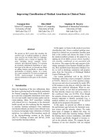
isolation of NPEV in Sistan & Balouchestan by Direct, Pellet
and Two-phase concentration methods were
11(12.79%),31(36.05%)and 44(51.16%)respectively (Fig.
1). Statistical analysis with SPSS13 software were reflected
that there was no significant correlation between Direct
method and Pellet & Two-phase concentration methods for
detection of NPEV in Sistan & Baluchestan Province. This
matter indicates the acceptability of Pellet and Two-phase
methods for isolation of NPEV. But there was significant
correlation (in 0.01 level) between Direct method and Pel-
let & Two-phase concentration methods in Fars and Tehran
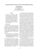
Provinces. According to the Fig. 2 Sistan & Baluchestan has
the greatest number of isolated N.P.E.V, as well as, the iso-
lation in the summer, autumn and winter were the same
(30.91%) and the lowest circulation related to spring
(7.27%). As the graph shows, the isolation of N.P.E.V in
spring and autumn were the same in Tehran Province.
Meanwhile the isolation in summer and winter revealed
the same pattern. Besides, the most isolation of NPEV in
Fars regarded to summer (4.16%), winter (6.25%) and
autumn(4.16%). As a whole, there was no significant cor-
relation between isolation of Enteroviruses and different
seasons. Moreover the isolation of NPEV in RD and HEp-2
cell lines indicate that, RD cell line is the best for detection
of NPEV in Sistan & Balouchestan Province with 53.94%
and also the detection was 10.47% in HEp-2 cell line. Hav-
ing applied SPSS 13 and ANOVA test, there was significant
correlation for isolation of NPEV between RD and HEp-2
cell lines. In Fars the best cell line for isolation of NPEV was
RD cell line with 16.6% and the isolation in HEp-2 cell line
was 4.17%, all of isolated virus in HEp-2 related to COX-B
virus, and the isolation of NPEV in Tehran were 44.66% in
RD and 15.87% in HEp-2 cell line too.
Discussion
Monitoring circulating enteroviruses is important because
individual serotypes have different temporal patterns of
circulation and the changes in predominant serotypes can
be accompanied by large-scale outbreaks of enteroviral ill-
nesses. Serotype-based enterovirus surveillance in the
United States has five objectives. First, NESS data help
public health practitioners determine long-term patterns
Table 1: Number of isolated Non-polio Enteroviruses in this study, Iran, Sistan and Balouchestan, Tehran and Fars
Serotype N.T.E.V E1 E3 E4 E6 E7 E11 E12 E13 E20 E21 E25 E27 E33 COX-B Total
Provinces n(%)
Sistan &
Baluchestan
7
(12.73)
2
(3.64)
3
(5.45)
11
(20)
4
(7.27)
6
(10.91)
8
(14.55)
4
(7.27)
0(0) 0(0) 0(0) 0(0) 0(0) 1
(1.82)
9
(16.36)
55
(100)
Tehran 7
(22.58)
1
(3.22)
0(0) 0(0) 1
(3.22)
2
(6.45)
4
(12.9)
0(0) 3
(9.68)
3
(9.68)
1
(3.22)
4
(12.9)
2
(6.45)
0(0) 3
(9.68)
31
(100)
Fars 2
(22.22)
0(0) 0(0) 0(0) 0(0) 1
(11.11)
4
(44.44)
0(0) 0(0) 0(0) 0(0) 0(0) 0(0) 0(0) 2
(22.22)
9
(100)
Total 16
(31.52)
3
(5 91)
3
(5 91)
11
(21.67)
1
(1 97)
9
(17.73)
16
(31.52)
4
(7.88)
3
(5 91)
3
(5 91)
1
(1.97)
4
(7.88)
2
(3.94)
1
(1.97)
14
(27.58)
95
(100)
Virology Journal 2009, 6:149 />Page 4 of 5
(page number not for citation purposes)
of circulation for individual enteroviruses. Moreover, the
data are used for interpreting trends in enteroviral dis-
eases, such as aseptic meningitis, by associating them with
circulating serotypes and can be helpful for studying the
association of enteroviruses with clinical manifestations.
Besides, the data are used to guide outbreak investigations
by enabling linkage of disease clusters; diagnosis by sero-
logic assay and clinical presentation, which varies by sero-
type; and timelier laboratory identification. Likewise,
because susceptibility to candidate anti-enterovirus drugs
varies by serotype, information on circulating serotypes
helps guide development of new diagnostic tests and ther-
apies. Finally, NESS monitors poliovirus detections,
thereby supplementing poliovirus surveillance in the
United States [3]. However, NPEV are occasionally related
to more serious illnesses, for example aseptic meningitis,
life-threatening myocarditis and hepatitis, and are proba-
bly associated with juvenile diabetes mellitus type 1.
Compared to the number of NPEV infections, these seri-
ous organ infections are rather rare events, with a fre-
quency quite similar to that of poliomyelitis anterior,
which is an infrequent organ manifestation of poliovirus
wild-type infection, and an extremely rare complication of
poliovirus vaccine strains. Nevertheless, such a strategy is
justified if the study investigates the association of NPEV
types with a certain disease [6].
In the context of poliomyelitis eradication, a reinforced
sentinel laboratory network for surveillance of enterovi-
ruses (RSE) was implemented in France in January 2000,
and the purpose of that report is to describe the results of
the five first years of surveillance. Over the 5 years of sur-
veillance, information was collected from 192,598 clini-
cal samples, including 39,276 cerebrospinal fluid
specimens, of which 14.7% were positive for enterovi-
ruses, 45,889 stool samples (4.3% positive for enterovi-
ruses), 70,330 throat swabs (2.2% positive) and 14,243
sera (1.4% positive). The ten main non-polio enterovi-
ruses typed were as follows, in decreasing order of fre-
quency: E-30, E-13, E-6, CV-B5, E-11, CV-B4, E-9, E-7, CV-
B1, and CV-B2. Continued surveillance of enteroviruses is
important to alert physicians and public health officials to
changes in disease trends. Although the geographical cov-
erage of the RSE network as well as the percentage of
enteroviruses identified must be improved, the large
number of samples tested for enteroviruses shows the
ability of virology laboratories to detect the circulation of
enteroviruses and to report the possible identification of
poliovirus (wild-type, vaccine-derived, or Sabin-like)
[14]. In several countries wild polioviruses have been
detected in the environment in the absence of reported
AFP cases. Thus, after eradication of wild polioviruses
from AFP cases in high risk areas, WHO has recom-
mended the complementary surveillance by using sewage
sample and stools of healthy children [11]. Therefore, Sis-
tan & Balouchestan, Tehran and Fars provinces were
selected for this research. Based on the recommendation
of WHO, a useful criterion of satisfactory overall perform-
ance of the surveillance is detection of non-polio Entero-
viruses in the samples. At least 30% of concentrated
sewage from grab samples should reveal NPEV [10,11].
In this study, for the first time, we suggested the Pellet
concentration method, and used the Two-phase concen-
tration method, simultaneously. From the total samples
in Sistan & Baluchestan, non-polio enteroviruses were iso-
lated from 11(12.79%), 31(36.05%) and 44 (51.16%)
samples by direct, pellet and two-phase methods, respec-
tively. These results confirm the efficiency of concentra-
tion methods, in enterovirus surveillance. Another
purpose of this study was evaluation of distribution and
analysis of environmental circulation of NPEVs. Japanes,
study on Enteroviruses shows that E6, E17, Cox-B5 in
1999, E9, E71, E25, E11 in 2000 and E11 and Cox-B5 in
2001 have played the main role in aseptic meningitis out-
break. In 2002, also E11 and E13 were the most frequently
isolated Enteroviruses from aseptic meningitis patients
[15]. During the seasons under study, E4 (20%), Cox-B
(16.36%) and E11 (14.55%) were the predominant sero-
types in Sistan & Baluchestan. But N.T.E.V(22.58%), E25
Number of isolated Non-polio Enteroviruses based on three concentration methods in Sistan and Balouchestan, Tehran and FarsFigure 1
Number of isolated Non-polio Enteroviruses based
on three concentration methods in Sistan and Balo-
uchestan, Tehran and Fars.
0
10
20
30
40
50
60
Sistan &
Baluchestan
Tehran Fars
Direct
Pellet
Two-phase
Number of Non-polio Enteroviruses based on different sea-sons in Sistan & Baluchestan, Tehran and FarsFigure 2
Number of Non-polio Enteroviruses based on differ-
ent seasons in Sistan & Baluchestan, Tehran and
Fars.
0
5
10
15
20
25
30
35
Spring Summer Fall Winter
Sistan &
Baluchestan
Tehran
Fars
Publish with BioMed Central and every
scientist can read your work free of charge
"BioMed Central will be the most significant development for
disseminating the results of biomedical research in our lifetime."
Sir Paul Nurse, Cancer Research UK
Your research papers will be:
available free of charge to the entire biomedical community
peer reviewed and published immediately upon acceptance
cited in PubMed and archived on PubMed Central
yours — you keep the copyright
Submit your manuscript here:
/>BioMedcentral
Virology Journal 2009, 6:149 />Page 5 of 5
(page number not for citation purposes)
and E11(12.9%) were the most serotypes in Tehran Prov-
ince. The epidemiological pattern of enterovirus infec-
tions varies by geographical region, climate, age and
season. Therefore, it is necessary to evaluate relationship
between non-polio enterovirus disease and environmen-
tal circulation of these viruses in different part of Iran.
Such studies can be perform for providing a suitable vac-
cine to prevent of enterovirus infections in high risk area.
Until now, the cell line that capable to isolation of all
enteroviruses has not identified. Several coxsackievirus A
(CAV) serotypes of the species Human enterovirus A are
hard to isolate on cell cultures and require animal experi-
ments with suckling mice for virus isolation. These are not
routinely performed in most laboratories. Fortunately, RD
cells are recommended by the World Health Organization
for poliovirus surveillance. Use of RD cells and of the shell
vial technique clearly improves isolation of CAV serotypes
but some serotypes and strains even fail to replicate on RD
cells. Thus poliovirus surveillance efforts may produce
some data on CAV circulation but some CAV types are still
overlooked by this approach, leaving the picture of enter-
ovirus surveillance somewhat incomplete [6]. However,
the use of L20B and RD cells without HEp-2, may have an
impact on the non-poliovirus enterovirus isolation rate,
especially during periods of Coxsackie B circulation in the
community [13,16,17]. Therefore, in this study RD and
HEp-2 cells were used for identification of more extend
spectrum of enteroviruses. Overall, 46 and 9 NPEVs were
detected in Sistan & Baluchestan, 28 and 10 NPEVs in
Tehran, 7 and 2 NPEVs in Fars, on RD and HEp-2 cells,
respectively [9,18]. Not isolating vaccine derived poliovi-
ruses (VDPV) and vaccine derived NPEV shows the proper
AFP surveillance and vaccination coverage in our country
at high risk areas. But, repeated sampling and environ-
mental surveillance will increase the probability of detect-
ing low level transmission of enteroviruses in population.
Competing interests
The authors declare that they have no competing interests.
Authors' contributions
MK carried out the design of the study, coordination and
performed the statistical analysis. SS participated in sam-
pling, concentration, cell culture and neutralization test.
RN participated in the scientific consultation of this
research project. All authors read and approved the final
manuscript.
Acknowledgements
The writers of this Article offer their thanks & appreciation to the scientific
& sanitary research Institute affairs of the medical university of Tehran for
their financial & executive protection of this project.
References
1. Pallansch M, Roos RP: Picornaviridae:The Viruses and Their
Replication. In Fileds virology 1st edition. Edited by: Filds BN, et al.
Lippincott Raven., New Yourk; 2001:723-776.
2. Paul AV, Wimmer E, Rieder E: Application Number: 09/282351,
Publication Date:, International Classes 2001, C12Q1/48;
G01N33/50; C12Q1/00; C12Q1/68. .
3. Centers for Disease Control and Pervention: Enterovirus Surveil-
lance United States, 2002-2004. JAMA 2006, 295:1993-1994.
4. World Health Organization: Enteroviruses-non polio. Media centre 2002
[ />].
5. World Health Organization: Polio laboratory case definition.
Australian Government 2000 [ />wcms/Publishing.nsf/Content/cda-phlncd-poio.htm/$FILE/polio.pdf].
6. Heim A: From poliovirus surveillance to enterovirus surveil-
lance: a complete picture? J Med Microbiol 2005, 54:1-2.
7. Harris BN, Durrheim DN, Ogunbanjo GA: Polio eradication - the
validity of surveillance indicators. Tropical Medicine & Interna-
tional Health 2003, 8:386-392.
8. Deshpande JM, Shetty SJ, Siddiqui ZA: Environmental Surveil-
lance Systemto Track Wild Poliovirus Transmission. Applied
and Environmental Microbiology 2003, 69:2919-2927.
9. Centers for Disease Control and Pervention: Progress Toward
Poliomyelitis Eradication Poliomyelitis Outbreak in Sudan,
2004. MMWR 2005, 54:97-99.
10. World Health Organization: Global Eradication Initiative Stra-
tegic plan 2004-2008. WHO Publication. Printed in Switzerland;
2003:1-40.
11. World Health Organization: Guidelines for environmental sur-
veillance of poliovirus circulation Vaccines and Biologicals.
(Ordering code:WHO/V&B/03.03) 2003.
12. Hovi T, Stenvik M, Partanen H: Poliovirus surveillance by exam-
ining sewage specimens. Quantitative recovery of virus after
introduction into sewerage at remote upstream location.
Epidemiol Infect 2001, 127:101-106.
13. World Health Organization: Polio Laboratory Manual. Depart-
ment of Vaccines and Biologicals 2004 [ />poliolab/WHOPolio-Manual-9.pdf].
14. Antona D, Lévêque N, Chomel JJ, Dubrou S, Lévy-Bruhl D, Lina B:
Surveillance of enteroviruses in France, 2000-2004. Eur J Clin
Microbiol Infect Dis 2007, 26:403-412.
15. Infectious Agents Surveillance Report: The trend of enterovirus
isolation in association with aseptic meningitis, 1999-2002.
IASR 2002, 23:193-194.
16. World Health Organization: Distribution of L20B cells is under-
way. Polio Lab Network 1998, 4:1-4.
17. Sedmak G, Bina D, MacDonald J: Assessment of an Enterovirus
Sewage Surveillance System by Comparison of Clinical Iso-
lates with Sewage Isolates from Milwaukee, Wisconsin, Col-
lected Aug 1994 to Dec 2002. Applied and Environmental
Microbiology 2003, 69:7181-7187.
18. Yang C, Naguib T, Yang S, Nasr E, Jorba J, Ahmed N, Campagnoil R,
Avoot H, Shimizu H, Yoneyama T, Miyamura T, Pallansch M, Kew O:
Circulation of Endemic Type 2 Vaccine-Derived Poliovirus in
Egypt from 1983 to 1993. Journal of Virology 2003, 77:8366-8377.