Báo cáo khoa học: "Bovine viral diarrhea virus NS4B protein is an integral membrane protein associated with Golgi markers and rearranged host membranes" ppsx
Bạn đang xem bản rút gọn của tài liệu. Xem và tải ngay bản đầy đủ của tài liệu tại đây (2.28 MB, 15 trang )
BioMed Central
Open Access
Page 1 of 15
(page number not for citation purposes)
Virology Journal
Research
Bovine viral diarrhea virus NS4B protein is an integral
membrane protein associated with Golgi markers and rearranged
host membranes
Erica Weiskircher
1,3
, Jason Aligo
1
, Gang Ning
2
and Kouacou V Konan*
1
Address:
1
Department of Biochemistry and Molecular Biology, The Pennsylvania State University, University Park, PA 16802, USA,
2
The Huck
Institutes of the Life Sciences, The Pennsylvania State University, University Park, PA 16802, USA and
3
Absorption Systems LP, Exton, PA 19341,
USA
Email: Erica Weiskircher - ; Jason Aligo - ; Gang Ning - ;
Kouacou V Konan* -
* Corresponding author
Abstract
Background: Very little is known about BVDV NS4B, a protein of approximately 38 kDa.
However, a missense mutation in NS4B has been implicated in changing BVDV from a cytopathic
to noncytopathic virus, suggesting that NS4B might play a role in BVDV pathogenesis. Though this
is one possible function, it is also likely that NS4B plays a role in BVDV genome replication. For
example, BVDV NS4B interacts with NS3 and NS5A, implying that NS4B is part of a complex, which
contains BVDV replicase proteins. Other possible BVDV NS4B functions can be inferred by analogy
to hepatitis C virus (HCV) NS4B protein. For instance, HCV NS4B remodels host membranes to
form the so-called membranous web, the site for HCV genome replication. Finally, HCV NS4B is
membrane-associated, implying that HCV NS4B may anchor the virus replication complex to the
membranous web structure. Unlike its HCV counterpart, we know little about the subcellular
distribution of BVDV NS4B protein. Further, it is not clear whether NS4B is localized to host
membrane alterations associated with BVDV infection.
Results: We show first that release of infectious BVDV correlates with the kinetics of BVDV
genome replication in infected cells. Secondly, we found that NS4B subcellular distribution changes
over the course of BVDV infection. Further, BVDV NS4B is an integral membrane protein, which
colocalizes mainly with the Golgi compartment when expressed alone or in the context of BVDV
infection. Additionally, BVDV induces host membrane rearrangement and these membranes
contain BVDV NS4B protein. Finally, NS4B colocalizes with replicase proteins NS5A and NS5B
proteins, raising the possibility that NS4B is a component of the BVDV replication complex.
Interestingly, NS4B was found to colocalize with mitochondria suggesting that this organelle might
play a role in BVDV genome replication or cytopathogenicity.
Conclusion: These results show that BVDV NS4B is an integral membrane protein associated
with the Golgi apparatus and virus-induced membranes, the putative site for BVDV genome
replication. On the basis of NS4B Colocalization with NS5A and NS5B, we conclude that NS4B
protein is an integral component of the BVDV replication complex.
Published: 3 November 2009
Virology Journal 2009, 6:185 doi:10.1186/1743-422X-6-185
Received: 31 August 2009
Accepted: 3 November 2009
This article is available from: />© 2009 Weiskircher et al; licensee BioMed Central Ltd.
This is an Open Access article distributed under the terms of the Creative Commons Attribution License ( />),
which permits unrestricted use, distribution, and reproduction in any medium, provided the original work is properly cited.
Virology Journal 2009, 6:185 />Page 2 of 15
(page number not for citation purposes)
Background
Bovine viral diarrhea virus, or BVDV, is a major viral path-
ogen in cattle and other ruminants [1]. BVDV is divided
into two different genotypes (genotypes I and II) based on
the genetic composition of the 5'-untranslated region
(UTR) of the viral genome [2]. These genotypes are dis-
tinct from one another [2], but they cause the same dis-
ease. BVDV pathogenicity is manifested in two biotypes:
noncytopathic (ncp) and cytopathic (cp). In the case of
ncp BVDV, the virus can cause an acute or persistent infec-
tion [3]. Infections with cp BVDV are acute and symptoms
can range from mild to severe, often leading to a fatal dis-
ease. A feature that often distinguishes cp from ncp BVDV
is the production of precursor and mature nonstructural
proteins, NS2-3 and NS3, respectively [4,5]. In ncp BVDV
infections, the junction between NS2 and NS3 is not
cleaved, yielding precursor NS2-3 protein. However, in cp
BVDV infections, NS3 is cleaved from NS2, yielding NS2-
3 and NS3 proteins. Many cytopathic laboratory strains of
BVDV, such as National Animal Disease Laboratory
(NADL) [6], are derived from genotype I. BVDV is a mem-
ber of the pestivirus genus, along with classical swine fever
virus and Border's disease virus [7]. The pestivirus genus
belongs to the Flaviviridae family of viruses, which also
includes the genera hepacivirus and flavivirus. Members
of these genera include hepatitis C virus (HCV), yellow
fever virus (YFV), Dengue fever virus (DFV), and West Nile
virus (WNV). Like the other family members, BVDV is an
enveloped, positive-sense RNA virus. All these viruses
share a similar genome organization and replication cycle
[8]. The N-terminal half of the genome contains structural
proteins involved in virus assembly whereas the C-termi-
nus contains the nonstructural (NS) proteins involved in
viral genomic RNA synthesis [9].
BVDV has a 12.3 kb positive-sense RNA genome, com-
posed of a long open reading frame flanked by 5'- and 3'-
UTR. The genome is translated into a polyprotein, which
is subsequently cleaved by host and viral proteases, result-
ing in mature viral proteins in the order: N
pro
-C-E
0
-E
1
-E
2
-
NS2-3-NS4A-NS4B-NS5A-NS5B. The 5' UTR contains an
internal ribosomal entry site (IRES), which promotes cap-
independent translation of the viral genome. The 3'UTR
contains cis-acting elements that are important for viral
genome replication [10]. The BVDV genome organization
is closely related to that of HCV [9]. Additionally, transla-
tion of BVDV and HCV genomes require an IRES whereas
members of the flavivirus genus use cap-dependent trans-
lation [11,12]. Further, both viruses have similar non-
structural proteins whereas flaviviruses have NS1 and
NS5, which has functions related to NS5A and NS5B. For
these reasons, BVDV has been proposed as a surrogate
model for understanding HCV replication [9].
Most positive-sense RNA viruses replicate their genome in
association with rearranged cytosolic membranes [13]. In
HCV and Kunjin Virus, the remodeled membranes have
been referred to as membranous webs, convoluted mem-
branes, or vesicle packets [13-17]. These structures are
usually derived from the endoplasmic reticulum (ER) or
the Golgi apparatus [13,18]. The viral replicase proteins as
well as the viral RNA are generally localized to these mem-
branes, suggesting that these structures are the site for viral
genome replication [19]. In the case of BVDV, ultrastruc-
tural studies have shown large sac-like vesicles containing
mature viral particles [20,21]. However, it is not clear
whether these sacs are only the vehicle for viral egress or if
they also serve as the site for viral RNA synthesis. Since,
these sac-like vesicles were observed in infected cells col-
lected at later time points post-infection (48 h), it is pos-
sible that early ultrastructural changes that might be
involved in viral genome replication could have been the
precursor to these vesicles.
No function has been ascribed to BVDV NS4B, a protein
of approximately 38 kDa [22]. However, a single point
mutation in NS4B (Y2441C) has been implicated in
changing the virus from cp to ncp, suggesting that NS4B
may play a role in BVDV pathogenesis [23]. Though this
is one possible function, it is also likely that BVDV NS4B
plays a greater role in the replication of the viral genome.
Other possible BVDV NS4B functions can be inferred by
analogy to HCV and DFV NS4B proteins. In these viruses,
NS4B protein is associated with replicase proteins NS3,
NS5A, and NS5B [24]. In addition, NS4B protein from
HCV and DFV is membrane-associated [23,25], suggest-
ing that NS4B may anchor the virus replication complex
to existing or rearranged intracellular membranes. Finally,
NS4B proteins from all these viruses are highly hydropho-
bic and have related membrane topology [23,25].
Expression of HCV NS4B has been associated with mem-
branous web formation [16,26], the site of HCV genome
replication [13]. Since HCV and BVDV NS4B proteins
share similar membrane topology, we hypothesized that
the two proteins have similar function. More specifically,
we postulate that BVDV NS4B induces the formation of a
novel membrane structure, which may serve as the site for
viral genome replication. In this report, we have used flu-
orescence microscopy and electron microscopy to exam-
ine NS4B in the context of BVDV infection. We show that
NS4B colocalizes with Golgi markers, but its subcellular
distribution appears to change in the course of BVDV
infection. We also show that NS4B is associated with rear-
ranged host membranes. The significance of such findings
will be discussed.
Results
Kinetics of BVDV RNA synthesis in infected MDBK cells
The function of NS4B protein in BVDV replication is
poorly understood. However, the findings that NS4B
interacts with NS3 and NS5A [27] may suggest that NS4B
Virology Journal 2009, 6:185 />Page 3 of 15
(page number not for citation purposes)
plays a role in BVDV genome replication. Unlike its HCV
counterpart, we know little about the subcellular distribu-
tion of BVDV NS4B protein. Further, it is not clear
whether NS4B is associated with BVDV-induced host
membranes. Thus, BVDV full-length RNA was electropo-
rated into MDBK cells and the resulting virus titer was
determined by plaque assay as shown in Fig. 1A. To exam-
ine the kinetics of BVDV replication, MDBK cells were
infected with cytopathic (cp) BVDV at a multiplicity of
infection (MOI) of 0.1. At various times post-infection,
the resulting virus was collected from the cell supernatant
(Media) and cell lysate (Lysate), and BVDV titer was deter-
mined via plaque assay. As seen in Fig. 1B, infectious
BVDV release (Media) began between 12 h and 18 h.p.i.,
and reached a plateau at 36 h.p.i., with a titer of 10
6
-10
7
plaque forming units per milliliter (pfu/ml). These results
are consistent with previous reports showing BVDV
growth kinetics in MDBK cells [27,28]. Additionally, virus
titers were consistently low (below 10
4
pfu/ml) in the cell
lysates (Fig. 1B). These data suggest that most of the virus
remaining in the cells may represent immature virus par-
ticles.
To ascertain the rate of RNA synthesis during BVDV infec-
tion, MDBK cells were infected at MOI of 0.1 and total cel-
lular RNA was collected at 0 h (after 1 h adsorption), 6 h,
12 h, 18 h, and 24 h.p.i. The RNA was subjected to Real-
Time PCR (RT-PCR) analysis with a probe specific to a
region of BVDV NS4B sequence. The RT-PCR results were
normalized using a probe specific to Glyceraldehyde-3-
phosphate dehydrogenase (GAPDH) mRNA. As displayed
in Fig. (1C &1D), BVDV genomic RNA was barely detect-
able in the cells at 6 h.p.i. However, by 12 h, there was a
50-fold increase in viral RNA production. BVDV RNA syn-
thesis continued to rise such that by 24 h.p.i., there was
almost a 500-fold increase in detectable viral genomic
RNA. These results are consistent with the kinetics of
infectious virus production and release from MDBK cells
(Fig. 1B).
Immunoblot analysis of NS3 protein in BVDV-infected
MDBK cells
To determine the kinetics of NS3 and NS4B expression,
MDBK cells were infected with BVDV at MOI of 5. This
MOI was chosen to ensure that approximately 99% of the
cells had the virus and to increase the expression levels of
NS3 or NS4B protein by immunoblotting. Infected cell
lysates were prepared at 6 h, 12 h, 18 h, 24 h, and 48 h.p.i.
BVDV NS3 and NS4B proteins were detected using rabbit
polyclonal antibodies specific to NS3 and NS4B proteins.
As seen in Fig. 2, BVDV NS3 protein, of approximately 80
kDa, was detectable in MDBK cells as early as 12 h.p.i.
NS3 expression increased over time and reached a maxi-
mum at approximately 24 h.p.i. These results are in agree-
ment with the kinetics of HCV RNA synthesis in Fig. (1C
and 1D). Western blot results of NS4B protein were incon-
clusive perhaps because the NS4B antibody used in this
study was not suitable for detecting NS4B protein via
immunoblotting.
Intracellular localization of BVDV NS4B in infected MDBK
cells
To ascertain NS4B subcellular distribution, MDBK cells
were plated on coverslips and infected with BVDV at an
MOI of 5. The cells were processed at 12 h, 18 h, and 24
h.p.i., and NS4B was detected with NS4B-specific anti-
body and Alexa fluor 488-conjugated secondary antibody.
As shown in Fig. 3, the NS4B distribution pattern
appeared to change over the course of BVDV infection. At
12 h.p.i., NS4B was observed in a Golgi-like staining pat-
tern (3A; i and ii). By 24 h.p.i., NS4B appeared to display
a heterogeneous staining pattern; some cells (ca. 75%)
had one or two punctate structures or foci, whereas others
(25%) had more than five large foci scattered in the cyto-
plasm (3A; v and vi). These results suggest a putative
change in NS4B intracellular localization during the
course of BVDV infection. Staining of mock-infected cells
resulted in little background (3A; vii), suggesting that
NS4B antibody was specific to BVDV NS4B protein.
To further assess the intracellular localization of NS4B
protein in BVDV-infected cells, MDBK cells were grown
on coverslips and infected with cp BVDV. Infected cells
were processed at 18 h.p.i., the earliest time when sub-
stantial viral RNA synthesis and virus release were
observed (Fig. 1B and 1C). The cells were then co-stained
with BVDV NS4B antibody and antibodies specific for var-
ious intracellular compartments, including the Golgi
apparatus (αTGN38 and αGolgin 97), the endoplasmic
reticulum or ER (αCalnexin), and the lysosome
(αLamp1). For each experiment, NS4B was detected with
Alexa fluor 488-conjugated secondary antibody whereas
the cellular marker was detected with Alexa fluor 594-con-
jugated secondary antibody. Colocalization of BVDV
NS4B (in green) with any cellular marker (in red) was
expected to yield yellow fluorescence. As shown in Fig. 3B,
the fluorescence pattern of NS4B appeared to partially
overlap with Golgi markers (TGN38; ii-iv, and αGolgin
97; vi-viii). These results suggest that BVDV NS4B protein
is associated with the Golgi compartment or Golgi mark-
ers. BVDV NS4B Colocalization with the lysosomal
marker, Lamp1, or ER-derived marker, calnexin, was
inconclusive (data not shown) because the antibodies to
Lamp1 and calnexin did not specifically detect these pro-
teins in MDBK cells.
Ultrastructural analysis of BVDV-infected MDBK cells
Like many positive-stranded RNA viruses, BVDV is pre-
dicted to replicate its genome in the cytosol in association
with host membranes. However, it is not clear whether
Virology Journal 2009, 6:185 />Page 4 of 15
(page number not for citation purposes)
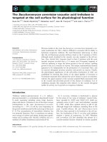
A. Representative plaque assays of cytopathic (cp) BVDV in MDBK cellsFigure 1
A. Representative plaque assays of cytopathic (cp) BVDV in MDBK cells. Cells were infected with 10-fold serial dilu-
tions of BVDV stocks from virus supernatant. After adsorption, monolayers were overlaid with DMEM/5% horse serum and
0.5% agarose plugs. After 72 h incubation, the plugs were removed and the monolayers stained with 1% crystal violet. B.
Growth Kinetics of cp BVDV in MDBK cells. Cells were infected with BVDV at MOI of 0.1. The supernatant (media, diamonds)
and cell lysates (lysate, squares) were harvested at the indicated time points. Viral titers were determined via plaque assay. The
results are given as log10 pfu/ml. C. BVDV RNA synthesis at various times post infection. MDBK cells were infected with
BVDV as above and total cellular RNA was collected at 0 h (after 1 h adsorption), 6 h, 12 h, 18 h, and 24 h.p.i. To determine
the amount of viral RNA in the cells, RT-PCR was performed with a probe specific to BVDV NS4B sequence. The amount of
BVDV RNA was determined relative to GAPDH. D. BVDV NS4B cDNA products from RT-PCR, prior to quantitation, were
run on 0.8% agarose gel and stained with ethidium bromide. Notice the increase in cDNA product from 6 to 24 h post BVDV
infection.
Virology Journal 2009, 6:185 />Page 5 of 15
(page number not for citation purposes)
BVDV replication complex is associated with virus-
induced membranes. To determine if BVDV infection
causes ultrastructural changes, MDBK cells were infected
at MOI of 10 to ensure that 100% of the cells were
infected. The cells were harvested at 18 h, 24 h and 48
h.p.i, fixed with glutaraldehyde, sectioned and examined
via transmission electron microscopy analysis (TEM). As
seen in Fig. 4, 5 and 6, mock-infected cells show different
types of vesicular structures indicated by the arrows and
arrowheads. These vesicles were not time-dependent, as
they were seen at 18 h, 24 h or 48 h post- seeding. More-
over, ultrastructural analysis of BVDV-infected cells
showed different membrane structures. Many of the vesi-
cles were similar to those found in uninfected cells, indi-
cating that these structures were not virally induced
[arrows, Fig. 4 and 5(B)]. However, we also observed
unique membrane structures at 18 h, 24 h and 48 h.p.i.
These structures (small and large stars) consist of vesicles
of various sizes enclosed in a much larger vesicle [Fig. (4B
and 4D); Fig. (5B and 5C); Fig. (6B and 6C)]. They do not
resemble the HCV-induced membranous web structure
[13]. Instead, they are more reminiscent of the vesicle
packets induced by Kunjin virus and shown to contain the
replicase proteins as well as the viral RNA [18].
To determine whether BVDV proteins were associated
with the induced membrane vesicles, MDBK cells were
infected with BVDV at MOI of 15. At 18 h.p.i., mock- and
BVDV-infected cells were fixed and stained with NS4B-
specific antibody and quantum dots (Qdots) 605-conju-
gated secondary antibody. As shown Fig. 7B, NS4B stain-
ing (red fluorescence) was observed in BVDV-infected
cells, and not in mock-infected cells (Fig. 7A), indicating
specificity of both the primary and secondary antibodies
used in this study. However, the lack of NS4B staining in
most of the BVDV-infected cells suggests, 1) differential
expression of NS4B in MDBK cells or, 2) an overestima-
tion of the BVDV titer.
When observed via TEM, the mock infected cells showed
no electron-dense Qdots staining (Fig. 7C and 7D). In
contrast, when BVDV-infected cells were examined at 18
h.p.i, electron-dense Qdots [Fig. 8(A-B); arrowheads in
8(C) and 8(D)] were found in vesicular structures similar
to those detected in Fig. (4B and 4D). These results suggest
that NS4B protein is associated with the vesicular struc-
tures observed at 18 h post BVDV infection.
BVDV NS4B is an integral membrane protein
Membrane floatation assay was performed to examine
BVDV NS4B association with intracellular membranes.
Since BVDV NS4B protein was not detected by immunob-
lotting (IB), we engineered an NS4B construct with a C-
Kinetics of BVDV NS3 protein expression in infected cellsFigure 2
Kinetics of BVDV NS3 protein expression in infected
cells. MDBK cells were infected with BVDV at an MOI of 5
and the cell lysates prepared at the indicated time points.
Rabbit anti-NS3 polyclonal antibody was used at 1/1000 dilu-
tion. Goat anti-rabbit alkaline phosphatase-conjugated sec-
ondary antibody was used to detect NS3 protein. M: Mock-
infected cell lysate collected at 24 h after incubation. Higher
NS3 expression levels are seen at 24 h and 48 h.p.i.
A. Localization of BVDV NS4B in infected MDBK cellsFigure 3
A. Localization of BVDV NS4B in infected MDBK
cells. Cells were grown on coverslips and infected with
BVDV at MOI of 10. At 12 h (i and ii), 18 h (iii and iv) and 24
h.p.i (v and vi), cells were processed for immunofluorescence
(IF) with NS4B-specific antibody (1/50 dilution) and Alexa
fluor 488-conjugated secondary antibody (1/500). Nuclei
were stained with DAPI. Notice the Golgi-like NS4B distri-
bution at 12 h and 18 h.p.i., whereas foci are seen at 24 h.p.i
No fluorescence is displayed in mock-infected cells (vii). Bars
= 10 μm. B. BVDV NS4B partially colocalizes with Golgi
markers. Cells were grown on coverslips and infected with
BVDV as above. At 18 h.p.i., cells were processed for IF with
NS4B- (ii and iv; vi and viii), TGN38- (iii and iv) and Golgin97
(vii and viii)- specific antibodies. Notice the colocalization of
NS4B with TGN38 or Golgin97 protein. Mock-infected cells,
stained with anti TGN38 (i) or Golgin97 (v), are also shown.
Virology Journal 2009, 6:185 />Page 6 of 15
(page number not for citation purposes)
terminal GFP tag (NS4B-GFP). When the construct was
transfected into MDBK cells followed by IB with GFP-spe-
cific antibody, NS4B-GFP protein was not detected (data
not shown) as a result of the low transfection efficiency of
MDBK cells. To circumvent this obstacle, we expressed
NS4B-GFP in BHK-21 cells which can also support BVDV
replication [29]. The cell extracts were collected at 48 h p.t.
and subjected to membrane floatation using a discontin-
uous iodixanol gradient [30]. Eight fractions were col-
lected, separated on 10% SDS-PAGE followed by IB with
GFP-specific antibody. If BVDV NS4B is a membrane-
associated protein, we predicted that NS4B would be
mostly found in the in the lower buoyant density, mem-
brane-enriched fractions (1 through 4). As shown in Fig.
9A, NS4B was mostly detected in the membrane-enriched
fractions. By contrast, control GFP was mostly found in
higher density, soluble fractions (5 through 8). These data
suggest that BVDV NS4B protein is membrane-bound.
To further characterize the nature of NS4B association
with internal membranes, NS4B-expressing BHK-21 cell
lysates were subjected to Triton X-100 (TX-100), 1 m NaCl
(high salt) or high pH (sodium carbonate, pH11.5) treat-
ment at 4°C for 30 min, followed by membrane floata-
tion assay and immunoblot detection of NS4B protein. As
shown in Fig. (9B and 9C), high salt or high pH had no
effect on NS4B membrane association. NS4B subcellular
distribution profile was similar to that of calnexin, a
membrane-bound protein, but different from GAPDH, a
soluble protein. Further, treatment with 0.5% TX-100
resulted in the redistribution of NS4B from the mem-
brane-bound fractions to the soluble fractions (Fig. 9C).
These findings indicate that BVDV NS4B protein is an
integral membrane protein.
Subcellular localization of BVDV NS4B protein
Two approaches were taken to determine the nature of the
NS4B-bound internal membranes. First, NS4B-expressing
BHK-21 cells were lysed in a hypotonic buffer, followed
by subcellular fractionation to obtain cytosolic, nuclear,
mitochondrial and microsomal fractions. Sixty micro-
grams of each fraction were separated on 10% SDS-PAGE,
followed by IB with GFP-specific antibody. As shown in
Fig. 9D, NS4B protein was mostly enriched in nuclear and
mitochondrial fractions as compared to control GFP
Ultrastructural analysis of MDBK cells examined at 18 h.p.i. MDBK cells were mock infected or infected with BVDV at MOI of 15Figure 4
Ultrastructural analysis of MDBK cells examined at
18 h.p.i. MDBK cells were mock infected or infected
with BVDV at MOI of 15. Cells were harvested at 18 h.p.i
and processed for TEM analysis. White arrows and arrow-
heads show the types of vesicles seen in mock-infected (A)
or infected cells (B). Stars indicate the vesicular structures
found solely in BVDV-infected cells (B). Higher magnifications
of the areas in mock-infected (C) and BVDV-infected cells
(D) are indicated by the rectangle boxes. Notice the pres-
ence of various size vesicles enclosed in the large vesicular
structures in (D). Bars = 1 μm.
Ultrastructural analysis of MDBK cells examined at 24 h.p.i. cells were infected and processed for TEM analysis as aboveFigure 5
Ultrastructural analysis of MDBK cells examined at
24 h.p.i. cells were infected and processed for TEM
analysis as above. White arrows show the types of vesicles
seen in mock infected (A) and BVDV-infected (B) cells. The
star indicates the vesicular structure found mainly in BVDV-
infected cells (B). A higher magnification of the area in
BVDV-infected cells (C) is indicated by the rectangle box.
Notice the presence of various size vesicles enclosed in the
large vesicular structures. Bars = 1 μm.
Virology Journal 2009, 6:185 />Page 7 of 15
(page number not for citation purposes)
which was prominent in the cytosolic fraction. To confirm
these results, NS4B-GFP was expressed in BHK-21 cells,
followed by fluorescence Colocalization of NS4B-GFP
with subcellular markers. NS4B-GFP was detected via GFP
fluorescence whereas intracellular markers were visual-
ized using ER-Tracker for ER membranes, Golgin-97 for
the Golgi apparatus, Rab5 for the early endosome, Lys-
oTracker for the lysosome and MitoTracker for mitochon-
dria. As shown in Fig. 10, NS4B-GFP subcellular
distribution merged well with Golgin-97 (iv-vi; b) and
MitoTracker (xiii-xv; e). Partial NS4B merging was
observed with ER-Tracker (i-iii; a) whereas Rab5 and Lys-
oTracker show no colocalization. These findings suggest
that NS4B is associated with the Golgi compartment and
mitochondria.
BVDV NS4B protein colocalizes with NS5A and NS5B
BVDV NS4B has been found to interact with NS3 and
NS5A proteins [27]. Further, nonstructural proteins (NS3,
NS4A, NS4B, NS5A and NS5B) are sufficient to promote
BVDV genome replication [31]. These findings suggest
that NS4B is a component of BVDV replication complex.
To test this hypothesis, we examined the subcellular dis-
tribution of NS4B, NS5A and NS5B proteins. Specifically,
BHK-21 cells wells were co-transfected with DNA con-
structs expressing NS4B-GFP and NS5A-His or NS4B-GFP
and NS5B-HA. At 48 h p.t., the cells were fixed and NS4B
was visualized via GFP fluorescence whereas NS5A and
NS5B were visualized via Alexa Fluor 594-conjugated sec-
ondary to Penta His antibody or HA antibody, respec-
tively. As shown in Fig. 11, NS4B colocalized with N5SA
and NS5B proteins. These data suggest that NS4B, NS5A
and NS5B have a similar subcellular distribution.
Discussion
NS4B proteins from hepaciviruses (HCV), pestiviruses
(e.g. BVDV) and flaviviruses (e.g. Dengue virus) show very
little conservation at the amino acid sequence level. How-
Ultrastructural analysis of MDBK cells examined at 48 h.p.i. cells were infected and processed for TEM analysis as aboveFigure 6
Ultrastructural analysis of MDBK cells examined at
48 h.p.i. cells were infected and processed for TEM
analysis as above. White arrow and arrowhead show the
types of vesicles seen in mock-infected cells (A). The star
indicates the vesicular structure found only in BVDV-infected
cells (B). A higher magnification of the area in BVDV-infected
cells (C) is indicated by the rectangle box. Notice the pres-
ence of various size vesicles enclosed in the large vesicular
structures. Bars = 1 μm.
Immunostaining of BVDV-infected MDBK cellsFigure 7
Immunostaining of BVDV-infected MDBK cells. Cells
were plated in 8-chamber slides, mock infected or infected
with BVDV. At 18 h.p.i, cells were fixed with 4% formalde-
hyde/0.1% glutaraldehyde for 10 min. Cells were permeabi-
lized with 0.05% Triton X-100, stained with NS4B-specific
antibody and Qdots 605-conjugated secondary antibody
(Molecular Probes, Invitrogen, CA), followed by fluorescence
microscopy. Nuclei were stained with DAPI. Notice the red
stain in BVDV-infected cells (B) and no stain in mock-infected
cells (A). Labeled cells were fixed with 2.5% glutaraldehyde
prior to sectioning and TEM analysis. Boxed area indicates
the vesicular structures in mock-infected cells (C). A higher
magnification of the boxed area is shown in (D). No electron
dense Qdots were observed in mock-infected cells (D). Bars
= 1 μm.
Virology Journal 2009, 6:185 />Page 8 of 15
(page number not for citation purposes)
ever, these proteins are highly hydrophobic, each having
at least four transmembrane domains [25,27,32]. Further,
NS4B C-terminal domains from HCV and BVDV are pre-
dicted to be on the cytosolic side of the ER membrane.
Finally, HCV or BVDV NS4B is associated with replicase
proteins [27,33], suggesting that NS4B plays a role in
BVDV RNA synthesis. In this study, we have taken the ini-
tial step to define the role of NS4B in BVDV replication by
examining NS4B subcellular distribution and its relation-
ship to BVDV-induced membrane alterations. We show
first that the release of infectious BVDV correlates with the
kinetics of BVDV genome replication in infected cells. Sec-
ondly, we found that NS4B subcellular distribution
changes over the course of BVDV infection. Further, we
show that BVDV NS4B protein is an integral membrane
protein, which is mostly associated with Golgi mem-
branes and mitochondria. Additionally, BVDV induces
host membrane remodeling and these membranes con-
tain BVDV NS4B protein. Finally, NS4B colocalizes with
replicase proteins NS5A and NS5B proteins, further rais-
ing the possibility that NS4B is a component of the BVDV
replication complex.
Despite its different host range, BVDV genome organiza-
tion is closely related to that of HCV. Thus, understanding
BVDV NS4B function in the context of BVDV infection
could shed some light on NS4B function during HCV rep-
lication. The findings that NS4B subcellular distribution
pattern changes during the course of BVDV infection sug-
gest some movement of NS4B-associated structures in the
cell and perhaps a change in the cellular composition of
these structures. Our results show that BVDV NS4B pro-
tein is mainly associated with the Golgi compartment, or
Golgi markers, when expressed singly or in the context of
the virus genome. Further, NS4B colocalizes with mito-
chondria when expressed alone. These results are in par-
tial agreement with the subcellular fractionation data
showing NS4B enrichment in nuclear and mitochondrial
fractions (Fig. 9D). However, when examined under fluo-
rescence microscopy, NS4B was not detected in the
nucleus during virus infection or when expressed alone.
Therefore, we propose that, 1) BVDV NS4B is transiently
incorporated into the nucleus, 2) the nuclear fraction may
contain whole cells, or 3) the nuclear fraction may pull
down ER that is contiguous with the nuclear membranes.
Finally, the colocalization of NS4B with the Golgi com-
partment occurs independently of NS5A and NS5B sug-
gesting that BVDV has a signal for Golgi translocation.
The role of NS4B protein in BVDV genome replication is
poorly understood. Our results indicate that BVDV NS4B
is an integral membrane protein. These data are in agree-
ment with the reported membrane topology model sug-
gesting that BVDV NS4B has at least four transmembrane
domains [27]. Since NS4B is likely to be translated on the
ER membranes, we propose that NS4B is inserted first into
the ER membranes before its transport to the Golgi and
mitochondria. If so, the predicted transmembrane
domains are anticipated to play a role in BVDV NS4B
insertion into the ER membrane. By analogy to HCV
NS4B protein whose replication complex is associated
with the ER and endosome-derived membranes
[30,34,35], we are tempted to speculate that BVDV repli-
cation complex is derived from the Golgi complex and
mitochondria. Indeed, the Golgi complex has been impli-
cated in the formation of the replication complex of Kun-
jin virus, a member of the Flaviviridae family [18]. In
addition, Flock House virus is known to replicate its
genome in association with the outer mitochondrial
membrane [36]. Nevertheless, the involvement of NS4B
in BVDV cytopathogenicity [27] and the induction of
apoptosis by cytopathic BVDV [37] suggest that NS4B
association with mitochondria might in part trigger apop-
tosis.
Since NS4B colocalizes with NS5A and NS5B in a Golgi-
like compartment, we are tempted to speculate that NS4B
may recruit NS5A and NS5B to form the BVDV replication
complex. This interpretation is in agreement with the
findings that BVDV NS4B interacts with replicase proteins
Immunodetection of NS4B protein in BVDV-induced mem-branesFigure 8
Immunodetection of NS4B protein in BVDV-induced
membranes. BVDV-infected cells were processed as above
for ultrastructural analysis. Notice the presence of electron
dense Qdots in vesicular structures from BVDV-infected
cells [rectangle areas in (A) and (B); arrowheads in (C) and
(D)]. Higher magnifications of the boxed areas are shown in
(C) and (D). Bars = 1 μm.
Virology Journal 2009, 6:185 />Page 9 of 15
(page number not for citation purposes)
Membrane association of BVDV NS4B proteinFigure 9
Membrane association of BVDV NS4B protein. A. BHK-21 cells were transfected with NS4B-GFP or GFP construct. At
48 h p.t., three hundred micrograms of cell extract were subjected to membrane floatation, followed by western blot with
GFP-specific antibody. Lysate refers to crude lysate. Lanes 1 to 4: membrane fractions and lanes 5 to 8: soluble fractions. B. and
C. Effect of detergent, high salt or high pH treatment on membrane localization of BVDV NS4B protein. BHK-21 cells were
transfected with NS4B-GFP as described above. Three hundred micrograms of cell extract were mixed with (B) 1 m sodium
chloride and (C) 0.5% TX-100 or 0.1 M sodium carbonate, pH 11.5. After incubation at 4°C for 30 min, the samples were sub-
jected to membrane floatation followed by immunobloting with GFP-, calnexin- or GAPDH-specific antibody. Notice that only
TX-100 treatment redistributes NS4B-GFP protein into the soluble fraction represented by lanes 4 through 8. D. Subcellular
distribution of NS4B protein. BHK-21 cells were transfected with NS4B-GFP or GFP construct. At 48 h p.t., the cell extracts
were separated into nuclear, mitochondrial microsomal and cytosolic fractions followed by immunobloting with GFP- specific
antibody. Notice that NS4B-GFP is more prominent in nuclear and mitochondrial fractions whereas GFP is mostly found in
cytosolic fractions.
Virology Journal 2009, 6:185 />Page 10 of 15
(page number not for citation purposes)
NS3 and NS5A [27] and is associated with BVDV non-
structural proteins involved in viral genome replication
[31]. In this context, our results indicate that NS4B is asso-
ciated with BVDV-induced membrane alterations. The
presence of rearranged membranes as early as 18 h.p.i
might indicate that these structures are involved in BVDV
genome replication. Further, the localization of NS4B to
these membrane vesicles suggests that NS4B might play a
role in the formation of these structures. However, it is
entirely possible that NS4B is just recruited into such
structures. Current studies are focused on testing, 1)
whether NS4B or other BVDV replicase proteins can
induce such structures and, 2) whether the remodeled
membranes contain all the replicase proteins as well as
viral RNA. It is important to note that NS4B expression is
not always associated with host membrane alterations.
For example, dengue virus NS4A, West Nile virus NS4A-
2K-NS4B proteins have been reported to induce mem-
brane alterations [38,39], but it is not clear whether these
membranes are required for virus genome replication.
Nevertheless, our findings further indicate that BVDV
NS4B protein might be an integral component of BVDV
replication complex.
Conclusion
We have shown that BVDV NS4B is an integral membrane
protein associated with the Golgi apparatus, mitochon-
dria and virus-induced membranes, the putative site for
BVDV genome replication. On the basis of NS4B Colocal-
ization with NS5A and NS5B, we conclude that NS4B pro-
tein is an integral component of the BVDV replication
complex and might play a role in BVDV cytopathogenicity
through mitochondrial dependent apoptosis.
Methods
Cells and Viruses
Madin-Darby bovine kidney (MDBK) cells were grown in
DMEM, supplemented with 10% heat-inactivated horse
serum (HS), sodium pyruvate (1 mM), nonessential
amino acids (0.1 mM), penicillin (100 units/ml) and
streptomycin (100 μg/ml). Baby hamster kidney (BHK-
21) cells were grown in DMEM, supplemented with 10%
heat-inactivated calf serum (or Advanced DMEM supple-
mented with 1.5% FBS), nonessential amino acids (0.1
mM), penicillin (100 units/ml) and streptomycin (100
μg/ml). Cells were maintained at 37°C in a 5% CO
2
incu-
bator. The cytopathic (cp) strain of bovine viral diarrhea
virus (BVDV), NADL, was generated through the use of a
cDNA clone, pNADLp15A [40], supplied graciously by
Ruben Donis, Center for Disease Control (CDC, Atlanta,
GA).
Antibodies
BVDV NS4B and NS3 polyclonal antibodies were kindly
supplied by Rubin Donis (CDC, Atlanta) and Charles Rice
(Rockefeller University), respectively. Alkaline phos-
phatase (AP)-conjugated anti-rabbit and anti-mouse sec-
ondary antibodies were from Vector Laboratories
(Burlingame, CA). TGN38 and GFP polyclonal antibodies
were from Santa Cruz Biotechnologies (Santa Cruz, CA).
Golgin-97 polyclonal antibody was from Abcam Inc,
(Cambridge, MA) and Alexa Fluor 488- or 594-conjugated
secondary antibodies were from Invitrogen (Carlsbad,
CA). Penta-His monoclonal antibody was from Qiagen
(Valencia, CA), whereas HA polyclonal antibody was
from Affinity Bioreagents (Golden, CO). For immuno-EM
studies, the secondary antibody used was conjugated to
electron-dense quantum dots (Q-dots) 605 (Molecular
Probes, Invitrogen, Carlsbad, CA).
Plasmids
To construct plasmids containing BVDV genes of interest,
the desired gene was amplified from pNADLp15A. For
recombinant vector containing NS4B-GFP, primers were
designed to introduce a BglII site at the 5' end of the gene,
a BamHI site at the 3' end, and an AUG start codon imme-
diately upstream of the BVDV NS4B coding region. The
resulting PCR product was cloned into pCR2.1 TOPO vec-
tor (Invitrogen, Carlsbad, CA) and the sequence was con-
firmed. Recombinant vector containing NS4B was cleaved
with EcoRI and BamHI and the purified fragment was sub-
cloned into EcoRI- and BamHI-cleaved pEGFP-N1 vector
(Clonetech, Palo Alto, CA). The resulting vector was
cleaved with XhoI and NotI and the purified NS4B-GFP
fragment was subcloned into SalI- and NotI-cleaved pIRES
vector (Clonetech, Palo Alto, CA). For subsequent plas-
mid construction requiring DNA amplification, the genes
of interest were cloned into pCR2.1 TOPO vector and
sequences were confirmed. To construct a plasmid con-
taining BVDV NS5A, NS5A was amplified with primers
that introduced an XhoI site at the 5' end, a NotI site and
6xHis epitope tag at the 3' end, and an AUG start codon
immediately upstream of the NS5A coding region.
Recombinant pCR2.1 plasmid with NS5A-His was cut
with XhoI and NotI and the purified NS5A-His fragment
was subcloned into an XhoI- and NotI-cleaved pIRES vec-
tor. To construct the plasmid containing BVDV NS5B,
NS5B was amplified with primers that introduced an
EcoRI site at the 5' end, a NotI site, an epitope HA tag at the
3' end, and an AUG start codon immediately upstream of
the NS5B coding region. Recombinant pCR2.1 plasmid
with NS5B-HA was cut with EcoRI and NotI and the puri-
fied NS5B-HA fragment was subcloned into an EcoRI- and
NotI-cleaved pIRES vector.
DNA transfection
For each experiment, BHK-21 cells were trypsinized and
grown overnight in 10 cm dishes to obtain 70-80% con-
fluent monolayer cells. Prior to transfection, the cells were
washed with phosphate-buffered saline (PBS) and fed
Virology Journal 2009, 6:185 />Page 11 of 15
(page number not for citation purposes)
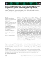
Subcellular distribution of BVDV NS4B in transfected cellsFigure 10
Subcellular distribution of BVDV NS4B in transfected cells. BHK-21 cells were transfected with NS4B-GFP. At 48 h
p.t., the cells were processed for fluorescence microscopy. ER-Tracker (i-iii; a), LysoTracker (x-xii; d) and MitoTracker (xiii-xv;
e) were used as markers for the ER, lysosome and mitochondria, respectively. Golgin-97 (iv-vi; b) and Rab5 (vii-ix; c) were used
as markers for the Golgi apparatus and early endosome, respectively. NS4B was detected via GFP fluorescence. Colocalization
of NS4B (green) with the cognate intracellular marker (red) results in yellow color. Notice the Colocalization of NS4B with
Golgin-97 (b) and MitoTracker (e). Bars = 10 μm.
Virology Journal 2009, 6:185 />Page 12 of 15
(page number not for citation purposes)
with 10 ml of fresh complete medium. Cells were trans-
fected according to the LipoD293 protocol from Signa-
Gen (Ljamsville, MD). Ten micrograms of DNA to were
added to 400 μl of OptiMEM, while 30 μl of LipoD293
were added to 400 μl OptiMEM. The LipoD293 mixture
was then added directly to the diluted DNA and incubated
for 15 min at room temperature. The DNA mixture was
then added to each dish and incubated at 37°C for 24 h
to 48 h.
In vitro transcription, electroporation and generation of
infectious BVDV
To linearize BVDV genome, pNADLp15A was digested
with SacII (New England Bio Labs, Ipswich, MA) at 37°C
for 1 h, followed by incubation at 70°C for 15 min to
inactivate SacII. pNADLp15A 3' overhangs were elimi-
nated following incubation with 5 mM dNTPs and T4
DNA polymerase at 16°C for 30 min. The linearized DNA
was then extracted using Phenol/Chloroform, followed by
ethanol precipitation at -20°C for 2 h., The samples were
resuspended in 10 μl RNase-free water. SacII-linearized
pNADLp15A was used as template for in vitro transcrip-
tion reaction with the T7 RiboMAX™ Kit (Promega, Madi-
son, WI). The RNA was then isolated using the RNeasy
miniprep kit (QIAGEN, Valencia, CA) and its integrity was
assessed on a 0.8% agarose gel.
Before electroporation, MDBK cells were trypsinized and
washed twice with PBS and resuspended to a final concen-
tration of 1 × 10
7
cells/ml in RNase-free PBS. Three micro-
grams of in vitro-transcribed BVDV genomic RNA were
mixed with 0.4 ml (4 × 10
6
) of the cell suspension in a 2
mm-gap electroporation cuvette and pulsed with a Bio-
Rad Gene Pulser (1 pulse; 125 μF; 0.28 kV). One milliliter
of complete DMEM, antibiotic-free, was added to the
cuvette and the resuspended sample was transferred to a
15 ml conical tube. Two milliliters of complete DMEM,
antibiotic-free, was used to wash the cuvette to recover the
remaining sample and was added to the same 15 ml con-
ical tube. The resuspended sample was then divided into
3 wells in a 6-well plate. Complete medium (without anti-
biotic) was added to each well to bring the final volume
to ca. 2 ml. Cells were incubated at 37°C in a 5% CO
2
incubator. At 12 h p.t., the floating, dead cells were
removed. Attached cells were washed twice with PBS and
fresh complete DMEM was added to each well. Cells were
observed at 24 h, 48 h, 72 h, and 96 h for cytopathic
effects.
Plaque Assay
MDBK cells were seeded in 6-well plates at 3 × 10
5
cells per
well. At the time of infection, cells were typically 70-80%
confluent. On the day of infection, medium was removed
and the monolayers were washed twice with PBS. The viral
stock was diluted in serum-free DMEM. Cells were
infected with 0.2 ml of the serially diluted virus (10-fold
dilutions). Following adsorption, the monolayers were
washed with 1 ml of complete DMEM and overlaid with
DMEM-5% horse serum/0.5% agarose plugs. Plates were
incubated for 15-30 min at RT to let the agarose solidify.
Plates were then incubated at 37°C for 72 h. At 72 h post-
infection, cells were fixed with 4% formaldehyde (in PBS)
for 2 h. The agarose plugs were removed and the fixed
monolayers were rinsed once with PBS. The monolayers
were stained with 1% crystal violet (in 50% ethanol) for
10 min. The plates were rinsed with distilled water and
plaques were counted. The viral titer was determined as
follows: number of plaques × 5 × dilution factor. The
resulting titer was expressed in plaque forming units per
ml (pfu/ml).
BVDV Growth Kinetics
MDBK cells were seeded in 60 mm dishes at 4 × 10
5
cells
per dish and grown overnight. The cell monolayers were
then washed twice with PBS and infected at an MOI of 0.1
[5]. After adsorption, the monolayer was washed with
PBS, and 5 ml fresh complete media was added to each
plate. For each time point (0 h, 6 h, 12 h, 18 h, 24 h, 36
h, and 48 h post-infection), the medium was harvested
from the plate and frozen. Fresh serum-free DMEM (5 ml)
was added to the monolayer and the cells were lysed via
two cycles of freeze/thaw. To determine the amount of
infectious virus particles in the medium and lysate at each
time point, plaque assays (as described above) were per-
Colocalization of BVDV NS4B with replicase proteins NS5A and NS5BFigure 11
Colocalization of BVDV NS4B with replicase pro-
teins NS5A and NS5B. BHK-21 cells were co-transfected
with NS4B-GFP and NS5A-His or NS5B-HA. At 48 h p.t., the
cells were processed for IF with either anti-His or anti-HA
antibody (dilution 1/50). NS4B was detected via GFP fluores-
cence. Colocalization of NS4B-GFP (green) and NS5AHis
(red) or NS5B-HA (red) results in yellow color. Bars = 10
μm.
Virology Journal 2009, 6:185 />Page 13 of 15
(page number not for citation purposes)
formed in duplicate. Each plaque assay was repeated three
times.
Quantitative Real-time PCR
To examine the kinetics of viral BVDV synthesis at various
times (0 h, 6 h, 12 h, 18 h, and 24 h) post infection, Real-
Time PCR was performed. First, MDBK cells were infected
with BVDV at MOI of 0.1. Total cellular RNA was collected
at each time point using the RNeasy Mini Kit (Qiagen,
Valencia, CA).
Total cellular RNA was prepared from virus-infected cells
by using the RNeasy Mini Kit (Qiagen) and was treated
with RNase-free DNase (Qiagen, Valencia, CA). First
strand cDNA was synthesized from the DNA-free RNA
using random primers and the High Capacity cDNA
Archive Kit (Applied Biosystems, Foster City, CA). Tripli-
cate samples of cDNA were mixed with a Taqman probe
and a set of forward and reverse primers specific for either
BVDV NS4B or GAPDH and the mixture was subjected to
real-time quantitative PCR using the ABI 7300 Sequence
Detection System (Applied Biosystems, Foster City, CA).
Immunoblot analysis of BVDV Proteins
Infected MDBK cells were lysed using RIPA buffer contain-
ing 150 mM NaCl, 50 mM Tris pH 8, 1 mM EDTA, 1% NP-
40, 0.1% SDS, 1 mM PMSF and protein concentrations
were measured via Bio-Rad Protein Assay (Bio-Rad Labo-
ratories, Hercules, CA). One hundred micrograms of total
protein were resuspended in 4x SDS loading buffer (240
mM Tris pH 6.8, 4% SDS, 40% glycerol, 4% β-mercap-
toethanol, 0.01% bromophenol blue) and boiled for 10
min, and centrifuged at 12000 × g for 10 min. Samples
were separated on a 10% sodium dodecyl sulfate-polyacr-
ylamide gel (SDS-PAGE), and transferred onto Immo-
bilon-P transfer membrane (Millipore, Billerica, MA).
Antibody-bound proteins were detected by chemifluores-
cence (ECF, Amersham/GE Healthcare, Piscataway, NJ)
and visualized on a phosphorimager (Typhoon 8600
Molecular Dynamics, Sunnyvale, CA).
Membrane Floatation Assay
For membrane floatation assay, BHK-21 cells were grown
overnight and transfected with BVDV NS4B-GFP or con-
trol GFP construct according to the conditions described
above. At 48 h p.t., the cells were resuspended in homog-
enization buffer (150 mM NaCl, 50 mM Tris pH 7.4, 2
mM EDTA) containing protease inhibitors (1 mM PMSF
and 1 tablet of Complete Mini; Roche, Nutley, NJ). The
cells were then lysed with 6-8 passages in a ball-bearing
homogenizer to ensure approximately 90% lysis. Cell
lysates were spun at 2500 × g/10 min at 4°C to pellet cel-
lular debris and nuclei. A discontinuous iodixanol gradi-
ent (5%, 25% and 30%) [30] was layered on the top of the
homogenate and the samples were spun at 120,000 × g for
4 h 25 min at 4°C in a Ti80 Rotor. A total of 8 fractions
(867 μl each) were collected from top to bottom. Each
fraction was precipitated with equal volume of 20% TCA,
separated on 10% SDS-PAGE and processed for western
blotting as described above. Typically, membrane-bound
proteins were associated with fractions 1 to 4 whereas sol-
uble proteins were prominent in fractions 5 to 8.
Subcellular fractionation of BVDV NS4B protein
Subcellular fractionation of BVDV NS4B protein was per-
formed as described by Hugle et al. [34]. BHK-21 cells
expressing BVDV NS4B were trypsinized at 48 h p.t. (p.t.)
and resuspended in complete medium on ice. The cells
were then spun at approximately 200 × g/5 min at 4°C,
followed by two washes in PBS. The cells were finally
resuspended in ice cold hypotonic buffer (10 mM Tris-Cl,
pH 7.5, 2 mM MgCl
2
) and lysed by 20 strokes of a dounce
homogenizer to ensure approximately 90-95% lysis. Next,
the lysate was spun at 1000 × g/5 min to pellet the nuclear
fraction. Sixty micrograms of the supernatant were resus-
pended in RIPA buffer and labeled "lysate". The remain-
der of this supernatant was adjusted to 0.25 m sucrose and
spun at 9000 × g/10 min to pellet the mitochondrial frac-
tion. The supernatant from the mitochondrial centrifuga-
tion was then spun at 105,000 × g/40 min to obtain the
microsomal pellet. Sixty micrograms of the remaining
supernatant was saved for immunoblot analysis and
labeled as "cytoplasmic".
Fluorescence Microscopy
MDBK cells were grown on coverslips and infected with
BVDV. The coverslips were washed with PBS and fixed for
10 min in 4% formaldehyde/PBS. Fixed cells were perme-
abilized for 6 min at room temperature (RT) in 0.05% Tri-
ton-X 100/PBS, followed by staining with the primary
polyclonal antibody (or antibodies in double labeling
experiments) and Alexa fluor 594- (or 488)-conjugated
secondary antibody. After three washes in PBS, the cells
were stained with 0.36 mM DAPI in PBS for 10 min at RT,
followed by three more washes in PBS. The coverslips
were mounted on slides using Vectashield (Vector Co.,
Burlingame, CA) and nail polish. The samples were ana-
lyzed by fluorescence microscopy (Zeiss Axiovert 200M)
at × 63 magnification and digital images were taken with
a CCD camera Axiocam MRm. An image stack was decon-
volved using the iterative mode of the Axiovision software
to exclude out-of-focus information. Images were saved as
TIFF files, imported and processed in Adobe Photoshop.
Colocalization of green (FITC) and red (Cy3) signals
results in yellow fluorescence.
For analysis of BVDV NS4B-expressing cells, BHK-21 cells
were grown on coverslips and transfected in 10 cm dishes
as described above. At 48 h p.t., the coverslips were
washed with PBS and the cells stained for 30 min with 100
Virology Journal 2009, 6:185 />Page 14 of 15
(page number not for citation purposes)
nm ER-Tracker, LysoTracker, or 1 μM MitoTracker (Invit-
rogen, Molecular Probes) in complete medium at 37°C in
a 5% CO2 incubator. The cells were then washed in PBS
and fixed for 10 min in 4% formaldehyde/PBS. For immu-
nostaining of BHK-21 cells, fixed cells were permeabilized
for 10 min at room temperature in 0.1% Triton-X 100/
PBS, washed three times in PBS, and stained with the
appropriate antibody for 1 h at room temperature, fol-
lowed by three more washes in PBS. The cells were then
immunostained with AlexaFluor 594-conjugated second-
ary antibody for 1 h followed by washing three times with
PBS. The cells were mounted on glass slides and processed
for fluorescence microscopy as described above.
Electron microscopy
MDBK cells were seeded at 6.8 × 10
5
cells per 100 mm
dish. Cells were infected at MOI of 10 and collected at var-
ious times (0 h, 12 h, 18 h, 24 h, 48 h, and 72 h) post-
infection. Briefly, at various times post-infection, the cells
were resuspended in 2% glutaraldehyde/0.1 m sodium
cacodylate buffer and incubated on ice for 30 min. After a
brief spin, fresh 2% glutaraldehde/0.1 m sodium
cacodylate was added to the pellet and the pellet was incu-
bated overnight at 4°C. The cell pellet was rinsed with 0.1
M sodium cacodylate prior to postfixation with 1%
osmium tetroxide/0.1 M cacodylate for 1-2 h at 4°C. After
rinsing and en bloc staining in aqueous uranyl acetate,
samples were dehydrated with graded ethanol concentra-
tions, infiltrated with eponate resin and embedded over-
night in eponate at 65°C. Ultrathin sections were cut on
Leica Ultracut UCT microtome (Wetzlar, Germany), col-
lected on copper grids and stained with 1% uranyl acetate-
1% lead citrate. The grids were double stained with uranyl
acetate and lead citrate and the sections were examined
with a JEOL 1200 EXII transmission electron microscope
(Peabody, MA) at 80 kV.
For Immuno-EM analysis of infected cells, MDBK cells
were plated in 8-chamber slides at 5.4 × 10
4
cells per
chamber. Cells were harvested at 18 h.p.i. and fixed to the
bottom of the chamber with 4% paraformaldehyde/0.1%
glutaraldehyde for 10 min. Cells were permeabilized with
0.05% Triton-X for 6 min at RT. After permeabilization,
cells were washed three times with PBS. Permeabilized
cells were then blocked with 3% BSA in PBS for 30 min at
RT. Immediately following blocking, anti-NS4B antibody,
diluted 1:50 in 3% BSA in PBS, was applied to the fixed
cells for 1 h at RT. The cells were washed three times in
PBS (15 min each). The secondary anti-Rabbit 605-Quan-
tum dots (Molecular Probes, Invitrogen, Carlsbad CA),
diluted 1:125 in 3% BSA in PBS, were incubated with the
cells for 2 h at 4°C, swirling gently. After incubation, cells
were washed three times in PBS, 15 min each. Finally,
nuclei were stained using 0.36 mM DAPI in PBS for 10
min at RT. Quantum dot labeling was observed via fluo-
rescence microscopy. Labeled cells were fixed with 2.5%
glutaraldehyde prior to sectioning and electron micros-
copy (see above).
Competing interests
The authors declare that they have no competing interests.
Authors' contributions
EW performed all the experiments except for the immuno-
EM, membrane floatation, subcellular distribution of
NS4B protein; she helped in writing the manuscript. JA
performed the membrane floatation, subcellular distribu-
tion of NS4B protein and subcellular fractionation of
NS4B protein; he helped in editing the manuscript. GN
performed the immuno-EM in collaboration with EW.
KVK supervised the project and wrote the manuscript. All
authors read and approved the final manuscript.
Acknowledgements
We are grateful to Charles Rice, and Ruben Donis for reagents, David
Manna for suggestions and critical reading of the manuscript. This work was
supported by K22 CA129241 from the National Institute of Health.
References
1. Brock KV: The persistence of bovine viral diarrhea virus. Bio-
logicals 2003, 31:133-135.
2. Ridpath JF, Bolin SR, Dubovi EJ: Segregation of bovine viral
diarrhea virus into genotypes. Virology 1994, 205:66-74.
3. Peterhans E, Jungi TW, Schweizer M: BVDV and innate immunity.
Biologicals 2003, 31:107-112.
4. Agapov EV, Murray CL, Frolov I, Qu L, Myers TM, Rice CM:
Uncleaved NS2 3 is required for production of infectious
bovine viral diarrhea virus. J Virol 2004, 78:2414-2425.
5. Mendez E, Ruggli N, Collett MS, Rice CM: Infectious bovine viral
diarrhea virus (strain NADL) RNA from stable cDNA clones:
a cellular insert determines NS3 production and viral
cytopathogenicity. J Virol 1998, 72:4737-4745.
6. Goens SD: The evolution of bovine viral diarrhea: a review.
Can Vet J 2002, 43:946-954.
7. Colett MS, Larson R, Gold C, Strick D, Anderson DK, Purchio AF:
Molecular cloning and nucleotide sequence of the pestivirus
bovine viral diarrhea virus. Virology 1988, 165:191-199.
8. Lindenbach BD, Thiel H-J, Rice CM: Flaviviridae: the viruses and
their replication. In Fields Virology Volume 1. 4th edition. Edited by:
DM Knipe PH, Griffin DE, Lamb RA, Martin MA, Roizman B. Philadel-
phia: Lohmann Williams & Wilkins; 2001:991-1041.
9. Buckwold VE, Beer BE, Donis RO: Bovine viral diarrhea virus as
a surrogate model of hepatitis C virus for the evaluation of
antiviral agents. Antiviral Res 2003, 60:1-15.
10. Yu H, Grassmann CW, Behrens SE: Sequence and structural ele-
ments at the 3' terminus of bovine viral diarrhea virus
genomic RNA: functional role during RNA replication. J Virol
1999, 73:3638-3648.
11. Bartenschlager R, Lohmann V: Replication of the hepatitis C
virus. Baillieres Best Pract Res Clin Gastroenterol 2000, 14:241-254.
12. Bartenschlager R, Lohmann V: Novel cell culture systems for the
hepatitis C virus.
Antiviral Res 2001, 52:1-17.
13. Egger D, Wolk B, Gosert R, Bianchi L, Blum HE, Moradpour D, Bienz
K: Expression of hepatitis C virus proteins induces distinct
membrane alterations including a candidate viral replication
complex. J Virol 2002, 76:5974-5984.
14. Kim M, Mackenzie JM, Westaway EG: Comparisons of physical
separation methods of Kunjin virus induced membranes. J
Virol Methods 2004, 120:179-187.
15. Mackenzie JM, Jones MK, Young PR: Immunolocalization of the
dengue virus nonstructural glycoprotein NS1 suggests a role
in viral RNA replication. Virology 1996, 220:232-240.
Publish with Bio Med Central and every
scientist can read your work free of charge
"BioMed Central will be the most significant development for
disseminating the results of biomedical research in our lifetime."
Sir Paul Nurse, Cancer Research UK
Your research papers will be:
available free of charge to the entire biomedical community
peer reviewed and published immediately upon acceptance
cited in PubMed and archived on PubMed Central
yours — you keep the copyright
Submit your manuscript here:
/>BioMedcentral
Virology Journal 2009, 6:185 />Page 15 of 15
(page number not for citation purposes)
16. Konan KV, Giddings TH Jr, Ikeda M, Li K, Lemon SM, Kirkegaard K:
Nonstructural protein precursor NS4A/B from hepatitis C
virus alters function and ultrastructure of host secretory
apparatus. J Virol 2003, 77:7843-7855.
17. Pfeifer U, Thomssen R, Legler K, Bottcher U, Gerlich W, Weinmann
E, Klinge O: Experimental non A, non B hepatitis: four types
of cytoplasmic alteration in hepatocytes of infected chim-
panzees. Virchows Arch B Cell Pathol Incl Mol Pathol 1980, 33:233-243.
18. Mackenzie JM, Jones MK, Westaway EG: Markers for trans Golgi
membranes and the intermediate compartment localize to
induced membranes with distinct replication functions in fla-
vivirus infected cells. J Virol 1999, 73:9555-9567.
19. Schmidt-Mende J, Bieck E, Hugle T, Penin F, Rice CM, Blum HE,
Moradpour D: Determinants for membrane association of the
hepatitis C virus RNA dependent RNA polymerase. J Biol
Chem 2001, 276:44052-44063.
20. Bielefeldt Ohmann H, Bloch B: Electron Microscopic Studies of
Bovine Viral Diarrhea Virus in Tissues of Diseased Calves
and in Cell Cultures. Archives of Viology 1982, 71:57-74.
21. Gray EW, Nettleton PF: The Ultrastructure of Cell Cultuers
Infected with Border Disease and Bovine Virus Diarrhoea
Viruses. J Gen Virol 1987, 68:2339-2346.
22. van Olphen AL, Donis RO: Identification of bovine viral diarrhea
virus nonstructural polypeptide NS4B/P38. Virus Res 1997,
51:197-201.
23. Qu L, McMullan LK, Rice CM: Isolation and characterization of
noncytopathic pestivirus mutants reveals a role for non-
structural protein NS4B in viral cytopathogenicity. J Virol
2001, 75:10651-10662.
24. Piccininni S, Varaklioti A, Nardelli M, Dave B, Raney KD, McCarthy JE:
Modulation of the hepatitis C virus RNA dependent RNA
polymerase activity by the non structural (NS) 3 helicase and
the NS4B membrane protein. J Biol Chem 2002,
277:45670-45679.
25. Miller S, Sparacio S, Bartenschlager R: Subcellular localization and
membrane topology of the Dengue virus type 2 Non struc-
tural protein 4B. J Biol Chem 2006, 281:
8854-8863.
26. Hugle T, Fehrmann F, Bieck E, Kohara M, Krausslich HG, Rice CM,
Blum HE, Moradpour D: The hepatitis C virus nonstructural
protein 4B is an integral endoplasmic reticulum membrane
protein. Virology 2001, 284:70-81.
27. Qu L, McMullan LK, Rice CM: Isolation and characterization of
noncytopathic pestivirus mutants reveals a role for non-
structural protein NS4B in viral cytopathogenicity. J Virol
2001, 75:10651-10662.
28. Mendez E, Ruggli N, Collett MS, Rice CM: Infectious bovine viral
diarrhea virus (strain NADL) RNA from stable cDNA clones:
a cellular insert determines NS3 production and viral
cytopathogenicity. J Virol 1998, 72:4737-4745.
29. Yu H, Isken O, Grassmann CW, Behrens SE: A stem loop motif
formed by the immediate 5' terminus of the bovine viral
diarrhea virus genome modulates translation as well as rep-
lication of the viral RNA. J Virol 2000, 74:5825-5835.
30. Aligo J, Jia S, Manna D, Konan KV: Formation and function of hep-
atitis C virus replication complexes require residues in the
carboxy terminal domain of NS4B protein. Virology 2009,
393:68-83.
31. Behrens SE, Grassmann CW, Thiel HJ, Meyers G, Tautz N: Charac-
terization of an autonomous subgenomic pestivirus RNA
replicon. J Virol 1998, 72:2364-2372.
32. Lundin M, Monne M, Widell A, Von Heijne G, Persson MA: Topol-
ogy of the membrane associated hepatitis C virus protein
NS4B. J Virol 2003, 77:5428-5438.
33. Dimitrova M, Imbert I, Kieny MP, Schuster C: Protein protein
interactions between hepatitis C virus nonstructural pro-
teins. J Virol 2003, 77:5401-5414.
34. Hugle T, Fehrmann F, Bieck E, Kohara M, Krausslich HG, Rice CM,
Blum HE, Moradpour D: The hepatitis C virus nonstructural
protein 4B is an integral endoplasmic reticulum membrane
protein. Virology 2001, 284:70-81.
35. Stone M, Jia S, Heo WD, Meyer T, Konan KV: Participation of
rab5, an early endosome protein, in hepatitis C virus RNA
replication machinery. J Virol 2007, 81:
4551-4563.
36. Miller DJ, Schwartz MD, Dye BT, Ahlquist P: Engineered retarget-
ing of viral RNA replication complexes to an alternative
intracellular membrane. J Virol 2003, 77:12193-12202.
37. Bendfeldt S, Grummer B, Greiser-Wilke I: No caspase activation
but overexpression of Bcl 2 in bovine cells infected with non-
cytopathic bovine virus diarrhoea virus. Vet Microbiol 2003,
96:313-326.
38. Miller S, Kastner S, Krijnse-Locker J, Buhler S, Bartenschlager R: The
non structural protein 4A of dengue virus is an integral
membrane protein inducing membrane alterations in a 2K
regulated manner. J Biol Chem 2007, 282:8873-8882.
39. Roosendaal J, Westaway EG, Khromykh A, Mackenzie JM: Regulated
cleavages at the West Nile virus NS4A 2K NS4B junctions
play a major role in rearranging cytoplasmic membranes
and Golgi trafficking of the NS4A protein. J Virol 2006,
80:4623-4632.
40. Vassilev VB, Collett MS, Donis RO: Authentic and chimeric full
length genomic cDNA clones of bovine viral diarrhea virus
that yield infectious transcripts. J Virol 1997, 71:471-478.