Báo cáo khoa học: " Retinoic acid inducible gene I Activates innate antiviral response against human parainfluenza virus type 3" pdf
Bạn đang xem bản rút gọn của tài liệu. Xem và tải ngay bản đầy đủ của tài liệu tại đây (986.07 KB, 7 trang )
BioMed Central
Page 1 of 7
(page number not for citation purposes)
Virology Journal
Open Access
Short report
Retinoic acid inducible gene I Activates innate antiviral response
against human parainfluenza virus type 3
Ahmed Sabbah and Santanu Bose*
Address: Department of Microbiology and Immunology, The University of Texas Health Science Center at San Antonio, San Antonio, Texas, USA
Email: Ahmed Sabbah - ; Santanu Bose* -
* Corresponding author
Abstract
Human parainfluenza virus type 3 (HPIV3) is a respiratory paramyxovirus that infects lung epithelial
cells to cause high morbidity among infants and children. To date, no effective vaccine or antiviral
therapy exists for HPIV3 and therefore, it is important to study innate immune antiviral response
induced by this virus in infected cells. Type-I interferons (IFN, interferon-α/β) and tumor necrosis
factor-α (TNFα activated by NFκB) are potent antiviral cytokines that play an important role
during innate immune antiviral response. A wide-spectrum of viruses utilizes pattern recognition
receptors (PRRs) like toll-like receptors (TLRs) and RLH (RIG like helicases) receptors such as
RIGI (retinoic acid inducible gene -I) and Mda5 to induce innate antiviral response. Previously it was
shown that both TNFα and IFNβ are produced from HPIV3 infected cells. However, the
mechanism by which infected cells activated innate response following HPIV3 infection was not
known. In the current study, we demonstrated that RIGI serves as a PRR in HPIV3 infected cells to
induce innate antiviral response by expressing IFNβ (via activation of interferon regulatory factor-
3 or IRF3) and TNFα (via activation of NF-κB).
Findings
Human parainfluenza virus type 3 (HPIV3) is an envel-
oped non-segmented negative-sense single stranded
(NNS) RNA virus that belongs to the paramyxovirus fam-
ily [1]. HPIV3 is a lung tropic virus known to cause severe
respiratory diseases (croup, bronchiolitis, pneumonia) in
infants, children, elderly and immuno-comprimised indi-
viduals [1,2]. Although HPIV3 causes life-threatening res-
piratory tract diseases, currently no effective vaccine or
antiviral therapy exists. Therefore, elucidation of innate
immune antiviral response induced by HPIV3 holds sig-
nificant potential for development of effective antiviral
therapies in the near future. The innate immune antiviral
response against viruses represents an important host
defense mechanism [3]. Innate immunity comprises the
first line of defense by the host to combat virus infection
before an orchestrated adaptive immune response is
launched. Two key molecules regulating the innate antivi-
ral function are interferon regulatory factors (IRFs) and
NFκB [3]. These two transcription factors are activated
either individually or together in infected cells, resulting
in the expression and production of potent antiviral
cytokines IFN-α/β (IFN) (type I interferon) [4,5]. IFNα/β
produced from infected cells binds to their cognate IFN
receptors on uninfected cells to induce the JAK/STAT anti-
viral pathway. Thus the paracrine action of IFN is abso-
lutely critical during innate antiviral defense [4-8].
Although the paracrine action of IFNs plays a critical role
in innate immune antiviral response, we have also identi-
fied an IFN independent antiviral pathway against HPIV3
Published: 17 November 2009
Virology Journal 2009, 6:200 doi:10.1186/1743-422X-6-200
Received: 15 September 2009
Accepted: 17 November 2009
This article is available from: />© 2009 Sabbah and Bose; licensee BioMed Central Ltd.
This is an Open Access article distributed under the terms of the Creative Commons Attribution License ( />),
which permits unrestricted use, distribution, and reproduction in any medium, provided the original work is properly cited.
Virology Journal 2009, 6:200 />Page 2 of 7
(page number not for citation purposes)
and human respiratory syncytial virus (RSV) that was
dependent on NF-κB activation and production of pro-
inflammatory cytokines like tumor necrosis factor-α
(TNFα) [9,10].
Infected cells utilize pattern recognition receptors (PRRs)
to recognize pathogen (virus) associated molecular pat-
terns (PAMPs) to trigger activation of the transcription
factors IRF3 and NF-κB, which then translocate to the
nucleus to transactivate antiviral genes like IFNα/β, TNFα
etc [11]. So far two classes of viral PRRs have been identi-
fied - toll-like receptors (TLRs) [12] and RLH (RIG like
helicases) receptors such as RIGI (retinoic acid inducible
gene - I) and Mda5 [13]. Recently, we also demonstrated
that NOD-like receptors such as NOD2 could act as a PRR
for RSV and influenza A virus [14]. TLRs are type I integral
transmembrane proteins that are utilized by various
viruses to activate NFκB and IRF3 in infected cells. Major-
ity of TLRs require MyD88 as an adaptor protein to induce
TLR-dependent signaling [12]. In contrast to membrane
bound TLR proteins, RLH receptors are cytoplasmic PRRs.
Both RIGI and Mda5 signals antiviral response via induc-
tion of IRF3 and NFκB pathways after binding to the sin-
gle stranded RNA genome of paramyxo, orthomyxo and
picarnoviruses [13]. Although TLRs and RLH receptors
were shown to induce innate antiviral response following
infection with various RNA and DNA viruses, the mecha-
nism by which HPIV3 activates the innate response is not
known. Moreover, there are no reports of any PRR that
play an important role in activation of IRF3/NF-κB during
HPIV3 infection.
The innate immune response to HPIV3 is not well under-
stood. HPIV3 has been shown to produce IFN-I in vivo and
in vitro [15-17] yet no reports of IRF3 activation by HPIV3
exist. In addition, previous studies have demonstrated
that HPIV3 activates NFκB to produce TNFα for establish-
ment of antiviral state [9]. Moreover, TLRs may be
involved in NFκB activation (at 24 h post-infection) since
blocking MyD88 function diminished NF-κB activation in
HPIV3 infected cells by 50%-55%. This suggested that
both TLRs and non-TLR molecules may be involved in
NFκB activation in HPIV3 infected cells. A similar sce-
nario has been reported for another paramyxovirus,
human respiratory syncitial virus (RSV). It was shown that
both TLR3 and RIGI are involved in NF-κB activation fol-
lowing RSV infection [18]. Therefore, we investigated
whether similar to RSV; HPIV3 can also utilize RIGI to
activate innate antiviral response. In the current studies
we have demonstrated that in A549 (A549 cells are
human respiratory epithelial cells that have been rou-
tinely used as a model of type II alveolar epithelial cells)
cells RIGI plays an important role in activation of innate
antiviral response during HPIV3 infection. Moreover,
RIGI was involved in activating both arms (NFκB/TNFα
and IRF3/IFNα/β) of innate immunity following HPIV3
infection.
In order to study the involvement of RIGI in antiviral
response stimulation via activation of IRF3/NFκB, we
expressed RIGI (FLAG tagged) in 293 cells (these cells
does not express majority of endogenous PRRs) and ana-
lyzed IFN/NFκB activation following HPIV3 infection. For
these experiments, 293 cells were transfected (by using
lipofectamine 2000 from Invitrogen) with FLAG-RIGI,
pcDNA, NF-κB-luciferase, and IRF3-luciferase plasmids.
At 24 h post-trasfection, cells were infected with HPIV-3
(0.5 MOI) and luciferase assay was performed at 8 h post-
infection as described previously [9,14]. The efficiency of
RIGI expression was confirmed by Western blotting (with
FLAG antibody) of lysate obtained from RIGI-FLAG trans-
fected 293 cells (Fig. 1A). As shown, in Fig. 1B, RIGI
expression resulted in drastic activation (measured by
luciferase assay) of both NF-κB and IRF3 in HPIV3
infected cells. The role of RIGI in activation of these tran-
scription factors were further confirmed by detecting
expression [reverse transcription or RT-PCR was per-
formed using RedMIX Plus (Gene Choice) with the fol-
lowing primers: GAPDH forward, 5'-
GTCAGTGGTGGACCTGACCT, GAPDH reverse, 5'-
AGGGGTCTACATGGCAACTG; ISG15 forward, 5'-CCGT-
GAAGATGCTGGCG, ISG15 reverse, 5'-CGAAGGT-
CAGCCAGAAC; IFN-
β
forward, 5'-
GATTCATCTAGCACTGGCTGG, IFN-
β
reverse,
5'CTTCAGGTAATGCAGAATCC; TNF-
α
forward, GAGT-
GACAAGCCTGTAGCCCATGTTGTAGCA, TNF-
α
reverse,
GCAATGATCCCAAAGTAGACCTGCCCAGACT] of their
target genes, TNF, IFN-β and ISG-15 (interferon stimu-
lated gene-15) in infected 293 cells expressing RIGI (Fig.
1C, 1D). These results demonstrated that RIGI is capable
of activating an innate antiviral response in HPIV3
infected cells.
RIGI protein consists of helicase and two CARD (caspase
recruitment domain) domains (Fig. 2A) domains. Previ-
ous studies have shown that CARD domains are required
for RIGI mediated signal transduction [19]; which consti-
tutes interaction of RIGI with the mitochondrial localized
adaptor protein MAVS (IPS-1) and activation of IRF3 and
NF-κB [18]. In order to investigate the role of caspase
domains in antiviral signaling, we co-expressed FLAG
tagged wild type (WT) and CARD deleted RIGI (ΔRIGI) in
293 cells, followed by HPIV3 infection. At 8 h post-infec-
tion, TNF and IFN-β induction was measured by RT-PCR.
Our result revealed that CARD domains are critical for
antiviral signaling, since co-expression of WT and ΔRIGI
resulted in loss of IFN-β and TNF induction in infected
cells (Fig. 2B). These results demonstrated that CARD
domains are important for RIGI signaling during HPIV3
infection. Furthermore, RIGI lacking the CARD domains
Virology Journal 2009, 6:200 />Page 3 of 7
(page number not for citation purposes)
(i.e. ΔRIGI) can act as a dominant negative molecule to
suppress the activity of functional RIGI during HPIV3
infection.
Since HPIV3 is a respiratory virus, we next evaluated the
role of RIGI in inducing IRF3/NF-κB in infected human
lung epithelial A549 cells. Infection of A549 cells with
HPIV3 resulted in induction of endogenous RIGI expres-
sion (RT-PCR was performed using the following primers:
RIGI forward, 5'-GCATATTGACTGGACGTGGCA, RIGI
reverse, 5'-CAGTCATGGCTGCAGTTCTGTC) during rela-
tively early infection time frame (8 h-12 h post-infection)
(Fig. 3A). Similarly, IRF3 and NF-κB (as assessed by luci-
ferase assay of infected A549 cells transfected with IRF3
and NF-κB luciferase) was induced by HPIV3 during early
infection (within 12 h post-infection) (Fig. 3B). Based on
similar induction/activation kinetics of RIGI and IRF3/
NF-κB, we speculated that endogenous RIGI may play a
role during IRF3/NF-κB activation. To examine such role
of RIGI, we initially utilized A549 cells expressing (follow-
ing transfection) ΔRIGI-FLAG, since ΔRIGI acted as a
dominant-negative molecule (Fig. 2). Efficient expression
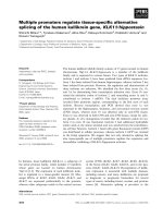
Activation of antiviral response by RIGI during HPIV3 infectionFigure 1
Activation of antiviral response by RIGI during HPIV3 infection. (a) Western blot analysis (with anti-FLAG antibody)
of RIGI expression in 293 cells transfected with pcDNA and RIGI (FLAG tagged) plasmids. (b) Activation of IRF3-luciferase and
NF-κB-luciferase in mock and HPIV3 infected (at 8 h post-infection) 293 cells transfected with either pcDNA or RIGI. The luci-
ferase assay results are presented as mean ± standard deviation from three independent experiments. (c) RT-PCR analysis of
TNF and IFN-β expression (at 4 h and 8 h post-infection) in mock and HPIV3 infected 293 cells transfected with either pcDNA
or RIGI. GAPDH served as a loading control. NS; non-specific. (d) RT-PCR analysis of ISG-15 expression (at 2 h, 4 h and 8 h
post-infection) in mock and HPIV3 infected 293 cells transfected with either pcDNA or RIGI
Virology Journal 2009, 6:200 />Page 4 of 7
(page number not for citation purposes)
of ΔRIGI (denoted as dominant negative RIGI or DN-
RIGI) is evident from the Western blotting of A549 cell
lysate with FLAG antibody (to detect ΔRIGI-FLAG) (Fig.
3C). In order to investigate the involvement of RIGI, A549
cells were transfected with ΔRIGI-FLAG, NF-κB- or IRF3-
luciferase. At 24 h post-transfection, cells were infected
with HPIV3 and luciferase activity was measured at 12 h
post-infection. Expression of ΔRIGI in A549 resulted in
drastic decline in both NF-κB and IRF3 activity following
HPIV3 infection (Fig. 3D). In addition, infected cells
expressing ΔRIGI lost expression of IRF3/NF-κB target
genes TNF and ISG-15 (Fig. 3E). These results demon-
strated that endogenously expressed RIGI plays a crucial
role in activation of innate antiviral response following
HPIV3 infection of human lung epithelial cells.
The role of endogenous RIGI was further confirmed by
silencing RIGI expression in A549 cells. The silencing was
performed by transfecting A549 cells with control or RIGI
specific siRNA [negative control siRNA and DDX58-1
(RIG-I) siRNA were ordered from Qiagen and cells were
transfected with 80 nM of siRNA using lipofectamine
2000]. The efficiency of silencing was validated in A549
cells, since HPIV3 failed to induce RIGI expression in cells
expressing RIGI-specific siRNA (Fig. 4A). We next utilized
the RIGI silenced A549 cells to examine the role of RIGI.
Analysis of TNF gene expression by RT-PCR revealed fail-
ure of HPIV3 to optimally induce TNF in RIGI silenced
cells, compared to cells transfected with control siRNA
(Fig. 4B). Similarly, expression of IFN-β following HPIV-3
infection was drastically reduced in the absence of RIGI
(RIGI silenced cells) protein (Fig. 4C). These results dem-
onstrated an involvement of endogenous RIGI in induc-
ing the innate antiviral pathway in HPIV3 infected human
lung cells.
Our results have demonstrated a role of RIGI as a HPIV3
specific PRR involved in inducing innate antiviral
response following activation of IRF3/IFN and NF-κB/
TNF. However, additional non-RIGI PRRs may also be
involved in inducing innate response following HPIV3
infection. In that context, well-established PRRs like
Mda5, TLR3 and TLR7 that are known to recognize viruses
and single-stranded RNA (ssRNA) are not involved during
CARD domains of RIGI are important for activation of antiviral responseFigure 2
CARD domains of RIGI are important for activation of antiviral response. (a) A schematic showing the RIGI con-
structs; WT RIGI (wild type RIGI)), ΔRIGI (RIGI lacking the two CARD domains). (b) RT-PCR analysis of TNF and IFN-β
expression (at 8 h post-infection) in mock and HPIV3 infected 293 cells transfected with the indicated plasmids. GAPDH
served as a loading control.
Virology Journal 2009, 6:200 />Page 5 of 7
(page number not for citation purposes)
innate response by HPIV3 infected lung epithelial cells.
Mda5 has previously been shown to specifically recognize
positive-sense single-stranded RNA viruses like picornavi-
ruses and alphaviruses [13]. Moreover, expression of TLR3
in 293 cells did not result in induction of IRF3 and NF-κB
activity following HPIV3 infection (data not shown). Our
observation is similar to previous studies showing the
non-involvement of TLR3 during induction of innate
response following infection with Sendai virus (a mouse
parainfluenza virus) [20]. TLR7 (which is capable of rec-
ognizing ssRNA) also do not function as HPIV3 specific
PRR in lung cells since, a) A549 cells (the lung epithelial
cells utilized in our current studies) lack expression of
TLR7 [21-23], b) TLR7 activating compound (e.g. R848)
failed to activate TLR7 in A549 cells [22,23], and c) TLR7
is not required for IRF3 and NF-κB activation in Sendai
Inactivation of endogenous RIGI in A549 cells by dominant-negative RIGI (ΔRIGI) abrogates innate antiviral responseFigure 3
Inactivation of endogenous RIGI in A549 cells by dominant-negative RIGI (ΔRIGI) abrogates innate antiviral
response. (a) RT-PCR analysis of endogenous RIGI expression in HPIV3 infected (at 4 h-12 post-infection) A549 cells. (b)
Activation of IRF3-luciferase and NF-κB-luciferase in mock and HPIV3 infected (at 12 h post-infection) A549 cells. The luci-
ferase assay results are presented as mean ± standard deviation from three independent experiments. (c) Expression of FLAG
tagged dominant-negative RIGI (DN-RIGI) in A549 cells was assessed by Western blotting A549 cell (transfected with either
pcDNA or FLAG-DN-RIGI) lysate with anti-FLAG antibody. (d) Activation of IRF3-luciferase and NFκ-B-luciferase in mock
and HPIV3 infected (at 12 h post-infection) A549 cells trasfected with either pcDNA or DN-RIGI. The luciferase assay results
are presented as mean ± standard deviation from three independent experiments. (e) RT-PCR analysis of TNF and ISG-15
expression (at 12 h and 16 h post-infection) in mock and HPIV3 infected A549 cells transfected with either pcDNA or DN-
RIGI.
Virology Journal 2009, 6:200 />Page 6 of 7
(page number not for citation purposes)
virus (a mouse parainfluenza virus) infected A549 cells
[21]. Our future studies will be directed in identifying and
characterizing additional PRRs that may play a role in acti-
vating innate response following HPIV3 infection.
Abbreviations
HPIV3: human parainfluenza virus type 3; RIGI: retinoic
acid inducible gene-I; TNF: tumor necrosis factor-α; IFN:
interferon; ISG15: interferon stimulated gene-15; IRF3:
interferon regulatory factor 3.
Competing interests
The authors declare that they have no competing interests.
Authors' contributions
AS and SB designed the experiments and prepared the
manuscript. AS performed the experiments. All authors
read and approved the final manuscript.
Acknowledgements
This work was supported by National Institutes of Health grants AI069062
(to SB), CA129246 (to SB), American Lung Association National Biomedi-
cal Research Grant-RG-49629-N (to SB). AS is supported by National Insti-
tutes of Health training grant T32-DE14318 (COSTAR program). We
would like to thanks Dr. Kui Li (University of Tennessee Health Science
Center) and Dr. Adolfo Garcia-Sastre (Mt. Sinai School of Medicine, New
York, NY) for providing several reagents used in the study.
Silencing of endogenous RIGI in A549 cells results in loss of induction of antiviral responseFigure 4
Silencing of endogenous RIGI in A549 cells results in loss of induction of antiviral response. (a) RT-PCR analysis of
RIGI expression in mock and HPIV3 infected (at 12 h post-infection) A549 cells transfected with either control siRNA or RIGI
specific siRNA. (b) RT-PCR analysis of TNF expression in mock and HPIV3 infected (at 12 h post-infection) A549 cells trans-
fected with either control siRNA or RIGI specific siRNA. (c) RT-PCR analysis of IFN-β expression in mock and HPIV3 infected
(at 12 h post-infection) A549 cells transfected with either control siRNA (Con siRNA) or RIGI specific siRNA (RIGI siRNA).
Publish with BioMed Central and every
scientist can read your work free of charge
"BioMed Central will be the most significant development for
disseminating the results of biomedical research in our lifetime."
Sir Paul Nurse, Cancer Research UK
Your research papers will be:
available free of charge to the entire biomedical community
peer reviewed and published immediately upon acceptance
cited in PubMed and archived on PubMed Central
yours — you keep the copyright
Submit your manuscript here:
/>BioMedcentral
Virology Journal 2009, 6:200 />Page 7 of 7
(page number not for citation purposes)
References
1. Chanock RM, Murphy BR, Collins PL: Parainfluenza viruses. In
Fields Virology 4th edition. Edited by: Knipe DM, Howley PM. Lippin-
cott Williams & Wilkins: Philadelphia, PA; 2001:1341-1380.
2. Hall CB: Respiratory syncytial virus and parainfluenza virus. N
Engl J Med 2001, 344:1917-1928.
3. Bose S, Banerjee AK: Innate immune response against nonseg-
mented negative strand RNA viruses. J Interferon Cytokine Res
2003, 23:401-412.
4. Stark GR, Kerr IM, Williams BRG, Silverman RH, Schreiber RD: How
cells respond to interferons. Annu Rev Biochem 1998, 67:227-264.
5. Biron CA, Sen GC: Interferons and other cytokines. In Fields
Virology 4th edition. Edited by: Knipe DM, Howley PM. Lippincott Wil-
liams & Wilkins: Philadelphia, PA; 2001:321-351.
6. Dong B, Zhou Q, Zhao J, Zhou A, Harty RN, Bose S, Banerjee A, Slee
R, Guenther J, Williams BR, Wiedmer T, Sims PJ, Silverman RH:
Phospholipid scramblase 1 potentiates the antiviral activity
of interferon. J Virol 2004, 78:8983-8993.
7. Basu M, Maitra RK, Xiang Y, Meng X, Banerjee AK, Bose S: Inhibition
of vesicular stomatitis virus infection in epithelial cells by
alpha interferon-induced soluble secreted proteins. J Gen Virol
2006, 87:2653-2662.
8. Zheng S, Xu W, Bose S, Banerjee AK, Haque J, Erzurum S: Impaired
nitric oxide synthase (NOS) 2 signaling pathway in cystic
fibrosis airway epithelium. Am J Physiol 2004, 287:L374-381.
9. Bose S, Kar N, Maitra R, DiDonato JA, Banerjee AK: Temporal acti-
vation of NF-kB regulates an interferon-independent innate
antiviral response against cytoplasmic RNA viruses. Proc Natl
Acad Sci USA 2003, 100:10890-10895.
10. Kota S, Sabbah A, Chang TH, Harnack R, Xiang Y, Meng Y, Bose S:
Role of human beta-defensin-2 during tumor necrosis factor-
α/NF-κB mediated innate anti-viral response against human
respiratory syncytial virus. J Biol Chem 2008, 283:22417-22429.
11. Uematsu S, Akira S: Toll-like receptors and Type I interferons.
J Biol Chem
2007, 282:15319-15323.
12. O'Neill LA: How Toll-like receptors signal: what we know and
what we don't know. Curr Opin Immunol 2006, 18:3-9.
13. Basler CF, Garcia-Sastre A: Sensing RNA virus infections. Nat
Chem Biol 2007, 3:20-21.
14. Sabbah A, Chang T, Harnack R, Frohlich V, Dube PH, Tominaga K,
Xiang Y, Bose S: Activation of innate immune antiviral
response by NOD2. Nat Immunol 2009, 10:1073-1080.
15. Gao J, De BP, Banerjee AK: Interferon type I downregulates
human parainfluenza virus type 3-induced major histocom-
patibility complex class II expression. Viral Immunol 2002,
15:85-93.
16. Krilov LR, Hendry RM, Godfrey E, McIntosh K: Respiratory virus
infection of peripheral blood monocytes: correlation with
ageing of cells and interferon production in vitro. J Gen Virol
1987, 68:1749-53.
17. Harmon AT, Harmon MW, Glezen WP: Evidence of interferon
production in the hamster lung after primary or secondary
exposure to parainfluenza virus type 3. Am Rev Respir Dis 1982,
125:706-11.
18. Liu P, Jamaluddin M, Li K, Garofalo RP, Casola A, Brasier AR: Retin-
oic acid-inducible gene I mediates early antiviral response
and Toll-like receptor 3 expression in respiratory syncytial
virus-infected airway epithelial cells. J Virol 2007, 81:1401-1411.
19. Yoneyama M, Kikuchi M, Natsukawa T, Shinobu N, Imaizumi T, Miy-
agishi M, Taira K, Akira S, Fujita T: The RNA helicase RIG-I has
an essential function in double-stranded RNA-induced
innate antiviral responses. Nat Immunol 2004, 5:730-7.
20. Elco CP, Guenther JM, Williams BR, Sen GC: Analysis of genes
induced by Sendai virus infection of mutant cell lines reveals
essential roles of interferon regulatory factor 3, NF-kappaB,
and interferon but not toll-like receptor 3. J Virol 2005,
79:3920-9.
21. Melchjorsen J, Jensen SB, Malmgaard L, Rasmussen SB, Weber F,
Bowie AG, Matikainen S, Paludan SR: Activation of innate defense
against a paramyxovirus is mediated by RIG-I and TLR7 and
TLR8 in a cell-type-specific manner.
J Virol 2005, 79:12944-51.
22. Matikainen S, Sirén J, Tissari J, Veckman V, Pirhonen J, Severa M, Sun
Q, Lin R, Meri S, Uzé G, Hiscott J, Julkunen I: Tumor necrosis fac-
tor alpha enhances influenza A virus-induced expression of
antiviral cytokines by activating RIG-I gene expression. J Virol
2006, 80:3515-22.
23. Tissari J, Sirén J, Meri S, Julkunen I, Matikainen S: IFN-alpha
enhances TLR3-mediated antiviral cytokine expression in
human endothelial and epithelial cells by up-regulating TLR3
expression. J Immunol 2005, 174:4289-94.