Báo cáo khoa học: " Hepatitis C virus core protein induces apoptosis-like caspase independent cell death" docx
Bạn đang xem bản rút gọn của tài liệu. Xem và tải ngay bản đầy đủ của tài liệu tại đây (1.06 MB, 13 trang )
BioMed Central
Page 1 of 13
(page number not for citation purposes)
Virology Journal
Open Access
Research
Hepatitis C virus core protein induces apoptosis-like caspase
independent cell death
Christoph P Berg, Stephan F Schlosser, Dorothee KH Neukirchen,
Costa Papadakis, Michael Gregor, Sebastian Wesselborg* and
GerburgMStein
Address: Department of Internal Medicine I, Medical Clinic, University of Tübingen, Germany
Email: Christoph P Berg - ; Stephan F Schlosser - ;
Dorothee KH Neukirchen - ; Costa Papadakis - ; Michael Gregor -
tuebingen.de; Sebastian Wesselborg* - ; Gerburg M Stein -
* Corresponding author
Abstract
Background: Hepatitis C virus (HCV) associated liver diseases may be related to apoptotic processes.
Thus, we investigated the role of different HCV proteins in apoptosis induction as well as their potency
to interact with different apoptosis inducing agents.
Methods and Results: The use of a tightly adjustable tetracycline (Tet)-dependent HCV protein
expression cell system with the founder osteosarcoma cell line U-2 OS allowed switch-off and on of the
endogenous production of HCV proteins. Analyzed were cell lines expressing the HCV polyprotein, the
core protein, protein complexes of the core, envelope proteins E1, E2 and p7, and non-structural proteins
NS3 and NS4A, NS4B or NS5A and NS5B. Apoptosis was measured mainly by the detection of hypodiploid
apoptotic nuclei in the absence or presence of mitomycin C, etoposide, TRAIL and an agonistic anti-CD95
antibody. To further characterize cell death induction, a variety of different methods like fluorescence
microscopy, TUNEL (terminal deoxynucleotidyl transferase (TdT)-catalyzed deoxyuridinephosphate
(dUTP)-nick end labeling) assay, Annexin V staining, Western blot and caspase activation assays were
included into our analysis.
Two cell lines expressing the core protein but not the total polyprotein exerted a strong apoptotic effect,
while the other cell lines did not induce any or only a slight effect by measuring the hypodiploid nuclei. Cell
death induction was caspase-independent since it could not be blocked by zVAD-fmk. Moreover, caspase
activity was absent in Western blot analysis and fluorometric assays while typical apoptosis-associated
morphological features like the membrane blebbing and nuclei condensation and fragmentation could be
clearly observed by microscopy. None of the HCV proteins influenced the apoptotic effect mediated via
the mitochondrial apoptosis pathway while only the core protein enhanced death-receptor-mediated
apoptosis.
Conclusion: Our data showed a caspase-independent apoptosis-like effect of the core protein, which
seems to be inhibited in the presence of further HCV proteins like the non structural (NS) proteins. This
observation could be of relevance for the viral spread since induction of an apoptosis-like cell death by the
core protein may have some impact on the release of the HCV particles from the host cell.
Published: 1 December 2009
Virology Journal 2009, 6:213 doi:10.1186/1743-422X-6-213
Received: 3 August 2009
Accepted: 1 December 2009
This article is available from: />© 2009 Berg et al; licensee BioMed Central Ltd.
This is an Open Access article distributed under the terms of the Creative Commons Attribution License ( />),
which permits unrestricted use, distribution, and reproduction in any medium, provided the original work is properly cited.
Virology Journal 2009, 6:213 />Page 2 of 13
(page number not for citation purposes)
Background
Hepatitis C virus (HCV) infection represents one of the
most important factors for the generation of chronic hep-
atitis, liver cirrhosis and hepatocellular carcinoma [1-3].
Since the identification of the virus in 1989 [4], an abun-
dance of investigations had contributed to decipher the
molecules and mechanisms involved in the pathogenesis
of the disease. However, the properties and signaling
mechanisms of the HCV proteins encoded by the viral
RNA are still not completely understood. It has been
reported that induction of apoptosis is of great impor-
tance for the pathogenesis, and two major problems of
HCV infection may be related to apoptosis, i.e. the viral
persistence and the direct or indirect destruction of liver
cells. Therefore, the study of host-virus interactions, espe-
cially the influence on the regulation of apoptotic proc-
esses by the different viral proteins is poorly defined but
may help explain these problems. Thus, if viral proteins
inhibit host cell apoptosis this effect may contribute to the
viral persistence since the virus escapes the immunologi-
cal attack. On the other hand, if viral proteins induce
apoptosis in the host cell, this may be an important factor
for liver cell destruction.
From a variety of viruses it is well known that they employ
different apoptotic signaling components in the host cell
for inhibition or activation of the endogenous suicide
program. Thus, some viruses are able to induce apoptosis
of the host cell via their newly synthesized virus-specific
proteins [5-7], while virus-specific proteins from other
viruses act as anti-apoptotic agents [8-12]. Similar obser-
vations were made for the hepatitis C virus, showing that
the virus may destroy hepatocytes by induction of apop-
tosis. In addition, CD4+ and CD8+ T-cells are involved in
the inflammatory process as well as the destruction of
these cells by directly inducing cytotoxic effects via apop-
tosis or indirectly by secretion of different cytokines [13].
On the other hand, inhibition of apoptotic processes cre-
ates a privileged milieu for the replication and propaga-
tion of HCV [14]. Furthermore, inhibition of apoptosis
may play a major role in the generation of hepatocellular
carcinoma [15,16].
In the past, the apoptotic and anti-apoptotic effects of dif-
ferent HCV proteins have been intensively studied. How-
ever, conflicting data were generated depending on the
experimental conditions, i.e. methods and cell lines used.
E.g. in transfected HepG2, Jurkat T or COS-7 cells endog-
enously expressing the core protein or the full length HCV
polyprotein, induction of apoptosis was observed [17-
19]. In contrast, stably transfected B cells expressing the
core protein did not exert any apoptotic effect [20]. In
addition, studying the effect of 'non-core' HCV proteins
conflicting results have also been found with respect to
their potency to stimulate apoptotic processes [21-23].
A similar situation could be observed studying the influ-
ence of the HCV on the extrinsic receptor-mediated and
intrinsic mitochondrial apoptosis pathway. Thus, a slight
inhibition of the death receptor-mediated apoptosis by
the endogenously expressed core protein was described
[24], while other authors found an increase of the Fas-
mediated apoptosis by the transfected cells expressing the
core protein using the same founder cell line [25,26].
These data demonstrate that the experimental settings like
the use of different vectors, different kinetics, cell cultures,
or detection methods may influence the results and
render a generalized statement more difficult. Thus, the
objective of our study was to investigate the effect of a
spectrum of HCV proteins and protein complexes in a
tightly adjustable HCV protein expression cell system
which allowed switch off and on of the endogenous pro-
duction of HCV proteins [27-31]. Using this tetracycline-
regulated (Tet-off) system we studied the influence of dif-
ferent HCV proteins on apoptosis induction and on the
receptor-mediated and mitochondrial pathway of apopto-
sis stimulated by different agents.
Methods
Tetracycline-regulated cell lines
All tetracycline-regulated cell lines (Table 1) were a kind
gift from Darius Moradpour, Division of Gastroenterol-
ogy and Hepatology, Centre Hospitalier Universitaire
Vaudois, Lausanne, Switzerland, and were generated
using the constitutively tetracycline-controlled transacti-
Table 1: HCV-proteins expressed in the different cell lines
cell lines expressed HCV-proteins clones Ref.
UHCV ORF UHCV-32 [30]
UC p21 (core-protein) UCcon-39 Moradpour, unpublished
UCp7 p21-p7
(core-protein-E1-E2-p7)
UCp7con-11.17 Moradpour, unpublished
UNS3-4A NS3, NS4A UNS3-4A-24 [31]
UNS4B NS4B UNS4Bcon-4 [27]
UNS5A NS5A UNS5A [32]
UNS5B p68 (NS5B) UNS5Bcon-5 [33]
* E: envelope, NS: non structural protein, ORF: open reading frame
Virology Journal 2009, 6:213 />Page 3 of 13
(page number not for citation purposes)
vator (tTA)-expressing U-2 OS osteosarcoma cell line
(ATCC HTB-96) as described [27-33] (Moradpour unpub-
lished). All cell lines were maintained in culture in Dul-
becco's MEM (invitrogen Life Technologies, Karlsruhe,
Germany) supplemented with 10% heat-inactivated fetal
calf serum (PAA laboratories, Cölbe, Germany), 500 μg/
ml Geneticin (G418; invitrogen), Glutamax 2 mM (invit-
rogen), 50 units/ml penicillin (invitrogen), 5 μg/ml strep-
tomycin (invitrogen), 1 μg/ml puromycin (Sigma,
Deisenhofen, Germany) and 1 μg/ml tetracycline (Tet,
Sigma) [29,30]. Cells were grown at 37°C in a 5% CO2
atmosphere in the log phase. Adding tetracycline to the
different cell lines blocks the expression of the HCV pro-
teins. On the other hand cells were washed twice with PBS
(invitrogen) and incubated in medium without tetracy-
cline to induce HCV protein expression.
Apoptosis and cell viability assays
Apoptosis was measured by flow cytometry using the
Nicoletti method to detect the leakage of fragmented DNA
from apoptotic nuclei [34,35]. Briefly, the different cell
lines were grown in the presence or absence of tetracycline
and/or in the presence or absence of different apoptosis
inducing agents for the indicated times at a concentration
of 1 × 10
5
/ml in 96-well (200 μl) or 24-well plates (1 ml)
and cultured for 48 h if not stated otherwise. In some
assays, cells were pre-incubated with the broad-range cas-
pase inhibitor benzyloxycarbonyl-Val-Ala-Asp-fluor-
omethylketone (zVAD-fmk; 100 μM; Bachem,
Heidelberg, Germany) for 24 h before the apoptotic stim-
uli were added for another 24 h. Apoptosis was induced
exogenously by TRAIL (TNF-receptor-associated apoptosis
inducing ligand; 40 ng/ml; R&D systems, Heidelberg, Ger-
many), anti-CD95 antibody (100 ng/ml; CH11; upstate/
Biomol, Hamburg, Germany), etoposide (400 ng/ml;
Sigma), or mitomycin C (50 μg/ml; Medac, Wedel, Ger-
many).
In further experiments a variety of protease inhibitors of
signal transduction were added to the cultures at day 0:
leupeptin (100 μM; Böhringer Mannheim, Mannheim,
Germany), pepstatin A (50 μM; Böhringer Mannheim),
cathepsin B inhibitor Ca-074 (30 μM; Calbiochem, Bad
Soden, Germany), calpain inhibitor II (N-Ac-L-Leuc-L-
Leucyl-L-methioninal; 10 μg/ml; Sigma), pefabloc (0.3
mM; Roche, Mannheim, Germany), oligomycin (10 μM;
Calbiochem), LY294002 (20 μM; inhibitor of PI3 kinase;
Cell signaling, Beverley, USA), and ROCK inhibitor Y-
27632 (100 μM; Calbiochem).
At the end of the incubation period, cells were collected
and lysed for 10 min in 100 μl of hypotonic buffer (0.1%
sodium citrate, 0.1% Triton X-100, 50 μg/ml propidium
iodide (PI)). Apoptotic nuclei were detected by flow
cytometry (FACSCalibur; BD, Heidelberg, Germany)
using the CellQuest analysis software. Nuclei to the left of
the 2 N peak containing hypodiploid DNA were consid-
ered apoptotic [35,36]. Analyses were performed in tripli-
cates and mean and standard deviation are provided in
the Figures.
Apoptosis was also detected by Annexin V/PI staining as
reported after trypsinization of the cells after a 48 h cul-
ture period [37].
For the determination of cell viability using the methyl-
tetrazolium salt (MTS) test, 1 × 10
5
cells/ml were incu-
bated in the presence or absence of Tet and the apoptotic
stimuli for the times indicated. Subsequently, MTS (450
μg/ml; 3-(4,5-Dimethylthiazol-2-yl)-2,5-diphenyltetrazo-
liumbromid, Sigma) was added to the cells for 4 h at
37°C. Resulting formazan crystals were dissolved in 4%
SDS and measured at 550 nm. Analyses were performed
in triplicates and mean and standard deviation are pro-
vided in the Figure.
Western blot analyses
For the detection of HCV and apoptosis-related proteins,
Western blot analyses were performed following the
method described previously with slight modifications
[35,36,38,39]. As primary antibodies mouse monoclonal
antibodies (moAbs) directed against caspase-8 (1:10 dilu-
tion of a hybridoma supernatant; Cell Diagnostica, Ger-
many), caspase-3 (1 μg/ml; Transduction Laboratory,
Heidelberg, Germany), PARP (poly-ADP-ribose polymer-
ase; 1:2,000; Alexis, Hiddenhausen, Germany), the core
protein and the NS3 protein (1:1,000) [31,40] were used.
HRPO-conjugated secondary antibodies to mouse IgG
(1:4,000; Biorad, Munich, Germany) allowed the use of
the ECL plus technique (Amersham-Buchler, Braunsch-
weig, Germany) to visualize the antigens after extensive
washing.
Fluorometric assay of caspase activity
Analyses of the caspase activity using cytosolic cell extracts
of 2 × 10
4
cells were performed as described [39].
Microscopy
To study morphological alterations of the cell lines,
microscopic analysis were performed. Therefore, 2 × 10
4
cells/well were cultured in chamber slides (Lab Tek, Brand
Products, Germany) in the presence or absence of Tet and
zVAD-fmk (100 μM) for 24 h. Afterwards, mitomycin C
(50 μg/ml), TRAIL (40 ng/ml), or anti-CD95 antibody
(100 ng/ml) were added for another 24 h. Nuclei were
stained with the cell permeable dye Höchst 33342 (2 μg/
ml; Sigma) for 10 min at 37°C and investigated by fluo-
rescence microscopy using the Axiovert 135 microscope
(Zeiss, Jena, Germany). Analyses were performed in tripli-
cates.
Virology Journal 2009, 6:213 />Page 4 of 13
(page number not for citation purposes)
TUNEL
To evaluate the induction of DNA-fragmentation by the
terminal deoxynucleotidyl transferase (TdT)-catalyzed
deoxyuridinephosphate (dUTP)-nick end labeling
(TUNEL) assay, 5 × 10
5
cells/ml were cultured for 24 h in
the presence and absence of Tet and zVAD-fmk (100 μM)
before mitomycin C (50 μg/ml) or TRAIL (40 ng/ml) were
added for another 24 h. DNA-fragments were detected
using the MEBSTAIN Apoptosis kit Direct (Coulter-Immu-
notech, Krefeld, Germany) following the instructions of
the manufacturer as described [41,42].
Results
1. Induction of hypodiploid nuclei by the HCV core protein
In order to compare the potency of the different HCV pro-
teins to induce apoptosis, we first studied the expression
of the proteins produced by the UHCV cell line coding for
the ORF and the UC cell line coding for the core protein.
Figure 1A demonstrates by Western blot analysis in a
kinetic study that in the absence of tetracycline (Tet) the
core protein is strongly synthesized in both cell lines,
while the NS3 protein, exemplary shown for the expres-
sion of further HCV proteins, is present only in the Tet-off
UHCV but not the UC cell culture (Figure 1B). Thus,
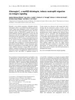
Expression of different HCV-proteins in the UHCV and UC cells (A, B) and their induction of apoptotic nuclei (C, D): 2 × 10
6
cells (A, B) or 1 × 10
4
, 2 × 10
4
, and 3 × 10
4
cells/well (C, D) of each cell line were cultured for the indicated time in the pres-ence or absence of tetracycline (Tet) to induce HCV-specific protein expressionFigure 1
Expression of different HCV-proteins in the UHCV and UC cells (A, B) and their induction of apoptotic nuclei
(C, D): 2 × 10
6
cells (A, B) or 1 × 10
4
, 2 × 10
4
, and 3 × 10
4
cells/well (C, D) of each cell line were cultured for the
indicated time in the presence or absence of tetracycline (Tet) to induce HCV-specific protein expression. A, B:
Cellular proteins were resolved by SDS-PAGE and HCV proteins were detected by immunoblotting with an antiserum gener-
ated against the core (A) or NS3 protein (B) of HCV. C, D: Induction of apoptosis was assessed by flow cytometric analysis of
propidium iodide staining of hypodiploid apoptotic nuclei. The mean values and standard deviation of triplicate cultures are
shown.
UC
UHCV
A
C :
D :
Tetracycline + + + + - - -
Culture period 0 1d 2d 3d 1d 2d 3d
Immunoblot: anti-NS3
UHCV
UC
p70
p70
B
p21
p21
UHCV
Immunoblot: anti-HCV-core
UC
Tetracycline
Culture period
++++
01d2d3d
-
1d
-
2d
-
3d
+
6h
-
6h
+
4d
-
4d
0123
20
40
60
80
100
10,000 cells/well
apoptotic nuclei [%]
culture period [d]
- Tet
+ Tet
0123
20
40
60
80
100
20,000 cells/well
apoptotic nuclei [%]
culture period [d]
- Tet
+ Tet
0123
0
20
40
60
80
100
30,000 cells/well
apoptotic nuclei [%]
culture period [d]
- Tet
+ Tet
0123
0
20
40
60
80
100
10,000 cells/well
apoptotic nuclei [%]
culture period [d]
- Tet
+ Tet
0123
0
20
40
60
80
100
20,000 cells/well
apoptotic nuclei [%]
culture period [d]
- Tet
+ Tet
0123
0
20
40
60
80
100
30,000 cells/well
apoptotic nuclei [%]
culture period [d]
- Tet
+ Tet
Virology Journal 2009, 6:213 />Page 5 of 13
(page number not for citation purposes)
within the UHCV cell line the polyprotein is cleaved to
release the single HCV proteins.
To study the effects of the core protein and the whole HCV
proteins on apoptosis induction, we analyzed the typical
apoptosis-associated leakage of fragmented DNA from
apoptotic nuclei by the Nicoletti method using flow
cytometry. As shown in Figure 1C in kinetic studies, there
was no apoptotic effect detectable in the polyprotein
expressing UHCV cell line, independent from the cell
number seeded. In contrast, the core protein expressed in
the UC cell line in the absence of Tet led to a strong leak-
age of fragmented DNA already after one day (Figure 1D).
The typical apoptotic effect depended on the expression
level of the core protein and not on the cell density
employed. Thus, testing two high and two low expression
cell lines from the UHCV and the UC cells, DNA fragmen-
tation was induced only in the UC cell line with an ele-
vated expression of the core protein (data not shown).
2. Cell death could not be induced by further HCV proteins
Next, we addressed the question, whether further HCV
proteins expressed in our test system also exert cell death
inducing properties. Therefore we tested a variety of cell
lines expressing different single HCV proteins or protein
groups by flow cytometry [27,31,32,43]. However, a
strong effect on the generation of hypodiploid nuclei
could only be observed in the cell line UCp7 expressing
the core, E1, E2 and p7 protein, whereas the other cell lines
did not exert any or only a slight (NS3-4A and NS4B pro-
teins) effect (Figure 2). For the NS3-4A cells the increase
of apoptotic cells after 3 days was independent from the
NS3-4A protein since the difference in the rate of apop-
totic nuclei between the induced and the non-induced
cells was constant from day 1 to day 3. Possibly, this is a
problem of the position of the insert coding for the HCV
protein in this cell line.
Since we did not observe any difference in the rate of
apoptotic nuclei in the absence of Tet in the NS5B cells
after 2 days, we further studied the activity after a quite
longer period, i.e. 6 days. However, we only found an
unspecific increase, most likely due to the consumption of
nutrients in the cell culture medium.
3. Apoptotic features induced by the HCV core protein
In order to characterize more precisely cell death induc-
tion by the core protein we analyzed the reactivity of the
UC cell line by different methods. Thus, we observed by
phase contrast and fluorescence microscopy (magnifica-
Induction of apoptosis in different HCV-protein expressing cell lines: 2 × 10
4
cells of the different cell lines UCP7, UNS3-4A, UNS4B, UNS5A, and UNS5B were cultured for the indicated times in the presence or absence of Tet to induce specific pro-tein expressionFigure 2
Induction of apoptosis in different HCV-protein expressing cell lines: 2 × 10
4
cells of the different cell lines
UCP7, UNS3-4A, UNS4B, UNS5A, and UNS5B were cultured for the indicated times in the presence or
absence of Tet to induce specific protein expression. Induction of apoptosis was assessed by propidium iodide staining
of hypodiploid apoptotic nuclei and flow cytometry. The mean values of triplicate cultures and standard deviation are shown.
0123
0
20
40
60
80
100
UNS5A
apoptotic nuclei [%]
culture period [d]
- Tet
+ Tet
0123
0
20
40
60
80
100
UNS3-4A
apoptotic nuclei [%]
culture period [d]
- Tet
+ Tet
0123
0
20
40
60
80
100
- Tet
+ Tet
UNS4B
apoptotic nuclei [%]
culture period [d]
0123
0
20
40
60
80
100
UCp7
apoptotic nuclei [%]
culture period [d]
- Tet
+ Tet
0126
20
40
60
80
100
UNS5B
apoptotic nuclei [%]
culture period [d]
- Tet
+ Tet
Virology Journal 2009, 6:213 />Page 6 of 13
(page number not for citation purposes)
tion 320×) that the core protein induced typical morpho-
logical features of apoptosis: Similar to mitomycin C and
TRAIL, which served as positive controls, the core protein
stimulated apoptotic blebs on the cell surface (Figure 3A).
In addition, nuclei were condensed and fragmented in
these cells as evidenced by the staining pattern with the
Hoechst dye 33342 (Figure 3A). However, in the TUNEL
assay detected by flow cytometry there was only a slight
increase in the amount of fragmented nuclei which were
accessible for the TdT in response to the core protein as
compared to the positive controls (Figure 3B).
4. Influence of the HCV proteins on death receptor-
mediated and mitochondrial apoptosis pathways
Since in our experiments the major effect was induced by
the core protein, we focused in our further studies on the
UC cell line. To investigate whether the HCV core protein
exerts an enhancing effect on the activation of the death
Different features of apoptosis induced by the HCV-core protein in UC cells cultured in the presence or absence of tetracycline to induce specific protein expressionFigure 3
Different features of apoptosis induced by the HCV-core protein in UC cells cultured in the presence or
absence of tetracycline to induce specific protein expression. A: membrane blebbing and nuclear condensation was
visualized in 2 × 10
4
UC cells cultured for 48 h. Apoptotic stimuli were added during the last 24 h: mitomycin C (50 μg/ml), and
TRAIL (40 ng/ml). Nuclei were stained with the cell permeable dye Höchst 33342 and cells were applied to phase contrast and
fluorescence microscopy (magnification: 320 ×). B: Detection of apoptosis by the TUNEL assay. 5 × 10
5
cells/ml were cultured
for 48 h and mitomycin C (50 μg/ml) and TRAIL (40 ng/ml) were added during the last 24 h and served as positive control.
TUNEL assay was measured by flow cytometry.
A
B
Medium
Tet +
Medium
Tet -
Mitomycin C
Tet +
TRAIL
Tet +
d-UTP-FITC
counts
Medium
Tet +
Medium
Tet -
Mitomycin C
Tet +
M1
0 200
0.5%
M1
0 200
0.5%
0 200
0.5%
0 200
0.5%
M1
0 200
14.4%
M1
0 200
14.4%
M1
0 200
14.4%
0 200
14.4%
0 200
14.4%
M1
0 200
1.9%
M1
0 200
1.9%
0 200
1.9%
0 200
1.9%
TRAIL
Tet +
M1
0 200
18.4%
Virology Journal 2009, 6:213 />Page 7 of 13
(page number not for citation purposes)
receptor pathway or the mitochondrial apoptosis pathway
we first stimulated the expression of the HCV proteins for
24 h and added a variety of apoptosis inducers to the cell
cultures for another 24 h. For stimulation of death recep-
tors we used agonistic anti-CD95 antibodies or the DR4
and DR5 ligand TRAIL and for the activation of the mito-
chondrial apoptosis pathway we used the anticancer drugs
mitomycin C and etoposide, as previously described [39].
As shown in Figure 4A, a costimulatory effect of the core
protein expressed by the UC cells on the rate of hypodip-
loid nuclei measured by flow cytometry could be
observed only in the TRAIL and anti-CD95 stimulated
cells as compared to the non-core expressing cells.
Figure 4B demonstrates that the core protein alone slightly
enhanced the phosphatidylserine (PS) externalization
and further enhanced the effect of the apoptotic agents
acting via the receptor-mediated pathway as measured by
Influence of HCV-core protein on the receptor-mediated and the mitochondrial apoptosis pathway studied in different assays: 1 × 10
5
UC cells/ml were cultured for 48 h in the presence or absence of Tet to induce specific protein expressionFigure 4
Influence of HCV-core protein on the receptor-mediated and the mitochondrial apoptosis pathway studied in
different assays: 1 × 10
5
UC cells/ml were cultured for 48 h in the presence or absence of Tet to induce specific
protein expression. Apoptotic stimuli were added during the last 24 h: mitomycin C (50 μg/ml), etoposide (400 ng/ml),
TRAIL (40 ng/ml) and anti-CD95 antibody (100 ng/ml). A: Induction of apoptosis was assessed by flow cytometric analysis of
propidium iodide staining of hypodiploid apoptotic nuclei. The mean values and standard deviation of triplicate cultures are
shown. B: Apoptosis was visualized by the externalization of PS which was stained with Annexin V and C: viability of the cells
was measured by staining of the cells with PI and subsequent detection by flow cytometry. Given are means of duplicates. D:
Metabolic activity of the UC cells was determined by the MTS test. Optical density was measured in an ELISA reader after incu-
bation of the cells with MTS for 4 h and suspension of crystals. Given are mean and standard deviation of triplicates.
Medium
Mito
Etopo
TRAIL
a-CD95
0
20
40
60
80
100
apoptotic nuclei [%]
+ Tet
- Tet
Medium
Mito
Etopo
TRAIL
a-CD95
0
20
40
60
80
Annexin V positive [%]
+ Tet
- Tet
Medium
Mito
Etopo
TRAIL
a-CD95
0
20
40
60
80
PI positive [%]
+ Tet
- Tet
Medium
Mito
Etopo
TRAIL
0.0
0.2
0.4
0.6
0.8
viability [OD]
+ Tet
- Tet
Virology Journal 2009, 6:213 />Page 8 of 13
(page number not for citation purposes)
the staining with Annexin V by flow cytometry. Similar
observations were made for the uptake of propidium
iodide that measures cell death in general and cannot dis-
criminate between apoptosis and necrosis (Figure 4C). In
addition, the viability of the cells expressing the core pro-
tein was reduced by the core protein as evidenced by a
diminished formazan crystallization in the MTS test (Fig-
ure 4D).
However, analyzing the UHCV, UNS4B, and NS5A cell
lines, there was no significant difference in response to the
exogenously added apoptotic stimuli between the cells
expressing the respective HCV proteins or not (data not
shown).
5. Cell death induction by the core protein is not caspase-
dependent
In order to study whether caspases are involved in the
process of cell death induction by the core protein, we first
stimulated the core expressing UC cell line in the presence
or absence of the broad spectrum caspase inhibitor zVAD-
fmk. As shown in Figure 5A, the core protein induced gen-
eration of hypodiploid nuclei was only partially affected
by zVAD-fmk, whereas zVAD-fmk clearly inhibited their
generation stimulated by mitomycin C, etoposide, TRAIL,
and anti-CD95 antibody in the Tet-on cells. In contrast, in
the polyprotein expressing UHCV cell line generation and
inhibition of apoptotic nuclei using different apoptotic
stimuli with or without zVAD-fmk was independent of
the Tet-off system (Figure 5B).
Despite the observation that the UC cell line was less sen-
sitive to the receptor-mediated apoptosis pathway, an
additional apoptotic effect could be observed by the core-
protein (Figure 5A). This effect could only partially be
inhibited by zVAD-fmk suggesting that a caspase-inde-
pendent mechanism may be responsible for the core pro-
tein induced cell death.
Studying in more detail the core protein mediated apopto-
sis it became evident that zVAD-fmk did not inhibit the
core protein-induced generation of hypodiploid nuclei, in
contrast to cell death induction due to Mitomycin C and
TRAIL which showed an almost complete inhibition fol-
lowing application of zVAD-fmk (Figure 5C). Interest-
ingly, most hypodiploid nuclei were very small in the core
protein expressing cells as compared to the nuclei arising
after stimulation with TRAIL. While zVAD-fmk did not
inhibit the core protein-induced generation of hypodip-
loid nuclei, it almost completely blocked the small nuclei
induced by mitomycin C.
To directly analyze the involvement of caspases in the
action of the core protein, Western blot analyses were per-
formed confirming that both, caspases-3 and -8, had not
been activated since neither caspase cleavage products
could be observed, nor did they comprise any activity, as
demonstrated by the lack of the cleavage of the caspase
substrate PARP (Figure 5D). In contrast, cultivation with
the typical apoptotic stimuli mitomycin C, TRAIL or the
stimulatory anti-CD95 antibody induced caspase activa-
tion that could be inhibited by zVAD-fmk.
In addition, using the fluorogenic substrate DEVD-AMC
in a fluorometric assay we could not observe any core pro-
tein related caspase activity (Figure 5E). Cell lysates of the
Tet regulated core expressing UC cell line did not possess
any caspase activity, in contrast to the lysates of cells incu-
bated with mitomycin C, TRAIL or the anti-CD95 anti-
body which showed a typical caspase activity. Similar
observations were made with the UHCV cell line (data not
shown).
Additional experiments were performed to study whether
ICAD (inhibitor of caspase activated DNAse) was cleaved
by the core protein which in turn may lead to the activa-
tion of the endonuclease CAD (caspase activated DNAse).
However, we could not observe any cleavage of ICAD by
the core protein (data not shown) which further confirms
a caspase-independent type of DNA cleavage.
6. Analysis of the involvement of a variety of protease
inhibitors in the apoptosis-like effect of the core protein
To study in more detail the mechanisms involved in the
apoptosis-like activity of the core protein, we tested a vari-
ety of broad-spectrum as well as specific protease inhibi-
tors for their ability to block the core protein-induced
generation of apoptotic nuclei (Figure 6). In these kinetic
studies, neither the cathepsin B inhibitor (Figure 6A) nor
the calpain inhibitors I (data not shown) and II (Figure
6B) exerted any effect on the core protein-induced apopto-
sis. In addition, none of the other specific and unspecific
inhibitors as leupeptin, pepstatin, pefabloc, ROCK inhib-
itor and oligomycin were able to block the apoptotic effect
after 48 h of cell culture, while the inhibitor of the PI3
kinase LY294002 and the calpain inhibitor I were toxic
(data not shown).
Discussion
The objective of our study was to investigate the potency
of endogenously expressed HCV proteins on apoptosis
induction and to analyze their influence on the death
receptor-mediated and the mitochondrial apoptosis path-
way. To address these questions, we used a recently estab-
lished tightly adjustable HCV protein expression cell
system which allowed switch off and on of the endog-
enous production of a broad spectrum of HCV proteins or
protein complexes (Tet-off system) [27-31]. Using this
system we compared the apoptosis-inducing effects of the
different single HVC proteins and protein complexes. This
Virology Journal 2009, 6:213 />Page 9 of 13
(page number not for citation purposes)
is of major importance since the literature presents con-
flicting data on that topic. It could be shown that e.g. the
receptor-mediated apoptosis was inhibited by the core
protein [24] while just the opposite effect was obtained by
different authors, even if the same cell line was used
[25,26]. These data demonstrate that the observed effects
strongly depend upon the experimental conditions. To
circumvent this problem by using cell lines inducible
expressing a broad spectrum of HCV proteins and protein
complexes it became evident that the cell lines expressing
the core protein (i.e. UC and UCp7) showed a strong
induction of apoptotic nuclei. The other HCV proteins
and protein complexes did not show any effect with the
exception of a very slight stimulation by the NS3-4A and
NS4B proteins.
Cell death induction of the core protein expressing cells
was evidenced by a variety of methods. Thus, typical
apoptosis-associated morphological alterations like the
loss of the contact to neighboring cells, formation of
apoptotic blebs and nuclear condensation could be
clearly detected. In addition, a slight externalization of
Influence of the HCV-core protein on caspase activationFigure 5
Influence of the HCV-core protein on caspase activation. 1 × 10
5
cells/ml of the UC (A, C, E) and UHCV cell lines (B)
or 1 × 10
6
UC cells (D) were cultured for the indicated times in the presence or absence of Tet to induce specific protein
expression and the broad spectrum caspase inhibitor zVAD (100 μM). The apoptotic stimuli mitomycin C (50 μg/ml), etopo-
side (400 ng/ml), TRAIL (40 ng/ml), and anti-CD95 antibody (100 ng/ml) were added during the last 24 h of the culture. The
broad spectrum caspase inhibitor zVAD was added at day 0 if indicated. A-C: Induction of apoptosis was assessed after 48 h by
flow cytometric analysis of propidium iodide staining of hypodiploid apoptotic nuclei. The mean values and standard deviation
of triplicate cultures are shown (A+B). D: Cleavage of caspases-3 and -8 as well as of PARP was detected in the cell lysates by
Western Blot analysis. E: Detection of the caspase activity in UC cells was performed by in vitro cleavage of the fluorogenic sub-
strate DEVD-AMC and was measured by fluorometry at the time indicated. Apoptotic stimuli were added for 24 h. The mean
values and standard deviation of triplicate cultures are given.
Virology Journal 2009, 6:213 />Page 10 of 13
(page number not for citation purposes)
phosphatidylserine as well as a diminished metabolic
activity induced by the core protein fit to these observa-
tions. The best read-out system for the analysis of the
apoptotic effect was the visualization of hypodiploid
nuclei. Interestingly, these nuclei were very small, similar
to those obtained by stimulation with mitomycin C but
their generation could not be blocked by the caspase
inhibitor zVAD-fmk in contrast to that observed for mito-
mycin C or TRAIL. In addition, the typical 'DNA-ladder'
obtained after internucleosomal cleavage of DNA could
not be observed in the UC cell line (data not shown).
Moreover, using the less sensitive TUNEL assay we did not
find any core protein related typical apoptosis-associated
DNA fragmentation pattern while mitomycin C and
TRAIL were active in this test system. The lack of reactivity
of the core protein in these two assays is in accordance
with the lack of caspase activation since the internucleo-
somal cleavage of DNA is mainly due to the activity of
CAD (caspase-dependent DNase) during apoptosis,
which is inhibited by ICAD (inhibitor of CAD) [44,45].
Consistently with the lack of caspase activation, we, in
contrast to Sacco et al. [46], did not observe alterations of
ICAD (data not shown). From all these data it is assumed
that the core protein stimulated apoptosis-like cell death is
mainly caspase-independent. Caspase-independent apop-
tosis pathways have been described and are now generally
accepted [47]. In contrast to our data, Moorman et al. and
Goh et al. observed an activation of caspases-3 and -8 by
the core protein [19] or the cleavage of PARP [17] which
may be strongly influenced by the experimental condi-
tions.
Analyzing the apoptotic effect of the UCp7 cells we cannot
completely exclude that it was influenced by the proteins
E1, E2 or p7, although it seems reasonable that the core
protein was responsible for the major effect. However, in
this respect two publications may be of relevance showing
apoptotic effects of the E1 [48] and the E2 protein [49].
Thus, further investigations should include cells express-
ing either protein in the same adjustable system.
In order to better define the mechanisms involved in the
apoptosis-like machinery stimulated by the core protein,
we tried to block the core protein induced generation of
hypodiploid nuclei by a variety of proteases via unspecific
and specific inhibitors. However, none of the different
protease inhibitors, like the specific inhibitor of cathepsin
B (Ca-074) or calpains or the unspecific inhibitors of pro-
teases like leupeptin and pepstatin could block cell death
induction. In future experiments further signal transduc-
tion cascades like the Akt/PKB and other signaling path-
ways have to be investigated.
One intriguing finding was that the polyprotein express-
ing UHCV cells did not exert any apoptotic effect although
they clearly expressed the core protein. This observation is
HCV core protein induced apoptosis could not be completely inhibited by a broad spectrum of protease inhibitorsFigure 6
HCV core protein induced apoptosis could not be completely inhibited by a broad spectrum of protease inhib-
itors. 1 × 10
5
UC cells/ml were cultured for the indicated times (A, B) in the presence or absence of Tet to induce specific
protein expression. The protease inhibitors were added at day 0: cathepsin inhibitor Ca-074 30 μM, and calpain inhibitor II 10
μM. Induction of apoptosis was assessed by propidium iodide staining of hypodiploid apoptotic nuclei and flow cytometry. The
mean values and standard deviation of triplicate cultures are given.
AB
123
0
20
40
60
80
100
apoptotic nuclei [%]
culture period [d]
Medium Tet +
Medium Tet -
Ca-074 Tet +
Ca-074 Tet -
123
0
20
40
60
80
100
apoptotic nuclei [%]
culture period [d]
Medium Tet +
Medium Tet -
Calp. Inh. II Tet +
Calp. Inh. II Tet -
Virology Journal 2009, 6:213 />Page 11 of 13
(page number not for citation purposes)
difficult to explain and may reflect the complexity of the
virus-specific reactions. It may be possible that both, the
core protein associated apoptotic and the possible anti-
apoptotic effects of further HCV proteins may act
together. Thus, in stably transfected cell lines, Chung et al.
found an anti-apoptotic effect of the NS5A protein while
the core protein exerted apoptotic potency, similar to our
data [50]. In addition, it has been described that different
HCV proteins like the NS3 and NS4A or NS4A, NS4B and
NS5A interact [51,52]. Thus, it cannot be excluded that
different proteins also bind to the core protein or signaling
molecules of the core protein induced cascade and block
its effect. This regulation of apoptosis may be of advantage
for the virus in order to circumvent a premature apoptosis
before the virus replication and assemblage has finished.
The second objective of our investigations was to study
the influence of the HCV proteins on apoptosis induced
by exogenous stimuli acting on the mitochondrial (mito-
mycin C and etoposide) or the death receptor-mediated
(TRAIL, agonistic anti-CD95 antibody) apoptosis path-
way [35,36,39]. None of the tested cell lines, i.e. the
UHCV, UNS4B and UNS5A cell lines exerted a significant
stimulatory or inhibitory effect on either apoptosis path-
way. In contrast, the UC cell line enhanced the TRAIL and
anti-CD95 mediated apoptosis as evidenced by an
increase of cell death-related features studied in different
test systems.
From the data presented here it appears that the core pro-
tein did not exert its major effect via the receptor mediated
pathway by inducing the respective ligands on the neigh-
boring cells. However, we cannot exclude that this mech-
anism is operative in the core protein mediated
enhancement of apoptosis induced by death receptor lig-
ands.
Comparing our data on the influence of the HCV proteins
on exogenous apoptotic stimuli with the data in the liter-
ature our results in part are in accordance with previously
published work since some authors did not find an influ-
ence of the core protein on the receptor-mediated apopto-
sis pathway [53], an increase [26,54] or an inhibition
[24].
In contrast to the available data on the core protein, the
data on the NS5A protein are more uniform demonstrat-
ing a rather anti-apoptotic effect for that protein
[22,23,55-58].
From the results obtained in our study it is evident that the
core protein exerted the strongest caspase-independent
direct apoptosis-like effect whereas none of the other HCV
proteins showed a clear-cut influence on exogenous apop-
totic stimuli. In this respect the localization of the mature
core protein at the outer mitochondrial membrane may be
of importance [59]. Although we did not observe the
release of cytochrome c in the Tet-off UC cell line (data
not shown) an interaction of the core protein with other
molecules of the apoptotic machinery localized at the
mitochondrial membranes may occur. Thus, further
investigations are necessary to better characterize the role
of the different HCV and host cell proteins in the apop-
totic processes. Since some of the proteins interact with
each other, more complex systems are needed for a more
precise evaluation of the pathology of the disease in order
to develop new remedies with an anti-viral effect to HCV.
Conclusion
In our experiments the non-structural proteins seem to
exert an anti-apoptotic effect since in the polyprotein
expressing cells no apoptosis-like features could be
observed. Thus, it is tempting to speculate that in vivo they
may inhibit early host cell death while core protein stimu-
lated caspase-independent apoptosis-like effect may fol-
low at later stages and, therefore, could be of relevance for
the release of the HCV particles from the host cell and the
viral spread.
List of abbreviations
CAD: caspase-dependent DNase; DEVDamc: N-acetyl-
Asp-Glu-Val-Asp-aminomethylcoumarin; E: envelope;
etopo: etoposide; HCV: hepatitis C virus; ICAD: inhibitor
of CAD; mito: mitomycin; MTS: methyltetrazolium salt;
NS proteins: non structural proteins, ORF: open reading
frame; Tet: tetracycline; TNF: tumor necrosis factor; TRAIL:
TNF-related apoptosis inducing ligand; TUNEL: terminal
deoxynucleotidyl transferase (TdT)-catalyzed deoxyurid-
inephosphate (dUTP)-nick end labeling; zVAD: benzy-
loxycarbonyl-Val-Ala-Asp-fluoromethylketone.
Competing interests
The authors declare that they have no competing interests.
Authors' contributions
CPB and SFS contributed equally to this paper and share
first authorship. GMS, CPB, DKHN and CP performed
research. Also GMS and SW contributed equally to this
paper and share senior authorship. GMS, CPB, SFS and
SW designed research, analyzed data and wrote the man-
uscript. MG helped discussing the data. They all read and
approved the final manuscript.
Acknowledgements
We would like to acknowledge Darius Moradpour, Division of Gastroen-
terology and Hepatology, Centre Hospitalier Universitaire Vaudois,
Lausanne, Switzerland, for providing us the cell lines expressing the differ-
ent HCV proteins and the monoclonal antibodies to the core and the NS3
protein. This work was kindly supported by grants from the Deutsche For-
schungsgemeinschaft (WE-1801/2-4, GRK 1302, SFB 685; SW), the German
Bundesministerium fuer Bildung und Forschung (Hep-Net; GMS and SW),
Virology Journal 2009, 6:213 />Page 12 of 13
(page number not for citation purposes)
the Wilhelm-Sander-Stiftung (2004.099.1; SW), the Federal Ministry of Edu-
cation, Science, Research and Technology (Fö. 01KS9602) and Interdiscipli-
nary Center of Clinical Research Tuebingen (IZKF) to SW, the
Landesforschungsschwer-punktprogramm of the Ministry of Science,
Research and Arts of the Land Baden-Wuerttemberg (1423-98101; SW)
and from the Fortune Program of the University of Tuebingen (No.
F1281399; CPB and GMS).
References
1. Brass V, Moradpour D, Blum HE: Molecular Virology of Hepatitis
C Virus (HCV): 2006 Update. Int J Med Sci 2006, 3:29-34.
2. Lauer GM, Walker BD: Hepatitis C virus infection. N Engl J Med
2001, 345:41-52.
3. Penin F, Dubuisson J, Rey FA, Moradpour D, Pawlotsky JM: Struc-
tural biology of hepatitis C virus. Hepatology 2004, 39:5-19.
4. Choo QL, Kuo G, Weiner AJ, Overby LR, Bradley DW, Houghton M:
Isolation of a cDNA clone derived from a blood-borne non-
A, non-B viral hepatitis genome. Science 1989, 244:359-62.
5. Desaintes C, Demeret C, Goyat S, Yaniv M, Thierry F: Expression
of the papillomavirus E2 protein in HeLa cells leads to apop-
tosis. Embo J 1997, 16:504-14.
6. Levine B, Huang Q, Isaacs JT, Reed JC, Griffin DE, Hardwick JM: Con-
version of lytic to persistent alphavirus infection by the bcl-2
cellular oncogene. Nature 1993, 361:739-42.
7. Marcellus RC, Teodoro JG, Wu T, Brough DE, Ketner G, Shore GC,
Branton PE: Adenovirus type 5 early region 4 is responsible for
E1A-induced p53-independent apoptosis. J Virol 1996,
70:6207-15.
8. Crook NE, Clem RJ, Miller LK: An apoptosis-inhibiting baculovi-
rus gene with a zinc finger-like motif. J Virol 1993, 67:2168-74.
9. Gratama JW, Oosterveer MA, Zwaan FE, Lepoutre J, Klein G, Ernberg
I: Eradication of Epstein-Barr virus by allogeneic bone mar-
row transplantation: implications for sites of viral latency.
Proc Natl Acad Sci USA 1988, 85:8693-6.
10. Miura M, Friedlander RM, Yuan J: Tumor necrosis factor-induced
apoptosis is mediated by a CrmA-sensitive cell death path-
way. Proc Natl Acad Sci USA 1995, 92:8318-22.
11. Neilan JG, Lu Z, Kutish GF, Sussman MD, Roberts PC, Yozawa T,
Rock DL: An African swine fever virus gene with similarity to
bacterial DNA binding proteins, bacterial integration host
factors, and the Bacillus phage SPO1 transcription factor,
TF1.
Nucleic Acids Res 1993, 21:1496.
12. Tewari M, Telford WG, Miller RA, Dixit VM: CrmA, a poxvirus-
encoded serpin, inhibits cytotoxic T-lymphocyte-mediated
apoptosis. J Biol Chem 1995, 270:22705-8.
13. Koziel MJ: The role of immune responses in the pathogenesis
of hepatitis C virus infection. J Viral Hepat 1997, 4(Suppl
2):31-41.
14. Patel T, Steer CJ, Gores GJ: Apoptosis and the liver: A mecha-
nism of disease, growth regulation, and carcinogenesis.
Hepatology 1999, 30:811-5.
15. Kountouras J, Zavos C, Chatzopoulos D: Apoptosis in hepatitis C.
J Viral Hepat 2003, 10:335-42.
16. Muratori L, Gibellini D: A new route to apoptosis in hepatitis C
virus infection. J Hepatol 2001, 35:814-5.
17. Goh PY, Tan YJ, Lim SP, Lim SG, Tan YH, Hong WJ: The hepatitis
C virus core protein interacts with NS5A and activates its
caspase-mediated proteolytic cleavage. Virology 2001,
290:224-36.
18. Kalkeri G, Khalap N, Garry RF, Fermin CD, Dash S: Hepatitis C
virus protein expression induces apoptosis in HepG2 cells.
Virology 2001, 282:26-37.
19. Moorman JP, Prayther D, McVay D, Hahn YS, Hahn CS: The C-ter-
minal region of hepatitis C core protein is required for Fas-
ligand independent apoptosis in Jurkat cells by facilitating
Fas oligomerization. Virology 2003, 312:320-9.
20. Giannini C, Caini P, Giannelli F, Fontana F, Kremsdorf D, Brechot C,
Zignego AL: Hepatitis C virus core protein expression in
human B-cell lines does not significantly modify main prolif-
erative and apoptosis pathways. J Gen Virol 2002, 83:1665-71.
21. Ciccaglione AR, Marcantonio C, Costantino A, Equestre M, Rapicetta
M: Expression of HCV E1 protein in baculovirus-infected
cells: effects on cell viability and apoptosis induction. Intervi-
rology 2003, 46:121-6.
22. Arima N, Kao CY, Licht T, Padmanabhan R, Sasaguri Y: Modulation
of cell growth by the hepatitis C virus nonstructural protein
NS5A. J Biol Chem 2001, 276:12675-84.
23. Polyak SJ, Paschal DM, McArdle S, Gale MJ Jr, Moradpour D, Gretch
DR: Characterization of the effects of hepatitis C virus non-
structural 5A protein expression in human cell lines and on
interferon-sensitive virus replication. Hepatology 1999,
29:1262-71.
24. Marusawa H, Hijikata M, Chiba T, Shimotohno K: Hepatitis C virus
core protein inhibits Fas- and tumor necrosis factor alpha-
mediated apoptosis via NF-kappaB activation. J Virol 1999,
73:4713-20.
25. Otsuka M, Kato N, Taniguchi H, Yoshida H, Goto T, Shiratori Y,
Omata M: Hepatitis C virus core protein inhibits apoptosis via
enhanced Bcl-xL expression. Virology 2002, 296:84-93.
26. Ruggieri A, Harada T, Matsuura Y, Miyamura T: Sensitization to
Fas-mediated apoptosis by hepatitis C virus core protein.
Virology 1997, 229:68-76.
27. Hugle T, Fehrmann F, Bieck E, Kohara M, Krausslich HG, Rice CM,
Blum HE, Moradpour D: The hepatitis C virus nonstructural
protein 4B is an integral endoplasmic reticulum membrane
protein. Virology 2001, 284:70-81.
28. Moradpour D, Englert C, Wakita T, Wands JR: Characterization of
cell lines allowing tightly regulated expression of hepatitis C
virus core protein. Virology 1996, 222:51-63.
29. Moradpour D, Wakita T, Wands JR, Blum HE: Tightly regulated
expression of the entire hepatitis C virus structural region in
continuous human cell lines. Biochem Biophys Res Commun 1998,
246:920-4.
30. Moradpour D, Kary P, Rice CM, Blum HE: Continuous human cell
lines inducibly expressing hepatitis C virus structural and
nonstructural proteins. Hepatology 1998, 28:192-201.
31. Wolk B, Sansonno D, Krausslich HG, Dammacco F, Rice CM, Blum
HE, Moradpour D: Subcellular localization, stability, and trans-
cleavage competence of the hepatitis C virus NS3-NS4A
complex expressed in tetracycline-regulated cell lines. J Virol
2000,
74:2293-304.
32. Brass V, Bieck E, Montserret R, Wolk B, Hellings JA, Blum HE, Penin
F, Moradpour D: An amino-terminal amphipathic alpha-helix
mediates membrane association of the hepatitis C virus non-
structural protein 5A. J Biol Chem 2002, 277:8130-9.
33. Schmidt-Mende J, Bieck E, Hugle T, Penin F, Rice CM, Blum HE,
Moradpour D: Determinants for membrane association of the
hepatitis C virus RNA-dependent RNA polymerase. J Biol
Chem 2001, 276:44052-63.
34. Nicoletti I, Migliorati G, Pagliacci MC, Grignani F, Riccardi C: A rapid
and simple method for measuring thymocyte apoptosis by
propidium iodide staining and flow cytometry. J Immunol Meth-
ods 1991, 139:271-9.
35. Wesselborg S, Engels IH, Rossmann E, Los M, Schulze-Osthoff K:
Anticancer drugs induce caspase-8/FLICE activation and
apoptosis in the absence of CD95 receptor/ligand interac-
tion. Blood 1999, 93:3053-63.
36. Stepczynska A, Lauber K, Engels IH, Janssen O, Kabelitz D, Wessel-
borg S, Schulze-Osthoff K: Staurosporine and conventional anti-
cancer drugs induce overlapping, yet distinct pathways of
apoptosis and caspase activation. Oncogene 2001, 20:1193-202.
37. Lauber K, Bohn E, Krober SM, Xiao YJ, Blumenthal SG, Lindemann
RK, Marini P, Wiedig C, Zobywalski A, Baksh S, et al.: Apoptotic
cells induce migration of phagocytes via caspase-3-mediated
release of a lipid attraction signal. Cell 2003, 113:717-30.
38. Berg CP, Stein GM, Klein R, Pascu M, Berg T, Kammer W, Priemer M,
Nordheim A, Schulze-Osthoff K, Gregor M, et al.: Demonstration
of PDC-E1 subunits as major antigens in the complement-
fixing fraction M4 and re-evaluation of PDC-E1-specific anti-
bodies in PBC patients. Liver Int 2006, 26:846-55.
39. Engels IH, Stepczynska A, Stroh C, Lauber K, Berg C, Schwenzer R,
Wajant H, Janicke RU, Porter AG, Belka C, et al.: Caspase-8/FLICE
functions as an executioner caspase in anticancer drug-
induced apoptosis. Oncogene 2000,
19:4563-73.
40. Moradpour D, Wakita T, Tokushige K, Carlson RI, Krawczynski K,
Wands JR: Characterization of three novel monoclonal anti-
bodies against hepatitis C virus core protein. J Med Virol 1996,
48:234-41.
41. Bussing A, Vervecken W, Wagner M, Wagner B, Pfuller U, Schietzel
M: Expression of mitochondrial Apo2.7 molecules and cas-
Publish with BioMed Central and every
scientist can read your work free of charge
"BioMed Central will be the most significant development for
disseminating the results of biomedical research in our lifetime."
Sir Paul Nurse, Cancer Research UK
Your research papers will be:
available free of charge to the entire biomedical community
peer reviewed and published immediately upon acceptance
cited in PubMed and archived on PubMed Central
yours — you keep the copyright
Submit your manuscript here:
/>BioMedcentral
Virology Journal 2009, 6:213 />Page 13 of 13
(page number not for citation purposes)
pase-3 activation in human lymphocytes treated with the
ribosome-inhibiting mistletoe lectins and the cell membrane
permeabilizing viscotoxins. Cytometry 1999, 37:133-9.
42. Figarella K, Rawer M, Uzcategui NL, Kubata BK, Lauber K, Madeo F,
Wesselborg S, Duszenko M: Prostaglandin D2 induces pro-
grammed cell death in Trypanosoma brucei bloodstream
form. Cell Death Differ 2005, 12:335-46.
43. Egger D, Wolk B, Gosert R, Bianchi L, Blum HE, Moradpour D, Bienz
K: Expression of hepatitis C virus proteins induces distinct
membrane alterations including a candidate viral replication
complex. J Virol 2002, 76:5974-84.
44. Zamzami N, Kroemer G: Condensed matter in cell death.
Nature 1999, 401:127-8.
45. Nagata S, Nagase H, Kawane K, Mukae N, Fukuyama H: Degrada-
tion of chromosomal DNA during apoptosis. Cell Death Differ
2003, 10:108-16.
46. Sacco R, Tsutsumi T, Suzuki R, Otsuka M, Aizaki H, Sakamoto S, Mat-
suda M, Seki N, Matsuura Y, Miyamura T, et al.: Antiapoptotic reg-
ulation by hepatitis C virus core protein through up-
regulation of inhibitor of caspase-activated DNase. Virology
2003, 317:24-35.
47. Lockshin RA, Zakeri Z: Caspase-independent cell death? Onco-
gene 2004, 23:2766-73.
48. Ciccaglione AR, Marcantonio C, Tritarelli E, Equestre M, Magurano F,
Costantino A, Nicoletti L, Rapicetta M: The transmembrane
domain of hepatitis C virus E1 glycoprotein induces cell
death. Virus Res 2004, 104:1-9.
49. Zhu LX, Liu J, Xie YH, Kong YY, Ye Y, Wang CL, Li GD, Wang Y:
Expression of hepatitis C virus envelope protein 2 induces
apoptosis in cultured mammalian cells. World J Gastroenterol
2004, 10:2972-8.
50. Chung YL, Sheu ML, Yen SH: Hepatitis C virus NS5A as a poten-
tial viral Bcl-2 homologue interacts with Bax and inhibits
apoptosis in hepatocellular carcinoma. Int J Cancer 2003,
107:65-73.
51. Lin C, Wu JW, Hsiao K, Su MS:
The hepatitis C virus NS4A pro-
tein: interactions with the NS4B and NS5A proteins. J Virol
1997, 71:6465-71.
52. Liang Y, Kang CB, Yoon HS: Molecular and structural character-
ization of the domain 2 of hepatitis C virus non-structural
protein 5A. Mol Cells 2006, 22:13-20.
53. Dumoulin FL, Leifeld L, Honecker U, Sauerbruch T, Spengler U: Int-
rahepatic expression of interleukin-1beta and tumor necro-
sis factor-alpha in chronic hepatitis C. J Infect Dis 1999,
180:1704-8.
54. Zhu N, Khoshnan A, Schneider R, Matsumoto M, Dennert G, Ware
C, Lai MM: Hepatitis C virus core protein binds to the cyto-
plasmic domain of tumor necrosis factor (TNF) receptor 1
and enhances TNF-induced apoptosis. J Virol 1998, 72:3691-7.
55. Bonte D, Francois C, Castelain S, Wychowski C, Dubuisson J, Meurs
EF, Duverlie G: Positive effect of the hepatitis C virus non-
structural 5A protein on viral multiplication. Arch Virol 2004,
149:1353-71.
56. He Y, Nakao H, Tan SL, Polyak SJ, Neddermann P, Vijaysri S, Jacobs
BL, Katze MG: Subversion of cell signaling pathways by hepati-
tis C virus nonstructural 5A protein via interaction with
Grb2 and P85 phosphatidylinositol 3-kinase. J Virol 2002,
76:9207-17.
57. Lan KH, Sheu ML, Hwang SJ, Yen SH, Chen SY, Wu JC, Wang YJ, Kato
N, Omata M, Chang FY, et al.: HCV NS5A interacts with p53 and
inhibits p53-mediated apoptosis. Oncogene 2002, 21:4801-11.
58. Street A, Macdonald A, Crowder K, Harris M: The Hepatitis C
virus NS5A protein activates a phosphoinositide 3-kinase-
dependent survival signaling cascade. J Biol Chem 2004,
279:12232-41.
59. Schwer B, Ren S, Pietschmann T, Kartenbeck J, Kaehlcke K, Barten-
schlager R, Yen TS, Ott M: Targeting of hepatitis C virus core
protein to mitochondria through a novel C-terminal locali-
zation motif. J Virol 2004, 78:7958-68.