Báo cáo khoa học:" Protein intrinsic disorder and influenza virulence: the 1918 H1N1 and H5N1 viruses" docx
Bạn đang xem bản rút gọn của tài liệu. Xem và tải ngay bản đầy đủ của tài liệu tại đây (1.36 MB, 12 trang )
BioMed Central
Page 1 of 12
(page number not for citation purposes)
Virology Journal
Open Access
Research
Protein intrinsic disorder and influenza virulence: the 1918 H1N1
and H5N1 viruses
Gerard Kian-Meng Goh*
1,4
, A Keith Dunker
1
and Vladimir N Uversky*
1,2,3
Address:
1
Center for Computational Biology and Bioinformatics, Indiana University School of Medicine, Indianapolis, Indiana 46202, USA,
2
Institute for Intrinsically Disordered Protein Research, Indiana University School of Medicine, Indianapolis, Indiana 46202, USA,
3
Institute for
Biological Instrumentation, Russian Academy of Sciences, 142290 Pushchino, Moscow Region, Russia and
4
Institute of Molecular and Cell
Biology, Singapore 138673, Republic of Singapore
Email: Gerard Kian-Meng Goh* - ; A Keith Dunker - ;
Vladimir N Uversky* -
* Corresponding authors
Abstract
Background: The 1918 H1N1 virus was a highly virulent strain that killed 20–50 million people.
The cause of its virulence remains poorly understood.
Methods: Intrinsic disorder predictor PONDR
®
VLXT was used to compare various influenza
subtypes and strains. Three-dimensional models using data from X-ray crystallographic studies
annotated with disorder prediction were used to characterize the proteins.
Results: The protein of interest is hemagglutin (HA), which is a surface glycoprotein that plays a
vital role in viral entry. Distinct differences between HA proteins of the virulent and non-virulent
strains are seen, especially in the region near residues 68–79 of the HA
2
. This region represents
the tip of the stalk that is in contact with the receptor chain, HA
1
, and therefore likely to provide
the greatest effect on the motions of the exposed portion of HA. Comparison of this region
between virulent strains (1918 H1N1 and H5N1) and less virulent ones (H3N2 and 1930 H1N1)
reveals that predicted disorder can be seen at this region among the more virulent strains and
subtypes but is remarkably absent among the distinctly less virulent ones.
Conclusion: The motions created by disorder at crucial regions are likely to impair recognition
by immunological molecules and increase the virulence of both the H5N1 and the 1918 H1N1
viruses. The results help explain many puzzling features of the H5N1 and the 1918 H1N1 viruses.
Summarizing, HA (and especially its intrinsically disordered regions) can serve as a predictor of the
influenza A virulence, even though there may be other proteins that contribute to or exacerbate
the virulence.
Background
Introducing influenza viruses
Influenza viruses belong to the Orthomyxoviridae family of
negative sense, single-stranded, segmented RNA viruses.
This family contains five genera, classified by variations in
nucleoprotein (NP and M) antigens: influenza A, influ-
enza B, influenza C, thogotovirus, and isavirus [1]. Influ-
enza A viruses, which are a major cause of influenza in
humans, have multiple subtypes that are labeled accord-
ing to an H number (for hemagglutinin) and an N
Published: 3 June 2009
Virology Journal 2009, 6:69 doi:10.1186/1743-422X-6-69
Received: 25 March 2009
Accepted: 3 June 2009
This article is available from: />© 2009 Goh et al; licensee BioMed Central Ltd.
This is an Open Access article distributed under the terms of the Creative Commons Attribution License ( />),
which permits unrestricted use, distribution, and reproduction in any medium, provided the original work is properly cited.
Virology Journal 2009, 6:69 />Page 2 of 12
(page number not for citation purposes)
number (for neuraminidase). There are 16 different
hemagglutinin (HA) antigens (H1 to H16) and nine dif-
ferent neuraminidase (NA) antigens (N1 to N9) for influ-
enza A [2]. The influenza A viral genome consists of 8
genes encoding 11 proteins, including: two nonstructural
proteins (NS1 and NS2), four transcriptase proteins (PB2,
PB1, PB1-F2, and PA), two surface glycoproteins (HA and
NA), two matrix proteins (M1 and M2), and one nucleo-
capsid protein (NP), where the 3 extra proteins arise from
the use of different reading frames.
The influenza A virion (i.e. a complete virus particle with
its RNA core and protein coat) is a globular particle
sheathed in a lipid bilayer derived from the plasma mem-
brane of its host. Two integral membrane proteins, HA
and NA, are studded in the lipid bilayer. They are distrib-
uted evenly over the virion surface, forming characteristic
spike-shaped structures. The matrix formed by matrix pro-
teins M1 and M2 is located beneath the envelope. This
matrix encompasses eight pieces of genomic RNA, each in
association with many copies of a nucleoprotein (NP),
some "non-structural" proteins with various functions
(e.g. NS1 and NS2) and several molecules of the three
subunits of its RNA polymerase. The influenza pandemics
(i.e. the epidemics of the influenza virus) of the recent era
include the Spanish influenza (H1N1, 1918), the Asian
influenza (H2N2, 1958), and the Hong Kong influenza
(H3N2, 1968) [3-6]. The human tolls resulting from the
pandemics have been very large. The Spanish influenza
pandemic of 1918–1919 alone caused acute illness in 25–
30% of the world's population and resulted in the death
of 40 million people worldwide. The number of the
deaths in the US have been estimated at 500,000, 70,000
and 34,000, respectively, for the Spanish influenza, the
Asian influenza, and the Hong Kong influenza pandemics
[3,4,6] (for more information see also http://
www.kdheks.gov/flu/download/avian_flu_facts.pdf).
More recently, various subtypes have afflicted smaller
numbers of humans mainly via avian species, especially
poultry. The subtypes include H5N1, H7N3, H7N7 and
H9N2. For instance, in 2003, an outbreak of H7N7
occurred in the Netherlands, where 89 people were con-
firmed to have H7N7 influenza virus infection following
an outbreak in poultry on several farms. One death was
recorded, whereas most of the 89 patients suffered only
conjunctivitis and mild influenza symptoms. As for the
related H7N3, which resulted in quarantine of 18 North
American farms in 2004 to halt the spread of the virus,
only two human cases were reported, but none resulted in
fatalities [7]. H5N1 subtypes, on the other hand, generally
have higher fatality rates of above 50% [3]. For example,
in 1997, the avian H5N1 virus was transmitted from poul-
try to humans in Hong Kong and caused 18 human cases
of influenza with a high mortality rate [8]. In February
2003, two cases of H5N1 infection in humans occurred in
Hong Kong, one of them fatal [9]. In 2004–2005, during
an extensive outbreak of H5N1 infection in poultry in
eight countries of Southeast Asia, over 100 human cases
occurred in Vietnam, Thailand and Cambodia, 50% of
them fatal [10]. A few human cases of H9N2 have been
reported [11]. It has been reported that H9N2 bears strik-
ing genetic resemblance to H5N1 [12].
In summary, different strains of influenza have led to
widely divergent fatality rates. Our goal here is to find
sequence features that correlate with, and therefore might
help to explain, this divergence.
Hemagglutinin
Specific surface glycoproteins are used by the enveloped
viruses, such as influenza, human immunodeficiency
virus-1 (HIV-1), and Ebola, to enter target cells via fusion
of the viral membrane with the target cellular membrane
[13-15]. One of the most well-studied membrane fusion
proteins is the influenza virus HA, which is a homot-
rimeric type I transmembrane surface glycoprotein
responsible for virus binding to the host receptor, inter-
nalization of the virus, and subsequent membrane-fusion
events within the endosomal pathway in the infected cell.
HA is also the most abundant antigen on the viral surface
and harbors the primary neutralizing epitopes for anti-
bodies. Each 70-kDa HA subunit contains two disulfide-
linked polypeptide chains, HA
1
and HA
2
, created by pro-
teolytic cleavage of the precursor protein HA
0
[16]. Such a
cleavage is absolutely crucial for membrane fusion [16].
During membrane fusion, HA binds the virus to sialic acid
receptors on the host cell surface and, following endocy-
tosis the acidic pH (pH 5–6) of endosomal compart-
ments, induces dramatic and irreversible reorganization
of the HA structure [17].
The HA trimer has a tightly intertwined "stem" domain at
its membrane-proximal base, which is composed of HA
1
residues 11 to 51 and 276 to 329, and HA
2
residues 1 to
176. The dominant feature of this stalk region in the HA
trimer is the three long, parallel α-helices (~50 amino
acids in length each), one from each monomer, that asso-
ciate to form a triple-stranded coiled coil. The membrane-
distal domain consists of a globular "head", which is
formed by HA
1
and which can be further subdivided into
the R region (residues 108–261), containing the receptor-
binding site and major epitopes for neutralizing antibod-
ies, and the E region (residues 56–108 and 262–274),
with close structural homology to the esterase domain of
influenza C HA esterase fusion (HEF) protein [18]. The
HA
2
chain contains two membrane-interacting hydropho-
bic peptide sequences: an N-terminal "fusion peptide"
Virology Journal 2009, 6:69 />Page 3 of 12
(page number not for citation purposes)
(residues 1–23), which interacts with the target mem-
brane bilayer [19], and a C-terminal transmembrane seg-
ment that passes through the viral membrane.
Crystallographic studies suggested that the interaction
with the host cell involves a dramatic structural reorgani-
zation of HA
2
, which moves the fusion peptide from the
interior approximately 100 Å toward the target membrane
[20,21]. In this process, the middle of the original long α-
helix unfolds to form a reverse turn, jack-knifing the C-ter-
minal half of the long α-helix backward toward the N-ter-
minus. These molecular rearrangements place the N-
terminal fusion peptide and the C-terminal transmem-
brane (TM) anchor at the same end of the rod-shaped HA
2
molecule [22,23], facilitating membrane fusion by bring-
ing the viral and cellular membranes together.
Causes of influenza virulence remain largely elusive
The understanding of the causes of virulence for the
respective subtypes of influenza viruses remains largely
incomplete. This is clearly illustrated by the example of
the H1N1 virus, for which several hypotheses have been
put forth to explain its virulence. One theory proposes
that the H1N1 virulence could be attributed to the effec-
tive inhibition of type I interferon by the NS1 protein
from the 1918 H1N1 virus. However, this model is unable
to account for the virulence of H5N1 subtypes that have
dissimilar NS1 sequences [24]. Multiple gene mutations
in various viral proteins (HA, NA and polymerases) have
also been suggested as a requirement for high virulence,
especially in mice [3]. Adding to the confusion is the find-
ing that viruses with 1918-H1N1 HA are highly patho-
genic to mice [3,25]. The reason for this remains elusive.
We hope that by using a relatively new set of computa-
tional tools (e.g. protein disorder prediction), we can pro-
vide suggested answers to some of these questions,
suggestions to be followed up by additional experiments.
Other puzzles of influenza
There is yet another mystery related to influenza and con-
nected to the mysterious way in which the 1918 H1N1
virus suddenly disappeared by 1920, after raging relent-
lessly in 1918 and 1919 [5,6,26]. Several strains of H1N1
appeared but none of them was as virulent as the 1918
virus [24,27,28]. How did these re-appearing strains differ
from the 1918 H1N1 virus? How did they evolve? Is the
H1N1 and the H5N1 virulence coming from the same
protein, e.g. HA? We report here that disorder prediction
can shed at least some light on these mysteries too.
Protein intrinsic disorder
Many proteins are intrinsically disordered; i.e. they exist
as dynamic ensembles of interconverting structures
instead of possessing rigid 3-D structures under physio-
logical conditions in vitro. Intrinsically disordered pro-
teins [29] are known by several names including
"intrinsically unstructured" [30] and "natively unfolded"
[31-33]. These proteins carry out numerous biological
functions that are unparalleled by ordered proteins
[29,30,32,34-51]. Importantly, it has been recently shown
that the availability and abundance of intrinsically disor-
dered proteins inside a cell is under tight control [52,53].
Intrinsically disordered proteins and regions differ from
structured globular proteins and domains with regard to
many attributes, including amino acid composition,
sequence complexity, hydrophobicity, charge, flexibility
[29,32,54], and type and rate of amino acid substitutions
over evolutionary time [55]. Many of these differences
between ordered and intrinsically disordered proteins
were utilized to develop various disorder predictors [56-
60]. The disorder predictors used in this work are two dif-
ferent Predictors of Naturally Disordered Regions,
PONDR
®
s VLXT and VL3 [61-64]. Earlier, we have shown
that different HA subtypes possess detectable differences
in predicted disorder [65]. Here we continue this investi-
gation to examine if the patterns of predicted disorder in
the HAs of the various subtypes show any correlation with
virulence.
Results
Summary of percentage predicted disorder of various HA
subtypes
Influenza A subtypes are traditionally identified by their
HA and NA proteins. Table 1 provides an overview of the
percentage of predicted disorder found in HA
1
and HA
2
chains of various influenza A subtypes whose crystal struc-
tures were resolved. Table 1 shows that H1N1 and H5N1
subtypes have several variants, such as 1918 H1N1 and
1930 H1N1.
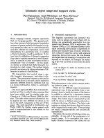
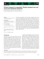
Three-dimensional presentation of predicted disorder in
various HA subtypes
In order to detect differences in the distribution of pre-
dicted intrinsic disorder in the various HA subtypes, a
series of three-dimensional models from proteins charac-
terized by X-ray crystallography were generated. Figure 1
shows a set of models generated by the combination of X-
ray data and results of the PONDR
®
VLXT analysis. The
HA1 and HA2 subunits are shown in light turquoise and
dark blue colors. The areas predicted to be disordered by
PONDR
®
VLXT are marked by red color. Figure 1 clearly
shows that various HA subtypes differ dramatically in the
amount and localization of the predicted intrinsic disor-
der.
PONDR
®
VLXT plots with normalized B-factor scores
Figure 2 compares the distributions of predicted disorder
within the sequences of HA
2
subunits from several influ-
enza A subtypes and strains with the distributions of nor-
malized B-factor scores evaluated from X-ray structures of
Virology Journal 2009, 6:69 />Page 4 of 12
(page number not for citation purposes)
the corresponding subunits. A residue with a PONDR
®
VLXT score at or above 0.5 is considered to be disordered.
For convenience, some areas predicted to be disordered
are marked by arrows and corresponding labels pointing
to the residues. Figure 2 shows that there is a general cor-
relation between the high level of predicted disorder in a
given HA region and its high B-factor scores. This indicates
that HA regions of predicted disorder can gain some fixed
structure during crystallization, although these regions
possess high mobility even in the crystal structures.
Figure 3 shows the peculiarities of the amino acid
sequences of HA
2
subunits from the various influenza
subtypes and strains in the vicinity of the residue 70. As
will be seen from the subsequent discussion, this region
might be related to the virulence of influenza viruses.
Discussion
H1N1 1918 viral strain has predicted disorder at a crucial
region of HA
A specific region is predicted to be disordered in the 1918 H1N1 but
not in other H1N1 strains
As mentioned in the introduction, the 1918 pandemic
caused global devastation in 1918–19, but became nonex-
istent by 1920 [24,27,28,66]. However, the H1N1 sub-
type of influenza A was still present in 1934. To answer
the question of why the 1918 strain was highly virulent,
we compared the intrinsic disorder distributions in amino
acid sequences of HA proteins from the 1918 strain and
later strains. Analysis of the data in Table 1 shows that the
overall percentage of predicted disorder does not vary
much for the different strains of H1N1, and that all of the
HA proteins could be considered as ordered by prediction.
However, a comparison of three-dimensional models
enhanced by the PONDR
®
VLXT annotations revealed
marked differences between various HA subtypes. The
HA
2
protein of the H1N1 1918 strain has two distinct
regions of predicted disorder (starting at positions 35 and
68, respectively, see Figures 1A and 2A), whereas two
other strains from the 1930s reveal only one disordered
region (starting at position 35, see Figures 1B–C and 2B–
C). The region that was predicted to be disordered in the
1918 strain but not in the H1N1 strains after 1919 is a
region at or around position number 68 of the HA
2
pro-
tein. The high level of predicted disorder in the region
around position 35 seems to be a common feature among
strains of the subtype H1N1. This region lies at the base of
the HA stalk. The missing region of predicted disorder
among the H1N1 strains in the 1930s, around position
68, is located at the higher end (the "tip") of the stalk. A
superficial analysis of the HA structure (Figure 1) suggests
that this region is likely to be located in an area close the
HA center of gravity, suggesting that motions of HA
2
at
this region are likely to have greatest impact on the HA
1
subunits, where the exposed area of the protein lies.
The crucial region of predicted disorder is associated with
virulent strains
A specific region of predicted disorder observed in 1918 H1N1 is
seen in both H5N1 and H9N2
A noticeable feature of the HA
2
subunit in the H5N1 sub-
type is the presence of two specific regions of predicted
disorder. The first region lies again at the base of the stalk,
even though on a different α-helix (see Figure 1). The
other region of predicted disorder coincides with the dis-
ordered fragment seen in the 1918 virus; i.e. this region is
positioned at the tip of the HA
2
stalk (around residue 68)
and is quite similar in size to that in the 1918 strain. This
region is also seen in the H9N2 subtype (See Figures 1E
Table 1: Summary Table of Percentage of Predicted Disorder in Each Chain of Influenza Hemagglutinins.
Viral Subtype Accessions Strain Description %VLXT
HA1 HA2 (HA1)
b
H1N1 1ruz A/South Carolina/1/18 Spanish Flu 1918 12 12
1ruy
A/Swine//Iowa/15/30 Swine 1930 15 5 Ordered
1rvx
A/Puerto Rico/3/34 1934 11 6
H3N2 1mqn A/Duck/Ukraine/63 Hong Kong Flu
1968
25 19 More Disordered
H5N1 2fk0 A/Vietnam/1203/2004 Avian Flu 15 18 Ordered
2ibx
A/Vietnam/1119/2004 12 12 Ordered
1jsn
A/Duck/Singapore/3/97 15 19 Ordered
H9N2 1jsd A/Swine/HongKong/9/98 Swine 1998 15 20 Ordered
The samples used are those elucidated using X-ray crystallography.
a
Percentage of residues that are predicted to be disordered by PONDR
@
VLXT in a given chain
b
Qualitative description based on predicted disorder for HA
1
Chain
Virology Journal 2009, 6:69 />Page 5 of 12
(page number not for citation purposes)
and 3E), but is noticeably shorter (residues 72–79).
The crucial region of predicted disorder in 1918 H1N1 is replaced
by more ordered regions in many less virulent strains
Given the data we have discussed thus far, one might sug-
gest that the region around position 68 could be com-
monly predicted to be disordered among all influenza
subtypes. However, the H7N3 subtype shown in Figures
1G and 2G does not have any predicted disorder near
position 68. This is also the case for the H3N2 subtype
shown in Figures 1F and 2F. Interestingly, the Hong Kong
H3N2 virus and the avian H7N3 virus, both of which have
ordered helical tips of HA
2
, were less virulent than the
Spanish influenza strain where this tip is predicted to be
disordered.
The low pathogenic avian influenza H7N3 strain does not have
predicted disorder at the helical tip
Infection of humans by avian influenza H7 subtypes has
been described in the Introduction. The human symp-
toms of the H7N3 have been largely mild, suggesting that
in the majority of cases we are dealing with low patho-
genic avian influenza (LPAI) strains. Although some
highly pathogenic avian influenza (HPAI) strains do exist,
the particular sample available via PDB (1ti8
.pdb, see Fig-
ures 1I and 2I); i.e. A/Turkey/Italy/02/, is a representative
Three-dimensional representation of the HA with predicted disorder annotationFigure 1
Three-dimensional representation of the HA with predicted disorder annotation. The regions in red represent
areas predicted to be disordered by VLXT. Conversely, the blue areas represent regions predicted to be ordered. A) 1918
H1N1 HA
2
, 1ruz.pdb B)1930 H1N1 1ruy.pdb, C) 1934 H1N1, 1rvx.pdb D) H5N1, 2fk0.pdb E) H5N1, 2ibx.pdb F) H5N1,
1jsn
.pdb G) H9N2.pdb, Swine H) H3N2, "Hong Kong Flu-1968 " Progenitor, Sample from 1963 I) H7N3, Avian.
Virology Journal 2009, 6:69 />Page 6 of 12
(page number not for citation purposes)
PONDR
@
VLXT and B-factor plots of the HA
2
Figure 2
PONDR
@
VLXT and B-factor plots of the HA
2
. The blue curves represent the VLXT score, while the black ones repre-
sent the normalized B-factor. A) 1918 H1N1 HA2, 1ruz
.pdb B)1930 H1N1 1ruy.pdb, C) 1934 H1N1, 1rvx.pdb D) H5N1,
2fk0
.pdb E) H5N1, 2ibx.pdb F) H5N1, 1jsn.pdb G) H9N2, 1jsd.pdb, Swine H) H3N2, "Hong Kong Flu-1968 " Progenitor, Sam-
ple from 1963, 1mqn
.pdb I) H7N3, Avian, H7N3, Italy, Avian, 1it8 [24,22,20].
Virology Journal 2009, 6:69 />Page 7 of 12
(page number not for citation purposes)
of the LPAI strains [67,68] (Table 1). A visual inspection
of the three-dimensional structure (Figure 1I) reveals
some interesting characteristics. The crucial region of pre-
dicted disorder observed in the highly virulent 1918
H1N1 strain (residues 68–79) is absent in this H7 sub-
type. An inspection of regions in close proximity to the
helical tip (HA
2
residues 68–79) reveals that the adjacent
loop (residues 61–68, see Figure 2I) is predicted to be dis-
ordered. This suggests that H7-related viruses could
become more pathogenic with mutations that extend the
disordered region to include the helical tip.
The H9N2 puzzle and modulation of virulence
A modulating "switch" in H9N2: infectivity versus immune evasion
The H9N2 virus is generally non-virulent for mammals
[69]. However, our analysis revealed that it has a region of
predicted disorder at the tip of its helical stalk. This region
of predicted disorder in the H9N2 HA
2
is noticeably
shorter than that in the 1918 H1N1 protein. On the other
hand, studies on H9N2 strains with low virulence
revealed that a single point mutation can make these
viruses more virulent [69]. In fact, Leu226 in the HA
receptor-binding site (RBS), responsible for human virus-
like receptor specificity, was found to be important for
transmission of the H9N2 viruses in ferrets [69]. Interest-
ingly, earlier it has been shown that changes in the HA
receptor-binding site of H9N2 isolates allow adaptation
to mammalian-type sialic receptors [70] and that a change
at position 227 of the HA
1
subunit of H5N1 had a notice-
able effect on its virulence in mice [71]. This suggests a
mechanism by which the virus can potentially modulate
its virulence. The virulence arising from mutations near
the receptor binding site may work in tandem with the
immune evasion arising from tip of the HA
2
stalk. One
way that the former can modulate the effects of the latter
is by controlling the infectivity of the virus. A virus that
binds to its host with great efficiency and is also effective
in evading the immune systems creates the "perfect
storm" for virulence.
A secondary switch for virulence of H9N2 and H5N1via
oligosaccharides
It has been suggested that immune pressure caused by the
use of a vaccine could create a survival advantage for those
influenza viruses that undergo antigenic variation [72].
This evolution produces low-virulence escape mutants,
many of which were associated with the acquisition of
new glycosylation sites in the HA
1
subunit, at positions
198 and 131 in H9N2 and H5N1, respectively [73,74]. It
has been reported that the restored pathogenicity of low-
virulence H5 and H9 escape mutants by lung-to-lung pas-
sage in mice could be attributed not only to reacquisition
of the wild-type HA gene sequence, but was also associ-
ated either with the removal of a glycosylation site (the
one acquired previously by the escape mutant) without
the exact restoration of the initial wild-type amino acid
sequence, or, for an H5 escape mutant that had no newly
acquired glycosylation sites, with an additional amino
acid change in a remote part of the HA molecule (at posi-
tion 156 of HA
1
) [75]. This clearly shows the crucial role
of the loss of glycosylation sites in restoration of the viru-
lence of H9 and H5 readaptants. It should be noted that
mutations affecting the glycosylation of HA are likely to
affect virulence, since it has been proposed that it is com-
mon for the glyco-conjugate to act in tandem with intrin-
sic disorder of viral proteins in immune evasion [76,77].
Therefore, it should not be surprising that modulation of
the virulence will often involve changes at the glyco-con-
jugate.
Possible mechanism for initial wave of non-virulent strain in 1918
By analogy, this may also help explain why and how the
first wave of the 1918 H1N1 virus was not as virulent as
the second [24,28]. As little as a single mutation could
have set off the virulent potential of the second wave of
the 1918 H1N1 virus.
Immune evasion, virulence, and the 'cytokine storm'
Avian influenza viruses and antigenic shift
It is believed that one of the major reasons for the inability
of the global population to have effective neutralizing
antibodies against the viruses (including the influenza
virus A) is the predisposition of the viral genomes for the
genetic reassortment, which results from the segmented
structure of genomes. As a result of this genome segmen-
tation, shuffling of gene segments can occur if two differ-
ent subtypes of influenza A virus infect the same host cell
[78]. This is an important mechanism, as all combina-
tions of the 16 different HA antigens (H1 to H16) and 9
different NA antigens (N1 to N9) are found in water fowl,
Protein sequence of HA
2
around tesidue 68Figure 3
Protein sequence of HA
2
around tesidue 68. Vertical
lines show where mutations have taken place. The numbers
denote the residue numbers.
Virology Journal 2009, 6:69 />Page 8 of 12
(page number not for citation purposes)
whereas only H1 to 3 and N1 to 2 viral subtypes are com-
monly found in humans with influenza. Therefore, if a
human H3N2 virus and an avian H5N1 virus co-infect a
human, the genetic reassortment can produce a novel
H5N2 virus, which then can be efficiently transmitted
from human to human because the majority of the gene
segments apart from H5 come from the human virus. This
shuffling of gene segments obviously leads to significant
antigenic changes, known as antigenic shift, as a result of
which most of the population would not have any effec-
tive neutralizing antibodies against the new virus subtype
[78]. In turn, this lack of effective antibodies can be
responsible for high virulence of new virus subtype.
Crucial disordered regions associated with avian influenza viruses
A consistent trend that has been observed is the tendency
for the crucial disordered HA
2
region around positions
67–79 to be seen among avian or avian-related viruses
(Figures 1A, D–G, I). In fact, all H5N1 and H9N2 samples
that we analyzed have this predicted disordered region
(although it is shortened in the H9N2 (Figure 1G)). Even
the avian-related H7 subtype (Figures 1I, 2I) has a tinge of
disordered at the edge of this region. This suggests that
avian-related influenza viruses depend on this region for
their fitness. They possibly utilize disorder in this region
to evade the immune systems of various hosts that they try
to move into.
Crucial disordered regions not commonly associated with human
influenza viruses
By contrast, except for the 1918 H1N1 strain, most human
influenza A viruses have not been observed to have this
disordered region at the HA
2
(Figures 1B, C, H and 2B, C,
H). This is especially the case for viruses that have been
circulating in humans for long period of time. A hint for
such pattern can be seen in the evolution of the H1N1
virus after 1918 [5,6]. The virulent 1918 H1N1 virus
became extinct by 1920 [18]. Subsequent H1N1 viruses
were not as virulent as the 1918 H1N1 strain [6]. This cor-
relates with our inability to find any H1N1 samples from
dates after 1918 with the "fangs"(i.e. crucial disordered
region around positions 67–79, Figure 1B, C), even
though some of the samples from the 1930s were proba-
bly descendents of the 1918 virus given the similarity of
the predicted disorder patterns of their HA
1
(Figure 1A–
C).
"Cytokine storm" and intrinsic disorder in HA
Most subtypes of avian influenza virus, such as H9, cause
very mild disease in poultry. However, the H5 and H7
subtypes are known to cause outbreaks involving massive
deaths in domestic poultry [78]. What is even worse avian
influenza A viruses can be zoonotically transmitted to
humans leading to serious outbreaks of human sickness
caused by avian virus. Such outbreaks can be very deadly.
For example, the 1997 outbreak of H5N1 influenza in
Hong Kong Special Administrative Region (HKSAR)
involved 18 human cases, with six fatalities [79,80].
The fatality rate of avian influenza epidemic (>50%)
occurred in Southeast Asia in 1997 was significantly
higher compared to the pandemic caused by the 1918
H1N1 (5–10%). When considering the fatal/total case
numbers (208/340) reported by World Health Organiza-
tion in respect of December 14th, 2007, the mortality rate
has reached to 61 percent [81].
One of the reasons for the high mortality rates associated
with certain subtypes of avian influenza were attributed to
so-called "cytokine storm" or hypercytokinemia, which is
characterized by the extremely enhanced production and
secretion of large numbers and excessive levels of pro-
inflammatory cytokines [81]. This hypercytokinemia is a
result of the overactive inflammatory response related to
the virus-induced cytokine dysregulation. In fact, H5N1
viruses were shown to serve as very strong inducers of var-
ious cytokines and chemokines, such as TNF-α, IFN-γ,
IFN-α/β, IL-1, IL-6, IL-8, MIP-1, MIG, IP-10, MCP-1, and
RANTES, leading to the "cytokine storm". This "cytokine
storm" is believed to be responsible for the development
of lethal clinical symptoms such as extensive pulmonary
oedema, acute bronchopneumoniae, alveolar haemor-
rhage, reactive haemophagocytosis, and acute respiratory
distress syndrome, associated with necrosis and tissue
destruction [81]. It has been reported than mutations in
NS1, PB2, HA and NA could be responsible for the initia-
tion of the cytokine storm. These mutations can increase
the viral replication rate, expend the tissue tropism, facili-
tate the systemic invasion and increase the resistance of a
virus against the host antiviral response [81]. We believe
that the crucial disordered region around positions 67–79
discussed in our paper can contribute to the virus resist-
ance toward the host immune system and therefore can be
one of the factors provoking the "cytokine storm" in
humans affected by avian H5N1 (or related) influenza.
Sequential analysis: shuffling and grouping by residues of
the same polarity around position 68 of HA
2
Shuffling by polarity in virulent strains
An analysis of the HA
2
sequence of the 1918 H1N1 strain
shows that a set of polar residues located in the vicinity of
residue 68 make this segment more polar and, thus,
potentially more disordered. Figure 2 shows that the nor-
malized B-factor values vary with PONDR
®
VLXT values.
However, the effects seem to be reduced in many cases.
For example, it can be seen that a peak in the normalized
B-factor curve near position 60 in the 1918 H1N1 HA
2
plot (Figure 2A, 1ruz.pdb), corresponds to the PONDR
®
VLXT maximum in the vicinity of residues 68–79. This
crucial PONDR
®
VLXT maximum, seen in the 1918 H1N1
Virology Journal 2009, 6:69 />Page 9 of 12
(page number not for citation purposes)
and H5N1 HA
2
, is also seen on a smaller scale in the
1930s H1N1 HA
2
. The peak of the 1918 H1N1 HA
2
is rel-
atively higher than those from the 1930s, suggesting that
this region in the 1918 H1N1 HA
2
is more flexible. This is
further supported by the corresponding B-factor curves
(see Figure 2).
Swine versus avian influenza viruses: patterns of predicted
disorder
Predicted disorder patterns at the base of the stalk seem to be
dependent on the hosts
Another puzzle is the presence of predicted disorder at the
bottom of the stalk. Often the predicted disorder appears
at the bottom of the longer alpha helix, but sometimes it
appears at the base of the smaller helix. An example of the
former is the H1N1 subtype, whereas an example of the
latter is H5N1. In yet other cases, such as H7N3 and
H3N2, predicted disorder appears at the bases of both hel-
ices. A hint for a possible explanation of this behavior is
provided by the analysis of the swine variant of the H9N2
subtype (see Figures 1G and 2G) [82]. In this instance, a
region of predicted disorder appears at the bottom of the
longer α-helix, just as in H1N1. This is of particular inter-
est since H1N1 is believed to be of swine-host origin
[28,82]. Therefore, we suggest that the position of this pre-
dicted disorder region at the stalk bottom is dependent on
the host type. Interestingly, the 1918 H1N1 is suspected to
be of both avian and swine origins [24,83-85] just as the
swine strain of the H9N2 subtype is of avian origin
[82,86]. The pattern of predicted disorder of the 1918
H1N1 HA, which resembles that of H9N2, may therefore
cast more light on the origin of the 1918 H1N1 virus. The
theory that the 1918 H1N1 virus is of avian origin but
evolved in swine [24,83] seems to be supported here.
Conclusion
Virulence of influenza A and intrinsic disorder
Virulence tied to the predicted disorder at segment at to the tip of
the
α
-helix stalk
It can be seen that all the virulent strains analysed in this
study have a region of predicted disorder at the top of the
largest alpha helix of the HA stalk (near the residues 68–
79, see Figures 1 and 2). This is the case for the 1918
H1N1 strain and for all the virulent samples of the H5N1
and H9N2 subtypes. Although the similarity between
H9N2 and H5N1 is striking, the pathogenesis of H9N2 is
still relatively unknown since few human cases have been
reported so far. Our data suggest that the H9 subtypes are
likely to be highly virulent as their HA
2
contains the pre-
dicted disorder region similar to segment of predicted dis-
order seen in all of the virulent strains studied.
Conversely, the subtypes and strains that are known to be
less virulent than the 1918 H1N1 and H5N1 have no pre-
dicted disorder at the tip of the stalk. Examples of this
behavior are H3N2 and the particular strain of H7N3.
H9N2 and 1918 H1N1: masking the virulence
The H9N2 subtype is potentially able to modulate its vir-
ulent nature by affecting the efficiency of binding to the
host cells, thus controlling its ability to evade the immune
system. This is supported by the observation that a single
mutation near the receptor binding site is generally suffi-
cient to convert H9N2 and some H5N1 strains from non-
virulent to highly pathogenic. This also represents a
potential mechanism by which the second wave of the
1918 H1N1 virus became more virulent than the first
wave.
Virulence, evasion of immune response and intrinsic disorder
We propose that the high mobility of the exposed region
of the HA trimer accounts for the evasion of the initial
immune response. This highly dynamic nature of the
potentially immunogenic region weakens the binding of
antibodies and other immune response-related molecules
to the HA molecule. In this way, the virus buys some time
to invade the host. It is also possible that the immune sys-
tem does not elicit the adequate immune response
throughout all stages of the disease as tight binding to the
highly dynamic HA is difficult. Evidence supporting both
hypotheses can be found in the observation that when the
1918 H1N1 virus was introduced to mice, unusually high
viral loads were seen within a short period of time [25].
The crucial region of predicted disorder at the helical tip is
seen in all HAs of influenza viruses of avian origin ana-
lysed thus far. It is highly plausible that this disordered
region is needed for the virus to move between various
species of birds by allowing sufficient copies of the virion
to avoid immune detection in the host's body in order to
increase the odds of infections. Therefore, the increased
intrinsic disorder may be associated with increased fitness
of the virus.
Strategies for vaccine development against the pathogenic viruses
Development of vaccine for highly virulent viruses such as
1918 H1N1 or H5N1 may be fraught with dangers as a
result of the ability of their proteins to trigger fatal
immune responses [87]. The results here provide new
potential strategies to develop vaccines for virulent influ-
enza strains. Relatively non-virulent immunogenic pro-
teins could be developed in either of two ways. One way
is to use disorder predictors such as PONDR
®
VLXT to
quickly identify variants of the virulent viruses that are
predicted to be non-virulent by lacking the region of pre-
dicted disorder at the tip of the stalk. Another strategy
would be to mutate the crucial region in the viruses them-
selves at the crucial sequence in the HA
2
protein.
Virology Journal 2009, 6:69 />Page 10 of 12
(page number not for citation purposes)
Methods
Implementation details
The list of viral proteins of interest incorporates the pro-
teins of Orthomyxoviruses. Searches were done on the list
using the Entrez website. Available samples were ran-
domly chosen with preferences given to those with longer
chains and those with binding partners. Whenever possi-
ble, homologous proteins from different virus strains
were included as samples and annotated. The respective
FASTA and PDB [88] files were downloaded and stored
using a JAVA
®
program. The database design has been
described in a previous paper [77].
PONDR
®
VLXT and B-factor normalization plots
The B-Factor values were retrieved from the respective
PDB files and placed in the respective table in the MYSQL
database already designed and populated [77]. The
PONDR
®
VLXT and normalized B-factor graphs were plot-
ted using MS-EXCEL
®
via output files obtained from SQL.
The normalized B-factor values were calculated using
EXCEL spreadsheet.
Graphics tools
Three-dimensional graphics was developed utilizing Jmol
[89] in conjunction with JAVA programming. The JAVA-
JDBC program reads from the prediction information
stored in the MYSQL
®
database and generates the corre-
sponding Jmol script, which creates the necessary molec-
ular graphics for the protein.
Competing interests
The authors declare that they have no competing interests.
Authors' contributions
GKMG proposed the idea of the study, implemented the
experiments, carried out the analyses, and drafted the
manuscript. AKD helped to design experiments and par-
ticipated in the manuscript drafting. VNU coordinated the
studies, participated in their design and helped to draft
the manuscript. All authors read and approved the final
manuscript.
Acknowledgements
We express our deepest gratitude to Prof. Ed Harper for careful reading
and editing the manuscript. This work was supported in part by the grants
R01 LM007688-01A1 (to A.K.D and V.N.U.) and GM071714-01A2 (to
A.K.D and V.N.U.) from the National Institutes of Health and the Program
of the Russian Academy of Sciences for the "Molecular and cellular biology"
(to V.N.U.). We gratefully acknowledge the support of the IUPUI Signature
Centers Initiative.
References
1. Brooks GF, Butel JS, Morse SA: Jawetz, Melnick and Adelberg's medical
microbiology 23rd edition. New York: Lange/McGraw-Hill; 2004.
2. Fouchier RA, Munster V, Wallensten A, Bestebroer TM, Herfst S,
Smith D, Rimmelzwaan GF, Olsen B, Osterhaus AD: Characteriza-
tion of a novel influenza A virus hemagglutinin subtype
(H16) obtained from black-headed gulls. J Virol 2005,
79:2814-2822.
3. Garcia-Sastre A, Whitley RJ: Lessons learned from reconstruct-
ing the 1918 influenza pandemic. J Infect Dis 2006, 194(Suppl
2):S127-132.
4. Reid AH, Taubenberger JK, Fanning TG: Evidence of an absence:
the genetic origins of the 1918 pandemic influenza virus. Nat
Rev Microbiol 2004, 2:909-914.
5. Reid AH, Taubenberger JK, Fanning TG: The 1918 Spanish influ-
enza: integrating history and biology. Microbes Infect 2001,
3:81-87.
6. Kilbourne ED: Influenza pandemics of the 20th century. Emerg
Infect Dis 2006, 12:9-14.
7. Tweed SA, Skowronski DM, David ST, Larder A, Petric M, Lees W, Li
Y, Katz J, Krajden M, Tellier R, et al.: Human illness from avian
influenza H7N3, British Columbia. Emerg Infect Dis 2004,
10:2196-2199.
8. Update: isolation of avian influenza A(H5N1) viruses from
humans – Hong Kong, 1997–1998. MMWR Morb Mortal Wkly Rep
1998, 46:1245-1247.
9. Wuethrich B: Infectious disease. An avian flu jumps to people.
Science 2003, 299:1504.
10. World Health Organization: Avian influenza, Viet Nam –
update. Wkly Epidemiol Rec 2005, 80:233-234.
11. Guan Y, Shortridge KF, Krauss S, Webster RG: Molecular charac-
terization of H9N2 influenza viruses: were they the donors of
the "internal" genes of H5N1 viruses in Hong Kong? Proc Natl
Acad Sci USA 1999, 96:9363-9367.
12. Lin YP, Shaw M, Gregory V, Cameron K, Lim W, Klimov A, Subbarao
K, Guan Y, Krauss S, Shortridge K, et al.: Avian-to-human trans-
mission of H9N2 subtype influenza A viruses: relationship
between H9N2 and H5N1 human isolates. Proc Natl Acad Sci
USA 2000, 97:9654-9658.
13. Skehel JJ, Wiley DC: Coiled coils in both intracellular vesicle
and viral membrane fusion. Cell 1998, 95:871-874.
14. Skehel JJ, Wiley DC: Receptor binding and membrane fusion in
virus entry: the influenza hemagglutinin. Annu Rev Biochem
2000, 69:531-569.
15. Eckert DM, Kim PS: Mechanisms of viral membrane fusion and
its inhibition. Annu Rev Biochem 2001, 70:777-810.
16. Wiley DC, Skehel JJ: The structure and function of the hemag-
glutinin membrane glycoprotein of influenza virus. Annu Rev
Biochem 1987, 56:365-394.
17. Skehel JJ, Bayley PM, Brown EB, Martin SR, Waterfield MD, White JM,
Wilson IA, Wiley DC: Changes in the conformation of influenza
virus hemagglutinin at the pH optimum of virus-mediated
membrane fusion. Proc Natl Acad Sci USA 1982, 79:968-972.
18. Stevens J, Corper AL, Basler CF, Taubenberger JK, Palese P, Wilson
IA: Structure of the uncleaved human H1 hemagglutinin
from the extinct 1918 influenza virus. Science 2004,
303:1866-1870.
19. Durrer P, Galli C, Hoenke S, Corti C, Gluck R, Vorherr T, Brunner J:
H+-induced membrane insertion of influenza virus hemag-
glutinin involves the HA2 amino-terminal fusion peptide but
not the coiled coil region. J Biol Chem 1996, 271:13417-13421.
20. Wilson IA, Skehel JJ, Wiley DC: Structure of the haemagglutinin
membrane glycoprotein of influenza virus at 3 A resolution.
Nature 1981, 289:366-373.
21. Bullough PA, Hughson FM, Skehel JJ, Wiley DC: Structure of influ-
enza haemagglutinin at the pH of membrane fusion. Nature
1994, 371:37-43.
22. Weber T, Paesold G, Galli C, Mischler R, Semenza G, Brunner J: Evi-
dence for H(+)-induced insertion of influenza hemagglutinin
HA2 N-terminal segment into viral membrane. J Biol Chem
1994,
269:18353-18358.
23. Wharton SA, Calder LJ, Ruigrok RW, Skehel JJ, Steinhauer DA, Wiley
DC: Electron microscopy of antibody complexes of influenza
virus haemagglutinin in the fusion pH conformation. Embo J
1995, 14:240-246.
24. Taubenberger JK, Reid AH, Janczewsk TA, Fanning TG: Integrating
historical, clinical and molecular genetic data in order to
explain the origin and virulence of the 1918 Spanish influenza
virus. Philos Trans R Soc Lond B Biol Sci 2001, 356:1829.
25. Kobasa D, Takada A, Shinya K, Hatta M, Halfmann P, Theriault S,
Suzuki H, Nishimura H, Mitamura K, Sugaya N, et al.: Enhanced vir-
Virology Journal 2009, 6:69 />Page 11 of 12
(page number not for citation purposes)
ulence of influenza A viruses with the haemagglutinin of the
1918 pandemic virus. Nature 2004, 431:703-707.
26. Reid A: The effects of the 1918–1919 influenza pandemic on
infant and child health in Derbyshire. Med Hist 2005, 49:29-54.
27. Kilbourne ED: Influenza pandemics of the 20th century. Emerg
Infect Dis 2006, 12:9-14.
28. Reid AH, Taubenberger JK, Fanning TG: The 1918 Spanish influ-
enza: Integrating history and biology. Microbes Infect. 2001,
3(1):81-87.
29. Dunker AK, Lawson JD, Brown CJ, Williams RM, Romero P, Oh JS,
Oldfield CJ, Campen AM, Ratliff CM, Hipps KW, et al.: Intrinsically
disordered protein. J Mol Graph Model 2001, 19:26-59.
30. Wright PE, Dyson HJ: Intrinsically unstructured proteins: re-
assessing the protein structure-function paradigm. J Mol Biol
1999, 293:321-331.
31. Weinreb PH, Zhen W, Poon AW, Conway KA, Lansbury PT Jr:
NACP, a protein implicated in Alzheimer's disease and
learning, is natively unfolded. Biochemistry 1996,
35:13709-13715.
32. Uversky VN, Gillespie JR, Fink AL: Why are "natively unfolded"
proteins unstructured under physiologic conditions? Proteins
2000, 41:415-427.
33. Uversky VN, Gillespie JR, Millett IS, Khodyakova AV, Vasiliev AM,
Chernovskaya TV, Vasilenko RN, Kozlovskaya GD, Dolgikh DA, Fink
AL, et al.: Natively unfolded human prothymosin alpha adopts
partially folded collapsed conformation at acidic pH. Biochem-
istry 1999, 38:15009-15016.
34. Dunker AK, Garner E, Guilliot S, Romero P, Albrecht K, Hart J, Obra-
dovic Z, Kissinger C, Villafranca JE: Protein disorder and the evo-
lution of molecular recognition: theory, predictions and
observations. Pac Symp Biocomput 1998:473-484.
35. Dunker AK, Brown CJ, Lawson JD, Iakoucheva LM, Obradovic Z:
Intrinsic disorder and protein function. Biochemistry 2002,
41:6573-6582.
36. Uversky VN, Oldfield CJ, Dunker AK: Showing your ID: intrinsic
disorder as an ID for recognition, regulation and cell signal-
ing. J Mol Recognit 2005, 18:343-384.
37. Daughdrill GW, Pielak GJ, Uversky VN, Cortese MS, Dunker AK:
Natively disordered proteins. In Protein Folding Handbook Edited
by: Buchner J, Kiefhaber T. Weinheim, Germany: Wiley-VCH, Verlag
GmbH & Co. KGaA; 2005:271-353.
38. Dunker AK, Cortese MS, Romero P, Iakoucheva LM, Uversky VN:
Flexible nets. The roles of intrinsic disorder in protein inter-
action networks. FEBS J 2005, 272:5129-5148.
39. Xie H, Vucetic S, Iakoucheva LM, Oldfield CJ, Dunker AK, Obradovic
Z, Uversky VN: Functional anthology of intrinsic disorder. 3.
Ligands, post-translational modifications, and diseases asso-
ciated with intrinsically disordered proteins. J Proteome Res
2007, 6:1917-1932.
40. Vucetic S, Xie H, Iakoucheva LM, Oldfield CJ, Dunker AK, Obradovic
Z, Uversky VN: Functional anthology of intrinsic disorder. 2.
Cellular components, domains, technical terms, develop-
mental processes, and coding sequence diversities corre-
lated with long disordered regions. J Proteome Res 2007,
6:1899-1916.
41. Xie H, Vucetic S, Iakoucheva LM, Oldfield CJ, Dunker AK, Uversky
VN, Obradovic Z: Functional anthology of intrinsic disorder. 1.
Biological processes and functions of proteins with long dis-
ordered regions. J Proteome Res 2007, 6:1882-1898.
42. Uversky VN: What does it mean to be natively unfolded? Eur J
Biochem 2002, 269:2-12.
43. Uversky VN: Natively unfolded proteins: a point where biol-
ogy waits for physics. Protein Sci 2002, 11:739-756.
44. Uversky VN: Protein folding revisited. A polypeptide chain at
the folding-misfolding-nonfolding cross-roads: which way to
go? Cell Mol Life Sci 2003, 60:1852-1871.
45. Tompa P: Intrinsically unstructured proteins. Trends Biochem Sci
2002, 27:527-533.
46. Dyson HJ, Wright PE: Intrinsically unstructured proteins and
their functions. Nat Rev Mol Cell Biol 2005, 6:197-208.
47. Tompa P: The interplay between structure and function in
intrinsically unstructured proteins. FEBS Lett 2005,
579:3346-3354.
48. Galea CA, Wang Y, Sivakolundu SG, Kriwacki RW: Regulation of
cell division by intrinsically unstructured proteins: intrinsic
flexibility, modularity, and signaling conduits. Biochemistry
2008, 47:7598-7609.
49. Dunker AK, Oldfield CJ, Meng J, Romero P, Yang JY, Chen JW, Vacic
V, Obradovic Z, Uversky VN: The unfoldomics decade: an
update on intrinsically disordered proteins. BMC Genomics
2008, 9(Suppl 2):S1.
50. Dunker AK, Silman I, Uversky VN, Sussman JL: Function and struc-
ture of inherently disordered proteins. Curr Opin Struct Biol
2008, 18:756-764.
51. Oldfield CJ, Meng J, Yang JY, Yang MQ, Uversky VN, Dunker AK:
Flexible nets: disorder and induced fit in the associations of
p53 and 14-3-3 with their partners. BMC Genomics 2008,
9(Suppl 1):S1.
52. Gsponer J, Futschik ME, Teichmann SA, Babu MM: Tight regulation
of unstructured proteins: from transcript synthesis to pro-
tein degradation. Science 2008, 322:1365-1368.
53. Uversky VN, Dunker AK: Biochemistry. Controlled chaos. Sci-
ence 2008, 322:1340-1341.
54. Campen A, Williams RM, Brown CJ, Meng J, Uversky VN, Dunker AK:
TOP-IDP-scale: a new amino acid scale measuring propen-
sity for intrinsic disorder. Protein Pept Lett 2008, 15:956-963.
55. Brown CJ, Takayama S, Campen AM, Vise P, Marshall TW, Oldfield
CJ, Williams CJ, Dunker AK: Evolutionary rate heterogeneity in
proteins with long disordered regions. J Mol Evol 2002,
55:104-110.
56. Ferron F, Longhi S, Canard B, Karlin D: A practical overview of
protein disorder prediction methods. Proteins 2006, 65:1-14.
57. Radivojac P, Iakoucheva LM, Oldfield CJ, Obradovic Z, Uversky VN,
Dunker AK: Intrinsic disorder and functional proteomics. Bio-
phys J 2007, 92:1439-1456.
58. Uversky VN, Radivojac P, Iakoucheva LM, Obradovic Z, Dunker AK:
Prediction of intrinsic disorder and its use in functional pro-
teomics. Methods Mol Biol 2007, 408:69-92.
59. Dosztanyi Z, Sandor M, Tompa P, Simon I: Prediction of protein
disorder at the domain level. Curr Protein Pept Sci 2007,
8:161-171.
60. Dosztanyi Z, Tompa P: Prediction of protein disorder. Methods
Mol Biol 2008, 426:103-115.
61. Li X, Romero P, Rani M, Dunker AK, Obradovic Z: Predicting pro-
tein disorder for N-, C-, and internal regions. Genome Inform
Ser Workshop Genome Inform 1999, 10:30-40.
62. Romero P, Obradovic Z, Li X, Garner EC, Brown CJ, Dunker AK:
Sequence complexity of disordered protein. Proteins 2001,
42:38-48.
63. Vucetic S, Brown CJ, Dunker AK, Obradovic Z: Flavors of protein
disorder. Proteins 2003, 52:573-584.
64. Obradovic Z, Peng K, Vucetic S, Radivojac P, Brown CJ, Dunker AK:
Predicting intrinsic disorder from amino acid sequence. Pro-
teins 2003, 53(Suppl 6):566-572.
65. Goh GK-M, Dunker AK, Uversky VN: Protein intrinsic disorder
toolbox for comparative analysis of viral proteins. BMC
Genomics 2008, 9:S4.
66. Reid A: The effects of the 1918–1919 influenza pandemic on
infant and child health in Derbyshire. Med Hist 2005, 49:29-54.
67. Russell RJ, Gamblin SJ, Haire LF, Stevens DJ, Xiao B, Ha Y, Skehel JJ:
H1 and H7 influenza haemagglutinin structures extend a
structural classification of haemagglutinin subtypes. Virology
2004, 325:287-296.
68. Di Trani LBB, Cordioli P, Muscillo M, Vignolo E, Moreno A, Tollis M:
Molecular characterization of low pathogenicity H7N3 avian
influenza viruses isolated in Italy.
Avian Dis. 2004,
48(2):376-383.
69. Wan H, Sorrell E, Song H, Hossain M, Ramirez-Nieto G, Monne I, Ste-
vens J, Cattoli G, Capua I, Chen L, et al.: Replication and transmis-
sion of H9N2 influenza viruses in ferrets: evaluation of
pandemic potential. PLoS ONE 2008, 3:e2923.
70. Matrosovich MN, Krauss S, Webster RG: H9N2 influenza A
viruses from poultry in Asia have human virus-like receptor
specificity. Virology 2001, 281:156-162.
71. Hatta M, Gao P, Halfmann P, Kawaoka Y: Molecular basis for high
virulence of Hong Kong H5N1 influenza A viruses. Science
2001, 293:1840-1842.
72. Guan Y, Poon LL, Cheung CY, Ellis TM, Lim W, Lipatov AS, Chan KH,
Sturm-Ramirez KM, Cheung CL, Leung YH, et al.: H5N1 influenza:
a protean pandemic threat. Proc Natl Acad Sci USA 2004,
101:8156-8161.
Publish with BioMed Central and every
scientist can read your work free of charge
"BioMed Central will be the most significant development for
disseminating the results of biomedical research in our lifetime."
Sir Paul Nurse, Cancer Research UK
Your research papers will be:
available free of charge to the entire biomedical community
peer reviewed and published immediately upon acceptance
cited in PubMed and archived on PubMed Central
yours — you keep the copyright
Submit your manuscript here:
/>BioMedcentral
Virology Journal 2009, 6:69 />Page 12 of 12
(page number not for citation purposes)
73. Kaverin NV, Rudneva IA, Ilyushina NA, Lipatov AS, Krauss S, Webster
RG: Structural differences among hemagglutinins of influ-
enza A virus subtypes are reflected in their antigenic archi-
tecture: analysis of H9 escape mutants. J Virol 2004,
78:240-249.
74. Kaverin NV, Rudneva IA, Ilyushina NA, Varich NL, Lipatov AS, Smir-
nov YA, Govorkova EA, Gitelman AK, Lvov DK, Webster RG: Struc-
ture of antigenic sites on the haemagglutinin molecule of H5
avian influenza virus and phenotypic variation of escape
mutants. J Gen Virol 2002, 83:2497-2505.
75. Rudneva I, Ilyushina N, Timofeeva T, Webster R, Kaverin N: Resto-
ration of virulence of escape mutants of H5 and H9 influenza
viruses by their readaptation to mice. J Gen Virol 2005,
86:2831-2838.
76. Goh GKM, Uversky V, Dunker AK: A Comparative Analysis of
Viral Matrix Proteins Using Disorder Predictor. Virol J
2008:126.
77. Goh GKM, Dunker AK, Uversky V: Protein Intrinsic Disorder
Toolbox for Comparative Analysis of Viral Proteins. BMC
Genomics. 2008, 9(Suppl 2):S4-S7.
78. Yuen KY, Wong SS: Human infection by avian influenza A
H5N1. Hong Kong Med J 2005, 11:189-199.
79. Yuen KY, Chan PK, Peiris M, Tsang DN, Que TL, Shortridge KF, Che-
ung PT, To WK, Ho ET, Sung R, Cheng AF: Clinical features and
rapid viral diagnosis of human disease associated with avian
influenza A H5N1 virus. Lancet 1998, 351:467-471.
80. Chan PK: Outbreak of avian influenza A(H5N1) virus infection
in Hong Kong in 1997. Clin Infect Dis 2002, 34(Suppl 2):S58-64.
81. Us D: [Cytokine storm in avian influenza]. Mikrobiyol Bul 2008,
42:365-380.
82. Ha Y, Stevens DJ, Skehel JJ, Wiley DC: X-ray structures of H5
avian and H9 swine influenza virus hemagglutinins bound to
avian and human receptor analogs. Proc Natl Acad Sci USA 2001,
98:11181-11186.
83. Reid AH, Taubenberger JK, Fanning TG: Evidence of an absence:
the genetic origins of the 1918 pandemic influenza virus. Nat
Rev Microbiol 2004, 2:909-914.
84. Webster RG, Sharp GB, Claas EC:
Interspecies transmission of
influenza viruses. Am J Respir Crit Care Med 1995 1995, 152:S25-23.
85. Gamblin SJ, Haire LF, Russell RJ, Stevens DJ, Xiao B, Ha Y, Vasisht N,
Steinhauer DA, Daniels RS, Elliot A, et al.: The structure and
receptor binding properties of the 1918 influenza hemagglu-
tinin. Science 2004, 303:1838-1842.
86. Lin YP, Shaw M, Gregory V, Cameron K, Lim W, Klimov A, Subbarao
K, Guan Y, Krauss S, Shortridge K, et al.: Avian-to-human trans-
mission of H9N2 subtype influenza A viruses: relationship
between H9N2 and H5N1 human isolates. Proc Natl Acad Sci
USA 2000, 97:9654-9658.
87. Loo YM, Gale M: Influenza: Fatal immunity and the 1918 virus.
Nature 2007, 445:267-268.
88. Berman HM, Westbrook Z, Feng G, Bhat H, Weissig H, Sindyalov LN,
Bourne PE: The Protein Data Bank. Nucleic Acids Res 2000,
28:235-242.
89. Herráez A: Biomolecules in the computer: Jmol to the rescue.
Biochemistry and Molecular Biology Education 2006, 34:255-261.