Báo cáo khoa học: " Genetic diversity of the E Protein of Dengue Type 3 Virus" pps
Bạn đang xem bản rút gọn của tài liệu. Xem và tải ngay bản đầy đủ của tài liệu tại đây (368.82 KB, 13 trang )
BioMed Central
Page 1 of 13
(page number not for citation purposes)
Virology Journal
Open Access
Research
Genetic diversity of the E Protein of Dengue Type 3 Virus
Alberto A Amarilla
1
, Flavia T de Almeida
2
, Daniel M Jorge
3
, Helda L Alfonso
1
,
Luiza A de Castro-Jorge
1
, Nadia A Nogueira
4
, Luiz T Figueiredo
1
and
Victor H Aquino*
2
Address:
1
Virology Research Center, School of Medicine of Ribeirão Preto/USP, Ribeirão Preto – SP, Brazil,
2
Department of Clinical, Toxicological
and Bromatological Analysis, FCFRP/USP, Ribeirão Preto – SP, Brazil,
3
Bioinformatics Laboratory, Department of Genetics, School of Medicine of
Ribeirão Preto/USP, Ribeirão Preto – SP, Brazil and
4
Department of Toxicological and Clinical Analysis, Federal University of Ceara, Brazil
Email: Alberto A Amarilla - ; Flavia T de Almeida - ;
Daniel M Jorge - ; Helda L Alfonso - ; Luiza A de Castro-
Jorge - ; Nadia A Nogueira - ; Luiz T Figueiredo - ;
Victor H Aquino* -
* Corresponding author
Abstract
Background: Dengue is the most important arbovirus disease in tropical and subtropical
countries. The viral envelope (E) protein is responsible for cell receptor binding and is the main
target of neutralizing antibodies. The aim of this study was to analyze the diversity of the E protein
gene of DENV-3. E protein gene sequences of 20 new viruses isolated in Ribeirao Preto, Brazil, and
427 sequences retrieved from GenBank were aligned for diversity and phylogenetic analysis.
Results: Comparison of the E protein gene sequences revealed the presence of 47 variable sites
distributed in the protein; most of those amino acids changes are located on the viral surface. The
phylogenetic analysis showed the distribution of DENV-3 in four genotypes. Genotypes I, II and III
revealed internal groups that we have called lineages and sub-lineages. All amino acids that
characterize a group (genotype, lineage, or sub-lineage) are located in the 47 variable sites of the E
protein.
Conclusion: Our results provide information about the most frequent amino acid changes and
diversity of the E protein of DENV-3.
Background
During the first decades of the 20
th
century, dengue was
considered a sporadic disease, causing epidemics at long
intervals. However, dramatic changes in this pattern have
occurred and, currently, dengue is the most important
mosquito-borne viral disease worldwide. Approximately,
3 billion people are at risk of acquiring dengue viral infec-
tions in more than 100 countries in tropical and subtropi-
cal regions. Annually, it is estimated that 100 million
cases of DF and half a million cases of dengue DHF/DSS
occur worldwide resulting in approximately 25,000
deaths [1]. Dengue disease can be caused by any of the
four antigenically related viruses named dengue virus type
1, 2, 3 and 4 (DENV-1, -2, -3 and -4). All of these serotypes
can cause a large spectrum of clinical presentations, rang-
ing from asymptomatic infection to dengue fever (DF)
and to the most severe form, dengue haemorrhagic fever/
dengue shock syndrome (DHF/DSS). Early diagnosis of
Published: 23 July 2009
Virology Journal 2009, 6:113 doi:10.1186/1743-422X-6-113
Received: 28 April 2009
Accepted: 23 July 2009
This article is available from: />© 2009 Amarilla et al; licensee BioMed Central Ltd.
This is an Open Access article distributed under the terms of the Creative Commons Attribution License ( />),
which permits unrestricted use, distribution, and reproduction in any medium, provided the original work is properly cited.
Virology Journal 2009, 6:113 />Page 2 of 13
(page number not for citation purposes)
dengue virus infection, which can be achieved by detect-
ing a viral protein or genome, is important for patient
management and control of dengue outbreaks [2].
Dengue is an enveloped virus with a single-stranded, pos-
itive-sense RNA genome of about 11 kb containing a sin-
gle open reading frame, flanked by untranslated regions
(5' and 3' UTR) [3]. The viral RNA encodes a single poly-
protein, which is co- and pos-translationally cleaved into
3 structural (C, prM and E) and 7 nonstructural proteins
(NS1-NS2A-NS2B-NS3-NS4A-NS4B-NS5) proteins [4].
The envelope (E) glycoprotein is the major component of
the virion external surface, responsible for important phe-
notypic and immunogenic properties. E protein is a mul-
tifunctional protein, which is involved in cell receptor
binding and virus entry via fusion with host cell mem-
branes. Thus, E protein is the main target of neutralizing
antibodies [5-10]. The crystal structure analysis of this
protein revealed that it includes three domains (I, II, and
III) that exhibit significant structural conservation when
compared to other flaviviruses [11]. For flaviviruses, most
of amino acid residues related to host range determinant,
tropism and virulence are located in domain III [12,13].
Similar to other RNA viruses, DENV exhibit a high degree
of genetic variation due to the non-proofreading activity
of the viral RNA polymerase, rapid rates of replication,
immense population size, and immunological pressure
[14]. Historically, variants within each DENV serotype
have been classified in different ways, accompanying tech-
nological progress. Studies from the seventies showed the
existence of antigenic variants within DENV-3 showing
that DENV-3 strains from Puerto Rico and Tahiti were
antigenically and biologically different from those of Asia
[15]. In the eighties, the term "topotype", based on RNA
fingerprinting, was used to define five genetic variants
within DENV-2 [16,17]. Other molecular methods such
as cDNA-RNA hybridization, hybridization using syn-
thetic oligonucleotides, and restriction endonuclease
analysis of RT-PCR products were also used to demon-
strate the existence of genetic variability within each sero-
type [18-22]. In the nineties, the use of nucleic acid
sequencing methods and phylogenetic analysis allowed
the identification of different genomic groups, called
"genotypes" or "subtypes", within each DENV serotype
[23-25]. Today, several geographically distinct genotypes
are described within each serotype. Thus, DENV-1
includes five genotypes: genotype I contains viruses from
the Americas, Africa, and Southeast Asia; genotype II
includes a single isolate from Sri Lanka; genotype III
includes a strain from Japan isolated in 1943; genotype IV
includes strains from Southeast Asia, the South Pacific,
Australia, and Mexico; and genotype V group contains
viruses from Taiwan and Thailand [23,26,27]. DENV-2
encompasses six genotypes denominated Asian I, Asian II,
American, American/Asian, Cosmopolitan and Sylvatic
[23,24,28]. DENV-3 was classified into four genotypes:
genotype I comprises viruses from Indonesia, Malaysia,
Philippines and the South Pacific islands; genotype II
comprises viruses from Thailand; genotype III is repre-
sented by viruses from Sri Lanka, India, Africa and Amer-
ica; genotype IV comprises Puerto Rican viruses. Recently,
it has been suggested that exist an additional group that
was named genotype V [25,29]. DENV-4 was classified
into two genetically distinct genotypes. Genotype I
includes viruses from the Philippines, Thailand and Sri
Lanka; genotype II includes viruses from Indonesia,
Tahiti, Caribbean Islands (Puerto Rico, Dominica) and
Central and South America [30]. A third genotype of
DENV-4 was identified which includes sylvatic isolates
that formed a distinct genotype [27].
Increased numbers of DENV sequences in the GenBank
has given a better picture of the genetic diversity of these
viruses, suggesting the existence of intragenotipic groups
within each genotype. Identification of these groups will
lead to a better understanding of the migration pattern of
the viruses, as well as the detection of emergent viruses
with altered antigenicity, virulence, or tissue tropism. In
this study, we have analyzed the variability of the E pro-
tein gene of DENV-3 by comparison of new and GenBank
deposited sequences and found several lineage and sub-
lineages within the different genotypes.
Results
Nucleotide sequences of the E protein gene (1479 bp) of
20 DENV-3 strains isolated in Ribeirao Preto and 427
sequences retrieved from the GenBank were included in
this study. These sequences represent viruses isolated
between 1956 and 2007. After an initial analysis, 75 iden-
tical sequences, three recombinant strains, two mutants,
one rare, and five sequences corresponding to the same
five strains deposited with different access codes were
excluded from the study (Additional file 1) [29,31]. Thus,
361 sequences were used to analyze the E protein diversity
and the phylogenetic relationship of the viruses.
To analyze the diversity of the E protein, nucleotide
sequences were aligned and compared. Any of the 1479
sites in the alignment were considered a variable site when
at least one virus showed a nucleotide substitution at that
site. By this criteria, 634 variable sites were found to be
evenly distributed in the E protein gene; 157 of these
showed non-synonymous substitutions (substitutions in
the codon that induce amino acid changes) (Additional
file 2). Seventy non-synonymous substitutions sites were
observed only in one virus, 28 sites in two viruses and 59
sites in three or more viruses.
Virology Journal 2009, 6:113 />Page 3 of 13
(page number not for citation purposes)
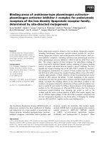
Based on the aligned nucleotide sequences, several phylo-
genetic analysis including maximum parsimony and dis-
tance methods were performed and all approaches
yielded identical or nearly identical topologies. The phyl-
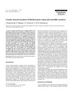
ogenetic tree showed four genetic groups within the
DENV-3 (Figure 1) where genotype I was represented by
strains from Indonesia, Malaysia, Philippines and the
South Pacific islands; genotype II included mainly isolates
from Thailand; genotype III was represented mainly by
viruses from Sri Lanka and Latin America and genotype IV
comprised Puerto Rican viruses.
For a better characterization of the genetic groups, E pro-
tein gene sequences of all viruses were compared manu-
ally. As mentioned above, 634 variable sites were
observed within the 1479 nucleotides of the E protein
gene (Additional file 2). Variable sites with nucleotide
substitutions in at least 90% of the members of any geno-
type were considered informative sites. Thus, 95 of the
634 were considered informative sites. Among these 95,
18 sites were in the domain I of E protein, 28 in domain
II, 27 in domain III, and 22 in the transmembrane
domain (Additional file 3). Each genotype showed a char-
acteristic nucleotide sequence when the informative sites
were analyzed. Nucleotide substitution in the informative
sites was mostly due to transitions (80 sites, 81%) rather
than transversions (21 sites, 19%). Nucleotide substitu-
tion were more frequent in the 3rd position (74 sites,
78%) of the codon, followed by the first position (15
sites, 16%) and finally, the second position (6 sites, 6%).
Non-synonymous substitutions were observed in 14
(15%) of the 95 informative sites (residues 22, 81, 132,
154, 160, 270, 301, 302, 380, 383, 386, 430, 452 and
459). Three non-synonymous substitutions were identi-
fied in domain I, three in domain II, five in domain III,
and three in the transmembrane domain (Additional file
3). Based on the tertiary structure of the E protein of
DENV-3 (36), it was observed that amino acid residues
81, 132, 154, 270, 301, 302, 380, and 383 were located in
solvent-exposed loops. Residues 22 and 386 were located
in β-strands exposed on the viral surface. The residue 160
was located in a hydrophobic region. Residues 430, 452
and 459 were located in the transmembrane region (Addi-
tional file 4A).
Intragenotipic groups
Careful analysis of the topology of the phylogenetic tree
suggests the existence of intragenotipic groups (Figure 1).
To better characterize these internal groups, protein E
gene sequences of members of each genotype were inde-
pendently analyzed.
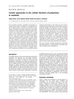
Genotype I
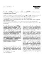
A phylogenetic tree was constructed using 76 protein E
gene sequences of genotype I viruses (Figure 2). The tree
showed that these viruses form two different clades that
were denominated lineage I and II. The nucleotide
sequence comparison showed the presence of 348 varia-
ble sites in the 1479 nucleotides of the E protein gene with
40 of them considered informative sites. Non-synony-
mous substitutions were observed in seven informative
sites (Table 1). Amino acid residues 231, 303 and 391
were found to be located in solvent-exposed loops, resi-
dues 68 and 169 in hydrophobic regions (Additional file
4B). Residues 479 and 489 were located in the transmem-
brane region.
The phylogenetic tree showed that lineage II included two
sub-lineages (Figure 2). The comparison of nucleotide
sequences (n = 68) showed the presence of 318 variable
sites within members of this lineage, six of them being
informative sites with synonymous substitutions (Table
1).
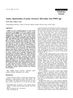
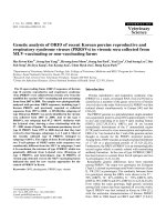
Genotype II
Genotype II included 144 viruses that were grouped into
two lineages (Figure 3). Comparison of these sequences
showed 392 variable sites; four of them being informative
sites with synonymous substitutions (Table 2). Lineage I
included 62 sequences that form two sub-lineages with
255 variable sites; 17 of them were considered informa-
tive sites and three had non-synonymous substitutions
(Table 3). The amino acid residue 140 was located in a β-
strand exposed in the surface of the protein; residues 447
and 489 were in the transmembrane domain (Additional
file 4C). Lineage II included 83 viruses distributed in two
sub-lineages. The comparison of these sequences showed
275 variable sites with only two informative sites, which
showed synonymous substitutions (Table 2).
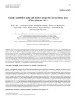
Genotype III
Genotype III was composed of 138 sequences grouped in
two lineages (Figure 4). Sequences comparison showed
321 variable sites with 11 informative sites, all of them
with synonymous substitutions. Lineage I included 29
sequences grouped into sub-lineage I and II with 123 var-
iable sites with only one of them considered as informa-
tive site, which showed a synonymous substitution (Table
3). The lineage II included 108 sequences forming two
groups, sub-lineage I and II; these sequences showed 250
variable sites and only seven of them were considered as
informative sites, all of them were synonymous substitu-
tions (Table 3). The sub-lineage II of lineage II included
the 20 viruses isolated in Ribeirao Preto, SP, Brazil,
between 2006–2007. These viruses were more closely
related to those isolated in other regions of Brazil than to
viruses that circulated in Ribeirao Preto, in 2003 (D3BR/
RP1/2003 and D3BR/RP2/2003). They formed two
groups, one more closely related to the strain D3BR/CU6/
2002 isolated in Cuiabá close to the border with Bolivia
Virology Journal 2009, 6:113 />Page 4 of 13
(page number not for citation purposes)
DENV-3 phylogenetic tree based on the E gene sequencesFigure 1
DENV-3 phylogenetic tree based on the E gene sequences. The three was constructed using the method of Neighbor-
joining with 1000 bootstrap replications. The genotypes are labeled according to the scheme of Lanciotti (1994) and the amino
acid changes distinguishing each genotype are shown on the tree. Protein E gene sequences of DENV-1, DENV-2 and DENV-4
were used as outgroup. Branch lengths are proportional to percentage of divergence. Tamura Nei (TrN+I+G) nucleotide sub-
stitution model was used with a proportion of invariable sites (I) of 0.3305 and gamma distribution (G) of 0.9911. Bootstrap
support values are shown for key nodes only.
VietN BID V1014 2006
TW 05 807KH0509a Tw
VietN BID V1018 2006
Viet0310b Tw
Viet0507a Tw
VietN BID V1015 2006
VietN BID V1017 2006
VietN BID V1016 2006
VietN BID V1009 2006
VietN BID V1011 2006
VietN BID V1012 2006
VietN BID V1008 2006
Viet0409a Tw
VietN BID V1010 2006
Viet9809a Tw
Viet9609a Tw
VietN BID V1013 2006
ThD3 1959 01
ThD3 0835 01
ThD3 0377 98
ThD3 0092 98
ThD3 0058 97
ThD3 0115 99
ThD3 0595 99
ThD3 1017 00
Thail 03 0308a Tw
ThD3 0903 98
ThD3 0650 97
ThD3 1687 98
Thal D93 044 93
ThD3 0240 92
Thail D94 283 94
Thail D95 0014 95
ThD3 0123 95
Thail D92 423 92
ThD3 0989 00
Ja 00 40 1HuNIID 00
ThD3 0328 02
ThD3 0723 99
Thail 02 0211a Tw
ThD3 1094 01
ThD3 1283 98
ThD3 0343 98
ThD3 0006 97
ThD3 0411 97
ThD3 1309 97
Tw 98 701TN9811a
98TWmosq 98
98TW368 98
98TW407 98
Thail 97 9709a Tw
ThD3 0005 96
Thail 98 9807a
Ja 96 17 1HuNIID 96
ThD3 0472 93
Thail D96 330 96
ThD3 0195 94
Thail D97 0144 97
ThD3 0546 98
Thail C0360 94
ThD3 0808 98
ThD3 0514 98
ThD3 0436 97
ThD3 1465 97
Thail 98 KPS 4 0657 207
Thail D96 313 96
Thail D97 0106 97
ThD3 0810 98
Thail D97 0291 97
Thail C0331 94 94
ThD3 0396 94
ThD3 0104 93
ThD3 0077 98
Thail D93 674 93
Thail D94 122 94
ThD3 0654 01
ThD3 0111 02
ThD3 0089 95
ThD3 0969 01
Thail D95 0400 95
ThD3 0182 96
ThD3 0188 91
Indo 98 98901590
Indo 98 98901640
BDH02 2 02
BDH02 5 02
BDH02 6 02
BDH Jacob 01
Bang0108a Tw
BDH02 3 02
BDH02 7 02
BDH02 4 02
BDH02 1 02
BDH02 8 02
BDH Apu 01
BDH 058 00
BDH 114 00
BDH 165 00
Ja 00 27 1HuNIID 00
Myan 05 0508a Tw
Thail 87
ThD3 0040 80
ThD3 0012 90
ThD3 0029 90
My 31985KLA 88
98TW182 98
Thail D91 393 91
Thail D92 431 92
ThD3 0396 88
Mal LN7029 94
Mal LN7933 94
ThD3 0213 88
Thail D91 538 91
ThD3 87
Thail 87 1384 87
ThD3 0220 85
ThD3 0065 86
ThD3 0134 83
ThD3 0402 85
ThD3 0183 85
ThD3 1035 87
Ma LN5547 92
Ma LN2632 93
Ma LN6083 94
Ma LN1746 93
Mal LN8180 94
Sing 8120 95
Thail PaH 881 88
ThD3 0010 87
Thail D88 086 88
Thail D89 273 89
ThD3 0796 87
ThD3 0033 74
ThD3 73 CH53489D 73 1
ThD3 0273 80
ThD3 0059 81
ThD3 0649 80
ThD3 285M 77
ThD3 0059 82
ThD3 0046 83
ThD3 0177 81
ThD3 86
ThD3 0137 84
ThD3 0140 84
In KJ30i 04
In TB55i 04
In TB16 04
NAMRU 2 98901620
In 98901403 DSS DV 3 98
ET D3 Hu Indonesia NIID02 2005
Indo9804a Tw
In 98901437 DSS DV 3 98
In 98901517 DHF DV 3 98
NAMRU 2 98901413
In den3 98
In FW01 04
Indo0312a Tw
In KJ71 04
In PH86 04
In PI64 04
Indo0508a Tw
In FW06 04
In KJ46 04
In BA51 04
ET SV0194 05
ET SV0171 05
ET D3 Hu TL018N IID 2005
ET SV0160 05
ET SV0186 05
ET SV0177 05
ET D3 Hu TL129N IID 2005
ET SV0193 05
ET D3 Hu TL109N IID 2005
ET D3 Hu OPD007NIID 2005
ET SV0153 05
ET SV0174 05
ET D3 Hu TL029N IID 2005
ET209 00
In den3 88
Indo9909a Tw
Indo85
Indo9108a Tw
Thail D88 303 88
In 98902890 DF DV 3 98
ET D3 Hu Indonesi a NIID 01 2005
ET D3 Hu Indonesi a NIID 04 2005
PF92 2986 92
PF92 4190 92
PF92 2956 92
PF89 320219 89
PF89 27643 89
PF90 6056 90
PF90 3056 90
Fiji 92
PF90 3050 90
PF94 136116 94
In Sleman 78
Indo73
Malasya 81
Malasya 74
Indo78
Philp 96 9609a Tw
Philp 98 9809a Tw
95TW466 95
Tw 94 813KH9408a Tw
Tw 05 812KH0508a Tw
Philip 05 0508a TW
Philp 98 9808a Tw
Philp 97 9711a Tw
Taiwan 739079A
Philip 83
In InJ 16 82
M25277 DENSP5AA
M93130 strai n H87
China 80 2
BR DEN3 RO1 02
BR H87
AJ563355
Philp 56 H87
Ja D3 73NIID 73
BR D3BR MA1 02
BR D3BR SG2 02
BR D3BR ST14 04
BR D3BR RP2 03
BR DEN3 290 02
BR D3BR GO5 03
D3 BR RP AAF 2007
BR D3BR RP1 03
PY D3PY AS10 03
BR D3BR IG10 03
BR D3BR SL3 02
PY D3PY PJ4 03
PY D3PY PJ5 03
PY D3PY PJ6 03
BR D3BR PV1 03
BR D3BR PV3 03
BR D3BR PV4 03
BR D3BR PV5 02
BR DEN3 97 04
BR DEN3 95 04
BR DEN3 98 04
D3 H IMTSSA MART 2000 1567
D3 H IMTSSA MART 2000 1706
Cuba116 00
BR D3BR BV4 02
D3 H IMTSSA M ART 2001 2336
D3 H IMTSSA MART 2001 2012
D3 H IMTSSA MART 1999 1243
D3 BR RP 2404 2006
D3 BR RP 2591 2006
D3 BR RP Val 2006
D3 BR RP 2198 2006
D3 BR RP 1651 2006
BR D3BR BR8 04
BR D3BR MR9 03
D3BR RP 1690 2006
D3 BR RP 1573 2006
D3 BR RP 2131 2006
D3 BR RP 1604 2006
D3 BR RP 2065 2006
D3 BR RP 554 2006
PY D3PY AS12 02
PY D3PY YA2 03
Bv FSB 439 2003
PY D3PY FM11 03
BR D3BR CU6 02
PtoR BID V1043 2006
PtoR BID V1078 2003
D3 H IMTS SA MART 2001 2023
BR74886 02
Peru FST312 Tumbes 2004
Peru OBT2812 Piur a 2003
Peru FST145 Tumbes 2003
Peru FSP581 Piur a 2001
Peru OBS8852 2000
Peru OBS8857 2000
Peru OBT1467 Tumbes 2001
Peru FST289 Tumbes 2004
Peru FST 346 Tumbes 2004
Cuba580 01
Cuba21 02
Peru FSL706 Loreto 2002
Peru FSL1212 Yuri maguas 2004
Peru IQD5132 Iquitos 2003
Peru IQD1728 Iquitos 2002
Peru MFI624 Iquitos Jan.2005
Peru OBT4024 Lima C omas 2005
BR Bel73318
BR GOI1099
BR MTO3103
BR 68784 00
BR GOI1100
Venz LARD5990 00
Venz LARD6667
VEN BID V906 2001
Venz LARD6315 00
Venz LARD6722
Venz LARD6666
Venz C02 003 Marac ay 2001
Venz C09 006 Marac ay 2001
VEN BID V904 2001
Venz LARD7110
VEN BID V913 2001
Venz LARD6411
Venz LARD6668
Venz LARD6318 00
Venz LARD7812
Venz LARD7984
Venz LARD6397 00
Venz C29 008 Marac ay 2003
Venz C23 009 Marac ay 2003
PtoR BID V858 2003
PtoR BID V1049 1998
PtoR BID V1050 1998
PtoR BID V859 1998
PtoR BID V1075 1998
6889 QUINTAN A ROO MX 97
MEX6097 95
6883 YUCATAN M X 97
6584 YUCATAN M X 96
MX 00 OAXACA
4841 YUCATAN M X 95
PANAMA 94
Nicarag ua24 94
BR CEA4739
BR RGN576
BR AM2394
BR ROR3832
Srilanka 89
Srilanka 91
SOMALIA 93 S142
Ja 00 28 1HuNIID 00
Srilanka 81
Srilanka 85
Samoa 86
India 84
D3 SG 05K3325DK1 2005
D3 SG 05K3912DK1 2005
D3 SG 05K3329DK1 2005
D3 SG 05K3887DK1 2005
D3 SG SS710 2004
D3 SG 05K3316DK1 2005
D3 SG 05K2406DK1 2005
D3 SG 05K3913DK1 2005
D3 SG 05K3927DK1 2005
D3 SG 05K2918DK1 2005
D3 SG 05K2933DK1 2005
D3 SG 05K791DK1 2005
D3 SG 05K802DK1 2005
D3 SG 05K3312DK1 2005
D3 SG 05K4648DK1 2005
D3 SG 05K2418DK1 2005
D3 SG 05K2899DK1 2005
D3 SG 05K3923DK1 2005
Singapore
SriLan 99 9912a
D3 H IMTS SA SRI 2000 1266
99TW628 99
PtoRico 63 BS PR ico63
Tahiti 65
PtoRico 77 1339
JAM1983 D2
1503 YUCATAN M X 84 D4
ThD1 0127 80 D1
5 changes
Genotype II
Genotype I
Genotype III
Genotype IV
DENV-2
DENV-4
DENV-1
88
73
100
100
99
100
100
22:E
81:*
132:*
154:*
160:A
270:I
301:S
302:G
380:T
383:K
386:R
430:F
452:I
459:I
22:D
81:I
132:H
154:E
160:A
270:T
301:*
302:N
380:I
383:K
386:K
430:L
452:I
459:V
22:D
81:*
132:*
154:D
160:V
270:N
301:L
302:N
380:I
383:K
386:K
430:L
452:I
459:V
22:D
81:V
132:Y
154:E
160:A
270:N
301:T
302:N
380:*
383:N
386:K
430:L
452:V
459:V
Virology Journal 2009, 6:113 />Page 5 of 13
(page number not for citation purposes)
Genotype I phylogenetic tree constructed using the method of Neighbor-joining with 1000 bootstrap replicationsFigure 2
Genotype I phylogenetic tree constructed using the method of Neighbor-joining with 1000 bootstrap replica-
tions. Sequences of each genotype II, III and IV were used as outgroup. Branch lengths are proportional to percentage diver-
gence. Tamura Nei (TrN+I+G) nucleotide substitution model was used with a proportion of invariable sites (I) of 0.5420 and
gamma distribution (G) of 2.6122. The lineage and sub-lineages are marked. Amino acids changes are indicated on the tree.
Bootstrap support values are shown for key nodes only.
In 98901437 DSS DV 3 98
In 98901517 DHF DV 3 98
NAMRU 2 98901413
In den3 98
In FW01 04
Indo0312a Tw
In KJ46 04
In KJ71 04
In PH86 04
In PI64 04
Indo0508a Tw
In FW06 04
In KJ30i 04
In TB55i 04
In TB16 04
NAMRU 2 98901620
ET D3 Hu Indonesia NIID02 2005
Indo9804a Tw
In 98901403 DSS DV 3 98
In BA51 04
ET SV0194 05
ET SV0171 05
ET D3 Hu TL018NIID 2005
ET SV0160 05
ET SV0186 05
ET SV0177 05
ET D3 Hu TL129NIID 2005
ET SV0193 05
ET D3 Hu TL109NIID 2005
ET D3 Hu OPD007NIID 2005
ET SV0153 05
ET SV0174 05
ET D3 Hu TL029NIID 2005
ET209 00
ET D3 Hu Indonesia NIID01 2005
ET D3 Hu Indonesia NIID04 2005
In den3 88
Indo9909a Tw
Indo85
Indo9108a Tw
Thail D88 303 88
In 98902890 DF DV 3 98
Malasya 74
Philp 96 9609a Tw
Philp 98 9809a Tw
95TW466 95
Tw 94 813KH9408a Tw
Tw 05 812KH0508a Tw
Philip 05 0508a TW
Philp 98 9808a Tw
Philp 97 9711a Tw
In InJ 16 82
Indo78
PF92 2986 92
PF92 4190 92
PF92 2956 92
PF89 320219 89
PF94 136116 94
PF89 27643 89
PF90 6056 90
PF90 3056 90
PF90 3050 90
Fiji 92
In Sleman 78
Indo73
Malasya 81
Taiwan 739079A
Philip 83
M25277 DENSP5AA
M93130 strain H87
China 80 2
BR DEN3 RO1 02
BR H87
Philp 56 H87
AJ563355
Ja D3 73NIID 73
BDH02 1 02
BDH Apu 01
Puerto Rico 1963
BR D3BR RP1 03
BR D3BR RP2 03
5 changes
Sub-Lineage I
Lineage I
Lineage II
Sub-Lineage II
Genotype II
Genotype I
V
Genotype III
82
100
100
69
85
97
99
96
100
96
47
68:I
169:A
231:R
303:T
391:R
479:A
489:V
68:V
169:V
231:K
303:A
391:K
479:V
489:A
Virology Journal 2009, 6:113 />Page 6 of 13
(page number not for citation purposes)
Table 1: Nucleotide and amino acid substitutions in the informative sites of genotype I.
Nucleotide Protein Domains
Genotype I
Position Lineage Lineage II Position Lineagen Type of amino acid Changes
Sub-Lineage
Gene Codon I II I II Protein I II I
48 3 G A
135 3 T C
174 3 G A II
202 1 A G 68 I V Conservative
219 3 A G
222 3 T C
282 3 T C
342 3 G A
366 3 A G
393 3 A G
441 3 T C I
474 3 T C
506 2 C T 169 A V Conservative
516 3 T C
537 3 C T
588 3 A G II
633 3 C T
640 1 T C
645 3 C T
663 3 A G
684 3 T C
692 2 G A 231 R K Conservative
714 3 T C
735 3 G A
759 3 A G
777 3 T C
849 3 T C I
909 1 A G 303 T A Nonconservative III
912 3 C T
1101 3 T A
1153 1 C T
1172 2 G A 391 R K Conservative
1269 3 G A TM
1281 3 G A
1302 3 C G
1317 3 G A
1329 3 A G
1380 3 C T
1436 2 C T 479 A V Conservative
1466 2 T C 489 V A Conservative
Domain I: 1–156
nt
(1–52
aa
); 397–573
nt
(133–191
aa
); 835–882
nt
(279–294
aa
)
Domain II: 157–396
nt
(53–132
aa
); 574–834
nt
(192–278
aa
)
Domain III: 883–1176
nt
(295–392
aa
)
Domain TM: 1177–1479
nt
(393–493
aa
)
nt:are indicated the nucleotide positions
aa::are indicated the amino acid positions
Virology Journal 2009, 6:113 />Page 7 of 13
(page number not for citation purposes)
(Group A) and another more closely related to the strain
D3BR/BR8/2004 isolated in northern Brazil (Group B).
Only the strain D3BR/RPAAF/2007 isolated in 2007 was
more closely related to D3BR/RP1/2003 strain.
Discussion
The comparison of E protein gene sequences of DENV-3
revealed many variable sites; however, only 47 of them
showed nucleotide substitutions that induced amino acid
changes in a significant number of viruses (Additional file
5). Therefore, the E protein of DENV-3 showed 47 sites
with variable amino acid residues, which were located
mainly on the viral surface. Our molecular modeling anal-
ysis showed that all the amino acid substitutions do not
interfere with the conformational structure of the E pro-
tein. These polymorphic amino acid residues could be
involved in cell attachment, viral pathogenesis, and recog-
nition by neutralizing antibodies [12,13,32]. Recently, it
was shown that a panel of sera collected from DF and
DHF patients 16–18 month after illness had different lev-
els of neutralizing antibodies to different DENV-3 strains
[33]. Those authors used in the neutralization tests iso-
lates from Cuba and Puerto Rico, which showed amino
acid substitutions at several of the 47 variable sites (Addi-
tional file 6). This suggests that those residues may be
involved in neutralization differences, but further studies
are necessary to confirm this hypothesis.
The phylogenetic analysis, based on E protein gene
sequences, presented in this study showed that DENV-3
are distributed into four genotypes which is supported by
complete mapping of this gene, and is in agreement with
previous studies [25,34]. In addition, internal groups (lin-
eages and sub-lineages) were observed within genotypes I,
II and III. It was not possible to confirm internal sub-
grouping within the genotype IV due to the low number
of sequences available in the GenBank. All amino acids
that characterize a group (genotype, lineage, or sub-line-
age) are located in the 47 variable sites of the E protein.
Characteristic amino acid residues corresponding to the
different DENV-3 genotypes, lineages, and sub-lineages
are evenly distributed in the E protein, and most of them
are exposed on the viral surface.
Recently, it has been reported the existence of a group of
virus forming another genotype (genotype V) within
DENV-3 [29]. However, our phylogenetic and nucleotide/
amino acid substitution analysis suggest that those viruses
of genotype V form a sub-group within the clade of geno-
type I and for this reason we have name this subgroup as
lineage I. The phylogenetic trees generated in other studies
using maximum likelihood and bayesian methods
showed that the so-called genotype V is in the same clade
of genotype I [35,36]. Therefore, we propose the mainte-
nance of the classification of DENV-3 into four genotypes
as previously suggested [25,34].
Other authors have also observed the existence of some of
the intragenotypic groups described in this study. It has
been observed that genotype I includes three groups of
viruses: South Pacific, Philippines, and East Timor viruses
[37]. South Pacific viruses are included in the sub-lineage
I, while Philippines and East Timor are internal groups
within our sub-lineage II of genotype I. It has also been
suggested that genotype II includes two groups of viruses
called: pre- and post-1992 [29]. These groups correspond
to our lineages I and II of genotype II, respectively. The
post-1992 viruses include groups A and B, which corre-
spond to our sub-lineages I and II of lineage II. In addi-
tion, it has been suggested that isolates from Bangladesh
form a distinct group within genotype II [38]. This group
corresponds to our sub-lineage II of lineage I. Another
study has also found three internal groups within geno-
type II: Malaysia, Bangladesh and Vietnam viruses [37].
These groups correspond to our sub-lineage I of lineage I,
sub-lineage II of lineage I, and sub-lineage II of lineage II,
respectively. The genotype III viruses have been classified
into four groups: Latin America, East Africa and groups A
and B from Sri Lanka viruses [39]. Our analysis showed a
similar distribution of genotype III viruses; however, we
found that Latin America viruses (lineage II) form two
groups that we called sub-lineages I and II. These sub-lin-
eages showed also internal monophyletic groups, which
were omitted to simplify the classification. However,
other authors have identified these internal groups within
sub-lineages I and II [37,40-42].
All the DENV-3 isolated in Ribeirao Preto between 2006–
2007 were grouped within the sub-lineage II/lineage II of
genotype III. They were more closely related to viruses iso-
lated in other cities than to those that were previously
reported at Ribeirao Preto in 2003, suggesting that DENV-
3 is constantly moving within the country [43]. Brazil is a
large tropical country with optimal conditions for the
spread of dengue virus making difficult the control of the
disease.
In summary, our results provide information about the
most frequent amino acid changes in the E protein of
DENV-3. These amino acids could be involved in cell
attachment, virus pathogenesis, and recognition by neu-
tralizing antibodies. However, further studies are needed
to confirm these hypotheses. The phylogenetic relation-
ship suggested the existence of only four genotypes of
DENV-3. In addition, we observed internal groups within
genotypes I, II and III.
Virology Journal 2009, 6:113 />Page 8 of 13
(page number not for citation purposes)
Genotype II phylogenetic tree constructed using the method of Neighbor-joining with 1000 bootstrap replicationsFigure 3
Genotype II phylogenetic tree constructed using the method of Neighbor-joining with 1000 bootstrap replica-
tions. Sequences of each genotype I, III and IV were used as outgroup. Branch lengths are proportional to percentage diver-
gence. Tamura Nei (TrN+I+G) nucleotide substitution model was used with a proportion of invariable sites (I) of 0.5041 and
gamma distribution (G) of 1.3902. The lineage and sub-lineages are marked. Amino acids changes are indicated on the tree.
Bootstrap support values are shown for key nodes only.
VietN BID V1014 2006
TW 05 807KH0509a Tw
VietN BID V1018 2006
VietN BID V1015 2006
VietN BID V1017 2006
VietN BID V1016 2006
Viet0310b Tw
Viet0507a Tw
VietN BID V1009 2006
VietN BID V1011 2006
VietN BID V1012 2006
VietN BID V1008 2006
Viet0409a Tw
VietN BID V1010 2006
Viet9809a Tw
Viet9609a Tw
VietN BID V1013 2006
ThD3 1959 01
ThD3 0835 01
ThD3 0377 98
ThD3 0092 98
ThD3 0058 97
ThD3 0115 99
ThD3 0595 99
ThD3 1017 00
Thail 03 0308a Tw
ThD3 0903 98
ThD3 0650 97
ThD3 1687 98
Thal D93 044 93
ThD3 0240 92
Thail D94 283 94
Thail D95 0014 95
ThD3 0123 95
Thail D92 423 92
ThD3 0989 00
Ja 00 40 1HuNIID 00
ThD3 0328 02
ThD3 0723 99
Thail 02 0211a Tw
ThD3 1094 01
ThD3 1283 98
ThD3 0343 98
ThD3 0006 97
ThD3 0411 97
ThD3 1309 97
Tw 98 701TN9811a
98TWmosq 98
98TW368 98
98TW407 98
Thail 97 9709a Tw
ThD3 0005 96
Thail 98 9807a
Ja 96 17 1HuNIID 96
Thail D96 330 96
ThD3 0195 94
Thail D97 0144 97
ThD3 0546 98
Thail C0360 94
ThD3 0808 98
ThD3 0514 98
ThD3 0436 97
ThD3 1465 97
Thail 98 KPS 4 0657 207
ThD3 0472 93
Thail D96 313 96
Thail D97 0106 97
ThD3 0810 98
Thail D97 0291 97
Thail C0331 94 94
ThD3 0396 94
ThD3 0104 93
ThD3 0077 98
Thail D93 674 93
Thail D94 122 94
ThD3 0654 01
ThD3 0111 02
ThD3 0089 95
ThD3 0969 01
Thail D95 0400 95
ThD3 0182 96
ThD3 0188 91
ThD3 0033 74
ThD3 73 CH53489D73 1
ThD3 0273 80
ThD3 0059 81
ThD3 0649 80
ThD3 285M 77
ThD3 0059 82
ThD3 0046 83
ThD3 86
ThD3 0137 84
ThD3 0140 84
ThD3 0177 81
Thail PaH881 88
ThD3 0010 87
Thail D88 086 88
ThD3 0796 87
Thail D89 273 89
Ma LN5547 92
Ma LN2632 93
Ma LN6083 94
Ma LN1746 93
Mal LN8180 94
Sing 8120 95
Thail 87 1384 87
ThD3 0220 85
ThD3 0065 86
ThD3 0402 85
ThD3 0183 85
ThD3 1035 87
ThD3 0134 83
ThD3 87
Thail 87
ThD3 0040 80
ThD3 0012 90
ThD3 0029 90
My 31985KLA 88
98TW182 98
Thail D91 393 91
Thail D92 431 92
ThD3 0396 88
Mal LN7029 94
Mal LN7933 94
ThD3 0213 88
Thail D91 538 91
BDH02 2 02
BDH02 5 02
BDH02 6 02
BDH Jacob 01
Bang0108a Tw
BDH02 3 02
BDH02 7 02
BDH02 4 02
BDH02 1 02
BDH02 8 02
BDH Apu 01
BDH 058 00
BDH 114 00
BDH 165 00
Ja 00 27 1HuNIID 00
Myan 05 0508a Tw
Indo 98 98901590
Indo 98 98901640
BR D3BR RP1 03
BR D3BR RP2 03
ET SV0174 05
ET SV0153 05
Puerto Rico 1963
5 changes
Lineage II
Lineage I
Sub-Lineage II
Sub-Lineage I
Sub-Lineage I
Sub-Lineage II
Genotype III
Genotype I
Genotype IV
100
57
99
66
68
48
40
140:I
447:S
489:A
140:T
447:G
489:T
Virology Journal 2009, 6:113 />Page 9 of 13
(page number not for citation purposes)
Methods
Virus and RNA purification
Twenty DENV-3 strains isolated in C6/36 cells (passage
number 2) from DF and DHF/DSS patients, between
2006–2007, in Ribeirao Preto city, Brazil, were included
in this study. Viral RNA was purified from 140 μl of cul-
ture fluid with the QIAamp Viral RNA kit (Qiagen, Ger-
many), following manufacturer's recommendations.
RT-PCR and sequencing
The E protein gene of the samples were reverse-transcribed
and amplified by polymerase chain reaction (RT-PCR),
using consensus primers, as previously described [43].
The amplicons were purified from agarose gel using the
QIAquick Gel Extraction Kit (Qiagen, USA), and directly
sequenced in an ABI PRISM
®
3100 Genetic Analyzer
(Applied Biosystems, USA). The sequences obtained in
this study were submitted to the GenBank and registered
with the following accession numbers: D3_BR/RP/1573/
2006 (EU617019
), D3_BR/RP/1604/2006 (EU617020),
D3_BR/RP/1625/2006 (EU617021
), D3_BR/RP/1651/
2006 (EU617022
), D3_BR/RP/2065/2006 (EU617023),
D3_BR/RP/2131/2006 (EU617024
), D3_BR/RP/2170/
2006 (EU617025
), D3_BR/RP/2198/2006 (EU617026),
Table 2: Nucleotide and amino acid substitutions in the informative sites of genotype II.
Nucleotide Protein Domains
Genotype II
Lineage I Lineage II Position Lineage I
Position Lineage Sub-Lineage Sub-Lineage Sub-Lineage Type of amino acid Changes
Gene Codon I II I II I II Protein I II
54 3 T A I
90 3 C T
96 3 T C
273 3 A G II
351 3 G A
419 2 T C 140 I T Nonconservative I
549 3 C T
525 3 A G
558 3 G C
609 3 A C II
708 3 G A
747 3 T C
834 3 T C
963 3 G A III
1002 3 T C
1134 3 G C
1176 3 T A
1188 3 C C TM
1233 3 A T
1339 1 T G 447 S G Nonconservative
1436 2 G C
1465 1 A A
1467 3 T T 489 A T Nonconservative
Domain I: 1–156
nt
(1–52
aa
); 397–573
nt
(133–191
aa
); 835–882
nt
(279–294
aa
)
Domain II: 157–396
nt
(53–132
aa
); 574–834
nt
(192–278
aa
)
Domain III: 883–1176
nt
(295–392
aa
)
Domain TM: 1177–1479
nt
(393–493
aa
)
nt:are indicated the nucleotide positions
aa::are indicated the amino acid positions
Virology Journal 2009, 6:113 />Page 10 of 13
(page number not for citation purposes)
D3_BR/RP/2404/2006 (EU617027), D3_BR/RP/2591/
2006 (EU617028
), D3_BR/RP/2604/2006 (EU617029),
D3_BR/RP/554/2006 (EU617030
), D3_BR/RP/590/2006
(EU617031
), D3_BR/RP/597/2006 (EU617032), D3_BR/
RP/AAF/2007 (EU617033
), D3_BR/RP/Val/2006
(EU617034
), D3BR/RP/549/2006 (EU617035), D3BR/
RP/1690/2006 (EU617036
), D3BR/RP/2121/2006
(EU617037
), D3BR/RP/2167/2006 (EU617038).
Phylogenetic analysis of sequences
The E protein gene sequences (1479 bp) obtained in this
study were analyzed using the Vector NTI software (Infor-
matix, USA) and then aligned with 427 sequences of
DENV-3 retrieved from GenBank (Additional file 1) using
the program CLUSTAL W software [44]. The alignment
was edited with the BioEdit software v7.0.0 and MEGA 3.1
[45,46]. Aligned sequences were analyzed in the Model-
test program to identify the best fit-model of nucleotide
substitution for phylogenetic reconstruction; in all the
analysis the Tamura and Nei (TrN+I+G) was the best
model [47]. The best fit-model was selected under the
hierarchical likelihood ratio test (hLTR). The phylogenetic
relationships among strains were reconstructed by the
neighbor-joining (NJ) and maximum parsimony (MP)
methods using the PAUP 4.0B10 program [48].
Structural analysis and comparisons
In order to identify location of the amino acid residues in
the E protein the putative E protein structure of different
isolates were compared with the E protein structure of
DENV-3 deposited in the Protein Data Bank (PDB) under
the access code 1UZG
[32]. Analysis of the structures and
construction of the illustrations were done using the
graphical program Pymol [49].
Table 3: Nucleotide and amino acid substitutions in the informative sites of genotype III.
Nucleotide Domains
Genotype III
Lineage I Lineage II
Position Lineage Sub-Lineage Sub-Lineage
Gene Codon I II I II I II
96 3 C T I
117 3 C A
121 1 C T
157 1 C T
312 3 T A
423 3 T C
588 3 A G II
633 3 C T
672 3 C T
784 1 C T
825 3 C T
1050 3 C T II
1131 3 A G
1170 3 C T
1185 3 G T TM
1314 3 T C
1356 3 G A
1374 3 T A
1473 3 A G
Domain I: 1–156
nt
(1–52
aa
); 397–573
nt
(133–191
aa
); 835–882
nt
(279–294
aa
)
Domain II: 157–396
nt
(53–132
aa
); 574–834
nt
(192–278
aa
)
Domain III: 883–1176
nt
(295–392
aa
)
Domain TM: 1177–1479
nt
(393–493
aa
)
nt:are indicated the nucleotide positions
aa::are indicated the amino acid positions
Virology Journal 2009, 6:113 />Page 11 of 13
(page number not for citation purposes)
Genotype III phylogenetic tree constructed using the method of Neighbor-joining with 1000 bootstrap replicationsFigure 4
Genotype III phylogenetic tree constructed using the method of Neighbor-joining with 1000 bootstrap replica-
tions. Some viruses of each genotype I, II and IV were used as outgroup. Branch lengths are proportional to percentage diver-
gence. Tamura Nei (TrN+G) nucleotide substitution model was used with gamma distribution (G) of 0.2796. The Lineage and
Sub-lineages are marked. Amino acids changes are indicated on the tree. Bootstrap support values are shown for key nodes
only.
D3 BR RP 2131 2006
D3 BR RP 1573 2006
D3BR RP 1690 2006
D3 BR RP 554 2006
D3 BR RP 1604 2006
D3 BR RP 2065 2006
BR D3BR CU6 02
PY D3PY AS12 02
PY D3PY YA2 03
Bv FSB 439 2003
PY D3PY FM11 03
PtoR BID V1043 2006
PtoR BID V1078 2003
D3 BR RP 2198 2006
D3 BR RP 2591 2006
D3 BR RP Val 2006
D3 BR RP 2404 2006
D3 BR RP 1651 2006
BR D3BR BR8 04
BR D3BR MR9 03
BR D3BR GO5 03
D3 BR RP AAF 2007
BR D3BR RP1 03
BR D3BR IG10 03
BR D3BR SL3 02
BR D3BR PV1 03
BR D3BR PV3 03
BR D3BR PV4 03
BR D3BR PV5 02
PY D3PY PJ4 03
PY D3PY PJ5 03
PY D3PY PJ6 03
BR DEN3 97 04
BR DEN3 95 04
BR DEN3 98 04
D3 H IMTSSA MART 2000 1567
D3 H IMTSSA MART 2000 1706
D3 H IMTSSA MART 1999 1243
D3 H IMTSSA MART 2001 2012
BR D3BR BV4 02
D3 H IMTSSA MART 2001 2336
BR D3BR MA1 02
BR D3BR SG2 02
BR D3BR ST14 04
BR D3BR RP2 03
BR DEN3 290 02
D3 H IMTSSA MART 2001 2023
PY D3PY AS10 03
BR74886 02
Cuba116 00
Peru FST312 Tumbes 2004
Peru OBT2812 Piura 2003
Peru FST145 Tumbes 2003
Peru FSP581 Piura 2001
Peru OBS8852 2000
Peru OBS8857 2000
Peru FST289 Tumbes 2004
Peru FST 346 Tumbes 2004
Cuba580 01
Cuba21 02
Peru FSL706 Loreto 2002
Peru FSL1212 Yurimaguas 2004
Peru IQD5132 Iquitos 2003
Peru IQD1728 Iquitos 2002
Peru MFI624 Iquitos Jan.2005
Peru OBT4024 Lima Comas 2005
Peru OBT1467 Tumbes 2001
BR Bel73318
BR GOI1099
BR MTO3103
BR 68784 00
BR GOI1100
BR CEA4739
BR RGN576
BR AM2394
BR ROR3832
Venz LARD5990 00
Venz LARD6667
Venz LARD6666
VEN BID V906 2001
Venz LARD7110
Venz LARD6315 00
Venz LARD6722
Venz C02 003 Maracay 2001
VEN BID V913 2001
VEN BID V904 2001
Venz C09 006 Maracay 2001
Venz C23 009 Maracay 2003
Venz C29 008 Maracay 2003
Venz LARD6411
Venz LARD6668
Venz LARD6318 00
Venz LARD7812
Venz LARD7984
Venz LARD6397 00
PtoR BID V858 2003
PtoR BID V1049 1998
PtoR BID V1050 1998
PtoR BID V859 1998
PtoR BID V1075 1998
6883 YUCATAN MX 97
6889 QUINTANA ROO MX 97
6584 YUCATAN MX 96
MX 00 OAXACA
MEX6097 95
4841 YUCATAN MX 95
PANAMA 94
Nicaragua24 94
D3 SG 05K3325DK1 2005
D3 SG 05K3912DK1 2005
D3 SG 05K3329DK1 2005
D3 SG 05K3887DK1 2005
D3 SG 05K3927DK1 2005
D3 SG SS710 2004
D3 SG 05K2406DK1 2005
D3 SG 05K3316DK1 2005
D3 SG 05K3913DK1 2005
D3 SG 05K2918DK1 2005
D3 SG 05K2933DK1 2005
D3 SG 05K791DK1 2005
D3 SG 05K802DK1 2005
D3 SG 05K3312DK1 2005
D3 SG 05K4648DK1 2005
D3 SG 05K2418DK1 2005
D3 SG 05K2899DK1 2005
D3 SG 05K3923DK1 2005
Singapore
SriLan 99 9912a
99TW628 99
D3 H IMTSSA SRI 2000 1266
Srilanka 81
Srilanka 85
Samoa 86
India 84
Srilanka 89
Srilanka 91
Ja 00 28 1HuNIID 00
SOMALIA 93 S142
BDH02 1 02
BDH Apu 01
Puerto Rico 1963
ET SV0174 05
ET SV0153 05
1 change
Lineage II
Lineage I
Sub-Lineage II
Sub-Lineage I
Sub-Lineage II
Sub-Lineage I
Genotype II
Genotype I
Genotype IV
100
100
62
83
85
80
90
99
44
41
A
B
Virology Journal 2009, 6:113 />Page 12 of 13
(page number not for citation purposes)
Competing interests
The authors declare that they have no competing interests.
Authors' contributions
AAA, FTA, DJ, HLA, LCA, NAN, LTF and VHA conceived of
the study, and participated in its design and coordination.
All authors read and approved the final manuscript.
Additional material
Acknowledgements
This work received financial support from Fundação de Amparo a Pesquisa
do Estado de São Paulo FAPESP), grants 05/04178-2. The authors are grate-
ful to Prof. Maria. Cristina Nonato and Matheus P. Pinheiros by the help in
structural analysis.
References
1. WHO: World Health Organization. Dengue and Dengue
Haemorrhagic Fever. Fact Sheet No. 117. Geneva. 2002.
2. Dos Santos HWG, Poloni T, Souza KP, Muller VDM, Tremeschin F,
Nali LC, Fantinatti LR, Amarilla AA, Castro HLA, Nunes MR, et al.: A
simple one-step real-time RT-PCR for diagnosis of dengue
virus infection. Journal of Medical Virology 2008, 80:1426-1433.
3. Henchal E, Putnak J: The dengue viruses. Clin Microbiol Rev 1990,
3:376-396.
4. Mackenzie J, Gubler D, Petersen L: Emerging flaviviruses: the
spread and resurgence of Japanese encephalitis, West Nile
and dengue viruses. Nat Med 2004, 10:S98-109.
5. Anderson R, King A, Innis B: Correlation of E protein binding
with cell susceptibility to dengue 4 virus infection. J Gen Virol
1992, 73(Pt 8):2155-2159.
6. He R, Innis B, Nisalak A, Usawattanakul W, Wang S, Kalayanarooj S,
Anderson R: Antibodies that block virus attachment to Vero
cells are a major component of the human neutralizing anti-
body response against dengue virus type 2. J Med Virol 1995,
45:451-461.
7. Chen Y, Maguire T, Marks R: Demonstration of binding of den-
gue virus envelope protein to target cells. J Virol 1996,
70:8765-8772.
8. Lindenbach B, Rice C: Flaviviridae: the viruses and their replica-
tion. In Fields virology Volume 1. Edited by: Knipe D, Howley P. Phila-
delphia: Lippincott Williams and Wilkins; 2001:991-1042.
9. Beasley D, Aaskov J: Epitopes on the dengue 1 virus envelope
protein recognized by neutralizing IgM monoclonal antibod-
ies. Virology 2001, 279:447-458.
10. Crill W, Roehrig J: Monoclonal antibodies that bind to domain
III of dengue virus E glycoprotein are the most efficient
blockers of virus adsorption to Vero cells. J Virol 2001,
75:7769-7773.
11. Modis Y, Ogata S, Clements D, Harrison S: Structure of the den-
gue virus envelope protein after membrane fusion. Nature
2004, 427:
313-319.
12. Jennings A, Gibson C, Miller B, Mathews J, Mitchell C, Roehrig J,
Wood D, Taffs F, Sil B, Whitby S: Analysis of a yellow fever virus
isolated from a fatal case of vaccine-associated human
encephalitis. J Infect Dis 1994, 169:512-518.
13. Rey F, Heinz F, Mandl C, Kunz C, Harrison S: The envelope glyco-
protein from tick-borne encephalitis virus at 2 A resolution.
Nature 1995, 375:291-298.
14. Twiddy S, Holmes E, Rambaut A: Inferring the rate and time-
scale of dengue virus evolution. Mol Biol Evol 2003, 20:122-129.
15. Russell P, McCown J: Comparison of dengue-2 and dengue-3
virus strains by neutralization tests and identification of a
subtype of dengue-3. Am J Trop Med Hyg 1972, 21:97-99.
16. Repik P, Dalrymple J, Brandt W, McCown J, Russell P: RNA finger-
printing as a method for distinguishing dengue 1 virus
strains. Am J Trop Med Hyg 1983, 32:577-589.
17. Trent D, Grant J, Rosen L, Monath T: Genetic variation among
dengue 2 viruses of different geographic origin. Virology 1983,
128:271-284.
18. Blok J: Genetic relationships of the dengue virus serotypes. J
Gen Virol 1985, 66(Pt 6):1323-1325.
19. Blok J, Henchal E, Gorman B: Comparison of dengue viruses and
some other flaviviruses by cDNA-RNA hybridization analysis
and detection of a close relationship between dengue virus
serotype 2 and Edge Hill virus. J Gen Virol 1984, 65(Pt
12):2173-2181.
20. Kerschner J, Vorndam A, Monath T, Trent D: Genetic and epide-
miological studies of dengue type 2 viruses by hybridization
using synthetic deoxyoligonucleotides as probes. J Gen Virol
1986, 67(Pt 12):2645-2661.
21. Vorndam V, Nogueira R, Trent D: Restriction enzyme analysis of
American region dengue viruses. Arch Virol 1994, 136:191-196.
Additional file 1
Database of the E protein gene sequences analyzed in this study. The
file provides details on all the sequences including in this study.
Click here for file
[ />422X-6-113-S1.xls]
Additional file 2
Alignment of nucleotide and amino acid sequences of the E protein of
the 361 strains of DENV-3. The file provides details on all the variable
sites distributed in the E protein gene.
Click here for file
[ />422X-6-113-S2.xls]
Additional file 3
Nucleotide and amino acid substitutions in the 95 informative sites of
the E gene of DENV-3. The file provides details on nucleotide and amino
acid substitutions in the informative sites of the E gene of DENV-3.
Click here for file
[ />422X-6-113-S3.xls]
Additional file 4
A stereoscopic drawing of the tertiary structure of E protein indicating
the location of the amino acid residues. Domains I, II and III are
colored in red, yellow and blue, respectively. The overlapping amino acids
are in gray. A) Location of amino acids that characterize the genotypes.
B) Location of amino acids that characterize the lineage I and II of the
genotype I. C) Location of amino acids that characterize the groups within
the lineage I of genotype II. D) Location of amino acids that characterize
the groups within the lineage I of genotype III.
Click here for file
[ />422X-6-113-S4.ppt]
Additional file 5
Comparison of the E protein amino acid sequence of the 361 viruses.
Details on the frequency of amino acids.
Click here for file
[ />422X-6-113-S5.xls]
Additional file 6
Comparison of E the protein amino acid sequence of the Cuba strains
and Puerto Rico. Sequence of isolates from Cuba and Puerto Rico, which
showed differences of amino acids in several sites of the E protein.
Click here for file
[ />422X-6-113-S6.xls]
Publish with BioMed Central and every
scientist can read your work free of charge
"BioMed Central will be the most significant development for
disseminating the results of biomedical research in our lifetime."
Sir Paul Nurse, Cancer Research UK
Your research papers will be:
available free of charge to the entire biomedical community
peer reviewed and published immediately upon acceptance
cited in PubMed and archived on PubMed Central
yours — you keep the copyright
Submit your manuscript here:
/>BioMedcentral
Virology Journal 2009, 6:113 />Page 13 of 13
(page number not for citation purposes)
22. Vorndam V, Kuno G, Rosado N: A PCR-restriction enzyme tech-
nique for determining dengue virus subgroups within sero-
types. J Virol Methods 1994, 48:237-244.
23. Rico-Hesse R: Molecular evolution and distribution of dengue
viruses type 1 and 2 in nature. Virology 1990, 174:479-493.
24. Lewis J, Chang G, Lanciotti R, Kinney R, Mayer L, Trent D: Phyloge-
netic relationships of dengue-2 viruses. Virology 1993,
197:216-224.
25. Lanciotti R, Lewis J, Gubler D, Trent D: Molecular evolution and
epidemiology of dengue-3 viruses. J Gen Virol 1994, 75(Pt
1):65-75.
26. Goncalvez A, Escalante A, Pujol F, Ludert J, Tovar D, Salas R, Liprandi
F: Diversity and evolution of the envelope gene of dengue
virus type 1. Virology 2002, 303:110-119.
27. Wang E, Ni H, Xu R, Barrett A, Watowich S, Gubler D, Weaver S:
Evolutionary relationships of endemic/epidemic and sylvatic
dengue viruses. J Virol 2000, 74:3227-3234.
28. Twiddy S, Farrar J, Vinh Chau N, Wills B, Gould E, Gritsun T, Lloyd
G, Holmes E: Phylogenetic relationships and differential selec-
tion pressures among genotypes of dengue-2 virus. Virology
2002, 298:63-72.
29. Wittke V, Robb T, Thu H, Nisalak A, Nimmannitya S, Kalayanrooj S,
Vaughn D, Endy T, Holmes E, Aaskov J: Extinction and rapid
emergence of strains of dengue 3 virus during an interepi-
demic period. Virology 2002, 301:148-156.
30. Lanciotti R, Gubler D, Trent D: Molecular evolution and phylog-
eny of dengue-4 viruses. J Gen Virol 1997, 78(Pt 9):2279-2284.
31. Worobey M, Rambaut A, Holmes E: Widespread intra-serotype
recombination in natural populations of dengue virus. Proc
Natl Acad Sci USA 1999, 96:7352-7357.
32. Modis Y, Ogata S, Clements D, Harrison S: Variable surface
epitopes in the crystal structure of dengue virus type 3 enve-
lope glycoprotein. J Virol 2005, 79:1223-1231.
33. Alvarez M, Pavon-Oro A, Rodriguez-Roche R, Bernardo L, Morier L,
Sanchez L, Alvarez A, Guzmán M: Neutralizing antibody
response variation against dengue 3 strains. J Med Virol 2008,
80:1783-1789.
34. Chungue E, Deubel V, Cassar O, Laille M, Martin P: Molecular epi-
demiology of dengue 3 viruses and genetic relatedness
among dengue 3 strains isolated from patients with mild or
severe form of dengue fever in French Polynesia. J Gen Virol
1993, 74(Pt 12):2765-2770.
35. Barrero P, Mistchenko A: Genetic analysis of dengue virus type
3 isolated in Buenos Aires, Argentina. Virus Res 2008,
135:83-88.
36. King C, Chao D, Chien L, Chang G, Lin T, Wu Y, Huang J: Compar-
ative analysis of full genomic sequences among different gen-
otypes of dengue virus type 3. Virol J 2008, 5:63.
37. Araújo J, Nogueira R, Schatzmayr H, Zanotto P, Bello G: Phylogeog-
raphy and evolutionary history of dengue virus type 3. Infect
Genet Evol 2009, 9:716-725.
38. Podder G, Breiman R, Azim T, Thu H, Velathanthiri N, Mai lQ, Lowry
K, Aaskov J: Origin of dengue type 3 viruses associated with
the dengue outbreak in Dhaka, Bangladesh, in 2000 and
2001. Am J Trop Med Hyg 2006, 74:263-265.
39. Messer W, Gubler D, Harris E, Sivananthan K, de Silva A: Emer-
gence and global spread of a dengue serotype 3, subtype III
virus. Emerg Infect Dis 2003, 9:800-809.
40. Fajardo A, Recarey R, de Mora D, D' Andrea L, Alvarez M, Regato M,
Colina R, Khan B, Cristina J: Modeling gene sequence changes
over time in type 3 dengue viruses from Ecuador. Virus Res
2009, 141:105-109.
41. de Mora D, Andrea L, Alvarez M, Regato M, Fajardo A, Recarey R,
Colina R, Khan B, Cristina J: Evidence of diversification of den-
gue virus type 3 genotype III in the South American region.
Arch Virol 2009, 154:699-707.
42. Kochel T, Aguilar P, Felices V, Comach G, Cruz C, Alava A, Vargas J,
Olson J, Blair P: Molecular epidemiology of dengue virus type
3 in Northern South America: 2000 – 2005. Infect Genet Evol
2008, 8:682-688.
43. Aquino V, Anatriello E, Gonçalves P, DA Silva E, Vasconcelos P, Vieira
D, Batista W, Bobadilla M, Vazquez C, Moran M, Figueiredo L: Molec-
ular epidemiology of dengue type 3 virus in Brazil and Para-
guay, 2002–2004. Am J Trop Med Hyg 2006, 75:710-715.
44. Thompson J, Gibson T, Plewniak F, Jeanmougin F, Higgins D: The
CLUSTAL_X windows interface: flexible strategies for mul-
tiple sequence alignment aided by quality analysis tools.
Nucleic Acids Res 1997, 25:4876-4882.
45. Hall T: : BioEdit: a user-friendly biological sequence align-
ment editor and analysis program for Windows 95/98/NT.
Nucl Acids Symp Ser 1999, 41:95-98.
46. Kumar S, Tamura K, Nei M: MEGA3: Integrated software for
Molecular Evolutionary Genetics Analysis and sequence
alignment. Brief Bioinform 2004, 5:150-163.
47. Posada D: ModelTest Server: a web-based tool for the statis-
tical selection of models of nucleotide substitution online.
Nucleic Acids Res 2006, 34:W700-703.
48. Swofford D: PAUP*: phylogenetic analysis using parsimony
(*and other methods). In Version 4.0b10a Sunderland, Mass: Sin-
auer Associates; 1998.
49. Delano W: The PyMOL Molecular Graphics System. 2002
[
]. San Carlos, CA, USA