Báo cáo khoa học: "Genetic characterization of porcine circovirus - 2 field isolates from PMWS pigs" potx
Bạn đang xem bản rút gọn của tài liệu. Xem và tải ngay bản đầy đủ của tài liệu tại đây (706.88 KB, 9 trang )
JOURNAL OF
Veterinary
Science
J. Vet. Sci. (2002), 3(1), 31-39
ABSTRACT
6)
PMWS is a new emerging disease in swine herds
worldwide. Field isolates of PCV-2, a putative major
causative agent of PMWS, were isolated and genetically
characterized. Viral genome of two field isolates
(PC201DJ and PC201SS) from pigs showing typical
PMWS was sequenced. The nucleotide sequence homology
with other PCV-2 isolates was ranging from 95% to
99% in complete viral genomic sequence. The highly
conserved nonanucleotide motif of replication origin
was identical to that of other PCV-2 isolates. To
determine the genetic heterogeneity of PCV-2 isolates,
thephylogenetictreebasedonthecompletegenome
of PCV-2 isolates were constructed. Two PCV-2 field
isolates were closely related to Canadian isolates of
PCV-2. PCV-2 isolated from field may have an origin
of North America and is possibly originated from
importation of breeding stocks. The result indicates
that although the genome of PCV-2 is relatively stable
in general, minor genetic variations exist among
PCV-2 isolates from the different geographic locations.
These differences of viral genome might have an
important implication for genetic characteristics of
PCV-2 infection. Three major immunorelevant epitopes
of capsid protein showed variations in amino acid
sequences. Also, the variance of amino acid sequence
in antigenic epitope existed between two Korean
PCV-2 isolates.
Key words: porcine circovirus, replication origin,
sequence homology, phylogenetic tree, epitope
Introduction
Porcine Circovirus (PCV) is a small non-enveloped virus
containing a single-stranded circular DNA genome. The
PCV belongs to the family Circoviridae that has two types
such as PCV-1 and PCV-2 [1, 5, 6, 12]. The two other
*
Corresponding author: Dr. Young S. Lyoo, Assistant Professor
College of Veterinary Medicine Konkuk University, Seoul Korea
143-701
Phone: +82-2-450-3719, Fax: +82-2-458-5113
E-mail:
animal circoviruses in this family are chicken anemia virus
(CAV), psittacine beak and feather disease virus (PBFDV).
The three plant circoviruses are known as banana bunchy
top virus, coconut foliar decay virus, and subterranean
clover stunt virus. Recently, a human circovirus, TT virus
(TTV), was identified from patients with post-transfusion
hepatitis. The human TTV has similarities to CAV in its
genomic organization [1, 9, 10, 35, 43]. Besides, circoviruses
show similarities to the family Geminiviridae with charac-
teristics of single stranded circular form of DNA genome
and using the rolling circle replication (RCR) strategy in its
replication [7, 15]. PCV-1 consisting of 1,759 nucleotides
shows neither cytopathic effects in tissue culture cells nor
any specific diseases [12, 32, 42]. In contrast, PCV-2 has
1,768 nucleotides of viral genome and is speculated as a
major agent causing post weaning multisystemic wasting
syndrome(PMWS)inpigs[4,6,11,26,18,19,20,24,25,
34, 36, 38, 40, 41].
PMWS, a newly emerging disease in pigs, usually occurs
in swine herds with good health condition and causes a low
rate of morbidity in Canada, the United States, Asia, and
many European countries. However, it affects weaners and
finishers from 5 to 12 weeks old with relatively high
mortality. PMWS pigs show clinical signs like dyspnea,
anemia, visibly enlarged lymph nodes, diarrhea, pallor,
progressive weight loss and jaundice. Histologically, main
lesions associated with PMWS are lymphadenopathy, gra-
nulomatous interstitial pneumonia, hepatitis, and nephritis.
Also, they include macrophage and lymphocytes infiltration
in affected organs.
PCV is regarded as not only a crucial agent causing
economical losses in swineherds, which is associated with
PMWS, but also potential hazard in human health when
xenotransplantation is addressed. Pig is a strong candidate to
be developed as future donors of tissues and organs for
those who need transplantation to replace impaired tissues
and organs.
Though PCV-2 seems to be a quite important pathogenic
agent, PCV-2 is not yet characterized in Korea. Presumably, this
characterization of Korean PCV-2 isolates is valuable for
developing diagnostic tools and vaccines. Consequently, in this
study, we purposed to isolate PCV-2 from PMWS pigs in
Korea and characterize PCV-2 isolates genetically by
nucleotide sequence analysis. And we determined the origin
Genetic characterization of porcine circovirus-2 field isolates from PMWS pigs
J
in H. Kim, Young S. Lyoo
*
I
mmunopathology laboratory, College of Veterinary Medicine, Konkuk University, Seoul, 143-701, Korea
32 Jin H. Kim, Young S. Lyoo
of Korean PCV-2 isolates and genetic similarity by homology
comparison and phylogenetic tree analysis.
Materials and Methods
Clinical samples
Pigs showing PMWS signs were submitted from swine farms
Korean nation-wide to the Immunopathology laboratory Konkuk
University. Tissue samples used for our research include lung,
lymph nodes, spleen, tonsil, kidney, and liver.
Polymerase chain reaction (PCR) and cloning
Primer sets were designed on the basis of the sequence of
PCV-1 (GenBank accession no. U49186) and PCV-2
(GenBank accession no. AF027217). DNA extraction was
performed by the commercial DNA extraction kit, DNAzol
(GIBCO BRL) according to manufactures instruction. Raw
materials for the DNA extraction include 100mg mixture of
lung, spleen, liver, kidney, inguinal lymph node, mesenteric
lymph node and tonsil of PMWS pigs. Five hundred㎕ cell
lysates of PK-15 cells as a control were used. Oligonucleotide
sequences the primers used for the amplification were
shown in Table 1 [26]. PCR product with 886 bp in length
specific for both PCV-1 and PCV-2 was amplified using
primersF1andR1.PrimersF2andR1specificforPCV-2
was used to amplify 469 bp of DNA fragments from the
samples collected in PMWS pigs. Full length PCV-2 genome
was amplified using specific primers F1 and 1768R. For the
sequencing of the complete genomic DNA, overlapping viral
gene from 433 bp to 1695 bp was amplified using internal
primers (Table 1). The direction of the amplification was
opposite to that of the first round full-length genomic DNA
amplification step. Annealing temperature for PCR was
52℃. The amplified linear forms of PCR products were
purified by GENECLEAN II Kit (Bio 101, Inc., USA) and
cloned into pGEM T-easy vector (Promega, U.S.A.). Plasmid
constructs containing viral gene were pGEM DJ1768, pGEM
DJ506 from PC201DJ and pGEM SS1768, pGEM SS506
from PC201SS, respectively. Plasmid DNA with insertion of
the PCV viral genes were prepared for the sequencing by
midi-prep using QIA filter Plasmid Midi Kit (QIAGEN).
Isolation of porcine circovirus associated with PMWS
PCV-2 positive samples such as inguinal lymph node,
lung, tonsil, spleen, and kidney by PCR were frozen in liquid
nitrogen, and homogenized in mortar with autoclaved sea
sands. The inocula composed of homogenized tissues and
minimum essential media (MEM, GIBCO BRL) containing
10% antibiotics were centrifuged and filtered through 0.22
㎛ filter to eliminate bacterial contaminant. The virus
isolation was performed in PK-15 cell line free from PCV-1
and PCV-2. Dr. Nayar G.P.S (University Crescent, Canada)
kindly provided PCV free PK-15 cells. The semi-confluent
PK-15 cells were inoculated with 3 ml of inoculum and
placed in a incubator for 90 minutes at 37℃ with 5 % CO
2
.
Then, fresh MEM containing 2 % fetal bovine serum, 1%
antibiotics and antimycotics, 2.5 % HEPES, 1 % non essential
amino acid and 1 % Na pyruvate was replaced. At twenty-four
hours post-inoculation, cells were washed with Hanks
balanced salt solution (HBSS, GIBCO BRL), treated with
300mM D-glucosamine for 30minutes and washed once with
HBSS only [35, 44]. Cells were incubated for 48 hours to
allow virus replication prior to further passage for cell
culture adaptation. Samples were passed three times with
D-glucosamine treatment and the presence of the PCV was
tested by PCR using PCV specific primers.
Sequencing and genetic analysis
The pGEM DJ1768, pGEM DJ506 and pGEM SS1768,
pGEM SS506 were sequenced by Sangers methods (Bionex,
Seoul Korea) using automated nucleotide sequencer. The
sequence of PCV-2 isolates, PC201SS and PC201DJ, were
analyzed with computer programs Clustal X 1.81 and GeneDoc
to construct phylogenetic tree for comparing with those of other
known PCV isolates. Sequence homology was searched by
BLAST from NCBI Genbank database.
Results
Isolation of porcine circovirus associated with PMWS
The PCV-2 virus PC201DJ and PC201SS were isolated
from pig tissue samples in PCV free PK-15 cells. But
cytopathic effect was not clearly detected in field virus after
inoculation.
PCR and cloning
Vero cells and PCV free PK-15 cells were used for
negative control in PCR. PK-15 cells (ATCC CCL-33)
Table 1. Sequence of oligomers used for confirmation of the viral presence in field samples, cloning and sequencing
Primer Sequence(5
′-
3
′
) Size
Position
in viral strand
Position
in complementary strand
F1
F2
R1
1768R
1696F
433R
ACCAGCGCACTTCGGCAG
TGAGTACCTTGTTGGAGAGC
GTAATCCTCCGATAGAGAGC
AATACTTACAGCGCACTTCTTTCG
GGTGTCTTCTTCTGCGGTAACG
TCCAACAAGGTACTCACAGCAG
18nt
20nt
20nt
24nt
22nt
22nt
1
∼
18
418
∼
437
1696
∼
1717
867
∼
886
1745
∼
1768
412
∼
433
Genetic characterization of porcine circovirus-2 field isolates from PMWS pigs 33
showed PCV-1 positive in PCR with 886 bp DNA product.
Two PCV-2 isolates, PC201DJ and PC201SS, showed both
DNA bands of 886 bp and 469 bp in 1.5% electrophoresis gel
(Fig.1). PCR products of 1768 bp and 506 bp using F1/1768R
and 1686F/433R were inserted into pGEM T-easy vector to
construct each of pGEM DJ1768, pGEM DJ506, pGEM
SS1768, pGEM SS506 hybrid plasmid. Restriction
endonuclease Not I was used to confirm insertion of the
PCV DNAs from hybrid plasmid since there is no restriction
endonuclease Not I in PCV-2 genomic sequence (Fig. 2).
Fig. 1. PCR amplification for differentiation and identification
ofPCV-1andPCV-2usingprimersetcommontoboth
PCV-1 and PCV-2, and primers specific for the PCV-2. PCR
productswith886bpforPCV-1andPCV-2wereamplified
with F1 and R1, and F2 and R1 primer set amplified 469
bp only in PCV-2. M: 1Kb DNA marker (Bioneer, Seoul,
Korea), lane 1 and lane 6: Vero cells, lane 2 and lane 7:
PK-15 cells free from PCV-1, lane 3 and lane 8: PK-15 cells
(ATCC CCL-33), lane 4 and lane 9: PC201DJ of PCV-2
isolate, lane 5 and lane 10: PC201SS of PCV-2 isolate.
Fig. 2. Digestion of restriction endonuclease Not I for hybrid
plasmid containing PCV DNA. PCR products of 1768 bp and
506 bp amplified by each primer sets of F1/1768R and
1696F/433R were inserted into pGEM T-easy vector.
Restriction endonuclease Not I digested pGEM DJ1768 (lane
1), pGEM DJ506 (lane 2), pGEM SS1768 (lane 3) and
pGEM SS506 (lane 4) released corresponding size of the
insert. M: 1Kb DNA size marker (Bioneer, Seoul, Korea).
Sequence analysis
Complete viral genomic sequence was generated from
sequence data obtained with overlapping sequencing
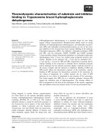
analysis using internal primer sets. The schematic diagram
of overlapping sequence is shown in Fig. 3. Complete viral
genomic sequences of PC201DJ and PC201SS were aligned
with PCV-2 (AF027217) and PCV-1 (U49186) as depicted in
Fig. 4. PCV-2 isolates showed identical genetic characteristics
known prototype PCV-2 such as overlapped putative eleven
ORFsasshowninFig.3.ThelargestORF1andORF2code
for Rep protein and viral capsid protein showed opposite
orientation. Nonanucleotide motif of replication origin is
observed in the same position with other PCV-2 strains, which
is an essential element for the rolling circle replication (Fig.
3 and Fig. 4) [21, 28, 30, 37]. The nonanucleotide motif of
5-AAGTATTAC-3, which is different from PCV-1s of
5-TAGTATTAC-3, was conserved in both PCV-2 field isolates.
Fig. 3. Schematic diagram of complete viral genome of PC201DJ.
Overlapped putative eleven ORFs were found in circular viral
genome. Replication origin composed of nonanucleotide motif was
located between ORFs 1 and 7. Two primer sets for sequencing
were displayed as overlapped sequencing.
Homology analysis
Sequence homology was compared with other known PCV
isolates in Table 2. Sequences of other known fifteen PCV-2
isolates and three PCV-1 isolates were downloaded from
GenBank [17, 25, 31, 34]. The geographic locations of PCV
isolates were varied as USA, Canada, France and Ireland.
Two Korean isolates, PC201DJ and PC201SS, have 97 % of
complete viral genomic sequence homology. Both PC201DJ
and PC201SS showed 95~99 % sequence homology of
complete viral genome in PCV-2 but 76~77 % in PCV-1.
Especially, PC201DJ shows 99 % complete sequence homology
Fig. 4. Complete viral genomic sequence alignments of PC201DJ and PC201SS with PCV-2 (AF027217, USA) and PCV-1
(U49186, PK-15). Conserved sequences were shaded in three levels according to identity. Asterisks show nonanucleotide
motif of PCV-2 as replication origin. Glycosylation sites were displayed by ^ marks.
34 Jin H. Kim, Young S. Lyoo
between strains
Amino acid position of ORF2 of PCV
-
2 PCV
-
2strains Putative antigenic epitope of ORF2 of PCV
-
2
69
∼
83
AF201311(France)
AF027217(USA)
PC201DJ(Korea)
PC201SS(Korea)
VDMM RFNINDFL PPG
VDMM RFNIDDFV PPG
VDMM RFKLDDFV PPG
VDML RFKIDDFV PPG
117
∼
131
AF201311(France)
AF027217(USA)
PC201DJ(Korea)
PC201SS(Korea)
G C GSS AVILDDNFVT
G V GST AVILDDNFVT
G V GST AVILDDNFVP
G V GSS AVILDDNFVP
169
∼
183
AF201311(France)
AF027217(USA)
PC201DJ(Korea)
PC201SS(Korea)
F TIDYFQPNNKENQL
S TIDYFQPNNKRTQL
S TIDYFQPNNKRNQL
G TIDYFQPNNKRNQL
Fig. 5. Phylogenetic tree of the 20 PCV isolates including two Korean PCV-2s was constructed using
computer analysis program Clustal X 1.81 on the basis of complete viral genome. Bootstrap neighbor joining
method with the option of exclusion of positions with gaps was used. Branch lengths are proportional to the
number of character-state changes. Scale bar: the number of character-state changes.
36 Jin H. Kim, Young S. Lyoo
Genetic characterization of porcine circovirus-2 field isolates from PMWS pigs 37
with AF118097. The amino acid sequence homology of both
Korean isolates was compared in the point of ORF1, ORF2,
ORF2 and ORF4. For both Korean isolates, the largest
ORF1 of PCV-2, 314 amino acids encoding Rep protein, had
over 98 % amino acid sequence homology in most case of
PCV-2 except some Canadian isolates. 233 amino acids of
ORF2, putative capsid protein, varied from 92 % to 97 % in
amino acid homology of Korean isolates, but PC201DJ had
exact consensus ORF2 with AF118097. The functions of 104
amino acids of ORF3 and 59 amino acids of ORF4 have been
not clearly reported. The rate of homology in ORF3 and
ORF4 of PCV-2 were varied from 92 % to 100%.
Immunorelevant epitopes in the viral capsid protein showed
considerable variations as shown in the Table 3. Especially
first epitope located between amino acid 69 and 83 had
higher mutation rate than others (Table 3). But the
variation of putative immunorelevant epitopes was not
consistent among strains.
Phylogenetic tree analysis
As shown in Fig. 5, phylogenetic tree was constructed on
basis of the complete viral genomic sequence of twenty PCV
isolates worldwide using computer analysis program Clustal
X 1.81. These sequences of eighteen PCV isolates available
in GenBank and two Korean PCV-2 isolates PC201DJ and
PC201SS were used for the analysis. Based on the
phylogenetic analysis, two major genotypes representing
PCV-1 and PCV-2 were distinct each other. Among PCV-1s,
AF071879 isolated from PK-15 cell and other PCV-1s
isolated in the European regions were also used for analysis.
PCV-2 has two distinct branches according to the geographic
regions. One major branch of PCV-2 genotype is found in
Europe and another major branch is present in North
America (USA and Canada) and Asia including Korea. Two
Korean PCV-2 field isolates of PC201DJ from mid-western
region of the Korean peninsula and PC201SS from Kyungki
province were closely related to Canadian isolates but were
clustered into different groups of PCV-2 genotypes. Bovine
isolate of circovirus was most closely related to the USA
isolates of PCV-2 [14].
Discussion
PMWS causes one of a major health problem in pig herds
worldwide. This is supposed to be caused by complex of
many different swine pathogens including porcine circovirus,
swine influenza, swine parvovirus, PRRS virus, Mycoplasma
hyopneumoniae, Haemophilus parasuis, etc. [1, 2, 3, 8, 13,
14, 22, 39]. There is no clear evidence supporting PCV as a
culprit in PMSW. Recent research data showed a strong
relationship between PCV-2 and PMWS [1, 4, 6, 16, 34, 36].
In this study, two Korean PCV-2 isolates was sequenced
and genetic characteristics were analyzed. Two Korean
PCV-2 isolates showed a high degree of sequence homology
with other PCV-2 strains available. The nonanucleotide
replication origin of PCV-2 was found as same conserved
sequence and position as other strains of PCV-2. This
sequence is critical in virus replication and the first
nucleotide of the conserved nonamer was mutated from T in
PCV-1 to A in PCV-2. When the first two nucleotide of the
nonanucleotide were altered in PCV-1, a total loss of repli-
cation function was found [29, 31]. Stem-loop around
nonanucleotide motif of replication origin has been reported
also. Andthree repetitions of the 6-bp motif CGGCAG seem
to be putative binding site for the Rep protein [28, 29].
ORF2 of PCV-2 corresponding to PCV-1s showed a homology
of about 63 %, but no detectable cross-reactivity could be
shown between ORF2 proteins of PCV-1 and PCV-2 [27].
ORF2 of Korean PCV-2 isolates showed over 98 % homology
with PCV-2 isolates from USA, Canada and France. But
Canadian isolates such as AF085695, AF086834, AF086835
and AF086836 showed slightly lower homology of 96 % with
two Korean isolates. Mache et al.suggestedthatthree
peptides of ORF2 of PCV-2 could be related to antigenic
epitopes on the base of the sequence of AF201311, which is
French PCV-2 isolate [27, 29, 45]. Amino acid variability in
antigenic epitope between Korean isolates and other PCV-2
strains may indicate that there is possible on going spon-
taneous genetic mutation, which could play an important
role in antigenicity of the virus. But there is no antigeni-
cally distinct PCV-2 has been reported. An ORF3 showed
slightly lower sequence homology than that of the ORF4 but
it was not significant. Four glycosylation sites in ORF1 and
one site in ORF2 existed in these two field isolates. By
phylogenetic analysis based on the complete viral genomic
sequence, it is assumed that two Korean PCV-2 isolates
might be originated from North American continent. Not
even live animals but other materials such as boar semen
imported from North America for the reproduction would be
a source of the virus transmission [23]. PC201DJ and
PC201SS were closely related with Canadian isolates, but
showed little divergence between the branch lengths. This
study suggests that although the genome of PCV-2 is
relatively conserved in general, but there are minor genetic
variations exist among PCV-2 isolates from the different
geographic locations.
Acknowledgment
We are grateful to Dr. Nayar G.P.S. (University Crescent,
Winnipeg, Manitoba, Canada) for providing PCV free PK-15
cells for this research and K.S. Kang for submitting field
samples for the research.
Reference
1. Allan G. M., and Ellis J. A. Porcine circoviruses: a
review.J.Vet.Diagn.Invest.2000,12,3-14.
2. Allan G. M., McNeilly F. Cassidy J. P., Reilly G. A.
C.,AdairB.,EllisW.A.,andMcNultyM.S.
38 Jin H. Kim, Young S. Lyoo
Pathogenesis of porcine circovirus; experimental infections
of colostrium deprived piglets and examination of pig
foetal material. Vet. Microbiol. 1995, 44,49-64.
3. Allan G. M., McNeilly F., Ellis J., Krakowka S.,
Meehan B., McNair I., Walker I., and Kennedy S.
Experimental infection of colostrums deprived piglets
with porcine circovirus 2 (PCV2) and porcine repro-
ductive and respiratory syndrome virus (PRRSV) poten-
tiates PCV2 replication. Arch. Virol. 2000, 145, 2421-
2429.
4. Allan G. M., McNeilly F., Meehan B. M., Kennedy
S., Mackie D. P., Ellis J. A., Espuna E., Saubi N.,
Riera P., Botner A., and Charreyre C. E. Isolation
and characterization of circoviruses from pigs with
wasting syndromes in Spain Denmark and Northern
Ireland. Vet. Microbiol. 1999, 66,115-123.
5. Allan G. M., Phenix K. V., Todd D., and McNulty M.
S. Some biological and physico-chemical properties of
porcine circovirus. J. Vet. Med. 1994, 41, 17~26.
6. Allan G., Meehan B., Todd D., Kennedy S. McNeilly
F.,EllisJ.,ClarkE.G.,HardingJ.,EspunaE.,
Botner A., and Charreyre C. Novel porcine circo-
viruses from pigs with wasting disease syndromes. Vet.
Rec.1998, 142,467-468.
7. Arguello-Astorga G., Herrera-Estrella L., Rivera-
Bustamante R. Experimental and theoretical definition
of geminivirus origin of replication. Plant Mol. Biol.
1994, 26,553-556.
8. Balasch M., Segales J., Rosell C., Domingo M.,
Mankertz A., Urniza A., and Plana-Duran J. Ex-
perimental inoculation of conventional pigs with tissue
homogenates from pigs with post-weaning multi-
systemic wasting syndrome. J. Comp. Path. 1999, 121,
139-148.
9. BassamiM.R.,BerrymanD.,WilcozG.E.,and
Raidal S. R. Psittacine beak and feather disease virus
nucleotide sequence analysis and its relationship to
porcine circoviruses, plant circoviruses, and chicken
Anaemia virus. Virol. 1998, 249, 453-459.
10. Bassami M. R., Ypelaar I., Berryman D., Wilcoz G.
E., and Raidal S. R. Genetic diversity of beak and
feather disease virus detected in psittacine species in
Australia. Virol. 2001, 279,392-400.
11. Choi C., Chae C., and Clark E. G. Porcine post-
weaning multisystemic wasting syndrome in Korean
pig: detection of porcine circovirus 2 infection by
immunohistochemistry and polymerase chain reaction.
J. Vet. Diagn. Invest. 2000, 12, 151-153.
12. Dulac G. C., and Afshar A. Porcine circovirus
antigens in PK-15 cell line(ATCC CCL-33) and evidence
of antibodies to circovirus in Canadian pigs. Can. J. Vet.
Res. 1989, 53,431-433.
13. Ellis J. A., Bratanich A., Clark E. G., Allan G.,
MeehanB.,HainesD.M.,HardingJ.,WestK.H.,
Krakowka S., Konoby C., Hassard L., Martin K.,
and McNeilly F. Coinfection by porcine circoviruses
and porcine parvovirus in pigs with naturally acquired
postweaning multisystemic wasting syndrome. J. Vet.
Diagn. Invest. 2000, 12,21-27.
14.FenauxM.,HalburP.G.,GillM.,TothT.E.,and
Meng X. Genetic characterization of type 2 porcine
circovirus (PCV-2) from pigs with postweaning
multisystemic wasting syndrome in different geographic
regions of north America and development of a diffe-
rential PCR-restriction fragment length polymorphism
assay to detect and differentiate between infections with
PCV-1 and PCV-2. J. Clin. Microbiol. 2000, 38(7),
2494-2503.
15. Gutierrez C., Geminivirus DNA replication. Cell. Mol.
Life. Sci. 1999, 56,313-329.
16.HamelA.L.,LinL.L.,andNayarG.P.S.
Nucleotide sequence of porcine circovirus associated
with postweaning multisystemic wasting syndrome in
pigs.J.virol.1998,72(6), 5262-5267.
17. Hamel A. L., Lin L. L., Sachvie C., Grudeski E.,
and Nayar G. P. S. PCR detection and characteri-
zation of type-2 porcine circovirus. Can. J. Vet. Rec.
2000, 64(1), 44-52.
18. Harms P. A. Field studies of postweaning multisystemic
wasting syndrome. Proceedings of swine disease
conference for swine practitioners, 1998, 5-7.
19.KennedyS.,AllanG.,McNeillyF.,AdairB.M.,
Hughes A., and Spillane P. Porcine circovirus
infection in Northern Ireland. Vet. Rec. 1998, 142,
495-496.
20.KiupelM.,StevensonG.W.,MittalS.K.,ClarkE.
G., and Haines D. M. Circovirus-like viral associated
disease in weaned pigs in Indiana. Vet. Pathol. 1998,
35,303-307.
21. Koonin E. V., and Ilyina T. V. Computer-assisted
dissection of rolling circle DNA replication. Bio. Systems.
1993, 30, 241-268.
22. Krakowka S., Ellis J. A., Meehan B., Kennedy S.,
McNeilly F., and Allan G. Viral wasting syndrome of
swine: Experimental reproduction of postweaning multi-
systemic wasting syndrome in gnotobiotic swine by
coinfection with porcine circovirus 2 and porcine
parvovirus. Vet. Path. 2000, 37, 254-263.
23. Larochelle R., Bielanski A., Muller P., and Magar
R. PCR detection and evidence of shedding of porcine
circovirus type 2 in boar semen. J. Clin Microbiol.
2000, 38(12), 4629-4632.
24. Larochelle R., Morin M., Antaya M., and Magar R.
Identification and incidence of porcine circovirus in
routine field cases in Quebec as determined by PCR.
Vet. Rec. 1999, 145,140-142.
25. LeCann P., Albina E., Madec F., Cariolet R., and
Jestin A. Pigletwastingdisease.Vet.Rec.1997,141,
660.
26.LyooY.S.,KimJ.H.,ParkC.K.Identification of
Genetic characterization of porcine circovirus-2 field isolates from PMWS pigs 39
porcine circoviruses with genetic variation from lymph
nodes collected in pigs with PMWS. Korean J. Vet. Res.
1999, 39(2), 353-358.
27. Mahe D., Blanchard P., Truing C., Arnauld C.,
LeCann P., Cariolet R., Madec F., Albina E., and
Jestin A. Differential recognition of ORF2 protein from
type 1 and type 2 porcine circoviruses and identification
of immunorelevant epitopes. J. Gen. Virol. 2000, 81,
1815-1824.
28. Mankertz A., and Hillenbrand B. Replication of
porcine circovirus type 1 require two protein encoded by
the viral rep gene.Virol.2001,279, 429-438.
29. Mankertz A., Domingo M., Folch J. M., LeCann P.,
Jestin A., Segales J., Chmielewicz B., Plana-Duran
J., and Soike D. Characterisation of PCV-2 isolates
from Spain, Germany and France. Virus Res. 2000, 66,
65-77.
30. Mankertz A., Mankertz J., Wolf K., and Buhk H J.
Identification of a protein essential for replication of
porcine circovirus. J. Gen. Virol. 1998, 79, 381-384.
31. Mankertz A., Persson F., Mankertz J., Blaess G.,
and Buhk H J. Mapping and characterization of the
origin of DNA replication of circovirus. J. Virol. 1997,
71(3), 2562-2566.
32. Mankertz J., Buhk H J., Blaess G, and Mankertz
A. Transcription analysis of porcine circovirus(PCV).
Virus Genes. 1998, 16(3),267-276.
33. Meehan B. M., Creelan J. L., McNulty M. S., and
Todd D. Sequence of porcine circovirus DNA: affinities
with plant circoviruses. J. Gen. Virol. 1997, 78, 221-227.
34. Meehan B. M., McNeilly F., Todd D., Kennedy S.,
JewhurstV.A.,EllisJ.A.,HassardL.E.,ClarkE.
G., Haines D. M., and Allan G. M. Characterization of
novel circovirus DNAs associated with wasting syndromes
inpigs.J.Gen.Virol.1998,79, 2171-2179.
35. Miyata H., Tsunoda H., Kazi A., Yamada A., Khan
M. A., Murakami J., Kamahora T., Shiraki K., and
Hino S. Identification of a novel GC-Rich 113-Nucleotide
Region to complete the circular, single-stranded DNA
genome of TT virus, the first human circovirus. J. Virol.
1999, 73(5), 3582-3586.
36. Morozov I., Sirinarumitr T., Sorden S. D., Halbur
P. G., Morgan M. K., Yoon K J., and Paul P. S.
Detection of a novel strain of porcine circovirus in pigs
with postweaning multisystemic wasting syndrome. J.
Clin. Microbiol. 1998, 36(9), 2535-3541.
37. Nawagitgul P., Morozov I., Bolin S. R. Harms P. A.,
Sorden S. D., and Paul P. S., Open reading frame 2 of
porcine circovirus type 2 encodes a major capsid protein.
J. Gen. Virol. 2000, 81, 2281-2287.
38. Nayar G. P. S., Hamel A., and Lin L. Detection and
characterization of procine circovirus associated with
postweaning multisystemic wasting syndrome in pigs.
Can. Vet. J. 1997, 38,385-386.
39. Rosell C., Segales J., Plana-Duran J., Balasch M.,
Rodriguez-Arrioja G. M., Kennedy S., Allan G. M.,
MeNeilly F., Latimer K. S., and Domingo M.
Pathological, immunohistochemical, and in-situ hybri-
dization studies of natural cases of postweaning
multisystemic wasting syndrome (PMWS) in pigs. J.
Comp. Path. 1999, 120, 59-78.
40. Segales J., Sitjar M., Domingo M., Dee S., Pozo M.
D., Noval R., Sacristan C., Heras A. D., Ferro A.,
and Latimer K. S. Firstreportofpost-weaning
multisystemic wasting syndrome in pigs in spain. Vet.
Rec. 1997, 141,600-601.
41. Sorden S. D. Postweaning multisystemic wasting syndrome
(PMWS)-a diagnostic update. Proceedings of swine
disease conference for swine practitioners, 1998, 1-4.
42. Stevenson G. W., Kiupel M., Mittad S. K., and
Kanitz C. L. Ultrastructure of porcine circovirus in
persistently infected PK-15 cell. Vet. Pathol. 1999, 36,
368-378.
43. Takahashi K., Iwasa Y., Hijikata M., and Mishiro
S. Identification of a new human DNA virus (TTV-like
mini virus, TLMV) intermediately related of TT virus
and chicken anemia virus. Arch. Virol. 2000, 145,
979-993.
44. Tischer I., Peters D., Rasch R. and Pociuli S.
Replication of porcine circovirus: induction by glucosamine
and cell cycle dependence. Arch. Virol. 1987, 96, 39-57.
45. Truong C., Mahe D., Blanchard P., LeDimna M.,
Madec F., Jestin A., and Albina E. Identification of
an immunorelevant ORF2 epitope from porcine circovirus
type 2 as a serological marker for experimental and
natural infection. Arch. Virol. 2001, 146, 1197-1211.