Báo cáo khoa học: "Intrinsic disorder in Viral Proteins Genome-Linked: experimental and predictive analyses" pot
Bạn đang xem bản rút gọn của tài liệu. Xem và tải ngay bản đầy đủ của tài liệu tại đây (1.06 MB, 13 trang )
BioMed Central
Page 1 of 13
(page number not for citation purposes)
Virology Journal
Open Access
Research
Intrinsic disorder in Viral Proteins Genome-Linked: experimental
and predictive analyses
Eugénie Hébrard*
1
, Yannick Bessin
2
, Thierry Michon
3
, Sonia Longhi
4
,
Vladimir N Uversky
5,6
, François Delalande
7
, Alain Van Dorsselaer
7
,
Pedro Romero
5
, Jocelyne Walter
3
, Nathalie Declerck
2
and Denis Fargette
1
Address:
1
UMR 1097 Résistance des Plantes aux Bio-agresseurs, IRD, CIRAD, Université de Montpellier II, BP 64501, 34394 Montpellier cedex 5,
France,
2
Centre de Biochimie Structurale, UMR 5048, 29 rue de Navacelles, 34090 Montpellier, France,
3
UMR1090 Génomique Diversité Pouvoir
Pathogène, INRA, Université de Bordeaux 2, F-33883 Villenave D'Ornon, France,
4
UMR 6098 Architecture et Fonction des Macromolécules
Biologiques, CNRS, Universités Aix-Marseille I et II, Campus de Luminy, 13288 Marseille Cedex 09, France,
5
Center for Computational Biology
and Bioinformatics, Department of Biochemistry and Molecular Biology, Indiana University School of Medicine, Indianapolis, IN 46202, USA,
6
Institute for Biological Instrumentation, Russian Academy of Sciences, 142290 Pushchino, Moscow Region, Russia and
7
Laboratoire de
Spectrométrie de Masse Bio-Organique, ECPM, 67087 Strasbourg, France
Email: Eugénie Hébrard* - ; Yannick Bessin - ; Thierry Michon - ;
Sonia Longhi - ; Vladimir N Uversky - ; François Delalande - ;
Alain Van Dorsselaer - ; Pedro Romero - ; Jocelyne Walter - ;
Nathalie Declerck - ; Denis Fargette -
* Corresponding author
Abstract
Background: VPgs are viral proteins linked to the 5' end of some viral genomes. Interactions
between several VPgs and eukaryotic translation initiation factors eIF4Es are critical for plant
infection. However, VPgs are not restricted to phytoviruses, being also involved in genome
replication and protein translation of several animal viruses. To date, structural data are still limited
to small picornaviral VPgs. Recently three phytoviral VPgs were shown to be natively unfolded
proteins.
Results: In this paper, we report the bacterial expression, purification and biochemical
characterization of two phytoviral VPgs, namely the VPgs of Rice yellow mottle virus (RYMV, genus
Sobemovirus) and Lettuce mosaic virus (LMV, genus Potyvirus). Using far-UV circular dichroism and size
exclusion chromatography, we show that RYMV and LMV VPgs are predominantly or partly
unstructured in solution, respectively. Using several disorder predictors, we show that both
proteins are predicted to possess disordered regions. We next extend theses results to 14 VPgs
representative of the viral diversity. Disordered regions were predicted in all VPg sequences
whatever the genus and the family.
Conclusion: Based on these results, we propose that intrinsic disorder is a common feature of
VPgs. The functional role of intrinsic disorder is discussed in light of the biological roles of VPgs.
Published: 16 February 2009
Virology Journal 2009, 6:23 doi:10.1186/1743-422X-6-23
Received: 26 January 2009
Accepted: 16 February 2009
This article is available from: />© 2009 Hébrard et al; licensee BioMed Central Ltd.
This is an Open Access article distributed under the terms of the Creative Commons Attribution License ( />),
which permits unrestricted use, distribution, and reproduction in any medium, provided the original work is properly cited.
Virology Journal 2009, 6:23 />Page 2 of 13
(page number not for citation purposes)
Background
The interactions between eukaryotic translation initiation
factors eIF4Es and Viral proteins genome-linked (VPgs)
are critical for plant infection by potyviruses (for review
see [1]). Mutations in plant eIF4Es result in recessive
resistances [2-7]. Mutations in VPgs of several potyviruses
result in resistance-breaking isolates [7-14]. These interac-
tions were demonstrated in vitro by interaction assays and
in planta by mean of co-localisation experiments [15-22].
Their exact roles are still unclear, although VPg/eIF4E
interactions had been suggested to be involved in protein
translation, in RNA replication and in cell-to-cell move-
ment (for review see [23]). A similar interaction has been
postulated in the rice/Rice yellow mottle virus (RYMV, Sobe-
movirus) pathosystem, involving the virulence factor VPg
and the resistance factor eIF(iso)4G [24].
Recently, Sesbania mosaic virus (SeMV, genus Sobemovirus),
Potato virus Y (PVY, genus Potyvirus) and Potato virus A
(PVA, genus Potyvirus) VPgs were reported to be "natively
unfolded proteins" [25-27]. Natively unfolded proteins,
also called intrinsically disordered proteins (IDPs), lack a
unique 3D-structure and exist as a dynamic ensemble of
conformations at physiological conditions. Proteins may
be partially or fully intrinsically disordered, possessing a
wide range of conformations depending on the degree of
disorder. Disordered domains have been grouped into at
least two broad classes – compact (molten globule-like)
and extended (natively unfolded proteins) [28,29]. IDPs
possess a number of crucial biological functions including
molecular recognition and regulation [30-37]. The func-
tional diversity provided by disordered regions is believed
to complement functions of ordered protein regions by
protein-protein interactions [38-40].
Intrinsically unstructured proteins and regions differ from
structured globular proteins and domains with regard to
many attributes, including amino acid composition,
sequence complexity, hydrophobicity, charge, flexibility,
and type and rate of amino acid substitutions over evolu-
tionary time. Many of these differences were utilized to
develop various algorithms for predicting intrinsic order
and disorder from amino acid sequences [41,42]. Bioin-
formatic analyses using disorder predictors showed that a
surprisingly high percentage of genome putative coding
sequences are intrinsically disordered. Eukaryotes
genomes would encode more disordered proteins than
prokaryotes having 52–67% of their translated products
containing segments predicted to have more than 40 con-
secutive disordered residues [43-47]. The highest propor-
tion of conserved predicted disordered regions (PDRs) is
found in protein domains involved in protein-protein
transient interactions (signalling and regulation). So far,
disorder prediction data for viral proteins are scarce,
although viruses have been shown to contain the highest
proportion of proteins containing conserved predicted
disordered regions (PDRs) compared to archaea, bacteria
and eukaryota [48].
The presence of VPgs is not restricted to poty- and sobe-
moviruses but is also found in animal viruses with double
or positive single strand (ss) RNA genome belonging to
several unrelated virus families and genera. The term
"VPg" refers to proteins highly diverse in sequence and in
size (2–4 kDa for Picornaviridae and Comoviridae mem-
bers, 10–26 kDa for Potyviridae, Sobemoviruses and Caliciv-
iridae members, and up to 90 kDa for Birnaviridae
members) [23]. High-resolution structural data are lim-
ited to 2–4 kDa VPgs. The 3D structures of synthetic pep-
tides corresponding to Picornaviridae VPgs are the only
ones available to date [49-51].
In this paper, we report the bacterial expression, purifica-
tion and biochemical characterization of VPgs from Rice
yellow mottle virus (RYMV) and Lettuce mosaic virus (LMV),
two viruses of agronomic interest related to SeMV (genus
Sobemovirus) or PVY and PVA (genus Potyvirus). We show
that they both contain disordered regions although at a
different extent. We next extend these results to a set of 14
VPg sequences representative of the various viral species.
In particular, we focused on viruses for which functional
VPg domains have been mapped, and in particular to
those viruses the VPgs of which are known to interact with
translation initiation factors. The disorder propensities of
the 14 VPg sequences were assessed in silico using several
complementary disorder predictors. Finally, the possible
implications of structural disorder of VPgs in light of to
their biological functions are discussed.
Results
Experimental evidences of intrinsic disorder in RYMV and
LMV VPgs
In order to assess the possible disordered state of RYMV
and LMV VPgs, two members of the sobemo- and potyvi-
ruses respectively, we undertook their bacterial expres-
sion, purification and biochemical characterization. For
this purpose, both proteins were produced as His-tagged
fusion in E. coli. By contrast to LMV VPg, most of the
recombinant RYMV VPg was produced as inclusion bod-
ies and only a small fraction could be recovered from the
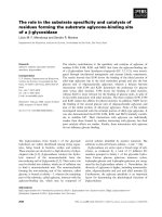
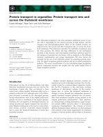
cell extract supernatant under native conditions (Figure
1A and 1C). Mass spectrometry confirmed that purified
RYMV and LMV VPgs have the expected molecular masses,
10.53 and 26.25 kDa respectively. However, their appar-
ent molecular masses turned out to be higher as judged by
SDS-PAGE and/or size exclusion chromatography (Figure
1). RYMV VPg migrated at around 15 kDa in denaturating
conditions whereas no such discrepancy was observed in
the case of LMV VPg (Figure 1A and 1C). Abnormal
mobility in denaturating electrophoresis has been already
Virology Journal 2009, 6:23 />Page 3 of 13
(page number not for citation purposes)
previously described for IDPs (see [52] and references
therein cited) and is due to their high proportion of acidic
residues (25% for RYMV VPg compared to 15% for LMV
VPg) [33]. Upon gel filtration, both RYMV and LMV VPgs
showed apparent larger molecular masses of 17 and 40
kDa respectively. Natively unfolded proteins have an
increased hydrodynamic volume compared to globular
proteins (see [52] and references therein cited). The elec-
trophoretic and hydrodynamic behaviors of RYMV and
LMV VPgs suggest that these proteins are not folded as
globular proteins.
The structural properties of the recombinant VPgs were
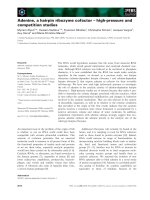
investigated by far UV-circular dichroism (far-UV CD).
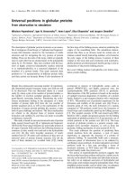
The CD spectrum of the RYMV VPg purified in non-dena-
turating conditions is typical of an intrinsically disordered
protein, as judged from its large negative ellipticity near
200 nm and from its low ellipticity at 190 nm (Figure 2A).
As reported by Uversky et al., far-UV CD enables discrim-
ination between random coils and pre-molten globules,
based on the ratio of the ellipticity values at 200 and 222
nm [28]. In the case of RYMV VPg, the ellipticity values of
-8830 and -3324 degrees cm
2
dmol
-1
at 200 and 222 nm
respectively are consistent with the existence of some
residual secondary structure, characteristic of the pre-mol-
Electrophoretic mobility and size-exclusion chromatography profile of RYMV and LMV VPgsFigure 1
Electrophoretic mobility and size-exclusion chromatography profile of RYMV and LMV VPgs. A, C. 15% SDS-
PAGE of recombinant His-tagged RYMV and LMV VPgs recovered from the supernatant (SN) and from the cell pellet (CP)
after E. coli cell extraction, and after imidazole gradient elution fractions (E1 to E5) obtained after loading a 1 ml affinity nickel
column (GE Healthcare) with the soluble fraction of the bacterial lysate. Low molecular weight (LMW) protein standards for
SDS PAGE (GE Healthcare) are shown. The expected molecular masses of 10.53 and 26.25 kDa respectively were indicated by
broken lines. The proteins in the major band (indicated by an arrow) migrate with an apparent molecular mass of about 15 and
27 kDa, respectively. B, D. Elution profile of purified His-tagged VPgs from a Superdex 75 HR10/30 column (GE Healthcare) in
50 mM Tris-HCl pH 8, 300 mM NaCl, at a flow rate of 0.5 ml/min. The proteins were eluted in a major peak with an apparent
molecular mass of about 17 and 40 kDa respectively as deduced from column calibration with low molecular weight protein
standards for gel filtration (GE Healthcare).
0
200
400
600
800
1000
1200
1400
1600
1800
6 8 10 12 14 16 18 20 22
Elution volume (ml)
A280nm (mAU)
67 43 25
13.7 kDa
0
100
200
300
400
500
600
700
800
900
6 8 10 12 14 16 18 20 22
Elution volume (ml)
A280nm (mAU)
67 43 25
13.7 kDa
14.4
kDa
20.1
45
30
97
66
SN CP E1 E2 E4E3
VPg
RYMV
LMW
kDa
20.1
45
30
97
66
SN CP E2 E3 E5E4
VPg
LMV
LMW
Virology Journal 2009, 6:23 />Page 4 of 13
(page number not for citation purposes)
ten globule state. The disordered state of LMV VPg is much
less pronounced (Figure 2B): indeed, the CD spectrum is
indicative of a predominantly folded protein, as judged
based on the presence of two well-defined minima at 208
and 222 nm and by the positive ellipticity at 190 nm. Nev-
ertheless, the relatively low ellipticity at 190 nm and the
slightly negative ellipticity near 200 nm of 621 and -1573
degrees cm
2
dmol
-1
respectively, are indicative of the pres-
ence of disordered regions (Figure 2B).
Previous secondary structure predictions have suggested
that both RYMV and LMV VPgs contain a high proportion
of α-helices, 35% and 33% respectively [21,24]. The sec-
ondary structure stabilizer 2,2,2-trifluoroethanol (TFE)
was therefore used to test the propensity of these proteins
to undergo induced folding into an α-helical conforma-
tion. The gain of α-helicity by both VPgs, as judged based
on the characteristic maximum at 190 nm and minima at
208 and 222 nm, parallels the increase in TFE concentra-
tion (Figure 2). The α-helical propensity of VPgs is
revealed at TFE concentrations as low as 5%. Further cal-
culations carried out with the K2d program [53] indicated
an α-helix content of 30% (± 4%) for RYMV VPg in the
presence of 30% TFE.
Disorder predictions in sobemoviral VPgs
The disorder propensities of VPgs from six sobemoviruses
including RYMV and SeMV were evaluated using five com-
plementary per-residue predictors of intrinsic disorder
(PONDR
®
VLXT, FoldIndex
©
, DISOPRED2, PONDR
®
VSL2
and IUPred). The amino acid sequences of sobemoviral
VPgs are highly diverse (20% identity between RYMV and
SeMV). Regions with a propensity to be disordered are
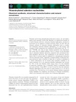
predicted in all VPgs (Figure 3). The boundaries of PDRs
varied depending on the virus and the prediction method.
However, according to PDR distribution within the
sequences, two groups of sobemoviral VPgs can be distin-
guished: RYMV/CoMV/RGMoV VPgs in one group and
SeMV/SBMV/SCPMV VPgs in the other group. This classi-
fication is consistent with the phylogenetic relationships
earlier described [54]. In the RYMV group, the N- and C-
terminus of the protein are predicted to be disordered.
The consensus secondary structure prediction in this
group indicates the presence of an α-helix followed by
two β-strands and another α-helix. Part of the terminal
regions of these VPgs are predicted to have propensities
both to be disordered and to be folded in α-helices. Resi-
dues 48 and 52, which are associated with RYMV viru-
lence, are located in the C-terminal region [55]. These
residues have been proposed to participate in the interac-
tion with two antiparallel helices of the eIF(iso)4G central
Far UV-CD spectra of RYMV and LMV VPgsFigure 2
Far UV-CD spectra of RYMV and LMV VPgs. CD spectra of purified RYMV (A) and LMV VPgs (B) in the absence (black
line) or in the presence of 5% (brown line), 10% (red line), 20% (orange line) and 30% (yellow line) of TFE.
-10000
-5000
0
5000
10000
15000
190 200 210 220 230 240 250 260
wavelength (nm)
mean residue molar ellipticity
(deg cm2 dmol-1)
-5000
0
5000
190 200 210 220 230 240 250 260
wavelength (nm)
Virology Journal 2009, 6:23 />Page 5 of 13
(page number not for citation purposes)
domain bearing E309 and E321, two residues involved in
rice resistance [24]. In the second group, the consensus is
more difficult to define and the PDRs are generally
shorter. Three conserved β-strands are predicted in the
members of this group. Despite the inconsistencies
among predictors and the intra-species differences, a pro-
pensity to structural disorder is predicted in all sobemov-
iral VPgs including the SeMV VPg, which had been
previously experimentally shown to be disordered [25].
Disorder predictions in potyviral VPgs
The disorder propensity of six potyviral VPgs for which
correlations between sequences and functions are well
documented was evaluated. The sequence identity of
these potyviruses ranges from 42% to 54%. Most of the
highly conserved regions are within domains predicted to
be ordered (Figure 4). However, PDRs were detected in
each potyviral VPg, including PVY and PVA which have
been shown to be intrinsically disordered [26,27]. The
length of the disordered regions varies among potyviruses
and discrepancies between results obtained with different
predictors are observed. Nevertheless, the N- and C-termi-
nal regions are predicted to be mainly disordered for all
proteins (Figure 4). They contain two highly conserved
segments spanning residues 43 to 45 and residues 165 to
170. Beyond the N- and C-terminus, the central region of
the VPgs is also predicted to be disordered by some predic-
tors. Several secondary structure elements are predicted
along the proteins including the central putative disor-
dered domain that is predicted to adopt an α-helical con-
formation. Interestingly, VPg sites involved in potyviral
virulence are generally located in this internal PDR (Figure
4). This region fits perfectly with the domain of LMV VPg
previously identified as a part of the binding site to HcPro
and eIF4E, two different VPg partners [21], and also par-
tially overlaps the TuMV VPg domain shown to be
involved in eIF(iso)4E binding [17]. The tyrosine residue
covalently linked to the viral RNA (position 60–64
depending on the virus) [56] is not located in a PDR.
Disorder predictions in caliciviral VPgs
The Caliciviridae family comprises four genera of human
and animal viruses [57] and possesses VPgs displaying
intermediary lengths between those of sobemoviral and
potyviral VPgs [23]. The VPg sequence of a member repre-
sentative of each genus was analysed. NV VPg, which is the
Disorder predictions of sobemoviral VPgsFigure 3
Disorder predictions of sobemoviral VPgs. Five predictors were used: PONDR
®
VLXT, FoldIndex
©
, DISOPRED2, VSL2,
IUPred. The location of predicted disordered regions (in the order provided by the above-listed predictors) was schematically
represented by lines along the VPg sequence. Numbering indicates the VPg length. The consensus predicted α-helices and β-
strands are indicated. The sites involved in RYMV virulence (*) are indicated. The VPgs experimentally demonstrated to be dis-
ordered are shaded. RYMV Rice yellow mottle virus, CoMV Cocksfoot mottle virus, RGMoV Ryegrass mottle virus, SBMV Southern
bean mosaic virus, SCPMV Southern cowpea mosaic virus, SeMV Sesbania mottle virus.
179
178
177
177
177
176
SBMV
SCPMV
RGMoV
CoMV
SeMV
RYMV
**
Virology Journal 2009, 6:23 />Page 6 of 13
(page number not for citation purposes)
longest caliciviral VPg, was predicted to be fully disor-
dered by most of the disorder predictors. For the three
other caliciviral VPgs, most PDRs are conserved although
the VPg sequence identities range from 25% to 36% (Fig-
ure 5). N-terminal extremities and C-terminal halves are
always predicted to be disordered. In addition, several
internal domains are also predicted to be disordered. The
tyrosine residues involved in urydylylation (position 20–
30 depending on the virus) [58] are generally not located
in PDRs.
α
-MoRF predictions
Often, intrinsically disordered regions involved in pro-
tein-protein interactions and molecular recognition
undergo disorder-to-order transitions upon binding [30-
32,35,59-63]. A correlation has been established between
the specific pattern in the PONDR
®
VLXT curve and the
ability of a given short disordered regions to undergo dis-
order-to-order transitions on binding [64]. Based on these
specific features, an α-MoRF predictor was recently devel-
oped [60,65].
The application of the α-MoRF predictor to the set of 16
VPgs reveals that helix forming molecular recognition fea-
Disorder predictions of potyviral VPgsFigure 4
Disorder predictions of potyviral VPgs. Five predictors
were used: PONDR
®
VLXT, FoldIndex
©
, DISOPRED2, VSL2,
IUPred. The location of predicted disordered (in the order
provided by the above-listed predictors) was schematically
represented by lines along the VPg sequence. Numbering
indicates the VPg length. Highly conserved regions (grey) and
consensus predicted α-helices and β-strands are indicated.
The conserved tyrosine (Y) involved in VPg urydylylation and
the sites (*) involved in virulence are indicated. The VPgs
experimentally demonstrated to be disordered are shaded.
LMV Lettuce mosaic virus, PVY Potato virus Y, PVA Potato virus
A, TEV Tobacco etch virus, TuMV Turnip mosaic virus, BYMV
Bean yellow mosaic virus.
***
*
PVY
PVA
1 188
1 188
1
188
1 192
1 191
TEV
TuMV
BYMV
Y
**
Y
*
Y
*
Y
*
Y
*
1LMV
Y
*
******
193
*
**
Disorder predictions of caliciviral VPgsFigure 5
Disorder predictions of caliciviral VPgs. Five predictors
were used: PONDR
®
VLXT, FoldIndex
©
, DISOPRED2, VSL2,
IUPred. The location of predicted disordered (in the order
provided by the above-listed predictors) was schematically
represented by lines along the VPg sequence. Numbering
represents the VPg length. The consensus predicted α-heli-
ces and β-strands are indicated. The conserved tyrosine resi-
due (Y) involved in VPg urydylylation is indicated. RHDV
Rabbit hemorrhabic disease virus (Lagovirus), VESV Vesicular
exanthema of swine virus (Vesivirus), SV Man Sapporo virus Man-
chester virus (Sapovirus) and NV Norwalk virus (Norovirus).
RHDV 1 114
1 113
1 138
1 114
VESV
SVMan
NV
Y
Y
Y
Y
Virology Journal 2009, 6:23 />Page 7 of 13
(page number not for citation purposes)
tures are highly abundant in these proteins. Table 1 shows
that there are 15 α-MoRFs in 12 VPgs. The regions of pot-
yviral VPgs spanning residues 24–26 and 41–43 are
always predicted to form α-MoRFs. By contrast, the puta-
tive α-MoRF regions are not conserved in sobemoviral
and caliciviral VPgs, likely reflecting lower sequence con-
servation among these proteins but also suggesting diver-
sity in the disordered state at intraspecies level. No α-
MoRFs were predicted in VESV, RGMoV, SBMV and
SCPMV VPgs. It should be pointed out, however, that not
all MoRF regions share these same features and some of
them may form β- or irregular structure rather than α-hel-
ices upon binding [61,62]. Therefore, predicted MoRFs
only represent a fraction of the total numbers of potential
MoRFs. According to secondary structure predictions,
SBMV and SCPMV would form more preferentially β-
MoRFs. In this respect, the prediction of α-MoRF in SeMV
VPg, which is related to SBMV and SCPMV, was not
expected.
CDF and CH-plot analyses
In order to compare the disordered state of VPgs from the
various viral genera, VPg sequences were analyzed by two
binary predictors of intrinsic disorder, charge-hydropathy
plot (CH-plot) [31,60] and cumulative distribution func-
tion analysis (CDF) [60]. These predictors classify entire
proteins as ordered or disordered, as opposed to the pre-
viously described disorder predictors, which output disor-
der propensity for each position in the protein sequence.
The usefulness of the joint application of these two binary
classifiers is based on their methodological differences
[60,66]. In Figure 6, each spot corresponds to a single pro-
tein and its coordinates are calculated as a distance of this
protein from the folded/unfolded decision boundary in
the corresponding CH-plot (Y-coordinate) and an average
distance of the corresponding CDF curve from the order/
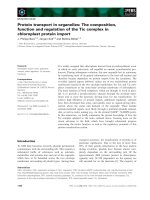
disorder decision boundary (X-coordinate). Figure 6
shows that the majority of VPgs are predicted to be disor-
dered: 11 VPgs including RYMV and LMV VPgs are located
within the (-, -) quadrant suggesting that they belong to
the class of native molten globules. Figure 6 shows that all
Caliciviridae VPgs are predicted to be native molten glob-
ules, whereas VPgs from Sobemoviruses and Potyviruses are
spread between different quadrants. Notably, PVA and
SeMV VPgs are located in the (+,-) quadrant of the ordered
proteins indicating that these binary methods failed to
detect the experimentally demonstrated disorder of these
two VPgs.
Discussion
In this paper, we provide experimental evidences that
RYMV and LMV VPgs contain intrinsically disordered
regions. These findings, together with the previous reports
documenting the disordered state of SeMV, PVY and PVA
VPgs [25-27], suggest that intrinsic disorder may be a
common and distinctive feature of sobemo- and potyviral
VPgs. By carrying out an in-depth in silico analysis, we
show that the disordered state of VPgs depend on the viral
genera. Sobemoviral SeMV and RYMV VPgs appeared
highly disordered with (i) 30% and 50% increases of their
molecular masses estimated from SDS-PAGE compared to
expected masses, respectively, and (ii) far-UV CD spectra
with large negative ellipticities near 200 nm and low ellip-
ticities at 190 nm. By contrast, the increase of the apparent
molecular masses of potyviral VPgs from SDS-PAGE are
moderate (<5% for LMV, approx. 10% for PVY and PVA)
and the trends of far-UV CD spectra indicate partial disor-
der better suggesting short disordered regions included in
globally ordered VPgs.
The experimentally observed disorder is also pointed out
by complementary in silico analyses. However, quantita-
tive assessment of disorder prediction strengths and pre-
cise location of consensus disordered regions turned out
to be hectic. While LMV, PVY and PVA VPgs showed
longer disordered segments, SeMV VPg showed short dis-
ordered segments whereas experimental results were sim-
ilar to RYMV VPg. Moreover, binary predictors which are
intended to allow a comparison of relative disordered
states failed to detect disorder in several VPgs, including
those for which the disordered state has been shown
experimentally such as SeMV and PVA. However, it is
important to notice that these predictors are meant to pre-
dict disorder on an entire protein basis, and SeMV and
PVA not only have substantial ordered regions, but their
disordered regions are in general shorter than those of the
other proteins studied. These features could have easily
tipped the balance towards an "ordered protein" predic-
tion. Otherwise, the use of complementary disorder pre-
dictors induces difficulties to precisely map consensus
Table 1: Location of predicted α-MoRFs in VPgs
Viral genus/family Viral species α-MoRFs
Sobemovirus RYMV 14–31
56–73
CoMV 1–18
SeMV 43–60
Potyvirus LMV 25–42
PVY 25–42
PVA 24–41
167–184
TEV 26–43
TuMV 25–42
BYMV 26–43
Caliciviridae RHDV 68–85
SV Man 14–31
NV 30–47
115–132
Virology Journal 2009, 6:23 />Page 8 of 13
(page number not for citation purposes)
disordered regions in VPgs, but this is due mainly to the
fact that different disorder predictors are built upon
slightly different definitions of disorder [41]. This is what
makes these predictions complementary of each other.
The presence of intrinsically disordered (ID) regions was
detected by five per-residue disorder predictors in 10–26
kDa VPgs. At intra-specific level in sobemo- and in poty-
viruses, the presence of intrinsic disorder regions was con-
served independently from sequence conservation.
Therefore, we enlarged our analysis to other genera,
namely caliciviral VPgs that had never been suggested
before to be disordered, and small VPgs (2 to 3 kDa) from
Picornaviridae and Comoviridae where ID was also pre-
dicted (data not shown). By contrast to several domains in
capsid and polymerase viral proteins, the disorder pro-
pensity had not been described so far as a common prop-
erty of VPgs [67]. The methodology used by Chen and
colleagues is likely not adapted to the highly diverse set of
VPg sequences because it includes a first step of conserved
domain identification before performing the disorder pre-
dictions.
Comparison of the PONDR
®
CDF and CH-plot analyses of whole protein order-disorder via distributions of VPgs within the CH-CDF phase spaceFigure 6
Comparison of the PONDR
®
CDF and CH-plot analyses of whole protein order-disorder via distributions of
VPgs within the CH-CDF phase space. Each spot represents a single VPg whose coordinates were calculated as a distance
of this protein from the boundary in the corresponding CH-plot (Y-coordinate) and an average distance of the corresponding
CDF curve from the boundary (X-coordinate). The four quadrants in the plot correspond to the following predictions: (-, -)
proteins predicted to be disordered by CDF, but compact by CH-plot; (-, +) proteins predicted to be disordered by both
methods; (+, -) contains ordered proteins; (+, +) includes proteins predicted to be disordered by CH-plot, but ordered by the
CDF analysis. Open circles correspond to caliciviral VPgs, gray circles represent sobemoviral VPgs, whereas black circles cor-
respond to potyviral VPgs.
CDF plot
-0.4 -0.2 0.0
CH-plot
-0.15
-0.10
-0.05
0.00
0.05
0.10
0.15
D, O
-, -
O, O
+, -
O, D
+, +
D, D
-, +
NV
SV
RHDV
VESV
RYMV
CfMV
RGMoV
SCPMV
SeMV
SBMV
TEV
TuMV
LMV
BYMV
PVY
PVA
Virology Journal 2009, 6:23 />Page 9 of 13
(page number not for citation purposes)
VPg ID was rather predicted in several small patches (<30
residues) than in few large domains, this trend is common
in short protein sequences with binding sites. These char-
acteristics of variable degree of disorder, together with the
complementarities of disorder definitions described
above, may explain why discrepancies in location of PDRs
were frequently observed. Still, all proteins showed a high
predicted disorder content (percentage of disordered resi-
dues), ranging in average from 44% for sobemoviral to
60% for caliciviral VPgs (PONDR
®
VSL2 predictions). Part
of the hydrophobic residues of VPgs would be involved in
the formation of additional secondary structure elements.
We performed in silico detection of α-helix-forming
molecular recognition features (α-MoRF) which mediate
the binding of initially disordered domains with interac-
tion partners [60]. Some α-MoRF domains were detected
in the N-terminal regions of VPgs which were not reported
to be interacting domains. By contrast, the first half of the
C-terminal domain of RYMV VPg and the central domain
of LMV VPg previously predicted to form α-helices [21,24]
were not identified as α-MoRFs. These domains were pre-
dicted both to be disordered and to form α-helices. The α-
helical propensities of RYMV VPgs, as observed in the
presence of TFE concentration as low as 5% (Figure 2),
suggest that some disordered regions in the isolated pro-
teins may undergo a disorder-to-order transition upon
association with a partner protein. Noteworthy, the only
VPg structures available to date (Picornaviridae) were
obtained either in the presence of a stabilizing agent [49]
or in association with the viral RNA-dependent RNA
polymerase (3D) which probably stabilized the VPg
folded state [50,51].
The property of proteins to be intrinsically disordered
confers to them the ability to bind to many different part-
ners. These characteristics likely explain why many pro-
teins critical in interaction networks (hub proteins) are
intrinsically disordered [36,45]. In RYMV VPg, the resist-
ance-breaking positions 48 and 52 suggested to be
involved in eIF(iso)4G interaction are located in a puta-
tive α-helix also predicted to be disordered. The same
result is obtained with LMV VPg where resistance-break-
ing sites involved in eIF4E interaction are located in the
central domain predicted to contain two α-helices and to
display disorder features. Analysis of other potyviral VPgs
suggests that domains associated with virulence are often
disordered with some residual structure. Besides their
interactions with eIF4Es, potyviral VPgs were found to
interact with a variety of host factors such as poly(A)-
binding protein [68,69], eIF4G [18] and eukaryotic elon-
gation factor eEF1A [70]. Multiple in vitro interactions of
VPgs with eIF4GI [71], eIF3 [72] and eIF4A [73], and oth-
ers proteins belonging to the translation initiation com-
plex, were also shown for Caliciviridae members. Potyviral
VPgs were also reported to interact with several viral pro-
teins such as NIb, HC-Pro, CI and CP [9,68,74].
As underlined in the introduction, VPgs are multifunc-
tional proteins. At least part of their functions implies
interactions with eIFs, with the VPg/eIF4E interaction hav-
ing been shown to enhance the in vitro translation of viral
RNA [22,75]. VPgs were suggested to mimic the mRNA 5'-
linked cap recruiting the translation initiation complex.
Besides, a ribonuclease activity of VPgs was reported. It
might contribute to host RNA translation shutoff [76].
VPg-eIF interactions were also suggested to be involved in
other key steps in the viral cycle [1]. In Picornaviridae, it
was established that VPg is involved in genome replica-
tion, its uridyl-form acting as primer for complementary
strand synthesis [77,78]. An additional role of potyviral
VPg-eIF4E interactions in plant cell-to-cell movement via
eIF4G and microtubules was also suggested [2,79]. VPg
could participate to a putative vascular movement com-
plex to cross the plasmodesmata and may facilitate virus
unloading [9,80]. Thus, VPg might be involved in key
steps of the viral cycle such as replication, translation and
movement. Additionally, ID VPg was reported to be nec-
essary to the processing of SeMV polyprotein by viral pro-
tease [25]. ID might explain how a unique protein can
perform and regulate these different biological functions.
PDRs might give to the VPg the necessary plasticity to fit
surface overlaps with various partners.
Conclusion
Experimentally, we showed that RYMV and LMV VPgs
contain both intrinsically disordered domains but with
different disordered states. Using in silico analyses, ID
domains were predicted to occur in 14 VPgs of sobemo-,
poty-and caliciviruses. Although highly diverse, VPgs
share the common feature of possessing ID domains.
These structural properties of VPgs are more conserved
than what could be anticipated from their sequence
homologies. However, comparative analyses at intra-and
interspecies levels showed the diversity of intrinsic disor-
der in VPgs.
Like many IDPs, VPg ID domains may play a role in pro-
tein interaction networks, interacting in particular with
translation initiation factor eIFs to perform key steps of
the viral cycle (replication, translation and movement).
Methods
Purification of recombinant RYMV and LMV VPgs
The VPg-encoding region in the RYMV ORF2a was ampli-
fied by PCR from FL5 infectious clone [81] by using the
primers FCIaVPgH 5'ATATCCATGGGATCCCA TTTGA-
GATTTACGGC (containing a NcoI site and RYMV nucle-
otides 1587–1607) and RCIaVPgH
5'TGCAAGATCTCTCGATATCAACATCCTCGCC (con-
Virology Journal 2009, 6:23 />Page 10 of 13
(page number not for citation purposes)
taining a BglII site and sequence complementary to RYMV
nucleotides 1823–1803). The resulting fragment was
cloned into the NcoI and BglII sites of pQE60 as a 6-His C-
terminal fusion (Qiagen) and the construct was
sequenced. The resulting expression plasmid was used to
transform the E. coli strain M15-pRep4 (Qiagen). After
induction with 0.5 mM isopropyl-1-thio-β-D-galactopyra-
noside at 25°C for 5 h, the cells from 1 L culture in LB
medium were harvested by centrifugation and frozen at -
80°C. Cells were thawn, resuspended in 30 mL of purifi-
cation buffer (50 mM Tris-HCl, pH 8.0, 300 mM NaCl,
10% glycerol), disrupted with a French press (Thermo)
and centrifuged at 18000 rpm for 30 min. The superna-
tant was filtered (0.5 μm filters) and purification of the
VPg in native conditions was carried out using a nickel-
loaded HiTrap IMAC HP column (GE Healthcare) fol-
lowed by gel filtration step onto a HR10/30 Superdex 75
column (GE Healthcare) in 50 mM Tris-HCl, pH 8.0, 300
mM NaCl, 5% glycerol.
LMV VPg was produced in E. coli using the pTrcHis plas-
mid as expression vector as already described [18]. The N-
terminal His-tagged protein was found to be expressed in
the soluble fraction of the bacterial lysate and was purified
as described above, except that 50 mM Tris-HCl pH 8, 800
mM NaCl, 10% glycerol, 2 mM β-mercaptoethanol was
used as the affinity chromatography buffer, and 20 mM
Tris-HCl pH 8, 800 mM NaCl, 5% glycerol as gel filtration
buffer.
Circular dichroism analyses
Freshly purified protein samples were used for CD analy-
ses. Sample buffer was changed by eluting the protein
from a PD10 desalting column (GE Healthcare) using 10
mM sodium phosphate buffer (pH 8.0), supplemented
with 300 mM or 500 mM NaF for RYMV or LMV VPgs
respectively. After centrifugation, the protein concentra-
tion was determined using a ND-1000 Spectrophotome-
ter (NanoDrop Technologies) and an extinction
coefficient of 7,780 and 18,490 M
-1
cm
-1
for RYMV and
LMV VPgs respectively. Far UV-CD spectra were recorded
with a chirascan dichrograph (Applied Photophysics) in a
thermostated (20°C) quartz circular cell with a 0.5 mm
path length, in steps of 0.5 nm. All protein spectra were
corrected by subtraction of the respective buffer spectra.
The mean molar ellipticity values per residue were calcu-
lated using the manufacturer software. Structural varia-
tions of the native protein samples were monitored by
recording successive CD spectra after addition of 2,2,2-tri-
fluoroethanol (TFE, Sigma) in the 5–30% range (vol:vol).
VPg sequences
Sequences for this study were obtained from the viral
genome resources at NCBI http://
www.ncbi.nlm.nih.gogomes/gen
list.cgi?taxid=10239&type=5&name=Viruses. Sequence
accession numbers are: Sobemovirus (RYMV AJ608219,
CoMV NC_002618, RGMoV NP_736586, SBMV
NP_736583, SCPMV NP_736598, SeMV NP_736592),
Potyvirus (LMV NP_734159, PVY NP_734252, PVA
NC_004039, TEV NP_734204, TuMV NC_002509, BYMV
NC_003492), and Caliciviridae (RHDV NP_740330, VESV
NP_786894, SV Man X86560, NV NP_786948).
Disorder predictions
Seven programs were used to predict the disorder ten-
dency of VPgs. PONDR
®
, Predictors of Natural Disordered
Regions, version VLXT is a neural network principally
based on local amino acid composition, flexibility and
hydropathy [82]
. FoldIndex
©
is
based on charge and hydropathy analyzed locally using a
sliding window [83] />dex. DISOPRED2 is also a neural network, but incorpo-
rates information from multiple sequence alignments
generated by PSI-BLAST [44] />opred. PONDR
®
VSL2 has achieved higher accuracy and
improved performance on short disordered regions, while
maintaining high performance on long disordered
regions [84] />predictorVSL2.php. IUPred uses a novel algorithm that
evaluates the energy resulting from inter-residue interac-
tions [85]
. PONDR
®
VLXT and
VSL2 as well as DISOPRED2 were all trained on datasets
of disordered proteins, while FoldIndex
©
and IUPred were
not. Binary classifications of VPgs as ordered or disor-
dered were performed using CDF and CH-plot analyses.
Cumulative distribution function curves or CDF curves
were generated for each dataset using PONDR
®
VLXT
scores for each of the VPgs [60]. Charge-hydropathy distri-
butions (CH-plots) were also analyzed using the method
described in Uversky et al. [31].
α
-MoRF predictions
The predictor of α-helix forming Molecular Recognition
Features, α-MoRF, focuses on short binding regions
within regions of disorder that are likely to form helical
structure upon binding [60,65]. It utilizes a stacked archi-
tecture, where PONDR
®
VLXT is used to identify short pre-
dictions of order within long predictions of disorder and
then a second level predictor determines whether the
order prediction is likely to be a binding site based on
attributes of both the predicted ordered region and the
predicted surrounding disordered region. An α-MoRF pre-
diction indicates the presence of a relatively short (20 res-
idues), loosely structured helical region within a largely
disordered sequence [60,65]. Such regions gain stable
structure upon a disorder-to-order transition induced by
binding to partner.
Virology Journal 2009, 6:23 />Page 11 of 13
(page number not for citation purposes)
Competing interests
The authors declare that they have no competing interests.
Authors' contributions
EH carried out experiments and drafted the manuscript.
YB participated in the design, performed protein purifica-
tions and far UV-CD analyses. TM and JW participated in
LMV VPg analyses. SL participated in predictive analyses.
VNU performed CDF and CH-plot analyses. FD and AVD
performed the mass spectrometry analyses. PR performed
α-MoRF analyses. ND and DF participated in the study
design and coordination and helped to draft the manu-
script. All authors read and approved the final manu-
script.
Acknowledgements
We are grateful to Anne-Lise Haenni and Jean-François Laliberté for helpful
discussions. We thank Jean-Paul Brizard for technical advice.
This work was partially supported by the French National Agency for
Research ('Poty4E', ANR-05-Blan-0302-01).
References
1. Robaglia C, Caranta C: Translation initiation factors: a weak
link in plant RNA virus infection. Trends in Plant Science 2006,
11:40-45.
2. Gao Z, Johansen E, Eyers S, Thomas CL, Noel Ellis TH, Maule AJ: The
potyvirus recessive resistance gene, sbm1, identifies a novel
role for translation initiation factor eIF4E in cell-to-cell traf-
ficking. Plant J 2004, 40:376-385.
3. Kanyuka K, Druka A, Caldwell DG, Tymon A, Mc Callum N, Waugh
R, Adams MJ: Evidence that the recessive bymovirus resist-
ance locus rym4 in barley corresponds to the eukaryotic
translation initiation factor 4E gene. Molecular Plant Pathology
2005, 6:449-458.
4. Nicaise V, German-Retana S, Sanjuan R, Dubrana MP, Mazier M, Mai-
sonneuve B, Candresse T, Caranta C, LeGall O: The eukaryotic
translation initiation factor 4E controls lettuce susceptibility
to the Potyvirus Lettuce mosaic virus. Plant Physiol 2003,
132:1272-1282.
5. Ruffel S, Dussault MH, Palloix A, Moury B, Bendahmane A, Robaglia
C, Caranta C: A natural recessive resistance gene against
potato virus Y in pepper corresponds to the eukaryotic initi-
ation factor 4E (eIF4E). Plant J 2002, 32:1067-1075.
6. Stein N, Perovic D, Kumlehn J, Pellio B, Stracke S, Streng S, Ordon F,
Graner A: The eukaryotic translation initiation factor 4E con-
fers multiallelic recessive Bymovirus resistance in Hordeum
vulgare (L.). Plant J 2005, 42:912-922.
7. Bruun-Rasmussen M, Moller IS, Tulinius G, Hansen JK, Lund OS,
Johansen IE: The same allele of translation initiation factor 4E
mediates resistance against two Potyvirus spp. in Pisum sati-
vum. Mol Plant Microbe Interact 2007, 20:1075-1082.
8. Nicolas O, Dunnington SW, Gotow LF, Pirone TP, Hellmann GM:
Variations in the VPg protein allow a potyvirus to overcome
va gene resistance in tobacco. Virology 1997, 237:452-459.
9. Rajamaki ML, Valkonen JP: Viral genome-linked protein (VPg)
controls accumulation and phloem-loading of a potyvirus in
inoculated potato leaves. Molecular Plant Microbe Interactions
2002, 15:138-149.
10. Borgstrom B, Johansen IE: Mutations in pea seedborne mosaic
virus genome-linked protein VPg after pathotype-specific
virulence in Pisum sativum. Mol Plant Microbe Interact 2001,
14:707-714.
11. Moury B, Morel C, Johansen E, Guilbaud L, Souche S, Ayme V,
Caranta C, Palloix A, Jacquemond M: Mutations in Potato virus Y
genome-linked protein determine virulence toward reces-
sive resistances in Capsicum annuum and Lycopersicon hirsu-
tum. Molecular Plant Microbe Interactions 2004, 17:322-329.
12. Sato M, Masuta C, Uyeda I: Natural resistance to Clover yellow
vein virus in beans controlled by a single recessive locus. Mol
Plant Microbe Interact 2003, 16:994-1002.
13. Ayme V, Souche S, Caranta C, Jacquemond M, Chadoeuf J, Palloix A,
Moury B: Different mutations in the genome-linked protein
VPg of potato virus Y confer virulence on the pvr2(3) resist-
ance in pepper. Molecular Plant Microbe Interactions 2006,
19:557-563.
14. Rajamaki ML, Valkonen JP: The 6K2 protein and the VPg of
potato virus A are determinants of systemic infection in
Nicandra physaloides. Mol Plant Microbe Interact 1999,
12:1074-1081.
15. Wittmann S, Chatel H, Fortin MG, Laliberte JF: Interaction of the
viral protein genome linked of Turnip mosaic potyvirus with
the translational eukaryotic initiation factor (iso) 4E of Ara-
bidopsis thaliana using the yeast two-hybrid system. Virology
1997, 234:84-92.
16. Schaad MC, Anderberg RJ, Carrington JC: Strain-specific interac-
tion of the tobacco etch virus NIa protein with the transla-
tion initiation factor eIF4E in the yeast two-hybrid system.
Virology 2000, 273:300-306.
17. Leonard S, Plante D, Wittmann S, Daigneault N, Fortin MG, Laliberte
JF: Complex formation between potyvirus VPg and transla-
tion eukaryotic initiation factor 4E correlates with virus
infectivity. Journal of Virology 2000, 74:7730-7737.
18. Michon T, Estevez Y, Walter J, German-Retana S, Le Gall O:
The
potyviral virus genome-linked protein VPg forms a ternary
complex with the eukaryotic initiation factors eIF4E and
eIF4G and reduces eIF4E affinity for a mRNA cap analogue.
FEBS J 2006, 273:1312-1322.
19. Beauchemin C, Boutet N, Laliberte JF: Visualization of the inter-
action between the precursors of VPg, the viral protein
linked to the genome of turnip mosaic virus, and the transla-
tion eukaryotic initiation factor iso 4E in Planta. J Virol 2007,
81:775-782.
20. Khan MA, Miyoshi H, Ray S, Natsuaki T, Suehiro N, Goss DJ: Inter-
action of genome-linked protein (VPg) of Turnip mosaic virus
(TuMV) with wheat germ translation initiation factors
eIFiso4E and eIFiso4F. J Biol Chem 2006, 280:28002-28010.
21. Roudet-Tavert G, Michon T, Walter J, Delaunay T, Redondo E, Le
Gall O: Central domain of a potyvirus VPg is involved in the
interaction with the host translation initiation factor eIF4E
and the viral protein HcPro. J Gen Virol 2007, 88:1029-1033.
22. Goodfellow I, Chaudhry Y, Gioldasi I, Gerondopoulos A, Natoni A,
Labrie L, Laliberte JF, Roberts L: Calicivirus translation initiation
requires an interaction between VPg and eIF 4 E. EMBO Rep
2005, 6:968-972.
23. Sadowy E, Milner M, Haenni AL: Proteins attached to viral
genomes are multifunctional. Adv Virus Res 2001, 57:185-262.
24. Hébrard E, Pinel-Galzi A, Fargette D: Virulence domain of the
RYMV Genome-Linked Viral Protein VPg towards rice
rymv1-2-mediated resistance. Archives of Virology 2008,
153:1161-1164.
25. Satheshkumar PS, Gayathri P, Prasad K, Savithri HS: "Natively
Unfolded" VPg is essential for sesbania mosaic virus serine
protease activity. Journal of Biological Chemistry 2005,
280:30291-30300.
26. Grzela R, Szolajska E, Ebel C, Madern D, Favier A, Wojtal I, Zagorski
W, Chroboczek J: Virulence factor of potato virus Y, genome-
attached terminal protein VPg, is a highly disordered pro-
tein. J Biol Chem 2007, 283:213-221.
27. Rantalainen K, Uversky V, Permi P, Kalkkinen N, Dunker A, Mäkinen
K: Potato virus A genome-linked protein VPg is an intrinsi-
cally disordered molten globule-like protein with a hydro-
phobic core.
Virology 2008, 377:280-288.
28. Uversky VN: Natively unfolded proteins: A point where biol-
ogy waits for physics. Protein Sci 2002, 11:739-756.
29. Daughdrill GW, Pielak GJ, Uversky VN, Cortese MS, Dunker AK:
Natively disordered proteins. Handbook of Protein folding
2005:271-353.
30. Wright PE, Dyson HJ: Intrinsically unstructured proteins: re-
assessing the protein structure-function paradigm. J Mol Biol
1999, 293:321-331.
31. Uversky VN, Gillespie JR, Fink AL: Why are "natively unfolded"
proteins unstructured under physiologic conditions? Proteins
2000, 41:415-427.
Virology Journal 2009, 6:23 />Page 12 of 13
(page number not for citation purposes)
32. Dunker AK, Lawson JD, Brown CJ, Williams RM, Romero P, Oh JS,
Oldfield CJ, Campen AM, Ratliff CM, Hipps KW, et al.: Intrinsically
disordered protein. J Mol Graph Model 2001, 19:26-59.
33. Tompa P: Intrinsically unstructured proteins. Trends in Biochem-
ical Sciences 2002, 27:527-533.
34. Uversky VN: What does it mean to be natively unfolded? Eur J
Biochem 2002, 269:2-12.
35. Dyson HJ, Wright PE: Intrinsically unstructured proteins and
their functions. Nat Rev Mol Cell Biol 2005, 6:197-208.
36. Dunker AK, Cortese MS, Romero P, Iakoucheva LM, Uversky VN:
Flexible nets. The roles of intrinsic disorder in protein inter-
action networks. FEBS Journal 2005, 272:5129-5148.
37. Uversky VN, Oldfield CJ, Dunker AK: Showing your ID: intrinsic
disorder as an ID for recognition, regulation and cell signal-
ing. J Mol Recognit 2005, 18:343-384.
38. Xie H, Vucetic S, Iakoucheva LM, Oldfield CJ, Dunker AK, Uversky
VN, Obradovic Z: Functional anthology of intrinsic disorder. 1.
Biological processes and functions of proteins with long dis-
ordered regions. J Proteome Res 2007, 6:1882-1898.
39. Vucetic S, Xie H, Iakoucheva LM, Oldfield CJ, Dunker AK, Obradovic
Z, Uversky VN: Functional anthology of intrinsic disorder. 2.
Cellular components, domains, technical terms, develop-
mental processes, and coding sequence diversities corre-
lated with long disordered regions. J Proteome Res 2007,
6:1899-1916.
40. Xie H, Vucetic S, Iakoucheva LM, Oldfield CJ, Dunker AK, Obradovic
Z, Uversky VN: Functional anthology of intrinsic disorder. 3.
Ligands, post-translational modifications, and diseases asso-
ciated with intrinsically disordered proteins. J Proteome Res
2007, 6:1917-1932.
41. Ferron F, Longhi S, Canard B, Karlin D: A practical overview of
protein disorder prediction methods. Proteins 2006, 65:
1-14.
42. Bourhis JM, Canard B, Longhi S: Predicting protein disorder and
induced folding: from theoretical principles to practical
applications. Curr Protein Pept Sci 2007, 8:135-149.
43. Dunker A, Obradovic Z, Romero P, Garner EC, Brown CJ: Intrinsic
protein disorder in complete genomes. Genome Informatics
2000, 11:161-171.
44. Ward JJ, Sodhi JS, McGuffin LJ, Buxton BF, Jones DT: Prediction and
functional analysis of native disorder in proteins from the
three kingdoms of life. J Mol Biol 2004, 337:635-645.
45. Haynes C, Oldfield CJ, Ji F, Klitgord N, Cusick ME, Radivojac P, Uver-
sky VN, Vidal M, Iakoucheva LM: Intrinsic disorder is a common
feature of hub proteins from four eukaryotic interactomes.
PLoS Comput Biol 2006, 2:e100.
46. Radivojac P, Iakoucheva LM, Oldfield CJ, Obradovic Z, Uversky VN,
Dunker AK: Intrinsic disorder and functional proteomics. Bio-
phys J 2007, 92:1439-1456.
47. Tompa P, Dosztanyi Z, Simon I: Prevalent structural disorder in
E. coli and S. cerevisiae proteomes. J Proteome Res 2006,
5:1996-2000.
48. Chen JW, Romero P, Uversky VN, Dunker AK: Conservation of
intrinsic disorder in protein domains and families: I. A data-
base of conserved predicted disordered regions. J Proteome
Res 2006, 5:879-887.
49. Schein CH, Oezguen N, Volk DE, Garimella R, Paul A, Braun W:
NMR structure of the viral peptide linked to the genome
(VPg) of poliovirus. Peptides 2006, 27:1676-1684.
50. Ferrer-Orta C, Arias A, Agudo R, Perez-Luque R, Escarmis C,
Domingo E, Verdaguer N: The structure of a protein primer-
polymerase complex in the initiation of genome replication.
EMBO J 2006, 25:880-888.
51. Gruez A, Selisko B, Roberts M, Bricogne G, Bussetta C, Jabafi I, Cou-
tard B, De Palma AM, Neyts J, Canard B: The crystal structure of
coxsackievirus B3 RNA-dependent RNA polymerase in com-
plex with its protein primer VPg confirms the existence of a
second VPg binding site on Picornaviridae polymerases. J
Virol 2008, 82:9577-9590.
52. Receveur-Brechot V, Bourhis JM, Uversky VN, Canard B, Longhi S:
Assessing protein disorder and induced folding. Proteins 2006,
62:24-45.
53. Merelo JJ, Andrade MA, Prieto A, Morán F: Proteinotopic Feature
Maps. Neurocomputing 1994, 6:443-454.
54. Hull R, Fargette D: Sobemovirus. In Virus Taxonomy Eight Report of
the International Committee on Taxonomy of Viruses Edited by: Fauquet
C, Mayo MA, Maniloff J, Desselberger U, Ball LA. Academic Press,
Elsevier; 2005:885-890.
55. Pinel-Galzi A, Rakotomalala M, Sangu E, Sorho F, Kanyeka Z, Traoré
O, Sérémé D, Poulicard N, Rabenantaondro Y, Séré Y, et al.: Theme
and variations in the evolutionary pathways to virulence of
an RNA plant virus species. PLoS Pathogens 2007, 3:e180.
56. Murphy JF, Rychlik W, Rhoads RE, Hunt AG, Shaw JG: A tyrosine
residue in the small nuclear inclusion protein of tobacco vein
mottling virus links the VPg to the viral RNA. J Virol 1991,
65:511-513.
57. Koopmans MK, Green KY, Ando T, Clarke IN, Estes MK, Matson
DO, Nakata S, Neill JD, Smith AW, Studdert MJ, Thiel HJ: Caliciviri-
dae. In Virus Taxonomy Eight Report of the International Committee on
Taxonomy of Viruses Edited by: Fauquet C, Mayo MA, Maniloff J, Des-
selberger U, Ball LA. Academic Press, Elsevier; 2005.
58. Machin A, Martin Alonso JM, Parra F: Identification of the amino
acid residue involved in rabbit hemorrhagic disease virus
VPg uridylylation. J Biol Chem 2001, 276:27787-27792.
59. Dyson HJ, Wright PE: Coupling of folding and binding for
unstructured proteins. Curr Opin Struct Biol 2002, 12:54-60.
60. Oldfield CJ, Cheng Y, Cortese MS, Brown CJ, Uversky VN, Dunker
AK: Comparing and combining predictors of mostly disor-
dered proteins. Biochemistry 2005, 44:1989-2000.
61. Mohan A, Oldfield CJ, Radivojac P, Vacic V, Cortese MS, Dunker AK,
Uversky VN: Analysis of Molecular Recognition Features
(MoRFs). J Mol Biol 2006, 362:1043-1059.
62. Vacic V, Oldfield CJ, Mohan A, Radivojac P, Cortese MS, Uversky VN,
Dunker AK: Characterization of molecular recognition fea-
tures, MoRFs, and their binding partners. J Proteome Res
2007,
6:2351-2366.
63. Oldfield CJ, Meng J, Yang JY, Yang MQ, Uversky VN, Dunker AK:
Flexible nets: disorder and induced fit in the associations of
p53 and 14-3-3 with their partners. BMC Genomics 2008,
9(Suppl 1):S1.
64. Garner E, Romero P, Dunker AK, Brown C, Obradovic Z: Predict-
ing Binding Regions within Disordered Proteins. Genome
Inform Ser Workshop Genome Inform 1999, 10:41-50.
65. Cheng Y, Oldfield CJ, Meng J, Romero P, Uversky VN, Dunker AK:
Mining alpha-helix-forming molecular recognition features
with cross species sequence alignments. Biochemistry 2007,
46:13468-13477.
66. Mohan A, Sullivan WJ Jr, Radivojac P, Dunker AK, Uversky VN:
Intrinsic disorder in pathogenic and non-pathogenic
microbes: discovering and analyzing the unfoldomes of
early-branching eukaryotes. Mol Biosyst 2008, 4:328-340.
67. Chen JW, Romero P, Uversky VN, Dunker AK: Conservation of
intrinsic disorder in protein domains and families: II. func-
tions of conserved disorder. J Proteome Res 2006, 5:888-898.
68. Leonard S, Viel C, Beauchemin C, Daigneault N, Fortin MG, Laliberte
JF: Interaction of VPg-Pro of turnip mosaic virus with the
translation initiation factor 4E and the poly(A)-binding pro-
tein in planta. Journal of General Virology 2004, 85:1055-1063.
69. Beauchemin C, Laliberte J: The Poly(A) Binding Protein Is Inter-
nalized in Virus-Induced Vesicles or Redistributed to the
Nucleolus during Turnip Mosaic Virus Infection. J Virol 2007,
81:10905-10913.
70. Thivierge K, Cotton S, Dufresne PJ, Mathieu I, Beauchemin C, Ide C,
Fortin MG, Laliberte JF: Eukaryotic elongation factor 1A inter-
acts with Turnip mosaic virus RNA-dependent RNA
polymerase and VPg-Pro in virus-induced vesicles. Virology
2008, 377:216-225.
71. Daughenbaugh KF, Wobus CE, Hardy ME: VPg of murine norovi-
rus binds translation initiation factors in infected cells. Virol-
ogy Journal 2006, 3:33.
72. Daughenbaugh KF, Fraser CS, Hershey JW, Hardy ME: The
genome-linked protein VPg of the Norwalk virus binds eIF3,
suggesting its role in translation initiation complex recruit-
ment.
Embo J 2003, 22:2852-2859.
73. Chaudhry Y, Nayak A, Bordeleau ME, Tanaka J, Pelletier J, Belsham GJ,
Roberts LO, Goodfellow IG: Caliciviruses Differ in Their Func-
tional Requirements for eIF4F Components. J Biol Chem 2006,
281:25315-25325.
74. Daros JA, Schaad MC, Carrington JC: Functional analysis of the
interaction between VPg-proteinase (NIa) and RNA
polymerase (NIb) of tobacco etch potyvirus, using condi-
tional and suppressor mutants. J Virol 1999, 73:8732-8740.
Publish with BioMed Central and every
scientist can read your work free of charge
"BioMed Central will be the most significant development for
disseminating the results of biomedical research in our lifetime."
Sir Paul Nurse, Cancer Research UK
Your research papers will be:
available free of charge to the entire biomedical community
peer reviewed and published immediately upon acceptance
cited in PubMed and archived on PubMed Central
yours — you keep the copyright
Submit your manuscript here:
/>BioMedcentral
Virology Journal 2009, 6:23 />Page 13 of 13
(page number not for citation purposes)
75. Khan MA, Miyoshi H, Gallie DR, Goss DJ: Potyvirus genome-
linked protein, VPg, directly affects wheat germ in vitro
translation: interactions with translation initiation factors
eIF4F and eIFiso4F. J Biol Chem 2008, 283:1340-1349.
76. Cotton S, Dufresne PJ, Thivierge K, Ide C, Fortin MG: The VPgPro
protein of Turnip mosaic virus: In vitro inhibition of transla-
tion from a ribonuclease activity. Virology 2006, 351:92-100.
77. Jang SK: Internal initiation: IRES elements of picornaviruses
and hepatitis c virus. Virus Res 2006, 119:2-15.
78. Strauss D, Wuttke D: Characterization of Protein-Protein
Interactions Critical for Poliovirus Replication: Analysis of
3AB and VPg Binding to the RNA-Dependent RNA Polymer-
ase. J Virol 2007, 81:6369-6378.
79. Lellis AD, Kasschau KD, Whitham SA, Carrington JC: Loss-of-sus-
ceptibility mutants of Arabidopsis thaliana reveal an essen-
tial role for eIF(iso)4E during potyvirus infection. Curr Biol
2002, 12:1046-1051.
80. Rajamaki ML, Valkonen JP: Localization of a potyvirus and the
viral genome-linked protein in wild potato leaves at an early
stage of systemic infection. Molecular Plant Microbe Interactions
2003, 16:25-34.
81. Brugidou C, Holt C, Yassi MN, Zhang S, Beachy R, Fauquet C: Syn-
thesis of an infectious full-length cDNA clone of rice yellow
mottle virus and mutagenesis of the coat protein. Virology
1995, 206:108-115.
82. Romero P, Obradovic Z, Li X, Garner E, Brown C, Dunker AK:
Sequence complexity of disordered protein. Proteins: Struct
Funct Gen 2001, 42:38-48.
83. Prilusky J, Felder CE, Zeev-Ben-Mordehai T, Rydberg EH, Man O,
Beckmann JS, Silman I, Sussman JL: FoldIndex: a simple tool to
predict whether a given protein sequence is intrinsically
unfolded. Bioinformatics 2005, 21:3435-3438.
84. Obradovic Z, Peng K, Vucetic S, Radivojac P, Dunker KA: Exploiting
heterogeneous sequence properties improves prediction of
protein disorder.
Proteins: Structure, Function, and Bioinformatics
2005, 61:176-182.
85. Dosztanyi Z, Csizmok V, Tompa P, Simon I: IUPred: web server
for the prediction of intrinsically unstructured regions of
proteins based on estimated energy content. Bioinformatics
2005, 21:3433-3434.