Báo cáo khoa học: " Development of TaqMan® MGB fluorescent real-time PCR assay for the detection of anatid herpesvirus 1" pdf
Bạn đang xem bản rút gọn của tài liệu. Xem và tải ngay bản đầy đủ của tài liệu tại đây (279.14 KB, 8 trang )
BioMed Central
Page 1 of 8
(page number not for citation purposes)
Virology Journal
Open Access
Research
Development of TaqMan
®
MGB fluorescent real-time PCR assay for
the detection of anatid herpesvirus 1
Yufei Guo
1,2
, Anchun Cheng*
1,2
, Mingshu Wang*
1,2
, Chanjuan Shen
2
,
Renyong Jia
1
, Shun Chen
1
and Na Zhang
1
Address:
1
Avian Disease Research Center, College of Veterinary Medicine, Sichuan Agricultural University, Yaan 625014, PR China and
2
Key
Laboratory of Animal Diseases and Human Health of Sichuan Province, Sichuan Agricultural University, Yaan 625014, PR China
Email: Yufei Guo - ; Anchun Cheng* - ; Mingshu Wang* - ;
Chanjuan Shen - ; Renyong Jia - ; Shun Chen - ; Na Zhang -
* Corresponding authors
Abstract
Background: Anatid herpesvirus 1 (AHV-1) is an alphaherpesvirus associated with latent infection
and mortality in ducks and geese and is currently affecting the world-wide waterfowl production
severely. Here we describe a fluorescent quantitative real-time PCR (FQ-PCR) method developed
for fast measurement of AHV-1 DNA based on TaqMan MGB technology.
Results: The detection limit of the assay was 1 × 10
1
standard DNA copies, with a sensitivity of 2
logs higher than that of the conventional gel-based PCR assay targeting the same gene. The real-
time PCR was reproducible, as shown by satisfactory low intra-assay and inter-assay coefficients of
variation.
Conclusion: The high sensitivity, specificity, simplicity and reproducibility of the AHV-1
fluorogenic PCR assay, combined with its wide dynamic range and high throughput, make this
method suitable for a broad spectrum of AHV-1 etiologically related application.
Background
China is currently holding the largest waterfowl popula-
tion in the world and its waterfowl production industry
has been characterized by an increasing expansion and
rapid development during the past decades [1]. However,
infectious diseases represent the biggest obstacle to suc-
cessful development of this business. Anatid herpesvirus 1
(AHV-1) infection alternatively known as duck virus
enteritis (DVE), or duck plague (DP) [2], is one of the
most widespread and devastating diseases of waterfowls
in the family Anatidae and has severally affected the
waterfowl industry since the early 1900s because relatively
high mortality could be observed and a wide host range
including domestic [3] and wild ducks [4,5], geese and
swans of all species as well as other birds like coots are sus-
ceptible. Furthermore, serious carcass condemnations and
decreased egg production were also observed in affected
waterfowls. Like other herpesvirus-induced diseases,
AHV-1 infection has latent form and the virus can be per-
sistently shed by birds that recover from the disease [6].
This complicates the control of the disease, particularly
under small-holder farming conditions prevalent in
China.
The causative agent of AHV-1 is grouped in the alphaher-
pesviridae subfamily of the herpesvirus family [7] and the
Published: 4 June 2009
Virology Journal 2009, 6:71 doi:10.1186/1743-422X-6-71
Received: 6 April 2009
Accepted: 4 June 2009
This article is available from: />© 2009 Guo et al; licensee BioMed Central Ltd.
This is an Open Access article distributed under the terms of the Creative Commons Attribution License ( />),
which permits unrestricted use, distribution, and reproduction in any medium, provided the original work is properly cited.
Virology Journal 2009, 6:71 />Page 2 of 8
(page number not for citation purposes)
viral genome is a linear, double-stranded DNA molecule
approximately 180 kb in size and its structure is similar to
other alphaherpesviruses [8]. The AHV-1 genomic DNA
has % G + C content of 64.3, which is the highest reported
for any avian herpesvirus in the alphaherpesviridae [9].
Since prevention and early detection are presently the
most logical strategies for virus control, various diagnostic
procedures including microscopic, immunological and
molecular methods have been developed for AHV-1
detection, of which the polymerase chain reaction (PCR)
is a powerful tool with exquisite sensitivity for detection
of minute amounts of nucleic acids, even against a high
background of unrelated nucleic acids. Fluorescent quan-
titative real-time PCR (FQ-PCR) technique has eliminated
the need of sample post-amplification handling required
by the conventional PCR assay and has paved the way
towards fully automated detection systems now that they
usually display very high sensitivity and broad dynamic
capacity after optimization [10-12]. Since virus load and
proliferation dynamics serve as indispensable indicators
of virus-host interaction, antiviral evaluation, active/
latent infection [13-15] and guidance for therapeutic
intervention, FQ-PCR is therefore of paramount impor-
tance by its exquisite virus detection and monitoring abil-
ity [16].
The detection of AHV-1 by TaqMan real-time PCR method
has only been reported by Yang [17] and with the devel-
opment of technology, TaqMan Minor Groove Binding
(MGB™) probes as an upgrade of the ordinary TaqMan
probe has been widely used during the recent years since
the following advantages: (1) The TaqMan MGB probe is
characterized by the conjugation of minor groove binders
which facilitates highly specific of the detection. (2) The
TaqMan MGB probe contains a quencher dye that does
not emit fluorescence within the detectable wavelength
range and results in greater accuracy in the measurement.
Therefore a TaqMan MGB-based real-time PCR method
for detection and quantitation of AHV-1 is developed to
serve as an alternative and improvement of the previously
developed ordinary TaqMan real-time PCR method.
Results
Development and optimization of FQ-PCR and
conventional PCR
Following the optimization of FQ-PCR, final concentra-
tions of primers each of 0.3 μmol/L and probe of 0.1
μmol/L were selected. The MgCl
2
concentration was bal-
anced to 6 mM that provided optimal AHV-1 amplifica-
tion. Therefore the optimized 25- μL real-time PCR
reaction system for AHV-1 detection could be summa-
rized as follows: 1 × PCR buffer, 6 mmol/L MgCl
2
, 0.2
mmol/L dNTPs, 0.3 μmol/L each primers, 0.1 μmol/L
probe, 1 U Taq and 1 μL DNA template.
Following the optimization of conventional PCR, the
MgCl
2
concentration was balanced to 2.5 mM and the
annealing temperature of 52°C was selected. Therefore
the optimized conventional PCR reaction system could be
summarized as follows: 1 × PCR buffer, 2.5 mmol/L
MgCl
2
, 0.2 mmol/L dNTPs, 0.5 μmol/L each primers, 1.25
U Taq and 1 μL DNA template. The optimized annealing
temperature was 52°C.
Fluorescent quantitative real-time PCR standard curve
establishment
The FQ-PCR amplification curves and the corresponding
fluorescent quantitative real-time PCR standard curve
(Figure 1) were generated by employing the successively
diluted known copy number of pAHV-1 for real-time PCR
reaction under the optimized conditions. From the results
of correlation coefficient (0.999) and PCR efficiency
(86.9%) of the standard curve by the established FQ-PCR,
it could be known that the standard curve and the estab-
lished FQ-PCR are excellent at performance.
Sensitivity, specificity, reproducibility and dynamic range
of the established FQ-PCR
Different 32 AHV-1 strains kindly provided by the Avian
Disease Research Center of Sichuan Agricultural Univer-
sity were examined with the established FQ-PCR method
and these specimens all tested positive in the FQ-PCR
assay, indicating that this method is sensitive and compat-
ible with wide range of AHV-1 viruses. Ten-fold dilution
series of pAHV-1 plasmid standard DNA were tested by
the established real-time PCR assay to evaluate the sensi-
tivity of the system and the detection limit was found to
be 1.0 × 10
1
copies/reaction. Comparisons were made
between conventional PCR and the established FQ-PCR
using dilution series to calculate the end point sensitivity
of each assay. The results indicate that the established FQ-
PCR is around 100 times more sensitive than the conven-
tional PCR method, detecting pAHV-1 down to dilutions
of 1.0 × 10
1
, compared to dilutions of only 1.0 × 10
3
for
conventional PCR.
The test using DNA from several other bacteria and viruses
used as template to examine the technique's specificity
showed that none of the bacteria, virus (other than AHV-
1) and duck embryo fibroblast tested gave any amplifica-
tion signal and the results demonstrated that the estab-
lished FQ-PCR assay is of highly specific.
The intra-assay and inter-assay CV of this established FQ-
PCR was in the range of 1–3% for most of the dynamic
range (from 1.0 × 10
9
to 1.0 × 10
2
pAHV-1 plasmid copies/
μL), but increased to more than 6% at viral DNA loads
lower than 1.0 × 10
2
pAHV-1 plasmid copies/μL and
increased to more than 4% at viral DNA loads more than
1.0 × 10
9
pAHV-1 plasmid copies/μL (Table 1). The results
Virology Journal 2009, 6:71 />Page 3 of 8
(page number not for citation purposes)
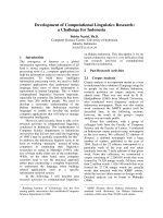
Establishment of the fluorescent quantitative real-time PCR standard curveFigure 1
Establishment of the fluorescent quantitative real-time PCR standard curve. Standard curve of the AHV-1 fluores-
cent quantitative real-time PCR. Ten-fold dilutions of standard DNA ranging from 1.0 × 10
9
to 1.0 × 10
2
copies/μL were used,
as indicated in the x-axis, whereas the corresponding cycle threshold (CT) values are presented on the y-axis. Each dot repre-
sents the result of triplicate amplification of each dilution. The correlation coefficient and the slope value of the regression
curve were calculated and are indicated.
Table 1: Intra- and inter-assay variability of the established FQ-PCR assay
Dilution of standard (copies/reaction) Intra-assay Inter-assay
Mean SD CV (%) Mean SD CV (%)
10
10
7.0 0.30 4.3 6.9 0.34 4.9
10
9
9.9 0.19 1.8 10.1 0.23 2.3
10
8
13.4 0.25 1.9 13.6 0.34 2.5
10
7
16.9 0.29 1.7 17.0 0.36 2.1
10
6
20.8 0.33 1.6 20.9 0.40 1.9
10
5
24.7 0.37 1.5 24.7 0.49 2.0
10
4
28.6 0.49 1.7 28.7 0.77 2.7
10
3
32.3 0.52 1.6 32.2 0.90 2.8
10
2
35.4 0.81 2.3 35.3 1.16 3.3
10
1
37.2 2.31 6.2 37.5 2.51 6.7
Virology Journal 2009, 6:71 />Page 4 of 8
(page number not for citation purposes)
demonstrated that the established fluorescent quantita-
tive real-time PCR method was characterized by a wide
dynamic range (8 logarithmic decades) of detection from
1.0 × 10
9
to 1.0 × 10
2
pAHV-1 plasmid copies/μL with
high precision. However, at lower and higher dilutions
quantitation was not always reproducible compared to
other properly diluted samples. Therefore the dynamic
range of the method was between 1.0 × 10
9
and 1.0 × 10
2
pAHV-1 plasmid copies/μL, which is relatively broad.
Test of established AHV-1 FQ-PCR assay using specimens
for practical applications
Viral load quantification through the established AHV-1
FQ-PCR demonstrated that the AHV-1 DNA copy number
of each sample could be calculated with the CT value
according to the standard curve and 100% of the samples
tested were quantifiable (Table 2) without the need for
further sample dilution or concentration.
Discussion
Conventional etiological, immunohistological and sero-
logical methods [18-20] were routinely used in AHV-1
identification. However, the sensitivity is usually not high
enough and the methods were time-consuming since
virus propagation in cell cultures is usually required and
the onset of virus-induced cytopathic effect (CPE) usually
requires at least 2–3 days to develop. Titration of infec-
tious virus in cell cultures is usually achieved by the end-
point dilution method in cell monolayer. Since titration
of the virus load is labor-consuming and requires about 5
days for evaluation of virus-induced CPE, distinguishing
between virus-induced CPE and non-specific cell altera-
tions may be difficult, the established real-time PCR assay
will be particularly suitable in these studies. In addition,
an even more important factor is that the virus from tis-
sues of infected birds is usually not readily adapted to cell
culture system during the initial several rounds of propa-
gations [21].
The PCR is a rapid, sensitive and specific nucleic acid
amplification technique and many conventional qualita-
tive PCR methods for revealing merely the presence or
absence of AHV-1 pathogen have been developed and
well documented [22-24]. However, the conventional
PCR assays are not sufficient in a variety of clinical situa-
tions. They frequently encountered problems including
the risk of cross-contamination (leading to false positives)
and poor quality of extracts (leading to false negatives).
Moreover, the lack of fluorogenic probes in the assay
results in relative lower specificity since the amplification
and detection of specific PCR products are determined
solely by the amplification primers. In this paper, the
development of a TaqMan MGB-based real-time PCR by
using fluorogenic labels and sensitive signal detection sys-
tem for detection and quantitation of AHV-1 is described.
The optimized FQ-PCR detection system presented in this
paper has been designed to address these issues and make
it even more applicable for routine diagnostic use with
several advantages over conventional PCR.
In this assay, the primers and probes have been selected
on conserved DNA segments of AHV-1 genome. TaqMan
Minor Groove Binding (MGB™) probes as target-specific
hydrolysis oligonucleotide employed in this assay are
characterized by the conjugation of minor groove binders
which increases the Tm of the hybridized probe and facil-
itates highly specific binding to the targeted sequence
[25]. Moreover this probe contains a quencher dye that
does not emit fluorescence within the detectable wave-
length range and results in greater accuracy in the meas-
urement. This improvement eliminates spectral overlaps
with fluorescence emitted by the reporter dye, and results
in greater accuracy in the measurement of reporter-spe-
cific signals.
In view of the great sensitivity of PCR, the occurrence of
false negative results is a highly underestimated problem.
So an artificial construct generated by cloning of the spe-
cific target sequence into a plasmid are often used as inter-
nal controls for the amplification step. This internal
positive control was incorporated into the reaction sys-
tem, thus improving diagnostic conclusions, especially
negative results, which is most important in the light of
quarantine programs.
By carrying out direct comparisons between the estab-
lished FQ-PCR method and the conventional PCR
method for AHV-1 detection, the results clearly showed
that overall the established FQ-PCR detection method is
more sensitive and reliable when compared to conven-
tional gel-based PCR, since it was able to detect as few as
Table 2: AHV-1 viral load in different clinical samples
Sample name DNA amounts (copies)
DEF cell culture supernatant 5.67 × 10
6
/μL
DEF cell culture 1.05 × 10
9
/μL
Allantoid fluid 2.85 × 10
6
/μL
Liver 2.73 × 10
9
/g
Brain 2.20 × 10
7
/g
Bursa of Fabricius 9.47 × 10
9
/g
Thymus 1.09 × 10
9
/g
Spleen 3.59 × 10
9
/g
Esophagus 9.56 × 10
9
/g
Duodenum 3.92 × 10
7
/g
Ileum 1.83 × 10
8
/g
Kidney 1.78 × 10
8
/g
Lung 3.15 × 10
8
/g
Peripheral blood 2.16 × 10
6
/μL
Cloacal swab 2.11 × 10
8
/swab
Oral swab 2.83 × 10
8
/swab
Virology Journal 2009, 6:71 />Page 5 of 8
(page number not for citation purposes)
1.0 × 10
1
DNA copies of template. Furthermore, this
established AHV-1 FQ-PCR method shows more excellent
characteristics such as dynamic range (from 1.0 × 10
9
to
1.0 × 10
2
pAHV-1 plasmid copies/μL, which is approxi-
mately 10
3
times broader) and sensitivity (detecting
pAHV-1 plasmid down to dilutions of 1.0 × 10
1
copies/μL,
which is about 2.3 times more sensitive) than other
reported method [17].
The high quality hot start Taq DNA polymerase used in
this assay could minimize unspecific amplifications and
increase the PCR cycling efficiency. In addition, FQ-PCR
reaction and detection is all done in a closed-tube system,
the need for post-amplification manipulation is removed
since the detection of the PCR products occurs online dur-
ing real-time PCR amplification, hence greatly reducing
the risk of cross-contamination and false positive results.
The optimization of the AHV-1 FQ-PCR assay was focused
on the concentration of primers and probe and Mg
2+
.
When all these different practical refinements are com-
bined, the final result is a molecular diagnostic method
that is not only rapid and reliable, but one that is also easy
to perform and applicable to use for testing large numbers
of samples since the FQ-PCR presented the benefits of
increased speed due to reduced cycle time and remove of
post-amplification process, offering considerable labor
savings and allowing higher throughput analysis than
conventional PCR assays and thus is favorable for the
transition of this method from research to routine use in
laboratories. This method was preliminarily mentioned in
a short report [26] but related details of primers and probe
sequence, specificity test, sensitivity test, reproducibility
analysis, dynamic range and internal control were una-
vailable. By contrast, great modification and optimization
have been made in this paper to improve the quality of
this study.
The AHV-1 FQ-PCR assay was highly reproducible and
linear over a range of eight orders of magnitude from 10
2
to 10
9
copies, allowing a precise calculation of viral DNA
load in samples containing a wide range of viral DNA
amounts, eliminating the need for sample dilution and
minimizing sample handling. The results for intra- and
inter-assay precision indicate that both intra-assay and
inter-assay CVs were satisfactorily low and the assay is
reproducible, even between different batches of reagents
used. Probability rather than sample quality variation is
the predominant cause of variability at low copy numbers
[27].
Conclusion
In conclusion, the FQ-PCR developed in this study is
highly specific and sensitive with better parameters than
conventional PCR method and is a valuable method for
the detection of AHV-1. The method described in this
study is especially helpful for high throughput analysis
such as evaluating the efficacy of antiviral drugs and
experimental vaccines for AHV-1. The research group of
authors is currently using this technique to study the AHV-
1 distribution characteristics in vaccinated birds and in
artificially infected birds. We believe that this method
could expedite related AHV-1 research in the AHV-1 viral
molecular biology.
Methods
Cell, virus and PCR template DNA preparation
Duck embryo fibroblast (DEF) monolayer was incubated
at 37°C with 5% CO
2
in tissue culture flasks with Minimal
Essential Medium (MEM) that contained 10% fetal
bovine serum (FBS), 100 U/mL penicillin, and 100 μg/mL
streptomycin.
Anatid herpesvirus 1 (AHV-1, CHv virulent strain) was
obtained from the Avian Disease Research Center of
Sichuan Agricultural University (Yaan, Sichuan, China).
Virus stock was added onto the surface of the cell layer
which was about 90% confluency at time of infection and
the maximum virus titers could usually be obtained 48 h
postinfection.
Table 3: Oligonucleotide sequences of primers and probes used in AHV-1 FQ-PCR detection
Name Type Sequences (5'to 3') Length (nt) Position Amplicon size (bp)
Real-F
a
Forward ttttcctcctcctcgctgagt 21 357–377 60
Real-P
a
Probe ccctgggtacaagcgc 16 383–398
Real-R
a
Reverse ggccgggtttgcagaagt 18 399–416
Con-F
b
Forward ggacagcgtaccacagataa 20 246–265 498
Con-R
b
Reverse acaaatcccaagcgtag 17 727–743
IC-F
c
Forward acgagcgcaacccttga 17 1054–1070 92
IC-P
c
Probe cggtttgtcaccggcagtcacct 23 1103–1125
IC-R
c
Reverse acgtcatccccaccttact 19 1127–1145
a
Based on the nucleotide sequence AF064639.
b
Based on the nucleotide sequence AF064639.
c
Based on the nucleotide sequence AJ971894.
Virology Journal 2009, 6:71 />Page 6 of 8
(page number not for citation purposes)
DNA extraction from AHV-1 infected DEF cells and tissues
of AHV-1 infected ducks were performed by using TIAN-
amp Genomic DNA extracting kit (Tiangen Corporation,
Beijing, China) according to the manufacture's instruc-
tions.
PCR primers and probe design
The FQ-PCR assay primers and probe (named Real-F,
Real-R and Real-P respectively) design was carried out
using the Primer Express™ software supplied by Applied
Biosystems and their sequences were listed in Table 3. The
forward and reverse primers amplified a 60 bp fragment
of AHV-1 DNA polymerase gene as described (GenBank
Accession No. AF064639
). The fluorogenic probe was
labelled at 5' with FAM (6-carboxyfluorescein) dye as
reporter and labelled at 3' with TAMRA (tetra-methylcar-
boxyrhodamine) as quencher and 3'with MGB™ (Minor
Groove Binder).
The conventional PCR amplification was carried out using
primers designed using the Primer Premier™ software
according to the sequence as described (GenBank Acces-
sion No. AF064639
). The forward primer and reverse
primer (named Con-F and Con-R respectively) sequences
were listed in Table 3 and this primer pair yielded a 498
bp amplicon, in which the 60 bp FQ-PCR fragment was
nested.
All probes and primers were synthesized by Genecore
Corporation (Shanghai, China) and purified by corre-
sponding HPLC system.
Development and optimization of fluorescent quantitative
real-time PCR and conventional PCR
The real-time PCR was carried out using the ABI AmpliTaq
Gold DNA polymerase system with an icycler IQ Real-
time PCR Detection System (Bio-Rad Corp., Hercules, CA)
according to the manufacturer's instructions. The reac-
tion, data acquisition and analysis were performed using
iCycler IQ optical system software. The Real-time PCR was
performed in an 25 μL reaction mixture containing 1 ×
PCR buffer, 0.2 mmol/L dNTPs, 1 U Taq and 1 μL DNA
template according to the manufacture's instructions.
Autoclaved nanopure water was added to bring the final
volume to 25 μL. The two-step PCR cycling condition was
as follows: initial denaturation and hot-start Taq DNA
polymerase activation at 95°C for 10 min, 50 cycles of
denaturation at 94°C for 15 s, primer annealing and
extension at 60°C for 20 s with fluorescence acquisition
during each annealing and extension stage. Real-time PCR
reactions were optimized in triplicate based on primer,
probe and MgCl
2
concentration selection criteria, which
was performed according to 4 × 4 × 4 matrix of primer
concentrations (0.2, 0.3, 0.4 and 0.5 μmol/L), probe con-
centrations (0.05, 0.1, 0.2, and 0.3 μmol/L) and MgCl
2
concentrations (2, 4, 6 and 8 mmol/L).
The conventional PCR was performed and optimized on
a Mycycler™ thermo cycler system (Bio-Rad Corp., Her-
cules, CA, USA) with a 50 μL PCR reaction system contain-
ing 1 × PCR buffer, 0.2 mmol/L dNTPs mixture, 1.25 U
rTaq (Takara Bio Inc., Shiga, Japan), 0.5 μmol/L each for-
ward and reverse primers and 1 μL template DNA. All PCR
experiments were carried out in 0.2 ml thin-walled tubes
with the following cycle parameters: The mixture was sub-
jected to initial denaturation at 95°C for 1 min, followed
by 50 cycles of 95°C for 60 s, annealing for 60 s, extension
at 72°C for 60 s, and one cycle of final extension at 72°C
for 5 min. The amplified 498 bp product then underwent
electrophoresis on 1.0% agarose gels. Electrophoresis was
carried out at 100 V in a Mini-sub (Bio-Rad Corp., Her-
cules, CA, USA) gel electrophoresis unit and gels were
viewed under a UV transilluminator. The conventional
PCR reactions were optimized based on MgCl
2
concentra-
tion and annealing temperature selection criteria in a sim-
ilar way as that of Real-time PCR and the selection was
made by the brightness of the amplified 498 bp fragments
on the agarose gel under a UV transilluminator.
An internal positive control was introduced into the FQ-
PCR assay to verify the absence of DNA losses during the
extraction step and of PCR inhibitors in the DNA tem-
plates. The internal positive control of pGM-T recom-
binant vector (designed as pB16S) consisting of Bacillus
16S rRNA gene (GenBank Accession No. AJ971894
)
sequence amplified with primers (IC-F and IC-R) listed in
Table 3 was added into the lysis buffer at the concentra-
tion of 1.0 × 10
6
copies/μL. Real-time PCR for IC detection
was carried out in a separate run, using primers and probe
(named IC-F, IC-R and IC-P respectively) listed in Table 3.
The fluorogenic probe was labelled at 5' with FAM as
reporter and labelled at 3' with TAMRA. The quantitative
real-time PCR protocol was the same as that of AHV-1
detection. From the ratio of the calculated amount of IC
to the actual amount of IC, which is shared by the speci-
men, the normalization could be achieved and the actual
amount of AHV-1 in the specimen could be obtained.
Actually this internally controlled method has been
widely used in other related detection assays [28,29].
Fluorescent quantitative real-time PCR standard curve
establishment
The 498 bp conventional PCR target amplicon band on
agarose gel was cut and the DNA was recovered and puri-
fied by TIANquick DNA Purification system (Tiangen
Corp., Beijing, China) according to the instruction man-
ual of the product. The product was ligated into pGM-T
vector (Tiangen Corp., Beijing, China) and transformed
into E.coli DH5α competent cells. Recombinant plasmid
Virology Journal 2009, 6:71 />Page 7 of 8
(page number not for citation purposes)
(designated as pAHV-1) was extracted using TIANprep
plasmid extraction kit (Tiangen Corp., Beijing, China).
Presence of the target DNA insert was confirmed by PCR
amplification and sequencing.
The standard curve of the FQ-PCR was generated by suc-
cessive dilutions of the known copy number of pAHV-1.
Recombinant plasmid pAHV-1 concentration was deter-
mined by taking the absorbance at 260 nm using a Smart-
spec 3000 spectrophotometer (Bio-Rad Corp., Hercules,
CA) and purity was confirmed using the 260/280 nm
ratio. Through its molecular weight, pAHV-1 copy
number was then calculated and the purified pAHV-1
plasmid DNA was then serially diluted 10-fold in TE
buffer, pH 8.0, from 1.0 × 10
9
to 1.0 × 10
2
plasmid copies/
μL. These dilutions were tested in triplicate and used as
quantitation standards to construct the standard curve by
plotting the plasmid copy number logarithm against the
measured CT values. The Bio-Rad iCycler IQ detection
software created the standard curve, calculated the corre-
lation coefficient (R
2
) of the standard curve, standard
deviations of triplicates.
FQ-PCR sensitivity, specificity, reproducibility and
dynamic range analysis
Different 32 AHV-1 strains (derived from a wide spectrum
of sources, subsequently confirmed through related etio-
logical methods, and then preserved by the Avian Disease
Research Center of Sichuan Agricultural University)
including virulent and avirulent strains were examined
with the established FQ-PCR method to test the sensitivity
and compatibility of this method. In addition, the sensi-
tivities of the conventional PCR and FQ-PCR were each
determined using triplicates of different concentrations of
recombinant plasmid pAHV-1. Template DNA was pre-
pared as follows: plasmids of pAHV-1 were diluted serially
in 10-fold steps from 10
10
copies/μL to 10
1
copies/μL
using sterile ultra pure water. One microliter from each
dilution was used as template and subjected to the con-
ventional PCR and FQ-PCR protocol respectively. The
detection limit of the conventional PCR was determined
based on the highest dilution that resulted in the presence
of clear and distinct amplified fragments (498 bp) on the
agarose gel. The detection limit of the FQ-PCR was deter-
mined based on the highest dilution that resulted in the
presence of CT value in real-time PCR detection.
DNA from duck embryo fibroblast (DEF) and several
other pathogens including duck hepatitis B virus, Salmo-
nella enteritidis, duck adenovirus, goose parvovirus,
Marek's disease virus, infectious laryngotracheitis virus
and Pasteurella multocida (kindly provided by Avian Dis-
eases Research Center of Sichuan Agricultural University)
were used as templates in triplicates to confirm the tech-
nique's specificity.
Within-run and between-run reproducibilities of the FQ-
PCR assay were assessed by multiple measurements of
pAHV-1 samples of different concentrations. The assay
was conducted by assessing the agreement between the
replicates in five replicates (within-run precision) and in
five separate experiments (between-run precision) of the
serially diluted pAHV-1 recombinant plasmid samples
through performing analysis of the mean coefficient of
variation (CV) values of each AHV-1 standard dilution.
Dilutions of pAHV-1 recombinant plasmid were used to
determine the dynamic ranges of the FQ-PCR assay. The
lower and upper limits of quantification were defined by
the pAHV-1 recombinant plasmid sample concentrations
possessing reasonable precision.
Test of established AHV-1 FQ-PCR assay using specimens
for practical applications
AHV-1 infected duck embryo fibroblast culture, allantoid
fluid and other specimens including liver, brain, Bursa of
Fabricius, thymus, spleen, esophagus, duodenum, ileum,
kidney, lung, peripheral blood each collected from AHV-
1 infected ducks were employed to assess the ability of the
established FQ-PCR to detect AHV-1 in a variety of usually
used samples. By this assay viral load quantification was
obtained.
Competing interests
The authors declare that they have no competing interests.
Authors' contributions
YG carried out most of the experiments and wrote the
manuscript. AC and MW critically revised the manuscript
and the experiment design. CS, RJ, SC and NZ helped with
the experiment. All of the authors read and approved the
final version of the manuscript.
Acknowledgements
This project was funded by a grant from the National Natural Science Foun-
dation of China (grant No. 30771598), the Cultivation Fund of the Key Sci-
entific and Technical Innovation Project, department of Education of
Sichuan Province (grant no. 07ZZ028), China Postdoctoral Science Foun-
dation (grant No. 20060391027), Program for Changjiang Scholars and
Innovative Research Team in University (grant No. IRT0848), Scientific and
Technological Innovation Major Project Funds in University (grant No.
706050), the earmarked fund for Modern Agro-industry Technology
Research System (nycytx-45-12) and Sichuan Province Basic Research Pro-
gram (grant No. 07JY029-016/07JY029-017/2008JO0003/2008JY0100/
2008JY0102). The authors wish to thank our colleagues for their profes-
sional assistance and technical support to this study.
References
1. Sluis W: Ducks are a flavour for the future. World Pou 2004,
20:41.
2. Sandhu TS, Shawky SA: Duck virus enteritis (Duck plague). In
Diseases of Poultry 11th edition. Edited by: Saif YM, Barnes HJ, Glisson
JR. Iowa State Press, Ames; 2003:354-363.
Publish with BioMed Central and every
scientist can read your work free of charge
"BioMed Central will be the most significant development for
disseminating the results of biomedical research in our lifetime."
Sir Paul Nurse, Cancer Research UK
Your research papers will be:
available free of charge to the entire biomedical community
peer reviewed and published immediately upon acceptance
cited in PubMed and archived on PubMed Central
yours — you keep the copyright
Submit your manuscript here:
/>BioMedcentral
Virology Journal 2009, 6:71 />Page 8 of 8
(page number not for citation purposes)
3. Montgomery RD, Stein G Jr, Novilla MN, Hurley SS, Fink RJ: An out-
break of duck virus enteritis (duck plague) in a captive flock
of mixed waterfowl. Avian Dis 1981, 25:207-213.
4. Brand CJ, Docherty DE: A survey of north American migratory
waterfowl for duck plague (duck virus enteritis) virus. J Wildl
Dis 1984, 20:261-266.
5. Brand CJ, Docherty DE: Post-epizootic surveys of waterfowl for
duck plague (duck virus enteritis). Avian Dis 1988, 32:722-730.
6. Burgess EC, Ossa J, Yuill TM: Duck plague: a carrier state in
waterfowl. Avian Dis 1979, 24:940-949.
7. Kaleta EF: Herpesviruses of birds. Avian Pathol 1990, 19:193-211.
8. Fukuchi K, Sudo M, Lee YS, Tanaka A, Nonoyama M: Structure of
Marek's disease virus DNA: detailed restriction map. J Virol
1984, 51:102-109.
9. Gardner R, Wilkerson J, Johnson JC: Molecular characterization
of the DNA of anatid herpesvirus 1. Intervirology 1993,
36:99-112.
10. Chen RW, Piiparinen H, Seppänen M, Koskela P, Sarna S, Lappalainen
M: Real-time PCR for detection and quantitation of hepatitis
B virus DNA. J Med Virol 2001, 65:250-256.
11. Jebbink J, Bai X, Rogers BB, Dawson DB, Scheuermann RH, Domiati-
Saad R: Development of real-time PCR assays for the quanti-
tative detection of Epstein-Barr virus and cytomegalovirus,
comparison of TaqMan probes, and molecular beacons. J Mol
Diagn 2003, 5:15-20.
12. Weinberger KM, Wiedenmnm E, Böhm S, Jilg W: Sensitive and
accurate quantitation of hepatitis B virus DNA using a
kinetic fluorescence detection system (TaqMan PCR). J Virol
Methods. 2000, 85(1-2):75-82.
13. Watzinger F, Suda M, Preuner S, Baumgartinger R, Ebner K, Baskova
L, Niesters HG, Lawitschka A, Lion T: Real-time quantitative
PCR assays for detection and monitoring of pathogenic
human viruses in immunosuppressed pediatric patients. J
Clin Microbiol 2004, 42:5189-5198.
14. Humar A, Kumar D, Boivin G, Caliendo AM: Cytomegalovirus
(CMV) virus load kinetics to predict recurrent disease in
solid-organ transplant patients with CMV disease. J Infect Dis
2002, 186:829-833.
15. Häusler M, Scheithauer S, Ritter K, Kleines M: Molecular diagnosis
of Epstein-Barr virus. Expert Rev Mol Diagn 2003, 3:81-92.
16. Watzinger F, Ebner K, Lion T: Detection and monitoring of virus
infections by real-time PCR. Mol Aspects Med 2006, 27:254-298.
17. Yang F, Jia W, Yue H, Luo W, Chen X, Xie Y, Zen W, Yang W:
Development of quantitative real-time polymerase chain
reaction for duck enteritis virus DNA. Avian Dis 2005,
49:397-400.
18. Salguero FJ, Sánchez-Cordón PJ, Núñez A, Gómez-Villamandos JC:
Histopathological and ultrastructural changes associated
with herpesvirus infection in waterfowl. Avian Pathol 2002,
31:133-140.
19. Islam MR, Nessa J, Halder KM: Detection of duck plague virus
antigen in tissues by immunoperoxidase staining. Avian Pathol
1993, 22:389-393.
20. Wolf K, Burke CN, Quimby MC: Duck viral enteritis: microtiter
plate isolation and neutralization test using the duck embryo
fibroblast cell line. Avian Dis 1974, 18:427-434.
21. Guo Y, Cheng A, Wang M, Jia R, Wen M, Zhou W, Chen X: Studies
on the growth characteristics of duck enteritis virus CH vir-
ulent strain on duck embryo fibroblast. Poult Husb dis control
2007, 4:2-4.
22. Hansen WR, Brown SE, Nashold SW, Knudson DL: Identification
of duck plague virus by polymerase chain reaction. Avian Dis
1999, 43(1):106-115.
23. Plummer PJ, Alefantis T, Kaplan S, O'Connell P, Shawky S, Schat KA:
Detection of duck enteritis virus by polymerase chain reac-
tion. Avian Dis 1998, 42:
554-564.
24. Pritchard LI, Morrissy C, Phuc KV, Daniels PW, Westbury HA:
Development of a polymerase chain reaction to detect Viet-
namese isolates of duck virus enteritis. Vet Microbiol 1999,
68:149-156.
25. Kutyavin IV, Afonina IA, Mills A, Gorn VV, Lukhtanov EA, Belousov
ES, Singer MJ, Walburger DK, Lokhov SG, Gall AA, Dempcy R, Reed
MW, Meyer RB, Hedgpeth J: 3'-minor groove binder-DNA
probes increase sequence specificity at PCR extension tem-
peratures. Nucleic Acids Res 2000, 28:655-661.
26. Guo Y, Cheng A, Wang M, Jia R, Wen M, Zhou W, Chen X: Estab-
lishment and application of quantitative real-time PCR assay
for detecting duck viral enteritis virus. Vet Sci chin 2006,
36:444-448.
27. De Vries TJ, Fourkour A, Punt CJA, Locht LTF van de, Wobbes T,
Bosch S van den, de Rooij MJM, Mensink EJBM, Ruiter DJ, van Muijen
GNP: Reproducibility of detection of tyrosine and MART-1
transcripts in the peripheral blood of melanoma patients: a
quality control study using real-time quantitative RT-PCR.
Br J Cancer 1999, 80:883-891.
28. Decaro N, Elia G, Campolo M, Desario C, Mari V, Radogna A, Cola-
ianni ML, Cirone F, Tempesta M, Buonavoglia C: Detection of
bovine coronavirus using a TaqMan-based real-time RT-PCR
assay. J Virol Methods. 2008, 151(2):167-171.
29. Elia G, Tarsitano E, Camero M, Bellacicco AL, Buonavoglia D, Cam-
polo M, Decaro N, Thiry J, Tempesta M: Development of a real-
time PCR for the detection and quantitation of caprine her-
pesvirus 1 in goats. J Virol Methods. 2008, 148(1-2):155-160.