Báo cáo khoa học: " Respiratory viral infections detected by multiplex PCR among pediatric patients with lower respiratory tract infections seen at an urban hospital in Delhi from 2005 to 2007" docx
Bạn đang xem bản rút gọn của tài liệu. Xem và tải ngay bản đầy đủ của tài liệu tại đây (373.95 KB, 11 trang )
BioMed Central
Page 1 of 11
(page number not for citation purposes)
Virology Journal
Open Access
Research
Respiratory viral infections detected by multiplex PCR among
pediatric patients with lower respiratory tract infections seen at an
urban hospital in Delhi from 2005 to 2007
Preeti Bharaj
1
, Wayne M Sullender
2
, Sushil K Kabra
3
, Kalaivani Mani
4
,
John Cherian
3
, Vikas Tyagi
3
, Harendra S Chahar
1
, Samander Kaushik
1
,
Lalit Dar
1
and Shobha Broor*
1
Address:
1
Department of Microbiology, All India Institute of Medical Sciences, Ansari Nagar, New Delhi, 110029, India,
2
Department of Pediatrics,
University of Alabama at Birmingham, Alabama, 35294-0011 USA,
3
Department of Pediatrics, All India Institute of Medical Sciences, Ansari
Nagar, New Delhi, 110029, India and
4
Department of Biostatistics, All India Institute of Medical Sciences, Ansari Nagar, New Delhi, 110029, India
Email: Preeti Bharaj - ; Wayne M Sullender - ; Sushil K Kabra - ;
Kalaivani Mani - ; John Cherian - ; Vikas Tyagi - ;
Harendra S Chahar - ; Samander Kaushik - ; Lalit Dar - ;
Shobha Broor* -
* Corresponding author
Abstract
Background: Acute lower respiratory tract infections (ALRI) are the major cause of morbidity
and mortality in young children worldwide. Information on viral etiology in ALRI from India is
limited. The aim of the present study was to develop a simple, sensitive, specific and cost effective
multiplex PCR (mPCR) assay without post PCR hybridization or nested PCR steps for the
detection of respiratory syncytial virus (RSV), influenza viruses, parainfluenza viruses (PIV1–3) and
human metapneumovirus (hMPV). Nasopharyngeal aspirates (NPAs) were collected from children
with ALRI ≤ 5 years of age. The sensitivity and specificity of mPCR was compared to virus isolation
by centrifugation enhanced culture (CEC) followed by indirect immunofluorescence (IIF).
Results: From April 2005–March 2007, 301 NPAs were collected from children attending the
outpatient department or admitted to the ward of All India Institute of Medical Sciences hospital
at New Delhi, India. Multiplex PCR detected respiratory viruses in 106 (35.2%) of 301 samples with
130 viruses of which RSV was detected in 61, PIV3 in 22, PIV2 in 17, hMPV in 11, PIV1 in 10 and
influenza A in 9 children. CEC-IIF detected 79 viruses only. The sensitivity of mPCR was 0.1TCID
50
for RSV and influenza A and 1TCID
50
for hMPV, PIV1, PIV2, PIV3 and Influenza B. Mixed infections
were detected in 18.8% of the children with viral infections, none detected by CEC-IIF.
Bronchiolitis was significantly associated with both total viral infections and RSV infection (p <
0.05). History of ARI in family predisposed children to acquire viral infection (p > 0.05).
Conclusion: Multiplex PCR offers a rapid, sensitive and reasonably priced diagnostic method for
common respiratory viruses.
Published: 26 June 2009
Virology Journal 2009, 6:89 doi:10.1186/1743-422X-6-89
Received: 18 March 2009
Accepted: 26 June 2009
This article is available from: />© 2009 Bharaj et al; licensee BioMed Central Ltd.
This is an Open Access article distributed under the terms of the Creative Commons Attribution License ( />),
which permits unrestricted use, distribution, and reproduction in any medium, provided the original work is properly cited.
Virology Journal 2009, 6:89 />Page 2 of 11
(page number not for citation purposes)
Background
Acute respiratory tract infections (ARI) are a leading cause
of morbidity and mortality in children worldwide [1]
accounting for about 30% of all childhood deaths in
developing world [2]. Viruses account for 50–90% of
acute lower respiratory tract infections (ALRI) in young
children [3] with respiratory syncytial virus (RSV), parain-
fluenza viruses (PIV), influenza viruses A and B and
human metapneumoviruses (hMPV) being most com-
monly identified [4-6].
Respiratory infections caused by above said viruses usu-
ally present with clinical features that are nearly indistin-
guishable [7]. The increased sensitivity of polymerase
chain reaction (PCR) over conventional methods for the
diagnosis of respiratory viral infections has been estab-
lished previously [8]. However, organism-specific RT-PCR
assays which require separate amplification of each virus
under investigation are resource intensive, time consum-
ing and labor intensive [9].
Multiplex PCRs (mPCR) detect multiple organisms in a
single assay and are available either as commercial assays
[9-12] or in-house assays [4,5,13-17]. Majority of the in-
house mPCR assays have not included recently discovered
respiratory pathogens and require validation of results by
post PCR hybridization or semi/nested PCR which make
the assay cost ineffective and increases chances of cross
contamination. Commercially available mPCR assays are
expensive and require dedicated instrumentation [18].
We developed a simplified and economical multiplex
PCR assay without any post PCR hybridization/nested
PCR steps for the detection of seven major respiratory
viruses.
(This material was presented in part at the 7
th
Asia Pacific
Congress of Medical Virology held at New Delhi, India in
November 2006 and Options for the Control of Influenza
VI held at Toronto, Canada in June 2007.)
Results
The primer set designed for PIV1 failed to amplify PIV1
RNA after repeated attempts. The primer set published by
Osiowy in 1998 [17] was found to amplify PIV1 N gene
successfully.
Standardization of cDNA synthesis
Ten Units of the AMV-RT enzyme, 500 ng of random hex-
amer primer (PdN
6
), 500 μM dNTP concentration and 8
U of RNAsin were found to be optimal for 25 μl cDNA
synthesis.
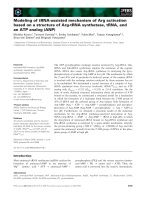
Standardization of multiplex PCR
All seven sets of primers when combined led to mispair-
ing and nonspecific amplification. After trying different
combinations, it was observed that RSV, Influenza A and
B viruses in one set and PIV1–3 and hMPV in another set
gave specific amplification for each virus (Figure 1).
Optimized reagent and PCR cycling conditions for first and
second tube multiplex PCR
The optimized reagent concentrations for each tube were
25 pM of each primer, 400 μM of dNTPs, 2 mM MgCl
2
and
6 U of Taq polymerase enzyme. The optimized cycling
conditions for both tubes were: 94°C for 3 min followed
by 35 cycles of 94°C for 1 min, 55°C for 1 min (53°C for
1 min for second tube) and 72°C for 1 min. Final exten-
sion was done at 72°C for 10 min for first tube and 7 min
for second set.
Sensitivity and specificity of multiplex PCR
The sensitivity of detection by two tube multiplex PCR
was 0.1TCID
50
for RSV, Influenza A and 1TCID
50
for
hMPV, PIV1, PIV2, PIV3 and Influenza B. There were no
non-specific amplification products against RNA from
heterologous sources.
Detection of seven respiratory viruses in clinical samples
Study group
Three hundred and one children from OPD and ward
with ALRI were enrolled in the study. Of the 166 children
seen in the outpatient department, 137 (82.5%) had
ALRI, 29 (17.5%) had severe ALRI and none had very
severe ALRI (as per WHO classification). Of the 135 chil-
dren admitted to pediatric ward, 35 (26%) had ALRI, 92
(68%) had severe ALRI and 8 (6%) had very severe ALRI
(Table 1). More number of children with ALRI were seen
in the OPD as compared to pediatric ward (p < 0.05, Pear-
son Chi square test). However, for severe ALRI, more chil-
dren were admitted than seen in OPD (p < 0.05, Pearson
Chi square test).
All the 301 children enrolled in the study were in the age
range of 1–72 months with the median age of 11 months.
The mean of their age was 15.6 ± 14 months. Among them
217 were males and 84 were females (Male: Female ratio
= 2.6:1). It was observed that there was no significant dif-
ference between the age range of children with ALRI or
severe ALRI from OPD or Ward (p > 0.05).
Detection of seven respiratory viruses in children with ALRI
Of the 301, 106 children (35.2%) had viral infections and
were positive for 130 respiratory viruses. Of these 106
children with ALRI, 64 presented to the OPD and 42 were
admitted to the ward. Of the 64 children who presented
to OPD, 52 had ALRI and 12 had severe ALRI. Of the 42
Virology Journal 2009, 6:89 />Page 3 of 11
(page number not for citation purposes)
children who were admitted, 8 had ALRI, 31 had severe
ALRI and 3 had very severe ALRI (Table 2).
In 106 children in whom respiratory viruses were
detected, the age range was 1–72 months with a median
of 12 months and the mean age was 15.8 ± 13.8 months.
In the PCR negative group, the age range was 1–61
months with a median of 10 months and the mean age
was 15.5 ± 14.1 months. This difference was not statisti-
cally significant between the two groups. There was no sig-
nificant difference in the male female ratio between the
two groups.
RSV was detected in 61, PIV3 in 22, PIV2 in 17, hMPV in
11, PIV1 in 10 and Influenza A in 9 children respectively
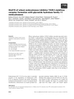
(Table 3). Figure 2 shows detection of single and mixed
infections in some samples on which two tube multiplex
PCR was applied. Of these, 86 were single virus infections
and mixed infections were seen in 20 children (18.8% of
the 106 children). Nested PCR for RSV identified the pres-
ence of RSV B in all 61 samples. Of the single infections,
RSV comprised 50, hMPV 9, Influenza A and PIV3 8 each,
PIV2 6 and PIV1 5.
It was seen that the percentage of virus detections by mul-
tiplex PCR were higher in the children with ALRI seen as
outpatients (37.2%) as compared to those admitted to the
ward (22.8%). Similarly, in the children with severe ALRI,
seen as outpatients were higher percentage was positive
for viruses (45%) as compared to those admitted to the
ward (33.7%). It was observed that of all the mixed infec-
tions, 5.8% of them had ALRI whereas 7.8% of them had
Standardization of two tube multiplex PCR for RSV, Influenza A&B viruses in first tube and PIV1–3 and hMPV in the second tubeFigure 1
Standardization of two tube multiplex PCR for RSV, Influenza A&B viruses in first tube and PIV1–3 and hMPV
in the second tube. Lane 1: Marker Ø X174 (Hae III digested). Lane 2: Amplicon forRSV showing band of 683 bp, Influenza A
of 105 bp, Influenza B of 503 bp. Lane 3: Marker 100 bp ladder. Lane 4: Amplicon for PIV1 showing band of 84 bp, PIV2 of 197
bp, PIV3 of 266 bp, and hMPV of 440 bp.
683bp
503bp
105bp
2 1
603bp
301bp
271/81
234bp
194bp
118bp
440bp
266bp
197bp
84bp
3 4
Table 1: Distribution of children with ALRI/Severe ALRI/very severe ALRI from OPD or pediatric ward
Site Clinical presentation Total
ALRI/172 (%) Severe ALRI/121 (%) Very Severe ALRI/8 (%)
OPD 137 (79.6)
a
29 (24)
b
0166
Pediatric Ward 35 (20.4)
a
92 (76)
b
8 (100) 135
Total 172 121 8 301
a, b
p value ≤ 0.05, Pearson chi square test
Virology Journal 2009, 6:89 />Page 4 of 11
(page number not for citation purposes)
severe and very severe ALRI. Although severe ALRI was
seen in higher number of children with mixed infections
as compared to those with ALRI with mixed infections, the
difference between the groups was not statistically signifi-
cant.
Co-relation between multiplex PCR and tissue culture
The "gold standard" isolation in tissue culture by CEC-IIF
detected 79 (61%) viruses as compared to 130 by multi-
plex PCR. CEC-IIF could not detect the presence of viruses
in samples with mixed infections (data not shown). The
sensitivity, specificity and likelihood ratio between the
two assays is shown in Table 4.
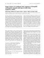
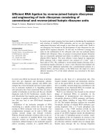
Temporal distribution of respiratory viruses
The number of RSV infections increased during late fall
and peaked between October and January during the first
year of the study. During the next year of the study, the
distribution of RSV was scattered. PIVs were detected dur-
ing the first year of the study, influenza A in winter
months and hMPV in spring season (Figure 3).
Cost effectiveness of the multiplex PCR assay
The cost per sample detected by two tube multiplex PCR
assay was USD16 (RNA extraction USD6, cDNA synthesis
USD2.5 and two tube multiplex PCR USD4.5, equipment
and personnel cost USD3) as compared to the cost per
sample by culture being USD24 (Sample collection
USD2, sample processing USD2, inoculation of sample
on to 3 different cell lines USD8, indirect immunofluores-
cence USD3, visualization under fluorescent microscope
USD3, equipment and personnel cost USD6).
Clinical symptoms
The clinical features, demographics and risk factors of
children with viral infections and RSV alone were com-
pared with the virus negative group (Table 5, 6). It was
observed that significantly higher number of children
below 12 months of age had RSV infection. Children pre-
senting with preceding bronchiolitis were significantly
associated with total viral infections and RSV infection (p
< 0.05). Runny nose was significantly present in children
with RSV infection (p < 0.05). Among risk factors, ARI in
family was found to be associated with virus positive chil-
dren (p < 0.005).
Discussion
The development of multiplex PCR for the detection of
respiratory viruses as a rapid, sensitive and time saving
technique has not gained priority in India even though
~0.5 million children die each year in this country due to
ALRI each year, accounting for one fourth of the 1.9 mil-
lion global ALRI deaths [19-21]. Among all the major
ALRI causing viruses namely RSV, PIVs and influenza
viruses A and B, the presence of RSV has been documented
to be the most commonly identified pathogen followed
by PIV3 [22]. In the present study, we standardized and
evaluated an economical two-tube multiplex PCR assay
devoid of any further confirmatory steps. The present
assay reagents costs were mere USD16/reaction in contrast
to USD90/reaction [18] reported for a commercial assay.
The sensitivity of our multiplex PCR assay was similar or
better than previously described mPCR assays for these
viruses [5,16,17,23,24]. We did not make direct compari-
sons of the performance of the different assays in our lab-
oratory.
Table 2: Distribution of children with ALRI/Severe ALRI/very severe ALRI from OPD or pediatric ward positive for different
respiratory viruses
Site Clinical presentation Total
ALRI/total ALRI (%) Severe ALRI/total severe ALRI (%) Very Severe ALRI/number (%)
OPD 52/60 (81.3) 12/43 (27.9) 0 64
Pediatric Ward 8/60 (18.7) 31/43 (70.1) 3/3 (100) 42
Total virus positive 60 43 3 106
Table 3: Virus identifications in children with ALRI detected
positive for viral infections by multiplex PCR
Viruses detected by mPCR Number of specimens
RSV 50
Influenza A 8
PIV1 5
PIV2 6
PIV3 8
hMPV 9
PIV2+PIV3 6
RSV+PIV2+PIV3 3
RSV+PIV3 3
RSV+PIV1 3
RSV+PIV2 1
PIV3+INFA 1
PIV1+PIV2+PIV3 1
RSV+hMPV 1
hMPV+PIV1 1
TOTAL 106
Virology Journal 2009, 6:89 />Page 5 of 11
(page number not for citation purposes)
In the present study we could culture majority of the
viruses detected by mPCR with the exception of RSV
which is known to be highly thermolabile [25]. The detec-
tion rate of viruses was similar to detection rate reported
earlier [16,17,24,26]. It was observed that a higher pro-
portion of virus positive children presented to the OPD
than the ward, similar to a study from Taiwan [26] and
could be due to the fact that the patients present earlier
after onset of symptoms to the OPD as compared to get-
ting admitted to the Ward. However, this could also be
because severe disease is more likely to be admitted to the
hospital and caused by bacteria than virus [27]. A higher
proportion of males were found to have infection with
respiratory viruses as compared to females as reported ear-
lier [28,29].
RSV was most commonly identified viral pathogen similar
to previously described viral identifications by mPCR
[16,17,28,29]. PIVs were the second most frequently iden-
tified pathogens [29] followed by hMPV [22,28-30] and
Influenza A virus infections [16,24,28].
The detection rate of co-infections was similar to previ-
ously reported multiplex PCR studies [5,14-17,28,29]. It
was observed that higher percentage of children with
mixed infections had severe and very severe ALRI as com-
pared to ALRI. Previous studies have shown that co-infec-
tion with different respiratory viruses might lead to a
more severe disease [31] or multiple viruses have been
detected from patients with severe disease [32].
ALRI caused by RSV was more common in younger chil-
dren as reported previously [28]. RSV and hMPV were
associated with bronchiolitis [28,29,33,34]. PIVs and
Influenza viruses were associated with pneumonia similar
to previous findings [28,29]. However, the number of all
Application of two tube multiplex PCR on clinical samplesFigure 2
Application of two tube multiplex PCR on clinical samples. Panel A. Lane 1: 100 bp DNA ladder. Lane 2: Clinical sam-
ple showing amplicon of 105 bp for FLU A. Lane 3: Clinical sample showing amplicon of 683 bp for RSV. Lane 4: Clinical sample
showing amplicon of 440 bp for hMPV. Lane 5: Clinical sample showing amplicon of 266 bp for PIV3. Lane 3: Negative clinical
sample. Panel B. Lane 1: 100 bp DNA adder. Lane 2: Clinical sample showing mixed infection of PIV1 (84 bp), PIV2 (197 bp)
and PIV3 (266 bp).
1 2 3
266bp
197bp
84bp
1 2 3 4 5
A
B
683bp
440bp
266bp
105bp
Table 4: Validity of multiplex PCR in comparison to CEC-IIF for the detection of respiratory viruses in children with ALRI
Results (RT-PCR/CEC-IIA) RSV INF A PIV1 PIV2 PIV3 hMPV
+/+ 27 8 5 10 18 11
+/- 34 1 5 7 4 0
-/- 274 293 296 291 283 290
-/+ 00 0000
Total samples 301 301 301 301 301 301
Sensitivity (%) 100 100 100 100 100 100
Specificity (%) 88.9 99.6 98.3 97.6 98.6 100
Likelihood ratio (positive) 9 250 59 42 71 -
Virology Journal 2009, 6:89 />Page 6 of 11
(page number not for citation purposes)
Monthly distribution of ALRI causing viruses detected during the studyFigure 3
Monthly distribution of ALRI causing viruses detected during the study.
0
5
10
15
20
25
AMJ J ASONDJFMAMJ J ASONDJFM
RSV
0
50
100
150
200
250
AMJ J ASONDJFMAMJ J ASONDJ FM
Humidity (%) Temp (°C) Rainfall (cm)
0
0.5
1
1.5
2
2.5
3
3.5
AMJ J ASOND J FMAMJ JASOND J FM
Influenza A
0
0.5
1
1.5
2
2.5
3
3.5
AMJ J A SONDJ FMAMJ J ASOND J FM
PIV1
0
1
2
3
4
5
6
AMJ J ASONDJFMAMJ J ASONDJ FM
PIV2
0
1
2
3
4
5
6
AMJJASONDJFMAMJJASONDJFM
PIV3
0
1
2
3
4
5
6
7
AMJ J ASOND J FMAMJ JASONDJFM
hMPV
Meteorological factors
Virology Journal 2009, 6:89 />Page 7 of 11
(page number not for citation purposes)
Table 5: Children with ALRI positive and negative for respiratory viruses by multiplex PCR
Variables Virus positive (n = 106) Virus negative (n = 195) p value OR (95% CI)
Median age (Mo) 12 (1–72) 10 (1–61) 0.36 -
Sex M:F 74:32 (2.3:1) 143:52 (2.8:1) 0.51 1.2 (0.68, 2.1)
Clinical symptoms
Cough 102 183 0.43 3.1 (0.99, 1.25)
Difficulty in breathing 70 113 0.17 1.4 (0.84, 2.4)
Runny nose 50 70 0.05 1.6 (0.96, 2.6)
Sore throat 5 2 0.10 4.8 (0.76, 51.2)
Fever 88 159 0.74 1.1 (0.57, 2.2)
Hoarseness 6 6 0.27 1.9 (0.49, 7.3)
Asthma 7 7 0.23 1.9 (0.55, 6.5)
Grunting 3 5 0.99 1.1 (0.17–5.8)
Nasal flaring 13 18 0.40 1.4 (0.59–3.1)
Stridor 3 5 0.99 1.1 (0.17–5.8)
Chest indrawing 40 80 0.57 0.87 (0.52–1.5)
Cyanosis 5 7 0.63 1.3 (0.32, 5.0)
Recurrent pneumonia 7 15 0.03 4.5 (1.1, 27.6)
Pneumonia 84 151 0.71 1.1 (0.60, 2.1)
Bronchiolitis 52 42 0.001 3.5 (2.0, 6.0)
Risk factors
ARI in family 51 62 0.005 1.9 (1.2, 3.3)
Prematurity 14 13 0.05 0.87 (0.41, 1.8)
Smokers in family 36 67 0.94 0.98 (0.58, 1.7)
Co-morbidity 19 66 0.003 0.43 (0.23, 0.78)
Table 6: Children with ALRI positive and negative for RSV by multiplex PCR
Variables RSV positive (n = 50)* Virus negative (n = 195) p value OR (95% CI)
Median age (Mo) 10.5 (2–48) 10 (1–61) 0.60 -
Less than 12 months 38 115 0.027 2.2 (1.1, 4.5)
Sex M:F 35:15 (2.3:1) 143:52 (2.8:1) 0.63 1.2 (0.55, 2.4)
Symptoms
Cough 49 183 0.47 3.2 (.45, 140.1)
Difficulty in breathing 30 113 0.79 1.1 (0.55, 2.2)
Runny nose 32 70 0.001 3.2 (1.6, 6.4)
Sore throat 0 2 0.99 -
Fever 44 159 0.27 1.7 (0.64, 5.1)
Hoarseness 4 6 0.12 2.7 (0.54, 12.0)
Asthma 4 7 0.24 2.3 (0.49, 9.6)
Grunting 2 5 0.63 1.6 (0.15, 10.0)
Nasal flaring 5 18 0.86 1.1 (0.30, 3.3)
Stridor 2 5 0.63 1.6 (0.15, 10.0)
Chest indrawing 19 80 0.69 0.88 (0.44, 1.7)
Cyanosis 0 7 0.35 -
Recurrent pneumonia 3 15 0.99 4.1 (0.53, 31.2)
Pneumonia 38 151 0.82 0.92 (0.43, 2.1)
Bronchiolitis 41 42 0.001 16.6 (7.1, 41.4)
Risk factors
ARI in family 23 62 0.06 1.8 (0.92, 3.6)
Prematurity 6 13 0.20 1.9 (0.56, 5.7)
Smokers in family 20 67 0.45 1.3 (0.63, 2.5)
Co-morbidity 5 66 0.001 0.22 (0.06, 0.59)
* episodes of co-infection with other viruses were excluded
Virology Journal 2009, 6:89 />Page 8 of 11
(page number not for citation purposes)
the viral detections except RSV was too few to comment
on the association of the virus with bronchiolitis or pneu-
monia.
In the present study, RSV was detected during the fall sea-
son similar to previously described studies from our labo-
ratory [35-37]. The rest of the virus identifications were
few and their seasonality cannot be commented upon.
Conclusion
In conclusion, we report a simplified multiplex PCR for
the detection of seven respiratory viruses in samples from
children with ALRI. This assay was found to be more sen-
sitive, less time consuming and economical than virus iso-
lation. Multiplex PCR format allowed the detection of co-
infections which cannot be done using monoplex PCR or
culture as shown in the present study.
Methods
Patient Specimens
Between April 2005 and March 2007, nasopharyngeal
aspirates (NPAs) were collected from children ≤ 5 years of
age with ALRI, severe ALRI and very severe ALRI as per
WHO criteria [38] and are shown in Table 7.
The children were either seen at the Outpatient Depart-
ment (OPD) or admitted to the Pediatric Ward of All
India Institute of Medical Sciences (AIIMS) Hospital, New
Delhi, India. The demographic profile of child, clinical
symptoms and risk factors were recorded in a predesigned
proforma. NPAs were collected and processed as
described earlier [39].
Standard strains of viruses
Standard strains of 9 viruses namely human respiratory
syncytial virus (A2 and 18537), PIV1 (Washington/1964),
PIV2 (Greer), PIV3 (D10025), influenza A {H1N1 (A/
New Caledonia/20/99) and H3N2 (A/Panama/2007/
99)} and B viruses and human metapneumovirus hMPV
(Can 97–83) were cultured in Hep-2, MDCK and LLCMK-
2 cells as described elsewhere [39-41].
RNA extraction
RNA was extracted from standard strain of the virus by
guanidinium thiocyanate method [42] and 500 μl of NPA
using RNeasy kit (Qiagen, GmBH, Germany) described
previously [39].
cDNA synthesis
cDNA synthesis was optimized using 5–20 units of AMV-
RT enzyme, 500 ng -1000 ng random hexamer primer
(PdN
6
), 0.1–2 mM dNTPs, 4–8 units of RNAsin (all rea-
gents from Promega, Madison, WI, USA) and 5–10 μl of
RNA in a 25 μl reaction volume.
Primer Designing
For RSV, PIVs and hMPV primers were designed from
nucleocapsid region and for Influenza (A and B) from the
matrix region using sequences available in GenBank,
using program OLIGO (Molecular Biology Insights, Cas-
cade, CO, USA,
) and oligonucleotide Tm
calculator (
). The sequence of all the
seven sets of primers and nested primers for RSV group A
and B are shown in Table 8.
Table 7: Classification of ARLI, severe ALRI and very severe ALRI in children from 2 months to 5 years of age
Signs Classify as
Fast breathing as per following criteria according to age ALRI
age less than 2 months: ≥ 60/minute
age 2–11 months: ≥ 50/minute
age 1–5 years: ≥ 40/minute.
Above symptoms with: Severe ALRI
Chest indrawing
Stridor
Nasal flaring
Grunting
Symptoms of severe pneumonia with: Very severe ALRI
central cyanosis
inability to breastfeed or drink
vomiting everything
convulsions, lethargy or unconsciousness
severe respiratory distress.
Adapted from POCKET BOOK of Hospital Care for Children Guidelines for the Management of Common Illnesses with limited resources
ISBN 92 4 154670 0 (NLM classification: WS 29)
Virology Journal 2009, 6:89 />Page 9 of 11
(page number not for citation purposes)
PCR standardization
cDNA was synthesized from pooled RNA of different
viruses to generate template for multiplex PCR. Parame-
ters that were optimized included different concentrations
of primers, dNTPs, magnesium chloride (MgCl
2
), Taq
polymerase, adjuvants (DMSO and glycerol) and cycling
conditions for a 25 μl reaction. If RSV was detected then
nested PCR was done for typing of RSV into group A or B.
All the PCR reactions were conventional block PCR assays,
carried out in GeneAmp
®
PCR System 9700 (Applied Bio-
systems, USA) using plasticware from Axygen Scientific,
USA.
An internal control glyceraldehyde-3-phosphate dehydro-
genase (GAPDH) was included to check the presence of
inhibitors of the RT-PCR assay.
Sensitivity and specificity of the Multiplex PCR
The sensitivity of the multiplex PCR assay was determined
by TCID
50
using Reed and Muench method [43]. Inter and
intra assay specificity of the primers was tested with RNA
extracted from RSV A and B, PIV1–3, Influenza A and B
viruses, hMPV, enteroviruses, cytomegalovirus, herpes
simplex virus 1 & 2.
Table 8: Sequences of oligonucleotides used for detection of viruses in the study
Target gene Primer Position (nucleotide) Sequence (5' to 3') Amplicon size
RSV N gene RSVNF 52–71 bp relative to RSV A (U39961) and
RSV B (AF013254)
CTGTCATCCAGCAAATACAC 683 bp
RSVNR 711–734 bp relative to RSV A (U39961) and
RSV B (AF013254)
ACCATAGGCATTCATAAACAA
TC
PIV1 N gene PIV1NF 64–89 bp primer location was relative to
NC003461, Washington 1964 strain
(Osiowy C 1998)
TCTGGCGGAGGAGCAATTATA
CCTGG
84 bp
PIV1NR 122–147 bp primer location was relative to
NC003461, Washington 1964 strain
(Osiowy C 1998)
ATCTGCATCATCTGTCACACT
CGGGC
PIV2 N gene PIV2NF 221–242 bp primer location was relative to
AF533012, Greer strain
GATGACACTCCAGTACCTCTT
G
197 bp
PIV2NR 395–416 bp primer location was relative to
AF533012, Greer strain
GATTACTCATAGCTGCAGAAG
G
PIV3 N gene PIV3NF 439–465 bp primer location was relative to
D10025 strain
GATCCACTGTGTCACCGCTCA
ATACC
266 bp
PIV3NR 680–705 bp primer location was relative to
D10025 strain
CTGAGTGGATATTTGGAAGTG
ACCTGG
hMPV N gene hMPVNF 79–104 bp primer location was relative to
hMPV 00–1 (AF371337) strain (Banerjee et
al., 2007)
AAGCATGCTATATTAAAAGAGT
CTCA
440 bp
hMPVNR 496–518 bp primer location was relative to
hMPV 00–1 (AF371337) strain (Banerjee et
al., 2007)
ATTATGGGTGTGTCTGGTGCT
GA
RSV N gene (nested primers) RSVAF 156–180 bp primer location was relative to
RSV A (U39961) strains
AAGCAAATGGAGTGGATGTAA
CAAC
260 bp
RSVAR 532–554 bp primer location was relative to
RSV A (U39961) strains
CTCCTAATCACAGCTGTAAGA
CCCA
RSVBF 135–160 bp primer location was relative to
RSV B (AF013254) strain
CAAACTATGTGGTATGCTATTA
ATCA
328 bp
RSVBR 463–486 bp primer location was relative to
RSV B (AF013254) strain
ACACAGTATTATCATCCCACA
GTC
Influenza A matrix gene Inf AF 119–140 bp primer location was relative to
NC003150 (A/New Caledonia/20/99) and
NC032261 (A/Panama/2007/99)
AGGYWCTYATGGARTGGCTAA
AG
105 bp
Inf AR 204–223 bp primer location was relative to
NC003150 (A/New Caledonia/20/99) and
NC032261 (A/Panama/2007/99)
GCAGTCCYCGCTCASTGGGC
Influenza B matrix gene Inf BF 54–76 bp primer location was relative to
CY018638 strain
GGAGAAGGCAAAGCAGAACTA
GC
503 bp
Inf BR 531–554 bp primer location was relative to
CY018638 strain
CCATTCCATTCATTGTTTTTGC
TG
GAPDH primers GAPDH1 Gueudin et al., 2003 TCA TCC ATG ACAACT TTG
GTA TCG TG
564 bp
GAPDH1 Gueudin et al., 2003 CTC TTC CTC TTG TGCTCT TG
Y, W, R, S are wobbles for C/T, A/T, A/G and G/C
Virology Journal 2009, 6:89 />Page 10 of 11
(page number not for citation purposes)
Virus isolation by centrifugation enhanced culture
Virus isolations were done using centrifugation enhanced
culture (CEC) followed by indirect immunofluorescence
(IIF) as described previously [35].
Costing methods
Costs are reported in this manuscript using United States
dollar values, with 2006 taken as the reference year for
reporting unit prices.
Metrological data
The environmental factors namely rainfall (cm), tempera-
ture (°C) and humidity (RH in %) were acquired from the
India Meteorological Department, Regional Meteorologi-
cal Centre, New Delhi, India.
Statistical analysis
Statistical analysis was carried out using STATA 9.0 (Col-
lege station, Texas, USA). Data were presented as number
or median (Range). Validity of multiplex PCR in compar-
ison to CEC-IIF was assessed using sensitivity (95% CI),
specificity (95% CI) and likelihood ratio. The association
between clinical features at the time of presentation and
virus detection was tested using Chi-square/Fisher's exact
test as appropriate and OR (95% CI) was also calculated.
A p value of < 0.05 was considered statistically significant.
Competing interests
The authors declare that they have no competing interests.
Authors' contributions
PB carried out all the molecular and culture based assays
and prepared the manuscript. WMS contributed in analy-
sis for the paper and drafting the manuscript. SKK, CJ and
VT clinically analyzed the pediatric patients and collected
samples from them. HSC, SK, LD helped in analyzing
data, drafting and critical reviewing of the manuscript. SB
conceived the idea, helped in analysis of the data, partici-
pated in its design and coordination and helped to frame
the manuscript. All the authors have contributed to, seen
and approved the final submitted version of the manu-
script.
Authors' information
Preeti Bharaj is a PhD scholar from Department of Micro-
biology, All India Institute of Medical Sciences, Ansari
Nagar, New Delhi, 110029, India.
Acknowledgements
The funding for the research was supported by NIH Project No. 1 R21
AI59792-01.
We acknowledge the Indian Council of Medical Research (ICMR), India for
supporting Preeti Bharaj via fellowship.
References
1. Hijazi Z, Pacsa A, Eisa S, el Shazli A, abd el-Salam RA: Laboratory
diagnosis of acute lower respiratory tract viral infections in
children. J Trop Pediatr 1996, 42(5):276-80.
2. Hinman AR: Global progress in infectious disease control. Vac-
cine 1998, 16:1116-21.
3. Glezen WP, Loda FA, Clyde WA Jr, Senior RJ, Sheaffer CI, Conley
WG, Denny FW: Epidemiologic patterns of acute lower respi-
ratory disease of children in a pediatric group practice. J Pedi-
atr 1971, 78(3):397-406.
4. Fan J, Henrickson KJ, Savatski LL: Rapid simultaneous diagnosis of
infections with respiratory syncytial viruses A and B, influ-
enza viruses A and B, and human parainfluenza virus types 1,
2, and 3 by multiplex quantitative reverse transcription-
polymerase chain reaction-enzyme hybridization assay
(Hexaplex). Clin Infect Dis 1998, 26(6):1397-1402.
5. Bellau-Pujol S, Vabret A, Legrand L, Dina J, Gouarin S, Petitjean-Lech-
erbonnier J, Pozetto B, Ginevra C, Freymuth F: Development of
three multiplex RT-PCR assays for the detection of 12 respi-
ratory RNA viruses. J Virol Methods 2005, 126(1–2):53-63.
6. Broor S, Bharaj P: Avian and human metapneumovirus. Ann N
Y Acad Sci 2007, 1102:66-85.
7. Debbia EA, Schito GC, Zoratti A, Gualco L, Tonoli E, Marchese A:
Epidemiology of major respiratory pathogens. J Chemother
2001, 1(1):205-10.
8. Weinberg GA, Erdman DD, Edwards KM, Hall CB, Walker FJ, Griffin
MR, Schwartz B, New Vaccine Surveillance Network Study Group:
Superiority of reverse-transcription polymerase chain reac-
tion to conventional viral culture in the diagnosis of acute
respiratory tract infections in children. J Infect Dis 2004,
189(4):706-10.
9. Templeton KE, Scheltinga SA, Beersma MF, Kroes AC, Claas EC:
Rapid and sensitive method using multiplex real-time PCR
for diagnosis of infections by influenza A and influenza B
viruses, respiratory syncytial virus, and parainfluenza viruses
1, 2, 3, and 4. J Clin Microbiol 2004, 42(4):1564-9.
10. Fan J, Henrickson KJ: Rapid diagnosis of human parainfluenza
virus type 1 infection by quantitative reverse transcription-
PCR-enzyme hybridization assay. J Clin Microbiol 1996,
34:1914-17.
11. Nolte FS, Marshall DJ, Rasberry C, Schievelbein S, Banks GG, Storch
GA, Arens MQ, Buller RS, Prudent JR: MultiCode-PLx System for
Multiplexed Detection of Seventeen Respiratory Viruses. J
Clin Microbiol 2007, 45(9):2779-86.
12. Marshall DJ, Reisdorf E, Harms G, Beaty E, Moser MJ, Lee WM, Gern
JE, Nolte FS, Shult P, Prudent JR: Evaluation of a Multiplexed PCR
Assay for Detection of Respiratory Viral Pathogens in a Pub-
lic Health Laboratory Setting Prudent. J Clin Microbiol 2007,
45(12):3875-82.
13. Aguilar JC, Perez-Brena MP, Garcia ML, Cruz N, Erdman DD, Echev-
arria JE: Detection and identification of human parainfluenza
viruses 1, 2, 3, and 4 in clinical samples of pediatric patients
by multiplex reverse transcription-PCR. J Clin Microbiol 2000,
38(3):1191-5.
14. Coiras MT, Perez-Brena P, Garcia ML, Casas I: Simultaneous
detection of influenza A, B, and C viruses, respiratory syncy-
tial virus, and adenoviruses in clinical samples by multiplex
reverse transcription nested-PCR assay. J Med Virol 2003,
69(1):132-44.
15. Coiras MT, Aguilar JC, Garcia ML, Casas I, Perez-Brena P: Simulta-
neous detection of fourteen respiratory viruses in clinical
specimens by two multiplex reverse transcription nested-
PCR assays. J Med Virol 2004, 72(3):484-95.
16. Grondahl B, Puppe W, Hoppe A, Kuhne I, Weigl JA, Schmitt HJ:
Rapid identification of nine microorganisms causing acute
respiratory tract infections by single-tube multiplex reverse
transcription-PCR: feasibility study. J Clin Microbiol 1999,
37(1):1-7.
17. Osiowy C: Direct detection of respiratory syncytial virus,
parainfluenza virus, and adenovirus in clinical respiratory
specimens by a multiplex reverse transcription-PCR assay. J
Clin Microbiol 1998, 36(11):3149-54.
18. Hindiyeh M, Hillyard DR, Carroll KC: Evaluation of the Prodesse
Hexaplex multiplex PCR assay for direct detection of seven
respiratory viruses in clinical specimens. Am J Clin Pathol 2001,
116(2):218-24.
Publish with BioMed Central and every
scientist can read your work free of charge
"BioMed Central will be the most significant development for
disseminating the results of biomedical research in our lifetime."
Sir Paul Nurse, Cancer Research UK
Your research papers will be:
available free of charge to the entire biomedical community
peer reviewed and published immediately upon acceptance
cited in PubMed and archived on PubMed Central
yours — you keep the copyright
Submit your manuscript here:
/>BioMedcentral
Virology Journal 2009, 6:89 />Page 11 of 11
(page number not for citation purposes)
19. Ahmad OB, Lopez AD, Inoue M: The decline in child mortality:
a reappraisal. Bull World Health Organ 2000, 78(10):1175-91.
20. Reddaiah VP, Kapoor SK: Acute respiratory infections in rural
underfives. Indian J Pediatr 1988, 55(3):424-426.
21. Williams BG, Gouws E, Boschi-Pinto C, Bryce J, Dye C: Estimates
of world-wide distribution of child deaths from acute respi-
ratory infections. Lancet Infect Dis 2002, 2(1):25-32.
22. Hall CB: Respiratory syncytial virus and parainfluenza virus. N
Engl J Med 2001, 21;344(25):1917-28.
23. Puppe W, Weigl JA, Aron G, Grondahl B, Schmitt HJ, Niesters HG,
Groen J: Evaluation of a multiplex reverse transcriptase PCR
ELISA for the detection of nine respiratory tract pathogens.
J Clin Virol 2004, 30(2):165-74.
24. Syrmis MW, Whiley DM, Thomas M, Mackay IM, Williamson J, Siebert
DJ, Nissen MD, Sloots TP: A sensitive, specific, and cost-effec-
tive multiplex reverse transcriptase-PCR assay for the
detection of seven common respiratory viruses in respira-
tory samples. J Mol Diagn 2004, 6(2):125-31.
25. Falsey AR, Walsh EE: Respiratory syncytial virus infection in
adults. Clin Microbiol Rev 2000, 3(3):371-84.
26. Tsai HP, Kuo PH, Liu CC, Wang JR: Respiratory Viral Infections
among Pediatric Inpatients and Outpatients in Taiwan from
1997 to 1999. J Clin Microbiol 2001, 39(1):111-8.
27. Huq F, Rahman M, Nahar N, Alam A, Haque M, Sack DA, Butler T,
Haider R: Acute lower respiratory tract infection due to virus
among hospitalized children in Dhaka, Bangladesh. Rev Infect
Dis 1990, 12 Suppl 8:S982-S987.
28. Choi EH, Lee HJ, Kim SJ, Eun BW, Kim NH, Lee JA, Lee JH, Song EK,
Kim SH, Park JY, Sung JY: The association of newly identified
respiratory viruses with lower respiratory tract infections in
Korean children, 2000–2005. Clin Infect Dis 2006, 43:585-92.
29. Thomazelli LM, Vieira S, Leal AL, Sousa TS, Oliveira DB, Golono MA,
Gillio AE, Stwien KE, Erdman DD, Durigon EL: Surveillance of
eight respiratory viruses in clinical samples of pediatric
patients in southeast Brazil. J Pediatr (Rio J) 2007, 83(5):422-8.
30. Banerjee S, Bharaj P, Sullender W, Kabra SK, Broor S: Human
metapneumovirus infections among children with acute res-
piratory infections seen in a large referral hospital in India. J
Clin Virol 2007, 38(1):70-2.
31. Kaida A, Kubo H, Goto K, Shiomi M, Kohdera U, Iritani N: Co-infec-
tion of human metapneumovirus with adenovirus or respira-
tory syncytial virus among children in Japan. Microbiol Immunol
2007, 51(7):679-83.
32. McNamara PS, Flanagan BF, Smyth RL, Hart CA: Impact of human
metapneumovirus and respiratory syncytial virus co-infec-
tion in severe bronchiolitis. Pediatr Pulmonol 2007, 42(8):740-3.
33. Williams JV, Harris PA, Tollefson SJ, Halburnt-Rush LL, Pingsterhaus
JM, Edwards KM, Wright PF, Crowe JE Jr: Human metapneumov-
irus and lower respiratory tract disease in otherwise healthy
infants and children. N Engl J Med 2004, 350(5):443-50.
34. Ebihara T, Endo R, Kikuta H, Ishiguro N, Ishiko H, Hara M, Takahashi
Y, Kobayashi K: Human metapneumovirus infection in Japa-
nese children. J Clin Microbiol 2004, 42:126-32.
35. Maitreyi RS, Broor S, Kabra SK, Ghosh M, Seth P, Dar L, Prasad AK:
Rapid detection of respiratory viruses by centrifugation
enhanced cultures from children with acute lower respira-
tory tract infections. J Clin Virol 2000, 16(1):41-7.
36. Broor S, Parveen S, Bharaj P, Prasad VS, Srinivasulu KN, Sumanth KM,
Kapoor SK, Fowler K, Sullender WM: A prospective three-year
cohort study of the epidemiology and virology of acute res-
piratory infections of children in rural India. PLoS ONE 2007,
2(6):e491.
37. Yusuf S, Piedimonte G, Auais A, Demmler G, Krishnan S, Van Cae-
seele P, Singleton R, Broor S, Parveen S, Avendano L, Parra J, Chavez-
Bueno S, Murguía De Sierra T, Simoes EA, Shaha S, Welliver R: The
relationship of meteorological conditions to the epidemic
activity of respiratory syncytial virus. Epidemiol Infect 2007,
135(7):
1077-90.
38. Integrated management of childhood illness. In World Health
Organization: WHO/FCH/CAH/00.12. HANDBOOK WHO; 2000.
39. Parveen S, Sullender WM, Fowler K, Lefkowitz EJ, Kapoor SK, Shobha
Broor S: Genetic Variability in the G Protein Gene of Group
A and B Respiratory Syncytial Viruses from India. J Clin Micro-
biol 2006, 44(9):3055-64.
40. Tao T, Durbin AP, Whitehead SS, Davoodi F, Collins PL, Murphy BR:
Recovery of a fully viable chimeric human parainfluenza
virus (PIV) type 3 in which the hemagglutinin-neuraminidase
and fusion glycoproteins have been replaced by those of PIV
type 1. J Virol 1998, 72(4):2955-61.
41. Hoogen BG van den, de Jong JC, Groen J, Kuiken T, de Groot R,
Fouchier RA, Osterhaus AD: A newly discovered human pneu-
movirus isolated from young children with respiratory tract
disease. Nat Med 2001, 7(6):719-24.
42. Chomczynski P, Sacchi N: Single-step method of RNA isolation
by acid guanidinium thiocyanate-phenol-chloroform extrac-
tion. Anal Biochem 1987, 162(1):156-9.
43. Reed LJ, Muench H: A simple method of estimating fifty per-
cent endpoints. The American Journal of Hygiene 1938, 27:493-7.
44. Gueudin M, Vabret A, Petitjean J, Gouarin S, Brouard J, Freymuth F:
Quantitation of respiratory syncytial virus RNA in nasal aspi-
rates of children by real-time RT-PCR assay. J Virol Methods
2003, 109(1):39-45.