báo cáo khoa học: " Storage protein profiles in Spanish and runner market type peanuts and potential markers" pps
Bạn đang xem bản rút gọn của tài liệu. Xem và tải ngay bản đầy đủ của tài liệu tại đây (952.55 KB, 9 trang )
BioMed Central
Page 1 of 9
(page number not for citation purposes)
BMC Plant Biology
Open Access
Research article
Storage protein profiles in Spanish and runner market type peanuts
and potential markers
XQ Liang
1,2
, M Luo
1,3
, CC Holbrook
4
and BZ Guo*
1
Address:
1
USDA-ARS, Crop Protection and Management Research Unit, Tifton, GA, USA,
2
Guangdong Academy of Agricultural Sciences, Institute
of Crop Sciences, Guangzhou, China,
3
University of Georgia, Department of Crop and Soil Sciences, Tifton, GA, USA and
4
USDA-ARS, Crop
Genetics and Breeding Research Unit, Tifton, GA, USA
Email: XQ Liang - ; M Luo - ; CC Holbrook - ;
BZ Guo* -
* Corresponding author
Abstract
Background: Proteomic analysis has proven to be the most powerful method for describing plant
species and lines, and for identification of proteins in complex mixtures. The strength of this
method resides in high resolving power of two-dimensional electrophoresis (2-DE), coupled with
highly sensitive mass spectrometry (MS), and sequence homology search. By using this method, we
might find polymorphic markers to differentiate peanut subspecies.
Results: Total proteins extracted from seeds of 12 different genotypes of cultivated peanut
(Arachis hypogaea L.), comprised of runner market (A. hypogaea ssp. hypogaea) and Spanish-bunch
market type (A. hypogaea ssp. fastigiata), were separated by electrophoresis on both one- and two-
dimensional SDS-PAGE gels. The protein profiles were similar on one-dimensional gels for all
tested peanut genotypes. However, peanut genotype A13 lacked one major band with a molecular
weight of about 35 kDa. There was one minor band with a molecular weight of 27 kDa that was
present in all runner peanut genotypes and the Spanish-derivatives (GT-YY7, GT-YY20, and GT-
YY79). The Spanish-derivatives have a runner-type peanut in their pedigrees. The 35 kDa protein
in A13 and the 27 kDa protein in runner-type peanut genotypes were confirmed on the 2-D SDS-
PAGE gels. Among more than 150 main protein spots on the 2-D gels, four protein spots that were
individually marked as spots 1–4 showed polymorphic patterns between runner-type and Spanish-
bunch peanuts. Spot 1 (ca. 22.5 kDa, pI 3.9) and spot 2 (ca. 23.5 kDa, pI 5.7) were observed in all
Spanish-bunch genotypes, but were not found in runner types. In contrast, spot 3 (ca. 23 kDa, pI
6.6) and spot 4 (ca. 22 kDa, pI 6.8) were present in all runner peanut genotypes but not in Spanish-
bunch genotypes. These four protein spots were sequenced. Based on the internal and N-terminal
amino acid sequences, these proteins are isoforms (iso-Ara h3) of each other, are iso-allergens and
may be modified by post-translational cleavage.
Conclusion: These results suggest that there may be an association between these polymorphic
storage protein isoforms and peanut subspecies fastigiata (Spanish type) and hypogaea (runner
type). The polymorphic protein peptides distinguished by 2-D PAGE could be used as markers for
identification of runner and Spanish peanuts.
Published: 12 October 2006
BMC Plant Biology 2006, 6:24 doi:10.1186/1471-2229-6-24
Received: 15 June 2006
Accepted: 12 October 2006
This article is available from: />© 2006 Liang et al; licensee BioMed Central Ltd.
This is an Open Access article distributed under the terms of the Creative Commons Attribution License ( />),
which permits unrestricted use, distribution, and reproduction in any medium, provided the original work is properly cited.
BMC Plant Biology 2006, 6:24 />Page 2 of 9
(page number not for citation purposes)
Background
There is considerable variation in Arachis hypogaea L. sub-
species hypogaea and fastigiata Waldron, which are further
classified into four market types including runner, Vir-
ginia, Spanish, and Valencia [1]. Most cultivated peanuts
belong to Spanish and runner types. They exhibit geneti-
cally-determined variation for a number of botanical and
agronomical traits including branching and flowering
habits, seed dormancy, and maturation time. However,
there are few categorical criteria for distinguishing subspe-
cies because of the limited detectable molecular polymor-
phism. Recently, several molecular approaches have been
employed to assess genetic diversity and taxonomic rela-
tionships. Among them are isozymes [2], restriction frag-
ment length polymorphisms (RFLP), random amplified
polymorphisms (RAPD), amplified fragment length poly-
morphisms (AFLP), and simple sequence repeats (SSR)
[3-6]. However, very little genetic polymorphism between
the two subspecies was detected. Singh et al. [7,8] and
Bianchi-Hall et al. [9] found very limited or no variation
among cultivated peanut based on seed protein profiles.
To date, proteomic analysis has proven to be the most
powerful method for describing plant species and lines
[10], and identification for proteins (especially protein
markers) in complex mixtures. The strength of this
method resides in high resolving power of two-dimen-
sional PAGE (2D-PAGE), coupled with polypeptide
sequencing by highly sensitive mass spectrometry (MS)
such as electrospray ionization tandem mass spectrometry
(ESI-MS/MS), and sequence homology search in data-
bases [11].
The aim of the research described in this paper was to
investigate the ability of proteomic analysis to assess
diversity of seed storage proteins in peanut for subspecies
or cultivar identification. Subspecies or cultivar-specific
proteins, if they exist, should be helpful for genetic stud-
ies, breeding, taxonomy and evolutionary relationships in
peanut.
Results
Analysis of gel electrophoresis
Total protein extracts from six runner and six Spanish-
bunch peanut cultivars and lines were separated by one-
dimensional SDS-PAGE, and the protein profiles revealed
few major difference among all tested peanut genotypes
(Fig. 1). Proteins were resolved as four groups
(conarachin, acidic arachin, basic arachin, and smaller
than 20 kDa). All but one peanut genotype had three
strong bands in the range of 35 to 45 kDa, which corre-
sponds to acidic arachins. Runner peanut A13 did not
have this 35 kDa polypeptide, a subunit of Ara h3 present
in other genotypes. This 35-kDa protein peptide was
reported as a 36-kDa protein associated with blanchabil-
ity in peanut [12]. A polymorphic protein band with a
molecular weight of about 26 kDa were present in all six
runner type genotypes and three Spanish derivatives GT-
YY7, GT-YY79, and GT-YY20, which all have a runner type
peanut, Induhuanpi, in their pedigrees (Fig. 1).
We used two-dimensional electrophoresis (2-D PAGE) to
achieve a better protein profile of each genotype (Fig. 2
and Fig. 3). Total protein from 12 peanut cultivars or
breeding lines was subjected to 2-D PAGE, resulting in
about 150 spots found in all cultivars. These protein pep-
tide spots covered a range of isoelectric points (pIs) (pH
3–10) and molecular masses (10 – 66 kDa). Many com-
ponents that were recorded on SDS-PAGE gel as a single
band (Fig. 1) were resolved into several distinct spots with
different pI values by 2-D PAGE gels (Fig. 2 and Fig. 3).
The conarachin group (Ara h1) with about 65 kDa molec-
ular weight by SDS-PAGE was separated into many spots
with different pIs. Interestingly, the acidic arachin group
with three clear bands ranging from 35 – 45 kDa for all
genotypes but A13 (Fig. 1) was resolved into two bands by
SDS-PAGE. There was additional polymorphism on 2-D
PAGE showing an additional spot in Spanish type peanut
as indicated by a arrow head (Fig. 2), which confirmed the
report by Bianchi-Hall et al. [9]. The 35 kDa and 26 kDa
protein bands, revealed on SDS-PAGE, were confirmed on
2-D PAGE. The basic arachin group with one heavy band
on SDS-PAGE at about 22 kDa was separated into several
spots or subunits on the 2-D PAGE with distinct isoelec-
tric points and slight differences in molecular weights
(Fig. 2 and Fig. 3). These patterns revealed polymor-
phisms between runner type and Spanish type genotypes.
There were four distinct protein spots labelled as spots 1–
4. Spot 1 (ca. 22.5 kDa, pI 3.9) and spot 2 (ca. 23.5 kDa,
pI 5.7) were observed in all Spanish-bunch genotypes, but
were not found in those of runner types. In contrast, spot
3 (ca. 23 kDa, pI 6.6) and spot 4 (ca. 22 kDa, pI 6.8) were
present in all runner genotypes but spot 3 was not in
Spanish-bunch type genotypes; spot 4 was present in
these accessions with lower concentration. The polymor-
phic patterns revealed on 2-D PAGE could be used to dif-
ferentiate subspecies fastigiata (Spanish type) (Fig. 2) and
subspecies hypogaea (runner type) (Fig. 3).
Polypeptide sequence analysis
Protein peptide sequence analysis was conducted. The
four polymorphic protein spots 1–4 were excised from the
2-D gels and PVDF membranes for peptide sequencing.
For internal sequencing, two to three peptides were ran-
domly picked and sequenced from each spot after in-gel
trypsin digestion. The internal and N-terminal peptide
sequences obtained for each spot and their homology
identified through database searches are summarized in
Table 2 and Fig. 4. All peptide fragments had significant
sequence homology to known peanut allergens, Ara h3,
BMC Plant Biology 2006, 6:24 />Page 3 of 9
(page number not for citation purposes)
Ara h4, and iso-Ara h3 [13] (Fig. 4). Interestingly, all
amino acid sequences of these 4 spots in Fig. 2 and Fig. 3
are present in different regions of peanut allergen proteins
as aligned with the published peanut allergen sequences
(Fig. 4).
Peptide sequence of spot 1 was unique, and present only
in Spanish-type peanuts. Two peptides sequenced after in-
gel trypsin digestion were the same, while one fragment
gave 100% (FYLAGNQEQEFLR) identity and another
fragment gave 88% (14 out of 16 amino acids) identity
with iso-Ara h3. The N-terminal sequence (VGQDDP-
SQQQ) of spot 1 was 100% identical with iso-Ara h3,
whereas Ara h3 and Ara h4 have two amino acids missing
in this region (Fig. 4). N-terminal sequencing for spot 2
and spot 3 resulted in the sequences containing VTFR-
QGG, identical with the sequence for iso-Ara h3 [13]. The
N-terminal sequence of spot 4 was GIEETICSASVK, 100%
identical with iso-Ara h3 and one amino acid (S/T) differ-
ent from Ara h3 and Ara h4, supporting that spot 4 is the
C-terminal part of this protein which always starts with
GIEETIC [13].
Discussion
The initial intention of this study was to profile the stor-
age proteins using improved protein extraction method
and to identify protein markers that could be used to sep-
arate subspecies of peanut, such as hypogaea and fastigiata,
in order to select diverse breeding lines for mapping pop-
ulation construction. Based on the preliminary protein
profiles [14], we selected Tifrunner and GT-YY20 for
development of recombinant inbred lines (RILs) for
genetic mapping. On 2-D PAGE gels, several proteins,
labelled as spots 1–4 with similar molecular mass and dif-
ferent pIs, were sequenced. The peptide sequences
obtained from these spots were all aligned to peanut aller-
gens, such as iso-Ara h3 (AAT39430), indicating that this
single gene encoded protein may be processed differently
in different peanut subspecies. The partial cDNA sequence
(accession number AY618460
) was deposited in GenBank
by Kang and Gallo-Meagher [15] in 2004. A full-length
cDNA sequence identified in our EST sequencing project
has been submitted to GenBank (DQ855115
). The inter-
nal and N-terminal sequences of peptide spot 1 suggest
that the apparent rearrangement of the amino acid
sequence has occurred (Fig. 4).
In peanut the majority of seed storage protein (about
87%) is globulin consisting of two major fractions,
arachin and conarachin [16]. The arachin subunits consist
of the acidic polypeptides and the basic polypeptides [17].
The uniformity of the one-dimensional SDS-PAGE pro-
tein profiles within the runner type and Spanish type cul-
tivars and breeding lines is in agreement with the studies
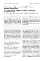
SDS-PAGE peanut seed total protein profilesFigure 1
SDS-PAGE peanut seed total protein profiles. One-dimensional SDS-PAGE of peanut seed protein of runner (R) and
Spanish (S) or Spanish derivatives (SD): R1 = A104, R2 = GK 7, R3 = A13, R4 = Tifrunner, R5 = A100, R6 = Georgia Green; S1
= ICGV 95435, S2 = MXHY, SD3 = GT-YY7, SD4 = GT-YY79, S5 = ZQ 48, SD6 = GT-YY20; M = molecular weight standards.
The arrow ( ) indicates the protein band with a molecular weight of 35 kDa and the arrow ( ) indicates the 26 kDa
protein band.
R1 R2 R3 R4 R5 R6 M S1 S2 SD3 SD4 S5 SD6 M
14.2
20.1
24
29
45
36
66
MW (kDa)
Acidic arachin
Basic arachin
BMC Plant Biology 2006, 6:24 />Page 4 of 9
(page number not for citation purposes)
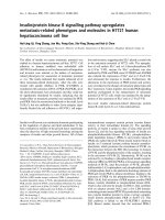
2-D SDS-PAGE peanut seed total protein profilesFigure 2
2-D SDS-PAGE peanut seed total protein profiles. Two-dimensional SDS-PAGE of peanut seed total protein profiles of
6 cultivated peanut genotypes, Spanish market type. Gels are oriented with the acid end of the isoelectric focusing separating
to left and the basic end to the right. The arrow ( ) indicates the protein band with a molecular weight of 35 kDa and the
arrow ( ) indicates the 27 kDa protein band (Fig. 1). The arrow head ( ) indicates the fourth band as reported for Span-
ish cultivars [9]. The numbered arrows ( ) pointing to cycled spots indicate the polymorphic polypeptide spots, which
were sequenced (Table 2).
4
1
2
3
14.2
6.5
20.1
66
45
36
29
24
MW
(kDa)
GT-YY79
pH 10.0
pH 3.0
4
1
2
3
GT-YY20
4
2
3
1
ICGV 95435
4
1
2
3
MXHY
4
2
3
1
GT-YY7
4
1
2
3
ZQ48
BMC Plant Biology 2006, 6:24 />Page 5 of 9
(page number not for citation purposes)
2-D SDS-PAGE peanut seed total protein profilesFigure 3
2-D SDS-PAGE peanut seed total protein profiles. Two-dimensional SDS-PAGE of peanut seed total protein profiles of
6 cultivated peanut genotypes, runner market type. Gels are oriented with the acid end of the isoelectric focusing separating to
left and the basic end to the right (Fig. 2). The arrow ( ) indicates the protein band with a molecular weight of 35 kDa and
the arrow ( ) indicates the 27 kDa protein band (Fig. 1). The numbered arrows ( ) pointing to cycled spots indicate
the polymorphic polypeptide spots, which were sequenced (Table 2).
3
4
2
1
GA Green
4
1
2
3
A100
3
2
4
1
GK 7
4
1
2
3
A104
3
2
4
1
A13
4
1
2
3
Tifrunner
BMC Plant Biology 2006, 6:24 />Page 6 of 9
(page number not for citation purposes)
[7-9], indicating that very low variation in protein profiles
was detected in cultivated peanut using SDS-PAGE gel
electrophoresis.
Generally, SDS-PAGE is not a sufficiently-powerful tech-
nique to distinguish a specific cultivar. Therefore, we
adopted the widely used protocol developed by Damerval
et al. [18] and introduced some modifications including a
preliminary de-fatting step of peanut seeds for 2-D PAGE
separation. We were able to generate 2-D electrophoresis
gel separations with superior resolution and recovery
from peanut seeds. Bianchi-Hall et al. [9] reported that the
polypeptides of acidic arachin using SDS-PAGE distin-
guish Spanish from other market type cultivars. In this
study, we did not identify the four bands in the range of
acidic arachin by SDS-PAGE (Fig. 1), but we could detect
the fourth spot of protein on 2-D PAGE for Spanish type
genotypes (Fig. 2). We also detected a 26 kDa polypeptide
by SDS-PAGE; this polypeptide could be used to differen-
tiate Spanish and runner.
Conclusion
This study demonstrated that two-dimensional electro-
phoresis (2-D PAGE) achieved a better resolution of pro-
tein profiles of peanut seeds, revealing polymorphisms
between runner and Spanish genotypes. The basic arachin
group, having one heavy band on SDS-PAGE gels at about
22 kDa, was resolved into several spots or subunits on the
2-D PAGE with distinct isoelectric points and slight differ-
ences in molecular weights. These proteins are isoforms
(iso-Ara h3) of each other and the iso-allergens may be
modified by post-translational cleavage. These results sug-
gest that there may be an association between these poly-
morphic storage protein isoforms and peanut subspecies
fastigiata (Spanish type) and hypogaea (runner type).
Future studies could be designed to test the allergenic
reactions of these peanut genotypes with different protein
profiles and association with the resistance to aflatoxin
contamination [19].
Methods
Plant materials
Twelve peanut genotypes were used in this study. There
were six runner-type peanut genotypes: Georgia Green,
A100, A104, GK7, A13 and Tifrunner, and six Spanish-
bunch type peanut genotypes: ICGV 95435 (International
Crops Research Institute for the Semi-Arid Tropics,
Patancheru, India), MXHY and ZQ48 (Chinese lan-
draces), and GT-YY20, GT-YY7 and GT-YY79 (Spanish
derivatives with runner type peanut in their pedigrees,
obtained from Crops Research Institute, Guangdong
Academy of Agricultural Sciences, China). To avoid the
effects of different locations, all genotypes were grown in
Tifton, GA in 2003. Seeds were harvested at full maturity
per normal production practices. After harvest, seeds were
air-dried at 40°C and stored at 4°C before use.
Table 1: List of cultivated peanut used in this study
Cultivars Subspecies Market type Origin
GT-YY20 fastigiata Spanish China
GT-YY79 fastigiata Spanish China
GT-YY7 fastigiata Spanish China
ZQ48 fastigiata Spanish China
MXHY fastigiata Spanish China
ICGV 95435 fastigiata Spanish India
Georgia Green [25] fastigiata Runner USA
A100 fastigiata runner USA
GK7 fastigiata runner USA
Tifrunner fastigiata runner USA
A104 fastigiata runner USA
A13 fastigiata runner USA
Table 2: Internal peptide and N-terminal sequences of some protein spots of cultivated peanut
Spot
1
Internal peptide sequence N-terminal peptide sequence
1 FYLAGNQEQEFLR NPDLEEFQCAGVALSR VGQDDPSQQQ
2 FYLAGNQEQEFLR NPDLEEFQCAGVALSR VTFRQGGEENEC
3 QGGEENECQFQR FHLAGNQEQEFLR IESEGGYIETWNPNNQEFECAGVALSR VTFRQGGEENEC
4 AQSENYEYIAFK VYDEELQEGHVLVVPQNFAVAAK GIEETICSASVK
1
Spot numbers are corresponding to the spot numbers in Fig. 2.
BMC Plant Biology 2006, 6:24 />Page 7 of 9
(page number not for citation purposes)
Total protein extraction
The total protein extraction was modified from TCA/Ace-
tone protein extraction protocol [18] with the first step of
de-fatting using hexane. Dry peanut kernels (20 g) of each
genotype were frozen in liquid nitrogen and ground to
powder in a mill and defatted with hexane (10 ml/g dry
weight) at -20°C overnight. The defatted samples were
collected by centrifugation (15,000 × g for 10 min at
4°C), air-dried, and ground to a fine powder in a pre-
chilled mortar and pestle in liquid nitrogen. Protein
Amino acid sequences alignmentFigure 4
Amino acid sequences alignment. Amino acid sequences alignment of peptide sequences (N = N-terminal sequences; I =
internal sequences by using in-gel trypsin digestion and sequencing), in bold-faced, of spots 1–4 with the published peanut aller-
gen sequences of Ara h4 (AAD47382), Ara h3 (AAC63045), and iso-Ara h3 (ABI17154) (26). Sequences obtained by N-terminal
sequencing are shaded in black. The different amino acid residues are colored in red. The amino acid sequences of Ara h3 IgE-
binding epitopes [24] are shaded in gray and the critical amino acids to IgE binding are colored in green and underlined.
Ara h4 1 MAKLLELSFC FCFLVLGASS ISFRQQPEEN ACQFQRLNAQ RPDNRIESEG GYIETWNPNN
Ara h3 1 RQQPEEN ACQFQRLNAQ RPDNRIESEG GYIETWNPNN
Iso-Ara h3 1 MAKLLALSLC FCVLVLGASS VTFRQGGEEN ECQFQRLNAQ RPDNRIESEG GYIETWNPNN
Spot 1&2 (I) NPDL
Spot 3 (I) QGGEEN ECQFQR IESEG GYIETWNPNN
Spot 2&3 (N) VTFRQGGEEN EC
Ara h4 61 QEFECAGVAL SRLVLRRNAL RRPFYSNAPQ EIFIQQGRGY FGLIFPGCPS TYEEPAQQGR
Ara h3 61 QEFECAGVAL SRLVLRRNAL RRPFYSNAPQ EIFIQQGRGY FGLIFPGCPRHYEEPHTQGR
Iso-Ara h3 61 QEFQCAGVAL SRTVLRRNAL RRPFYSNAPL EIYVQQGSGY FGLIFPGCPS TYEEPAQEGR
Spot 1&2 (I) EEFQCAGVAL SR
Spot 3 (I) QEFECAGVAL SR
Ara h4 121 RYQSQRPPRR LQE EDQSQ QQQDSHQKVH RFNEGDLIAV PTGVAFWLYN DHDTDVVAVS
Ara h3 121 RSQSQRPPRR LQG EDQSQ QQRDSHQKVH RFDEGDLIAV PTGVAFWLYN DHDTDVVAVS
Iso-Ara h3 121 RYQSQKPSRR FQVGQDDPSQ QQQDSHQKVH RFDEGDLIAV PTGVAFWMYN DEDTDVVTVT
Spot 1 (N) VGQDDPSQ QQ
Ara h4 181 LTDTNNNDNQ LDQFPRRFNL AGNHEQEFLR YQQQSRQSRR RSLPYSPYSP HSRPRREERE
Ara h3 181 LTDTNNNDNQ LDQFPRRFNL AGNTEQEFLR YQQQSRQSRR RSLPYSPYSP QSQPRQEERE
Iso-Ara h3 181 LSDTSSIHNQ LDQFPRRFYL AGNQEQEFLR YQQQQG -SRP HYRQ
Spot 1&2 (I) FYL AGNQEQEFLR
Spot 3 (I) FHL AGNQEQEFLR
Ara h4 241 FRPRGQHSRR ERAGQEEEDE GGNIFSGFTP EFLEQAFQVD DRQIVQNLWG ENESEEEGAI
Ara h3 241 FSPRGQHSRR ERAGQEEENE GGNIFSGFTP EFLEQAFQVD DRQIVQNLRG ETESEEEGAI
Iso-Ara h3 241 ISPR -VRGDEQENE GSNIFSGFAQ EFLQHAFQVD -RQTVENLRG ENEREEQGAI
Ara h4 301 VTVRGGLRIL SPDGTRGADE E EEYD EDQYEYHEQD GRRGRGSRGG GNGIEETICT
Ara h3 301 VTVRGGLRIL SPDRKRRADE E EEYD EDEYEYDEED RRRGRGSRGR GNGIEETICT
Iso-Ara h3 301 VTVKGGLRIL SPDEEDESSR SPPSRREEFD EDRSRP-QQR GKYDENRRGYKNGIEETICS
Spot 4 (N) GIEETICS
Ara h4 361 ACVKKNIGGN RSPHIYDPQR WFTQNCHDLN LLILRWLGLS AEYGNLYRNA LFVPHYNTNA
Ara h3 361 ASAKKNIGRN RSPDIYNPQA GSLKTANDLN LLILRWLGPS AEYGNLYRNA LFVAHYNTNA
Iso-Ara h3 361 ASVKKNLGRSSNPDIYNPQA GSLRSVNELD LPILGWLGLS AQHGTIYRNA MFVPHYTLNA
Spot 4 (N) ASVK
Ara h4 421 HSIIYALRGR AHVQVVDSNG NRVYDEELQE GHVLVVPQNF AVAGKSQSEN FEYVAFKTDS
Ara h3 421 HSIIYRLRGR AHVQVVDSNG NRVYDEELQE GHVLVVPQNF AVAGKSQSEN FEYVAFKTDS
Iso-Ara h3 421 HTIVVALNGR AHVQVVDSNG NRVYDEELQE GHVLVVPQNF AVAAKAQSEN YEYLAFKTDS
Spot 4 (I) VYDEELQE GHVLVVPQNF AVAAKAQSEN YEYLAFK
Ara h4 481 RPSIANFAGE NSFIDNLPEE VVANSYGLPR EQARQLKNNN PFKFFVPPF- QQSPRAVA
Ara h3 481 RPSIANLAGE NSVIDNLPEE VVANSYGLQR EQARQLKNNN PFKFFVPPS- QQSPRAVA
Iso-Ara h3 481 RPSIANLAGE NSIIDNLPEE VVANSYRLPR EQARQLKNNN PFKFFVPPFD HQSMREVA
BMC Plant Biology 2006, 6:24 />Page 8 of 9
(page number not for citation purposes)
extraction and precipitation were performed in 10% (w/v)
trichloroacetic acid in cold acetone with 0.07% (v/v) β-
mercaptoethanol at -20°C for 2 h, followed by centrifuga-
tion at 10,000 × g for 10 min at 4°C. The pellets were
washed twice with cold acetone containing 0.07% β-mer-
captoethanol, followed by washing twice with cold 80%
acetone and then centrifuged at 10,000 × g for 10 min at
4°C. The pellets were air dried and stored at 4°C over-
night. The total proteins were dissolved in lysis buffer (10
μl/mg) containing 9.5 M urea, 4% Igepal CA-360 (Sigma,
St. Louis, MO), 2.5% ampholytes (0.5% pH 3.0–10, 0.5%
pH 4–6, and 1.5% pH 6–8) (Sigma), 5% β-mercaptoeth-
anol, and kept at 35°C for 30 min. After centrifugation
(15,000 × g, 20 min, 25°C), the supernatant was collected
for loading in first-dimension gel electrophoresis, or alter-
natively, for storing at -20°C until use. The supernatant
protein concentration was determined using the Bradford
[20] assay. The experiment was conducted twice, and each
genotype was run at least three times.
SDS-PAGE and two-dimensional PAGE electrophoresis
Total protein samples from these twelve peanut genotypes
were first profiled using SDS-PAGE (15% separating gel
with 4% stacking gel) according to the method of Lae-
mmli [21] with the Mini-PROTEIN
®
II Dual Slab Cell Sys-
tem (BIO-RAD, Hercules, CA) [22]. Total proteins (100
μg) from each sample were loaded onto SDS-PAGE gels.
Low-range protein markers (Sigma) were used as molecu-
lar mass standard. The gels were electrophoresed (120 V,
1.5 h), stained with 0.125% Coomassie blue R-250 in
40% methanol and 10% acetic acid. For 2-D PAGE, total
seed proteins (1 mg) were loaded into tube gels (8 M urea,
4% acrylamide, 2% Igepal CA-630, 0.5% ampholyte pH
3.0–10, 0.5% ampholyte pH 4–6, 1.5% ampholyte pH 6–
8, 0.01% ammonium persulfate, and 0.1% TEMED), and
overlaid with 20 μl sample overlay buffer (4 M urea,
0.25% ampholyte pH 3.0–10, 0.25% ampholyte pH 4–6,
0.75% ampholyte pH 6–8, 2.5% β-mercaptoethanol, 1%
Igepal CA-360, and 0.05% Bromophenol blue). Isoelec-
tric focusing (IEF) was conducted by using Mini-Protean
®
2-D Electrophoresis Cell (BIO-RAD). The upper and lower
chamber buffers were 100 mM NaOH and 10 mM H
3
PO
4
respectively. IEF conditions were 200 V for 15 min, 300 V
for 15 min, 400 V for 30 min, and 750 V for 6 h. The
focused tube gels were equilibrated immediately for 30
min in 10 ml SDS equilibration buffer (60 mM Tris-HCl,
pH6.8, 2% SDS, 10% glycerol, and 0.05% Bromophenol
blue), or kept at -20°C until use. After equilibration, the
tube gels were embedded in a 1% agarose solution at the
top of the 2-D gel. The second dimension was run on 15%
polyacrylamide-SDS gels in a Mini-Protean
®
3 Cell (BIO-
RAD), 120 V for 90 min. The gels were stained with
Coomassie Brilliant Blue R250 and all gels were scanned
and the spot intensities were analyzed using the software
Image Master-2D (BIO-RAD). The interesting spots of
seed protein among the genotypes were identified by gel-
to-gel comparison. For molecular weight determination,
low molecular weight standard (Sigma) was used.
Peptide sequencing
Protein peptides were excised from the 2-D gels and PVDF
membranes for peptide sequencing using electrospray
ionization tandem mass spectrometry (ESI-MS/MS) to
obtain internal peptide sequences and using the conven-
tional Edman degradation method to obtain N-terminal
sequences. Protein spots from the gels were excised with
combined total protein amount up to 10 pg, and were
subjected to in-gel digestion and analysis by ESI-MS/MS
to obtain peptide sequence information at the Protein
Chemistry Core Facility, Baylor College of Medicine
(Houston, TX). When peptide sequences could not be
obtained unambiguously by using ESI-MS/MS, Edman
degradation was performed using an Applied Biosystems
Procise cLC sequencer to obtain sequence information for
protein identification.
Electrobloting and N-terminal sequence
To prevent N-terminal blockage during second-dimen-
sion gel electrophoresis, gels were poured at least 24 hr
prior to running and 0.1 mM thiodiglycolate was added as
a scavenger in the upper running buffer. 2-D gels were
equilibrated for 30 min in 25 mM Tris, 192 mM glycine,
10% MeOH (pH 8.3), and then electroblotted to Immo-
bilon-p PVDF-membrane (Millipore, Bedford, MA, USA)
at 300 mA for 4 hr in a Mini Trans-Blot
®
Electrophoretic
Transfer Cell (BIO-RAD). The membrane was subse-
quently equilibrated for 5 min in deionized water and
proteins stained with 0.05% Coomassie Blue in 1% acetic
acid and 50% methanol for a few min, destained in 50%
methanol until background was pale blue. The membrane
was rinsed for 5–10 min in deionized water and air-dried.
Spots were excised and used for N-terminal amino acid
microsequencing at Baylor Medical School (Houston,
TX).
Database sequence homology analysis
Internal and N-terminal peptide sequence homology
identification was performed using basic local alignment
search tool (BLAST) [23] against known or translated
open reading frames of expressed sequence tags (ESTs) in
the databases at the National Center for Biotechnology
Information (NCBI) and SWISS-Prot.
Authors' contributions
XQL performed the experiments and wrote the first draft
of the manuscript. ML performed the sequence search and
CCH provided plant materials. BZG conceived the
research and revised the manuscript. All authors read and
approved the final manuscript.
Publish with BioMed Central and every
scientist can read your work free of charge
"BioMed Central will be the most significant development for
disseminating the results of biomedical research in our lifetime."
Sir Paul Nurse, Cancer Research UK
Your research papers will be:
available free of charge to the entire biomedical community
peer reviewed and published immediately upon acceptance
cited in PubMed and archived on PubMed Central
yours — you keep the copyright
Submit your manuscript here:
/>BioMedcentral
BMC Plant Biology 2006, 6:24 />Page 9 of 9
(page number not for citation purposes)
Acknowledgements
We thank Ernest Harris and Kippy Lewis for technical assistance in the field
and the laboratory. This research was supported partially by funds provided
by USDA Agricultural Research Service and Peanut Foundation, and by
funds provided by Scientific Cooperation Research Program of U. S.
Department of Agriculture-Foreign Agricultural Service between U.S. and
China. Mention of trade names or commercial products in this publication
is solely for the purpose of providing specific information and does not
imply recommendation or endorsement by the U.S. Department of Agri-
culture.
References
1. Krapovickas SA, Rigoni VA: Taxonomia del genero Arachis
(Leguminosae). Bonplandia 1994, 8:1-186.
2. Stalker HT, Philips TD, Murphy JP, Jones TM: Variation of isozyme
patterns among Arachis species. Theor Appl Genet 1994,
87:746-755.
3. Kochert G, Halwart T, Branch WD, Simpson CE: RFLP variability
in peanut (Arachis hypogaea L.) cultivars and wild species.
Theor Appl Genet 1991, 81:565-570.
4. Halward TM, Stalker TH, LaRue E, Kochert G: Genetic variation
detectable with molecular markers among unadapted germ-
plasm resources of cultivated peanut and related wild spe-
cies. Genome 1991, 34:1013-1020.
5. He GC, Prakash S: Identification of polymorphic DNA markers
in cultivated peanut (Arachis hypogaea L.). Euphytica 1997,
97:143-149.
6. He G, Meng R, Gao H, Guo B, Gao G, Newman M, Pittman R, Prakash
CS: Simple sequence repeat markers for botanical varieties
of cultivated peanut (Arachis hypogaea L.). Euphytica 2005,
142:131-136.
7. Singh AK, Sivaramakrishnan S, Mengesha MH, Ramaiah CD: Polyge-
netic relation in section Arachis based on seed protein pro-
file. Theor Appl Genet 1991, 82:593-597.
8. Singh AK, Santosh G, Jambunathan R: Phylogenetic relationship in
the genus Arachis based on seed protein profiles. Euphytica
1994, 74:219-225.
9. Bianchi-Hall CM, Keys RD, Stalker TH: Use of seed protein pro-
files to characterize peanut cultivars. Peanut Sci 1994,
21:152-159.
10. Thiellemmet H, Bahrnman N, Damerval C, Plomion C, Rossignol M,
Santony V, Vienne D, De Zivy M: Proteomics for genetic and
physiological studies in plant. Electrophoresis 1999,
20:2013-2026.
11. Park OK: Proteomic studies in plants.
J Biochem Mol Biol 2004,
37:133-138.
12. Shokraii EH, Esen A, Mozingo RW: Relation of a 36,000-dalton
arachin subunit to blanch ability in peanut (Arachis hypogaea
L.). J Agric Food Chem 1985, 33:1114-1116.
13. Boldt A, Fortunato D, Conti A, Petersen A, Ballmer-Weber B, Lepp
U, Reese G, Becker W-M: Analysis of the composition of an
immunoglobulin E reactive high molecular weight protein
complex of peanut extract containing Ara h 1 and Ara h 3/4.
Proteomics 2005, 5:675-686.
14. Guo BZ, Liang XQ, Maleki SJ, Chung SY, Holbrook CC, Ozias-Akins
P: Characterization of five seed-proteins missing in one pea-
nut genotype and the allergic nature of these proteins. Pro-
ceeding of the thirty-sixth annual meeting of the American Peanut Research
and Education Society (APRES). San Antonio, Texas 2004:28. July 13–16,
2004
15. Kang I-H, Gallo-Meagher M: Cloning and characterization of a
novel ara h 3, a major peanut (Arachis hypogaea L.) allergen
gene. GenBank 2004 [.
]. National
Center for Biotechnology Information Accession Number AY618460
16. Basha SM, Pancholy SK: Polypeptide composition of arachin and
non-arachin proteins from early bunch peanut (Arachis
hypogaea L.) seed. Peanut Sci 1981, 8:82-88.
17. Krisha TG, Mitra R: Arachin polymorphism in groundnut (Ara-
chis hypogaea L.). Phytochemistry 1987, 26:897-902.
18. Damerval C, de Vienne D, Zivy M, Thiellement H: The technical
improvements in two-dimensional electrophoresis increase
the level of genetic variation detected in wheat-seedling pro-
teins. Electrophoresis 1986, 7:52-54.
19. Liang XQ, Guo BZ, Holbrook CC: Identification of peanut seed-
storage proteins associated with resistance against Aspergil-
lus flavus infection and aflatoxin production. Proceeding of the
thirty-sixth annual meeting of the American Peanut Research and Education
Society (APRES). San Antonio, Texas 2004:74. July 13–16, 2004
20. Bradford M: A rapid and sensitive method for the quantitation
of microgram quantities of protein utilizing the principal of
protein-dye binding. Anal Biochem 1976, 72:248-254.
21. Laemmli UK: Cleavage of structural proteins during the
assembly of the head of bacteriophage T4. Nature 1970,
227:239-251.
22. Guo BZ, Brown RL, Lax AR, Cleveland TE, Russin JS, Widstrom NW:
Protein profiles and antifungal activities of kernel extracts
from corn genotypes resistant and susceptible to Aspergillus
flavus. J Food Prot 1998, 61:98-102.
23. Altschul SF, Gish W, Miller W, Myers EW, Lipman DJ: Basic local
alignment search tool. J Mol Biol 1990, 215:403-410.
24. Rabjohn P, Helm EM, Stanley JS, West CM, Sampson HA, Burks AW,
Bannon GA: Molecular cloning and epitope analysis of the pea-
nut allergen Ara h3. J Clin Invest 1999, 103:535-542.
25. Branch WD: Registration of 'Georgia Green' peanut. Crop Sci
1996, 36:806.
26. Guo B, Chen H, Dang P, Chen X, Liang X, Holbrook C: Identifica-
tion of a new potential peanut seed allergen iso-Ara h3. Gen-
Bank 2006 [.
]. National Center for
Biotechnology Information Accession Number DQ855115