báo cáo khoa học: " The red fluorescent protein eqFP611: application in subcellular localization studies in higher plants" doc
Bạn đang xem bản rút gọn của tài liệu. Xem và tải ngay bản đầy đủ của tài liệu tại đây (662.76 KB, 12 trang )
BioMed Central
Page 1 of 12
(page number not for citation purposes)
BMC Plant Biology
Open Access
Research article
The red fluorescent protein eqFP611: application in subcellular
localization studies in higher plants
Joachim Forner and Stefan Binder*
Address: Molekulare Botanik, Universität Ulm, Albert-Einstein-Allee 11, 89069 Ulm, Germany
Email: Joachim Forner - ; Stefan Binder* -
* Corresponding author
Abstract
Background: Intrinsically fluorescent proteins have revolutionized studies in molecular cell
biology. The parallel application of these proteins in dual- or multilabeling experiments such as
subcellular localization studies requires non-overlapping emission spectra for unambiguous
detection of each label. In the red spectral range, almost exclusively DsRed and derivatives thereof
are used today. To test the suitability of the red fluorescent protein eqFP611 as an alternative in
higher plants, the behavior of this protein was analyzed in terms of expression, subcellular targeting
and compatibility with GFP in tobacco.
Results: When expressed transiently in tobacco protoplasts, eqFP611 accumulated over night to
levels easily detectable by fluorescence microscopy. The native protein was found in the nucleus
and in the cytosol and no detrimental effects on cell viability were observed. When fused to N-
terminal mitochondrial and peroxisomal targeting sequences, the red fluorescence was located
exclusively in the corresponding organelles in transfected protoplasts. Upon co-expression with
GFP in the same cells, fluorescence of both eqFP611 and GFP could be easily distinguished,
demonstrating the potential of eqFP611 in dual-labeling experiments with GFP. A series of plasmids
was constructed for expression of eqFP611 in plants and for simultaneous expression of this
fluorescent protein together with GFP. Transgenic tobacco plants constitutively expressing
mitochondrially targeted eqFP611 were generated. The red fluorescence was stably transmitted to
the following generations, making these plants a convenient source for protoplasts containing an
internal marker for mitochondria.
Conclusion: In plants, eqFP611 is a suitable fluorescent reporter protein. The unmodified protein
can be expressed to levels easily detectable by epifluorescence microscopy without adverse affect
on the viability of plant cells. Its subcellular localization can be manipulated by N-terminal signal
sequences. eqFP611 and GFP are fully compatible in dual-labeling experiments.
Background
Since the cloning of the green fluorescent protein (GFP)
cDNA and its first heterologous expression in the early
1990s [1,2], the use of intrinsically fluorescent proteins
(IFPs) has become one of the most powerful tools in
molecular and cell biology. These proteins are applied as
reporters in gene expression studies, as indicators of intra-
cellular physiological changes, for monitoring dynamics
of organelles and proteins, for investigation of protein-
Published: 6 June 2007
BMC Plant Biology 2007, 7:28 doi:10.1186/1471-2229-7-28
Received: 8 November 2006
Accepted: 6 June 2007
This article is available from: />© 2007 Forner and Binder; licensee BioMed Central Ltd.
This is an Open Access article distributed under the terms of the Creative Commons Attribution License ( />),
which permits unrestricted use, distribution, and reproduction in any medium, provided the original work is properly cited.
BMC Plant Biology 2007, 7:28 />Page 2 of 12
(page number not for citation purposes)
protein interactions in vivo and as fusion partners in stud-
ies of the subcellular localization of proteins [3,4].
From the very beginning, many efforts have been made to
optimize various features of the native GFP with the aim
to improve its application in biological research. These
modifications include for instance improved folding effi-
ciency, higher expression level or increased solubility [3].
Cyan and yellow fluorescent derivatives of GFP have been
created for investigations requiring the simultaneous dis-
tinguishable tagging of more than one protein at a time
[5,4]. These are used to compare the spatial distribution or
the expression pattern of two or more proteins and for the
analysis of protein-protein interactions by FRET. So far no
red fluorescent variant of GFP has been reported.
Recently, investigation of several non-bioluminescent
anthozoan species has led to the isolation of various true
red fluorescent proteins (RFPs) [6]. Among these, DsRed
and its derivatives are the most commonly used in molec-
ular and cell biological research [7].
Since plants contain a large number of multi-gene fami-
lies, comparisons of the subcelluar localizations of the
individual members are necessary as part of the compre-
hensive analysis of these proteins. The possibility to label
several proteins with different fluorescent proteins is a
great advantage when analyzing their respective subcellu-
lar localization. As a crucial prerequisite for such studies,
the compartments to which the fusion proteins are tar-
geted have to be unequivocally identified. This is often
done by staining with compartment-specific dyes. Mito-
chondria for instance can be visualized by staining with
the red fluorescent dye MitoTracker
®
Red CM-H2Xros
(Molecular Probes, Eugene, OR) which specifically inter-
acts with the respiratory chain. The staining procedure,
however, is time-consuming, invasive and short-lived and
can be replaced simply by co-expression of a spectrally dif-
ferent second fusion protein with a defined subcellular
localization. Additionally, the fused target sequence of the
fluorescent marker protein can be readily exchanged,
which allows selective labeling of nearly every subcellular
structure under investigation without the need to have a
specific dye for the different compartments.
Despite the discovery of a multiplicity of fluorescent pro-
teins in the red spectral range in recent years [6], so far
almost exclusively different forms of DsRed have been
used for studies in molecular cell biology in plants [8-12].
These proteins are applied in dual-labeling experiments
together with GFP or alone to report on promoter activity
or as a marker in transgenic plants. To introduce an alter-
native RFP for the application in plant cells and to expand
the palette of red fluorescent reporters for plant research,
we tested the suitability of the red fluorescent protein
eqFP611 from the sea anemone Entacmaea quadricolor as a
marker in subcellular localization experiments in plants.
eqFP611 shows far-red fluorescence with excitation and
emission maxima at 559 nm and 611 nm, respectively,
and therefore exhibits an extraordinarily large Stokes shift
of 52 nm [13]. In contrast, the respective values for DsRed
are 558 nm, 583 nm and 25 nm, respectively [13]. Both
eqFP611 and DsRed have comparable molecular masses
of 25.93 kDa and 26.05 kDa, respectively, for the mono-
mers. The extinction coefficient of eqFP611 (78,000 M
-1
*
cm
-1
) is slightly higher than that of DsRed (75,000 M
-1
*
cm
-1
). Fluorescence quantum yields for eqFP611 and
DsRed are 0.45 and 0.7 and the photobleaching quantum
yields are 3.5 * 10
-6
and 0.8–9.5 * 10
-6
, respectively. Sim-
ilar to DsRed, the emission of eqFP611 is constant
between pH 4 and 10. Though both form tetramers at
physiological concentrations, eqFP611 has a reduced ten-
dency to oligomerize and aggregate as compared to
DsRed. With a maturation half-time t
0.5
of 4.5 h at 24.5°C
[14], fluorophore maturation of eqFP611 is much faster
than that of DsRed (t
0.5
> 24 h at 24.5 °C) [13].
We demonstrate that native eqFP611 can be expressed in
plant cells. Fusions of this protein with respective N-ter-
minal signal sequences can be efficiently targeted to mito-
chondria and peroxisomes. We performed co-expression
experiments with eqFP611 and GFP and created vectors
for the straightforward application of the eqFP611 gene in
plants.
Results and Discussion
eqFP611 can be functionally expressed in plant cells
Recently, eqFP611, the gene for a red fluorescent protein
from the sea anemone Entacmaea quadricolor, has been
cloned and characterized [13,14]. This protein has been
succesfully expressed in bacteria and animal cells [13], but
has not yet been tested in plants.
To test its use as a marker in plants, the native eqFP611
cDNA was cloned into a pUC19-based vector. In the
resulting plasmid peqFP611, expression of this gene is
governed by the strong constitutive cauliflower mosaic
virus 35S promoter (CaMV 35S) and the nopaline syn-
thase terminator (NOS T) sequences. Upon inspection of
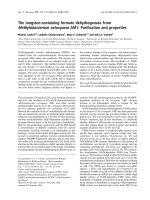
Nicotiana tabacum mesophyll cells transfected with this
plasmid in the epifluorescence microscope, the red fluo-
rescence was clearly detectable with a filter set (HQ545/
30/HQ 610/75) usually used for visualization of
MitoTracker Red and here later referred to as MitoTracker
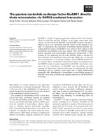
filter set (Fig. 1). The protein accumulates in the nucleus
and in the cytosol, where it is evenly distributed and does
not form any visible aggregates, but is clearly absent from
the chloroplasts. No such fluorescence was detectable in
untransfected control cells, confirming that the red fluo-
BMC Plant Biology 2007, 7:28 />Page 3 of 12
(page number not for citation purposes)
rescence indeed originates from the expression of the
introduced eqFP611. Protoplasts were analysed 16 hours
after transfection. Incubation for an additional 24 hours
did not markedly increase the intensity of the red fluores-
cence, suggesting the maximal level of mature protein to
be essentially reached within 16 hours after transfection.
Protoplasts expressing eqFP611 looked perfectly normal
and did not show any detrimental effects of this fluores-
cent protein.
These results show that eqFP611 can be readily used in
plants, since the functional protein accumulates to detect-
able levels without any obvious adverse effects. In contrast
to GFP, whose original jellyfish-derived cDNA was miss-
pliced specifically in plants at a cryptic splice site [15], no
modification of the eqFP611 coding sequence is necessary
for efficient expression in plants.
As expected from its spectral characteristics, the fluores-
cence is easily detectable with a filter set (see above) that
excludes the red autofluorescence of chlorophyll, a crucial
advantage for an RFP applied in mesophyll cells. Similar
to GFP [16], the native eqFP611 accumulates in the
nucleus and in the cytosol in plant cells. Thus, it should
be suited to investigate protein targeting into e.g. mito-
chondria, peroxisomes and plastids within plants. In
HeLa cells, native, unmodified eqFP611 was also found in
the nucleus and the cytosol [13].
Targeting eqFP611 to mitochondria
To investigate whether eqFP611 can indeed be used as
reporter protein for the analysis of subcellular protein
sorting, import into plant mitochondria was exemplarily
tested. To this end, the presequence of the mitochondrial
isovaleryl-CoA-dehydrogenase (IVD) was added to the N-
terminus of eqFP611 (plasmid pIVD145-eqFP611). The
IVD presequence was chosen because it has previously
been found to efficiently target a GFP fusion protein
exclusively to mitochondria [17]. In addition, the protein
has been repeatedly detected in proteomic analyses of this
organelle, demonstrating its unambiguous localization in
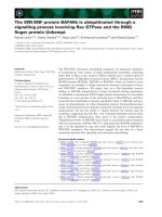
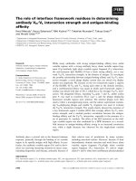
mitochondria [18-20]. Inspection of the protoplasts trans-
fected with pIVD145-eqFP611 using the MitoTracker filter
set revealed the red fluorescence to be restricted exclu-
sively to rod-shaped structures of 1 – 2 μm in length dis-
tributed throughout the cell (Fig. 2A). This pattern is
characteristic for a mitochondrial localization of the
fusion protein. No red fluorescence was detectable in
other parts of the protoplasts. Thus, eqFP611 can be effi-
ciently targeted to plant mitochondria, its subcellular
localization being exclusively determined by the targeting
information of the signal peptide fused to its N-terminus.
Furthermore, this result confirms that eqFP611 is effi-
ciently transported through two membranes while retain-
ing its ability to fold properly for effective fluorescence.
Similar to the native eqFP611, prolonged incubation of
the protoplasts did not increase the intensity of the fluo-
rescence.
The picture of the transfected protoplast displayed in Fig.
2A demonstrates nicely that the use of the MitoTracker fil-
ter set is appropriate to easily detect the red fluorescence
of eqFP611 while effectively blocking chlorophyll
autofluorescence. The latter is clearly visible through the
FITC (fluorescein isothiocyanate) filter set (HQ 470/40/
HQ 500 LP), which in turn blocks the fluorescence of
eqFP611 (Fig. 2B). This autofluorescence in the chloro-
plasts exactly fits to the areas without fluorescence in Fig.
2A. Furthermore, the untransfected cells surrounding the
eqFP611-expressing protoplast in Fig. 2A clearly show
that no other autogenous fluorescence is visible through
the MitoTracker filter set.
To assess the relative stability of the eqFP611 fluorescence
in plants, we qualitatively compared the time elapsed
until bleaching of the red fluorescence in protoplasts tran-
siently expressing IVD145-eqFP611 and of MitoTracker
®
Red CM-H2Xros (Molecular Probes, Eugene, OR) used for
staining of untransfected protoplasts. This latter mito-
chondria-specific fluorescent dye has excitation/emission
maxima of 579 nm and 599 nm, respectively. When indi-
vidual cells of both approaches were inspected under
identical light conditions in the fluorescence microscope,
the fluorescence of IVD145-eqFP611 was at least as stable
eqFP611 without presequenceFigure 1
eqFP611 without presequence. Transient expression of
original eqFP611 without presequence in N. tabacum wild-
type protoplasts. (A) Image taken through MitoTracker filter
set. Scale bar: 10 μm. (B) Plasmid peqFP611 used for trans-
fection. Black arrow, CaMV 35S: cauliflower mosaic virus 35S
promoter; red box, eqFP611: eqFP611 coding sequence;
black box, NOS T: nopaline synthase terminator. H: HindIII,
P: PstI, Xb: XbaI, B: BamHI, Sm: SmaI, Sa: SacI, E: EcoRI
restriction sites. Vector backbone: pUC19.
B
NOS TCaMV 35S
peqFP611
eqFP611
pUC19
HXb
B
Sm Sa EHP
A
BMC Plant Biology 2007, 7:28 />Page 4 of 12
(page number not for citation purposes)
as the fluorescence of MitoTracker, which further demon-
strates the usability of eqFP611 as marker at least in plant
mitochondria.
Co-expression of eqFP611 and smGFP4 in tobacco
protoplasts
Experiments like subcellular localization studies in which
one of the fluorescent proteins is used to mark a distinct
cellular compartment, require the simultaneous expres-
sion of two different fluorescent proteins. If eqFP611 is to
be used routinely in such applications, its expression must
be fully compatible with other IFPs, e.g. GFP. To test
whether co-expression of both fluorescent proteins is
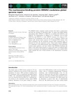
indeed useful, tobacco protoplasts were simultaneously
transfected with the constructs pIVD145-eqFP611 and
pIVD145-smGFP4. Both plasmids contain identical mito-
chondrial targeting sequences fused to the N-termini of
eqFP611 or smGFP4, respectively. Most of the succesfully
transfected protoplasts incorporated both plasmids and
expressed both eqFP611 and smGFP4. Identical patterns
of the red and the green fluorescence in these protoplasts
confirmed the co-expression of both proteins in the same
cell (Fig. 3). In addition to the GFP-derived green fluores-
cence in the mitochondria, the red chlorophyll autofluo-
rescence in the chloroplasts is seen with the FITC filter set
(Fig. 3B).
To examine whether the transport into mitochondria of
both fusion proteins occurs independently of each other
and to exclude a possible chance "piggy back" effect dur-
ing subcelluar transport of the two chimeric proteins,
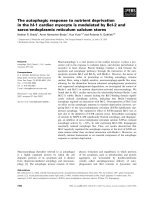
tobacco protoplasts were transfected with a different com-
bination of plasmids. This time, pIVD145-smGFP4 was
used for co-transfection with plasmid pKAT2-eqFP611,
which latter encodes a recombinant protein of the perox-
isomal targeting signal 2 (PTS2) [21] of 3-keto-acyl-CoA
thiolase 2 (KAT2) [22] N-terminally fused to the eqFP611
reading frame. Red and green fluorescences were again
found exclusively in the expected organelles (Fig. 4). The
green fluorescence is observed in mitochondria, while the
red fluorescence is visible in approximately 1 – 2 μm large
roundish structures, a shape expected for leaf peroxi-
somes. No green fluorescence is seen in these organelles
and conversely no red fluorescence is detected in mito-
chondria. This strongly suggests that if there is any inter-
ference, it does not disturb the correct targeting of the
Mitochondrially targeted eqFP611Figure 2
Mitochondrially targeted eqFP611. N. tabacum wild-type protoplast expressing a fusion protein of eqFP611 and the N-ter-
minal 48 amino acids of IVD. Pictures showing the same cell were taken through MitoTracker (A) and FITC (B) filter sets,
respectively. Scale bars: 10 μm. (C) Map of plasmid pIVD145-eqFP611 used for transfection. Black arrow, CaMV 35S: cauli-
flower mosaic virus 35S promoter; grey box, IVD(145): N-terminal 145 nucleotides of the IVD coding sequence; red box,
eqFP611: eqFP611 coding sequence; black box, NOS T: nopaline synthase terminator. H: HindIII, P: PstI, Xb: XbaI, B: BamHI,
Sm: SmaI, Sa: SacI, E: EcoRI restriction sites. Vector backbone: pUC19.
pIVD145-eqFP611
IVD(145)
NOS TCaMV 35S eqFP611
pUC19
HXbBBSm SaEHP
AB
C
BMC Plant Biology 2007, 7:28 />Page 5 of 12
(page number not for citation purposes)
individual fusion proteins. Thus, eqFP611 and smGFP4
can be used in parallel to study protein sorting to different
organelles within the same plant cell.
To verify that the KAT2-eqFP611 fusion protein was
indeed targeted to peroxisomes, pKAT2-eqFP611 was
used for co-transfection together with p35S-N-
TAP2(G)pex. The latter plasmid encodes a GFP fusion
protein targeted to peroxisomal membranes by the C-ter-
minal 36 amino acids of cotton ascorbate peroxidase
(APX). As shown in Fig. 5, the patterns of the green and
the red fluorescence overlap, indicating the correct perox-
isomal localization of KAT2-eqFP611. Green fluorescence
seems to be more intensive at the boundaries of the per-
oxisomes, while the red fluorescence is equally distributed
within the organelles. This is consistent with the predicted
intra-peroxisomal localization of the APX and KAT2 pro-
teins, respectively. No green or red fluorescence is visible
outside the peroxisomes. These experiments demonstrate
that the N-terminal peroxisomal targeting signal 2 effi-
ciently directs eqFP611 to the corresponding organelle
and that this RFP can thus be exployed to study protein
sorting into peroxisomes in plants.
Thus, as demonstrated by the expression in both mito-
chondria and peroxisomes, eqFP611 is a suitable partner
for GFP in double-labeling experiments. When the two
IFPs are co-expressed in the same cell, no mutual interfer-
ence regarding development of fluorescence or intracellu-
lar sorting is observed. Additionally, both eqFP611 and
GFP fluorescences can be easily distinguished by their
emission spectra. The previously reported minor green
Co-expression of eqFP611 and smGFP4 fusion proteins targeted to mitochondriaFigure 3
Co-expression of eqFP611 and smGFP4 fusion proteins targeted to mitochondria. Tobacco wild-type protoplasts
transfected with plasmids pIVD145-eqFP611 and pIVD145-smGFP4. The eqFP611 and smGFP4 fusion proteins contain the
mitochondrial presequence corresponding to the N-terminal 48 amino acids of IVD. Transfected protoplast seen through
MitoTracker (A) and FITC (B) filter sets, respectively. Scale bars: 10 μm. (C) Map of plasmids pIVD145-eqFP611 and pIVD145-
smGFP4. Black arrow, CaMV 35S: cauliflower mosaic virus 35S promoter; grey box, IVD(145): N-terminal 145 nucleotides of
the IVD coding sequence; red box, eqFP611: eqFP611 coding sequence; green box, smGFP4: smGFP4 coding sequence; black
box, NOS T: nopaline synthase terminator. H: HindIII, P: PstI, Xb: XbaI, B: BamHI, Sm: SmaI, Sa: SacI, E: EcoRI restriction sites.
Vector backbone: pUC19.
AB
IVD(145)
NOS TCaMV 35S
pIVD145-eqFP611
eqFP611
pUC19
HXbBBSm SaEHP
C
IVD(145)
NOS TCaMV 35S
pIVD145-smGFP4
smGFP4
pUC19
HXbBBSm SaEP
BMC Plant Biology 2007, 7:28 />Page 6 of 12
(page number not for citation purposes)
fluorescence of eqFP611 was undetectable under the con-
ditions used (Fig. 2B and 4B) [13].
Furthermore, despite the tendency of eqFP611 to form
tetramers [13], its fusion proteins can be efficiently and
reliably targeted to organelles. The transport across single
(peroxisomes) or double (mitochondria) membranes
does not interfere with the formation of the higher order
structure necessary for emitting fluorescence. In addition,
the fusion of a signal sequence to its N-terminus has no
negative influence on the red fluorescence of eqFP611.
Expression of both eqFP611 and smGFP4 from a single
plasmid
Transformation of Nicotiana benthamiana leaves by injec-
tion of Agrobacterium tumefaciens [23] containing IFP
fusion genes is another fast and simple method for the
analysis of the subcellular localization of a protein. This
procedure is presumably closer to the in vivo conditions
than protoplast transfection, since the transformed cells
remain in the original tissue context. In addition, this
approach does not require the relatively laborious prepa-
ration of protoplasts. In this case, expression of the two
fusion proteins from the same plasmid is advantageous,
since a single transformation event is sufficient to ensure
that every transformed cell contains both IFP genes. Apart
from that, expressing both fluorescent proteins from the
Co-expression of peroxisomally targeted eqFP611 and mitochondrially targeted smGFP4Figure 4
Co-expression of peroxisomally targeted eqFP611 and mitochondrially targeted smGFP4. Co-transfection of N.
tabacum wild-type protoplasts with two separate plasmids encoding eqFP611 with a peroxisomal targeting signal 2 (pKAT2-
eqFP611) and smGFP4 with a mitochondrial presequence (pIVD145-smGFP4). Images of a cell transfected with both con-
structs through MitoTracker (A) and FITC (B) filter sets, respectively. Scale bars: 10 μm. (C) Plasmids pKAT2-eqFP611 and
pIVD145-eqFP611 used for transfection. Black arrow, CaMV 35S: cauliflower mosaic virus 35S promoter; grey box, KAT2: N-
terminal 297 nucleotides of the KAT2 coding sequence; grey box, IVD(145): N-terminal 145 nucleotides of the IVD coding
sequence; red box, eqFP611: eqFP611 coding sequence; green box, smGFP4: smGFP4 coding sequence; black box, NOS T:
nopaline synthase terminator. H: HindIII, P: PstI, Xb: XbaI, Sm: SmaI, Sa: SacI, E: EcoRI, B: BamHI restriction sites. Vector back-
bone: pUC19.
AB
pIVD145-smGFP4
IVD(145)
NOS TCaMV 35S smGFP4
pUC19
HXbBBSm SaEP
pKAT2-eqFP611
KAT2
NOS TCaMV 35S eqFP611
pUC19
HXbSm SaEHP
C
BMC Plant Biology 2007, 7:28 />Page 7 of 12
(page number not for citation purposes)
same plasmid under identical promoters should generate
equal amounts of RFP and GFP within a cell. The entire
procedure should be easier since only a single construct
has to be handled. To investigate the feasibilty of this pro-
cedure, plasmid pIVD144-eqFP611-IVD145-smGFP4
containing both the eqFP611 and the smGFP4 genes with
mitochondrial presequences each under control of a
CaMV 35S promoter was constructed and first tested by
transfection of tobacco protoplasts. Again, both red and
green fluorescence could easily be detected in the same
cell (Fig. 6). The fluorescence is found exclusively in mito-
chondria, the patterns of both red and green fluorescence
being identical. This result is indistinguishable from the
experiment with the same eqFP611 and smGFP4 expres-
sion cassettes encoded on two different plasmids (Fig. 3),
but this time every transfected protoplast expressed both
eqFP611 and smGFP4.
For co-expression of eqFP611 and smGFP4 in N. bentha-
miana, a binary vector suitable for plant transformation by
agrobacteria was generated. The RFP-GFP-expression cas-
sette from pIVD144-eqFP611-IVD145-smGFP4 was trans-
ferred into pBI121, creating pIVD144-eqFP611-IVD145-
smGFP4-pBI121. A. tumefaciens containing the latter plas-
mid was then injected into N. benthamiana leaves. After
transformation, both red and green fluorescence were vis-
Co-transfection of tobacco protoplasts with plasmids encoding eqFP611 and GFP targeted to peroxisomesFigure 5
Co-transfection of tobacco protoplasts with plasmids encoding eqFP611 and GFP targeted to peroxisomes.
Transfection of N. tabacum wild-type protoplasts with two separate plasmids encoding eqFP611 with a peroxisomal targeting
signal 2 (pKAT2-eqFP611) and GFP targeted to the peroxisomal membrane (p35S-N-TAP2(G)pex). Pictures of the same pro-
toplast taken through MitoTracker (A) and FITC (B) filter sets, respectively. Scale bars: 10 μM (C) Plasmid maps. Black arrow,
CaMV 35S: cauliflower mosaic virus 35S promoter; grey box, KAT2: N-terminal 297 nucleotides of the KAT2 coding sequence;
grey box, TAP: chimeric sequence for tandem affinity purification; red box, eqFP611: eqFP611 coding sequence; green box,
GFP(S65T): GFP coding sequence including the S65T modification; grey box, APX: sequence encoding the C-terminal 36 amino
acids of cotton ascorbate peroxidase; black box, NOS T: nopaline synthase terminator. H: HindIII, P: PstI, Xb: XbaI, Sm: SmaI,
Sa: SacI, E: EcoRI, Xh: XhoI restrictions sites. Vector backbone: pUC19 and pGreenII, respectively.
AB
pKAT2-eqFP611
KAT2
NOS TCaMV 35S eqFP611
pUC19
HXbSm SaEHP
p35S-N-TAP2(G)pex
TAP
APX(36)CaMV 35S GFP (S65T)
pGreenII
HXhSm XbSa
C
BMC Plant Biology 2007, 7:28 />Page 8 of 12
(page number not for citation purposes)
ible in mitochondria of epidermal cell layers (Fig. 7),
demonstrating the convenient use of the corresponding
vector in this system.
Tobacco plants stably expressing mitochondrially targeted
eqFP611
A third way to use eqFP611 as a mitochondrial marker in
plant cells is the generation of transgenic plants constitu-
tively expressing mitochondrially targeted eqFP611. To
create such plants, the RFP-expression cassette of
pIVD145-eqFP611 was cloned into pBI121. The resulting
plasmid pIVD145-eqFP611-pBI121 was stably trans-
formed into tobacco by leaf disc transformation. Several
independent plant lines were regenerated from transgenic
calli and screened for bright red fluorescence in mito-
chondria. Red fluorescent mitochondria were observed in
all T
0
transformants, but expression levels varied between
individual plants. In addition, segregation was observed
in the next generation. Thus, only the offspring of the
most strongly fluorescent T
1
plant was used for propaga-
tion (Fig. 8). The transgenic plants completed their life
cycle like wild-type plants and the red fluorescence in
mitochondria was stably transmitted up to the T
3
genera-
tion, the last generation analyzed. No phenotypic differ-
ences were observed between the transgenic and wild-type
plants. Thus, eqFP611 obviously causes no cytotoxic or
other detrimental effects even upon constitutive expres-
sion over several generations.
Conclusion
Our results consistently demonstrate that eqFP611 meets
all requirements for a potential fluorescent reporter pro-
tein for application in plants. It can be expressed in plant
cells from the unmodified E. quadricolor cDNA sequence
to levels easily detectable by epifluorescence microscopy
without any adverse affect on viability. eqFP611 fluores-
Mitochondrially targeted eqFP611 and smGFP4 expressed from the same plasmidFigure 6
Mitochondrially targeted eqFP611 and smGFP4 expressed from the same plasmid. Transfection of N. tabacum
wild-type protoplasts with a construct encoding both eqFP611 and smGFP4 with mitochondrial presequences (pIVD144-
eqFP611-IVD145-smGFP4). Pictures of the same cell, taken through MitoTracker (A) or FITC (B) filter sets, respectively. Scale
bars: 10 μm. (C) Plasmid used for transfection. Black arrow, CaMV 35S: cauliflower mosaic virus 35S promoter; grey boxes,
IVD(144)/(145): N-terminal 145 and 144 nucleotides of the IVD coding sequence, respectively; red box, eqFP611: eqFP611
coding sequence; green box, smGFP4: smGFP4 coding sequence; black box, NOS T: nopaline synthase terminator; white box,
S: spacer sequence. H: HindIII, Sa: SacI, Sm: SmaI, Xh: XhoI, Xb: XbaI, P: PstI, B: BamHI, E: EcoRI restriction sites. Vector back-
bone: pUC19.
pIVD144-eqFP611-IVD145-smGFP4
AB
C
IVD(145) NOS TCaMV 35S
smGFP4
pUC19
Xb BBSmSaE
CaMV 35S
IVD(144)eqFP611NOS T
S
XbXhXhSmSaHP
BMC Plant Biology 2007, 7:28 />Page 9 of 12
(page number not for citation purposes)
cence can readily be separated from the red chlorophyll
autofluorescence by using appropriate filter sets. Its sub-
cellular localization can be efficiently controlled by N-ter-
minal signal sequences. eqFP611 and GFP are fully
compatible in dual-labeling experiments since there is no
cross-interference with regard to expression and intra-cel-
lular sorting and their fluorescence spectra can be clearly
distinguished.
In addition, the plasmids created in the course of this
work are convenient tools for the investigation of the sub-
cellular localization of proteins in plant cells. The con-
structs encoding IFP fusions proteins with mitochondrial
and peroxisomal targeting sequences can be used to
express markers for the visualization of the corresponding
organelles. The targeting sequences can also be easily
exchanged to create new IFP fusions with any protein. Fur-
thermore, all IFP expression cassettes can be transferred by
HindIII/EcoRI digestion into the plant transformation
vector pBI121 and derivatives thereof. Finally, the tobacco
line stably expressing eqFP611 targeted to mitochondria
is a useful source for protoplasts with an endogenous
mitochondrial marker.
In summary, eqFP611 represents a true alternative to
other RFPs and can be added into the tool box of red flu-
orescent proteins for use in plants.
Methods
Plasmid construction/cloning strategy
The eqFP611 wild-type coding sequence (696 bp) was
PCR amplified from a respective cDNA clone [13] with
primers eqFP611-H 5'-cacccgggatgaactcactgatcaagg-3' (in
which the EcoRI site at nucleotide position 4 relative to
the start codon was eliminated) and eqFP611-R 5'-
tcgagctctcaaagacgtcccagtttg-3'. The PCR product was
digested with XmaI and SacI and cloned into the respec-
tive site in the vector pIVD145-smGFP4 [17], in which
eqFP611 replaced the smGFP4 gene. The resulting plas-
Expression of eqFP611 and smGFP4 fusion proteins in N. benthamiana after leaf infiltrationFigure 7
Expression of eqFP611 and smGFP4 fusion proteins in N. benthamiana after leaf infiltration. Agrobacterium-medi-
ated transformation of N. benthamiana wild-type leaves with a construct encoding both eqFP611 and smGFP4 with mitochon-
drial presequences (pIVD144-eqFP611-IVD145-smGFP4-pBI121) on a single plasmid. Images of epidermal cell layers taken
through MitoTracker (A) and FITC (B) filter sets, respectively. (C) Image taken through FITC filter set with addition of white
light. Some mitochondria are examplarily indicated by a white arrow. Scale bars: 10 μm. (D) Representation of the plasmid
used for agroinfiltration. The IFP expression cassettes are identical with those in Figure 6, but have been inserted into pBI121.
Kan
r
: kanamycin resistance cassette (NOS promoter, neomycin phosphotransferase II, NOS terminator), RB: right border, LB:
left border. Vector backbone: pBI121.
ABC
D
pIVD144-eqFP611-IVD145-smGFP4-pBI121
NOS T
CaMV 35S
smGFP4
pBI121
Xb B Sa E
CaMV 35S
IVD(144)eqFP611NOS T
S
XbXhSaHP
XhSm
IVD(145)
BSm
Kan
r
RB LB
BMC Plant Biology 2007, 7:28 />Page 10 of 12
(page number not for citation purposes)
mid pIVD145-eqFP611 was used for studying mitochon-
drial targeting.
The plasmid peqFP611 for the expression of eqFP611
without presequence was obtained by excision of the IVD
presequence from pIVD145-eqFP611 by BamHI digestion
followed by religation.
To follow targeting into peroxisomes pKAT2-eqFP611 was
constructed as follows: Primers KAT2-5'-2 5'-tctagaccat-
ggagaaagcgatcgag-3' and KAT2-3'-2 5'-cccgggagggtcacctact-
tcacttgg-3' were used to amplify the N-terminal part (297
bp) of the 3-keto-acyl-CoA thiolase 2 (KAT2, At2g33150)
coding sequence using total oligo(dT) primed cDNA from
A. thaliana seedlings. The PCR product was cloned using
the pGEM
®
-T Vector System I kit (Promega), sequenced,
excised with XbaI and SmaI and ligated into plasmid
peqFP611. The 99 amino-acid long N-terminal part from
KAT2 including the peroxisomal targeting signal 2 (from
amino acids 1 to 34) is now fused in frame upstream the
eqFP611 coding sequence [21,22].
To study subcellular targeting of two fusion proteins
simultaneously, a plasmid carrying two genes for different
fluorescent proteins fused to identical mitochondrial tar-
geting sequences (pIVD144-eqFP611-IVD145-smGFP4)
was constructed. Briefly, IVD-eqFP611 and IVD-smGFP4
fusions both under control of a CaMV 35S promoter were
introduced into the same plasmid in head-to-head orien-
tation separated by a spacer sequence. Both presequences
can be exchanged separately by XhoI (eqFP611) and
BamHI (smGFP4) restriction digestion, respectively.
Cloning details are available on request.
For constitutive expression of eqFP611 and GFP fusion
proteins in plants, plasmids suitable for agrobacteria-
mediated transformation were constructed. To generate
pIVD145-eqFP611-pBI121, the HindIII-EcoRI fragment
containing the eqFP611 expression cassette was removed
from plasmid pIVD145-eqFP611 by cutting with EcoRI
and partial digestion with HindIII. This DNA fragment
was ligated into pBI121 digested with the same enzymes,
which replaces the GUS cassette in this vector.
An analogous approach was used to generate pIVD144-
eqFP611-IVD145-smGFP4-pBI121 from pIVD144-
eqFP611-IVD145-smGFP4 and pBI121, except that the
HindIII digestion was complete.
The vector backbone of psmGFP4 (sometimes also desig-
nated psmGFP) has been reported to be based on pUC118
and to contain the sequence ggatccaaggagatataacaatgagt
Constitutive expression of mitochondrially targeted eqFP611Figure 8
Constitutive expression of mitochondrially targeted eqFP611. Protoplasts derived from stably transformed N. taba-
cum plants constitutively expressing eqFP611 targeted to mitochondria (pIVD145-eqFP611-pBI121). (A) Image taken through
MitoTracker filter set. Scale bar: 10 μm. (B) Plasmid used for transformation. The IFP expression cassette is identical with that
in Figure 2, but has been inserted into pBI121. Kan
r
: kanamycin resistance cassette (NOS promoter, neomycin phosphotrans-
ferase II, NOS terminator). RB: right border, LB: left border. Vector backbone: pBI121.
A
pIVD145-
eqFP611-
pBI121
B
Kan
r
LB
IVD(145)
NOS TCaMV 35S eqFP611
pBI121
HXbBBSm SaEHP
RB
BMC Plant Biology 2007, 7:28 />Page 11 of 12
(page number not for citation purposes)
around the smGFP4 start codon (bold) [GenBank:
U70495
] [24]. Our plasmid psmGFP4 and all its deriva-
tives deviate from the published configuration in some
aspects. Sequencing of pIVD145-smGFP4 shows the
sequence downstream of the CaMV 35S promoter to be
tctagaggatcctatg (IVD) ggatcccgcccgggatg (smGFP4)
(start codons in bold). PCRs with one primer binding in
the vector backbone and the other one in the CaMV 35S
promoter or smGFP4 coding sequence in our psmGFP4
clearly show that the multiple cloning site is not orien-
tated like in pUC118 and pUC18 but like in pUC119 and
pUC19 (data not shown).
The absence of a 473 bp fragment in a digestion of the
plasmid pIVD144-eqFP611 with RsaI (data not shown)
rather indicates a pUC19-like instead of a pUC119-like
configuration of the psmGFP4-derived vector-backbone.
Polymerase chain reactions
All PCRs were performed with BD Advantage™ 2 Polymer-
ase Mix (Becton Dickinson GmbH, Heidelberg, Ger-
many), Phusion™ High-Fidelity DNA Polymerase (BioCat
GmbH, Heidelberg, Germany) or self-produced Taq
polymerase, respectively. Amplifications were done in 22
to 35 cycles under conditions recommended by the man-
ufacturer (BD Advantage 2, Phusion). Reactions with self-
produced Taq polymerase were done following standard
protocols [25].
All PCR-derived DNA fragments were sequenced after
cloning, except the RFP-expression cassette in pIVD144-
eqFP611-IVD145-smGFP4. In this case, only the IVD144
mitochondrial presequence was analyzed by sequencing.
Transformation procedures
PEG-mediated transient transfection of protoplasts was
essentially carried out as described previously [26]. For
transfection with single constructs, 60 μg DNA were used.
In case of simultaneous transfection with two separate
plasmids, 30 μg to 60 μg of each plasmid DNA were used.
Transgenic Nicotiana tabacum L., cv Petit Havana plants
were generated essentially as described elsewhere [27].
Expression of IVD145-eqFP611 in the T
0
, T
1
, T
2
and T
3
plants was followed by fluorescence microscopic analysis
of parts of the lower epidermis of leaves.
Agrobacteria-mediated transformation of N. benthamiana
by leaf infiltration was performed as described before
[23].
Strain GV2260 of A. tumefaciens was used for experiments
requiring T-DNA transfer.
Fluorescence microscopy
A Carl Zeiss Axioplan I microscope and the axiovision
software (Carl Zeiss, Oberkochen, Germany) were used
for visualization and documentation of eqFP611 and GFP
fluorescence. The microscope was equipped with FITC
(fluorescein isothiocyanate) (HQ 470/40/HQ 500 LP)
and MitoTracker (HQ545/30/HQ 610/75) filter sets
obtained from AHF (Tübingen, Germany) for GFP and
eqFP611 analysis, respectively.
Authors' contributions
JF designed and constructed the plasmids, carried out the
microscopic analyses and drafted the manuscript.
SB conceived and supervised the project and worked over
the draft version of the manuscript.
Acknowledgements
We thank Jörg Wiedenmann for providing the pQE32-based eqFP611
cDNA clone and critical reading of the manuscript, Jeff Harper for generous
gift of plasmid p35S-N-TAP2(G)pex and Edyta Bocian for subcloning of the
eqFP611 expression cassette from pIVD145-eqFP611 into pBI121. The
authors are also grateful to Bärbel Weber for excellent technical assistance
with protoplast preparation and transfection as well as fluorescence micro-
scopy and to Carmen Schilling-Kolle for transformation of pIVD145-
eqFP611-pBI121 into tobacco as well as cultivation of the wild-type tobacco
plants. This work was supported by the Deutsche Forschungsgemeinschaft,
the Rudolf und Clothilde Eberhardt-Stiftung and a fellowship of the Studi-
enstiftung des deutschen Volkes to JF.
References
1. Prasher DC, Eckenrode VK, Ward WW, Prendergast FG, Cormier
MJ: Primary structure of the Aequorea victoria green-fluo-
rescent protein. Gene 1992, 111:229-233.
2. Chalfie M, Tu Y, Euskirchen G, Ward WW, Prasher DC: Green flu-
orescent protein as a marker for gene expression. Science
1994, 263:802-805.
3. Yang TT, Cheng L, Kain SR: Optimized codon usage and
chromophore mutations provide enhanced sensitivity with
the green fluorescent protein. Nucleic Acids Res 1996,
24:4592-4593.
4. Lippincott-Schwartz J, Patterson GH: Development and use of flu-
orescent protein markers in living cells. Science 2003,
300:87-91.
5. Tsien RY: The green fluorescent protein. Annu Rev Biochem 1998,
67:509-544.
6. Verkhusha VV, Lukyanov KA: The molecular properties and
applications of Anthozoa fluorescent proteins and chromo-
proteins. Nat Biotechnol 2004, 22:289-296.
7. Shaner NC, Campbell RE, Steinbach PA, Giepmans BN, Palmer AE,
Tsien RY: Improved monomeric red, orange and yellow fluo-
rescent proteins derived from Discosoma sp. red fluorescent
protein. Nat Biotechnol 2004, 22:1567-1572.
8. Jach G, Binot E, Frings S, Luxa K, Schell J: Use of red fluorescent
protein from Discosoma sp. (DsRed) as a reporter for plant
gene expression. Plant J 2001, 28:483-491.
9. Dietrich C, Maiss E: Red fluorescent protein DsRed from Disco-
soma sp. as a reporter protein in higher plants. Biotechniques
2002, 32:286-293.
10. Goodin MM, Dietzgen RG, Schichnes D, Ruzin S, Jackson AO: pGD
vectors: versatile tools for the expression of green and red
fluorescent protein fusions in agroinfiltrated plant leaves.
Plant J 2002, 31:375-383.
11. Mirabella R, Franken C, van der Krogt GN, Bisseling T, Geurts R: Use
of the fluorescent timer DsRed-E5 as reporter to monitor
Publish with BioMed Central and every
scientist can read your work free of charge
"BioMed Central will be the most significant development for
disseminating the results of biomedical research in our lifetime."
Sir Paul Nurse, Cancer Research UK
Your research papers will be:
available free of charge to the entire biomedical community
peer reviewed and published immediately upon acceptance
cited in PubMed and archived on PubMed Central
yours — you keep the copyright
Submit your manuscript here:
/>BioMedcentral
BMC Plant Biology 2007, 7:28 />Page 12 of 12
(page number not for citation purposes)
dynamics of gene activity in plants. Plant Physiol 2004,
135:1879-1887.
12. Nishizawa K, Kita Y, Kitayama M, Ishimoto M: A red fluorescent
protein, DsRed2, as a visual reporter for transient expres-
sion and stable transformation in soybean. Plant Cell Rep 2006,
25:1355-1361.
13. Wiedenmann J, Schenk A, Röcker C, Girod A, Spindler KD, Nienhaus
GU: A far-red fluorescent protein with fast maturation and
reduced oligomerization tendency from Entacmaea quadri-
color (Anthozoa, Actinaria). Proc Natl Acad Sci USA 2002,
99:11646-11651.
14. Wiedenmann J, Vallone B, Renzi F, Nienhaus K, Ivanchenko S, Rocker
C, Nienhaus GU: Red fluorescent protein eqFP611 and its
genetically engineered dimeric variants. J Biomed Opt 2005,
10:14003.
15. Haseloff J, Siemering KR, Prasher DC, Hodge S: Removal of a cryp-
tic intron and subcellular localization of green fluorescent
protein are required to mark transgenic Arabidopsis plants
brightly. Proc Natl Acad Sci USA 1997, 94:2122-2127.
16. Köhler RH, Zipfel WR, Webb WW, Hanson MR: The green fluo-
rescent protein as a marker to visualize plant mitochondria
in vivo. Plant J 1997, 11:613-621.
17. Däschner K, Couée I, Binder S: The mitochondrial isovaleryl-
coenzyme a dehydrogenase of arabidopsis oxidizes interme-
diates of leucine and valine catabolism. Plant Physiol 2001,
126:601-612.
18. Kruft V, Eubel H, Jansch L, Werhahn W, Braun HP: Proteomic
approach to identify novel mitochondrial proteins in Arabi-
dopsis. Plant Physiol 2001, 127:1694-1710.
19. Millar AH, Sweetlove LJ, Giege P, Leaver CJ: Analysis of the Arabi-
dopsis mitochondrial proteome. Plant Physiol 2001,
127:1711-1727.
20. Heazlewood JL, Tonti-Filippini JS, Gout AM, Day DA, Whelan J, Millar
AH: Experimental analysis of the Arabidopsis mitochondrial
proteome highlights signaling and regulatory components,
provides assessment of targeting prediction programs, and
indicates plant-specific mitochondrial proteins. Plant Cell 2004,
16:241-256.
21. Kato A, Hayashi M, Kondo M, Nishimura M: Transport of peroxi-
somal proteins synthesized as large precursors in plants. Cell
Biochem Biophys 2000, 32:269-275.
22. Germain V, Rylott EL, Larson TR, Sherson SM, Bechtold N, Carde JP,
Bryce JH, Graham IA, Smith SM: Requirement for 3-ketoacyl-
CoA thiolase-2 in peroxisome development, fatty acid beta-
oxidation and breakdown of triacylglycerol in lipid bodies of
Arabidopsis seedlings. Plant J 2001, 28:1-12.
23. Voinnet O, Rivas S, Mestre P, Baulcombe D: An enhanced tran-
sient expression system in plants based on suppression of
gene silencing by the p19 protein of tomato bushy stunt
virus. Plant J 2003, 33:949-956.
24. Davis SJ, Vierstra RD: Soluble, highly fluorescent variants of
green fluorescent protein (GFP) for use in higher plants.
Plant Mol Biol 1998, 36:521-528.
25. Sambrook J, Russel DW: Molecular Cloning: A Laboratory Manual 3rd
edition. Cold Spring Harbor, Cold Spring Harbor Laboratory Press;
2001.
26. Koop HU, Steinmüller K, Wagner H, Rossler C, Eibl C, Sacher L:
Integration of foreign sequences into the tobacco plastome
via polyethylene glycol-mediated protoplast transformation.
Planta 1996, 199(2):193-201.
27. Horsch R, Fry J, Hoffman N, Wallroth M, Eichholtz D, Rogers S, Fraley
R: A simple and general method for transferring genes into
plants. Science 1985, 227:1229-1232.