Access for Dialysis: Surgical and Radiologic Procedures - part 10 pptx
Bạn đang xem bản rút gọn của tài liệu. Xem và tải ngay bản đầy đủ của tài liệu tại đây (2.53 MB, 44 trang )
381
Appendix I: Fifty Case Reports—Work in Progress
AI
Case Scenario #44: Poor Catheter Care
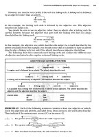
This somewhat debilitated patient came for placement of permanent access. On
removing the dressing covering the Tesio catheters this is what it looks like (Fig.
A.44.1). No signs of infection, and the catheter has worked just fine for now al-
most one year.
Fig. A.43.2.
Fig. A.44.1.
382
Access for Dialysis: Surgical and Radiologic Procedures
AI
Fig. A.45.1.
Comments
The author thinks it makes common sense for the dialysis unit (may it be the
nephrologist, RN, DON, technician) to change/check the catheter dressing, clean
the area, including the catheter; sutures should be removed 10-14 days after catheter
placement.
Case Scenario #45: Malplaced Dual Lumen
catheter
These two elderly ladies share a few problems in addition to advanced age (Fig.
A.45.1-2). Both have left subclavian vein cuffed, tunneled catheters that are
nonfunctioning (lady in Fig. A.45.1 also has a gastrostomy feeding tube). Catheter
383
Appendix I: Fifty Case Reports—Work in Progress
AI
in Figure A.45.1 is a split ash (poorly primed, blood in the red port) (same dialysis
unit as cases 43 and 44) and catheter in Figure A.45.2 is an OptiFlow sutured to the
wing. The long scar leading up to the exit site suggests some unusual surgical tech-
nique at time of placement (Fig. A.45.2). The main problem with these examples is
low and poorly positioned exit sites. This suggests inability to access the central vein
or in this case being occluded due to sucking against the lateral SVC wall (Fig.
A.45.3).
Fig. A.45.2.
384
Access for Dialysis: Surgical and Radiologic Procedures
AI
Comments
Much morbidity and suffering would be prevented by paying attention to a few
basic principles, and details of the steps for the specific procedure to be performed
(skills and knowledge), or is it a matter of attitude?
Consider each catheter placement a life saving procedure. Use micropuncture
technique, portable ultrasound and fluoroscopy. Also, mark the catheter tract and
exit site. The skin moves considerably from supine (head down) to standing, especially
in females and obese individuals (see Chapter 5 for catheter placement techniques).
Case Scenario #46: Catheter Infection
Patient, a 47 year old female, arrives in the office for permanent access place-
ment. The only access is a right IJ tunneled (2 cuff) OptiFlow catheter. She has a
temperature of 102 F, and has chills and sweatening. On exam there are no forearm
cephalic veins, but a possible L upper arm cephalic vein (to be worked up later!)
(Fig. A.46.1). She had not dialyzed in five days. The catheter tract is red, somewhat
tender (arrow) (Fig. A.46.2). The outer cuff is exposed.
Treatment
Patient was urgently taken to dialysis treatment, given 1g vancomycin IVPB at
completion. Then the catheter was removed; did not require any surgical incision.
Infected catheters can usually easily be pulled. One stitch was placed around the
tract at x (Fig. A.46.2) to prevent backbleeding. Blood cultures grew Staph aureus.
Fig. A.45.3.
385
Appendix I: Fifty Case Reports—Work in Progress
AI
Fig. A.46.1.
Fig. A.46.2.
After 48 hours of no fever, clinically improved, wbc = 6.7, a left IJ split ash catheter
was placed. A left arm AV access (Fig. A.46.1) is scheduled in 10-14 days.
Comments
The author strongly advises against suturing the wings to the skin. Only one
suture at exit site is needed, tied around catheter and into the groove remaining
when the wing is removed, i.e., split ash. To all manufacturers of catheters: Remove
the wings? To all surgeons: Do not suture the wing to the skin.
Case Scenario #47: Subclavian Vein Catheter
Patient (69 year old) with a left subclavian (!!) vein catheter as only access (Fig.
A.47.1). There is an old clotted left forearm AV graft. There are scars from previous
chest/neck catheters (arrows) (Fig. A.47.2). Duplex shows right IJ open, right sub-
clavian vein open.
386
Access for Dialysis: Surgical and Radiologic Procedures
AI
Fig. A.47.1.
Fig. A.47.2.
Fig. A.47.3.
387
Appendix I: Fifty Case Reports—Work in Progress
AI
Solution
1. A right forearm radio-cephalic primary AV fistula was created. The proximal
side branch was left intact (arrow) (Fig. A.47.3).
2. A right subclavian vein cuffed, tunneled catheter placed using the micro-
puncture technique, portable ultrasound and fluoroscopy (subcutaneous tract
drawn in Fig. A.47.2).
3. The left IJ cuffed tunneled catheter was removed.
Comments
A most burning question remains; why wasn’t her right cephalic vein used in the
first place; and how did it survive I.V.’s and blood draws for so long?
Case Scenario #48: The Elderly
This 94 year old mentally alert man had a right infected IJ catheter removed.
After one week (3 sessions) of femoral vein catheter dialysis a left IJ was placed, now
working well two weeks later. This visit is for preoperative permanent access place-
ment. His main concern was not to be able to have another catheter should this one
fail. After discussion with him and family we decide to just keep the current cath-
eter. Ten days later he suffered a stroke.
Fig. A.48.1.
388
Access for Dialysis: Surgical and Radiologic Procedures
AI
Comments
The management of the elderly with ESRD is challenging and requires much
common sense. Factors to be considered include: short survival, severe comorbidity,
mental capacity, fragile skin, social and family support. The author initially relies
more on temporary cuffed tunneled catheters.
Case Scenario #49: Osteomyelitis
of Clavicle-Sternal Joint
Patient is a 29 year old diabetic on dialysis for 6 months with a percutaneous
right IJ catheter as only access. She has a hard lump over the clavicle-sternal joint
since ~ 4-6 months after a SCV stick for I.V. access (Fig. A.49.1). She has been on
long term I.V. antibiotics (mostly in ICU). She comes for permanent access (will
need a PTFE graft). After discussion with referring doctor the left IJ was exchanged
with a split ash over a single guidewire technique (Chapter 5, Fig. 5.2). Permanent
access will be placed when infection free (negative blood cultures and normal white
cell count) and stable off antibiotics for >4 weeks.
Comments
A native AV fistula would be most desirable in this setting. Patient has no accept-
able peripheral veins at this time.
Fig. A.49.1.
389
Appendix I: Fifty Case Reports—Work in Progress
AI
Fig. A.50.1.
Case Scenario #50: Malplaced PD Catheter
Young girl with poor drainage of PD catheter, the pigtail was lodged in the right
upper quadrant (Fig. A.50.1).
Solution
The old catheter was removed and a new inserted at the periumbilical transrectus
(arrow) (see chapter 6 for surgical technique).
Comments
The below umbilicus midline incision for PD catheter insertion is suboptimal. It
also tends to misdirect the pigtail catheter into the right or left side of the abdomen,
to be engulfed by the omentum. (Chapter 6).
APPENDIX II
Access for Dialysis: Surgical and Radiologic Procedures, 2nd ed.,
edited by Ingemar J.A. Davidson. ©2002 Landes Bioscience.
Prescription Drug Administration in
ESRD and Dialysis: A Preliminary Guide
Mark Durso
This brief appendix is a guide to altered drug effects in renal failure patients. It is
not meant to be a prescription reference. For specific drug choices and dosing, refer
to your institution’s clinical pharmacist.
Prescription Drug Administration in ESRD and Dialysis
It is important to understand the physiologic and biochemical effects of drugs in
patients with renal disease. A decrease in glomerular filtration rate (GFR) associated
with ESRD impacts the metabolism and clearance of these drugs. To further com-
plicate matters, levels of many drugs are affected by the processes of CAPD or he-
modialysis. This chapter will briefly summarize some of the strategies used in
determining appropriate dosage and drug monitoring, as well as specific adjust-
ments and considerations of many commonly used drugs in a table.
Regardless of renal function status, drug concentration is a function of several
factors. Bioavailability is the rate and amount of a dose to enter systemic venous
circulation to yield the maximum drug concentration in the shortest time. While
drugs administered intravenously retain an essentially normal bioavailability in ESRD
patients, it is orally administered medications that can be affected at multiple levels
in ESRD patients.
Drug absorption across gastrointestinal membranes may be reduced or elimi-
nated in ESRD. Uremic gastrointestinal symptoms, including nausea and vomiting,
may prevent sufficient drug breakdown in the stomach. Enzymatic changes in ure-
mia, including those in intestinal epithelia and salivary urea may metabolize drug
compounds too early, interfere with acid hydrolysis or chelate active compounds.
Gastroparesis lengthens the time a drug remains in the stomach, which delays
bioavailability, while diarrhea shortens the intestinal contact time, reducing or elimi-
nating absorption by the small bowel.
Drugs metabolized or changed by the liver may be impacted by these gastrointes-
tinal factors, by diminished compound binding to the plasma protein or by de-
creased transformation of the drug on arrival to the liver, resulting in larger amounts
of nonmetabolized drug being cleared by the portal system, effectively diminishing
potential bioavailability.
Drug distribution may be highly variable in ESRD patients. Distribution is the
amount of dispersal of drug over a given time, and is a factor of drug amount,
plasma concentration and solubility factors. Protein-bound drugs are water soluble,
and demonstrate a narrow distribution in the extracellular fluid space. This can be
affected by patient fluid status (edema vs dehydration), ascites and muscle wast-
ing. Decreased binding of these drugs can occur in ESRD with low albumin
391Appendix II: Prescription Drug Administration in ESRD and Dialysis
AII
concentrations, resulting in higher amounts of unbound circulating compound,
which may be quickly eliminated or accumulate to toxic levels.
The overall drug metabolism and elimination is affected by ESRD. Drug end
products may be exclusively cleared by the kidney, representing a potentially toxic
metabolite build up with a regularly administered drug.
Calculations for Drug Dosing and Removal by Dialysis
Calculating creatinine clearance is the first step in safe and effective drug dosing
in patients with renal failure. There are many formulae for estimating creatinine
clearance, including the Cockcroft and Gault formula for patients with a steady
state serum creatinine level:
Creatinine Clearance (CrCl) = (140-age) x IBW (kg)
72 x serum creatinine (mg/dl)
Loading Dose (same for ESRD/nonESRD patients)
This calculation is most useful for drugs with long half lives.
LD = Vd x IBW x Cp
where LD is loading dose; Vd is volume of distribution in litres; and IBW is ideal
body weight in Kg, defined for men as 50Kg + 2.3Kg per inch for each inch above
5ft; and for women as 45.5Kg + 2.3Kg per inch for each inch above 5ft. Cp is
desired plasma concentration of the drug.
Drug Removal by Dialysis
Most drug removal during dialysis occurs by diffusion from plasma across the
dialysis membrane along a concentration gradient. Most drugs > 500D, those >90%
protein bound, or those with large tissue distribution volumes do not cross this
membrane.
The amount of drug removed during dialysis is increased by the use of porous
membranes (i.e., CVVHD, CAVHD), large surface area dialyzers or double dialyz-
ers, an increased blood flow rate, increased dialysate flow rate, or an increased treat-
ment duration.
Estimating Hemodialysis Drug Clearance
CL
HD
= CL
UREA
x (60/molecular weight of drug)
where CL
HD
is the hemodialysis drug clearance; CL
UREA
is the dialyzer urea
clearance (usually 150 ml/min) and (60/molecular weight of drug) is a standard
proportion.
Estimating Peritoneal Dialysis Drug Clearance
The peritoneal membrane is more porous than standard hemodialysis mem-
branes, but is still limited in effectiveness in clearing many drugs. The general rule
of thumb is if a drug is not cleared by hemodialysis, it will not be cleared by perito-
neal dialysis. Peritoneal dialysis is effective in clearing drugs that are not extensively
protein bound, those distributed in extracellular fluid, or with small molecular
weights. The rate and extent of small molecular weight drug removal depends on
the volume of peritoneal dialysate exchanged, as there is a large concentration gradi-
ent between the dialysate and blood. Therefore, concentration is greatly increased in
drugs added to the peritoneal dialysate.
392 Access for Dialysis: Surgical and Radiologic Procedures
AII
Continuous renal replacement therapy, including CAVH, CAVHD and CVVHD
involves the use of a large bore membrane with a lower, continuous blood flow
(15-20 ml/min in CAVHD and CVVHD and 10-30 ml/min in CAVH). Drugs >
80% protein bound are not removed, nor are drugs with a volume of distribution
>0.71/Kg.
393Appendix II: Prescription Drug Administration in ESRD and Dialysis
AII
% Usual Dose % Usual Dose % Usual Dose
for GFR for GFR for GFR
Drug Brand Name Usual Dose > 50 ml/min 10-50 ml/min < 10 ml/min
Immunosuppression
Anti T Cell (poly)
ATG (equine) Atgam 15 mg/kg/d over 4h 100 100 100
Anti T Cell (mono)
Muromonab CD3 OKT3 5 mg/d 100 100 100
Nuc Synthesis Inhib
Azathioprine Imuran 1-3 mg/kg/d 100 75 50
Mycophenolate Mofetil CellCept 1 g bid 100 100 NR
Cytokine Tran Inhib
Cyclosporine A Sandimmune 5 mg/kg bid 100 100 100
Cyclosporine emuls Neoral 5 mg/kg bid 100 100 100
Tacrolimus (FK506) Prograf 0.15-0.30 mg/kg bid 100
Steroids
Methylprednisolone Solumedrol 2 mg/kg/d tapered 100 100 100
Prednisone Deltasone 15 mg/d tapered 100 100 100
394 Access for Dialysis: Surgical and Radiologic Procedures
AII
% Usual Dose % Usual Dose % Usual Dose
for GFR for GFR for GFR
Drug Brand Name Usual Dose > 50 ml/min 10-50 ml/min < 10 ml/min
Antivirals
Acyclovir Zovirax 5 mg/kg q8h 5 mg/kg q8h 5 mg/kg q12-24h 2.5 mg/kg q24h
Ganciclovir Cytovene 5 mg/kg q12h q12h q24-48h q48-96h
CMV hyperimm glob Cytogam 150 mg/kg tapered 28 mg/kg 15 mg/kg 6 mg/kg
Antibiotics
TMP-SMX
160 mg-800 mg Bactrim DS 160/800 q12 160/800 q12 160/800 q18 160/800 q24
PCN + β Lactams
Amoxicillin/clavulanate Augmentin 250-500mg q8 q8h q8-12h q24h
Ticarcillin/clavulanate Timentin 3.1g q4h 100 2g q4-8h 2g q12h
Ampicillin/sulbactam Unasyn 1.5-3g q6 q6 Q8-12 q24
Quinolones
Levofloxacin Levaquin 500mg q24 100 500mg load 500mg load
250mg q24-48 250mg q48
Ciprofloxacin Cipro 250-500mg q12 po 100 50-75 50
Cephalosporins
Cefazolin Ancef 1-2g q8 q8 q12 q24-48
Ceftriaxone Rocephin 250mg-2g q12 100 100 100
395Appendix II: Prescription Drug Administration in ESRD and Dialysis
AII
% Usual Dose % Usual Dose % Usual Dose
for GFR for GFR for GFR
Drug Brand Name Usual Dose > 50 ml/min 10-50 ml/min < 10 ml/min
Aminoglycosides
Amikacin Amikin 7.5mg/kg q12 CONSIDER DOSING BASED ON DRUG LEVELS AND PHARMACO KINETICS
Tobramycin Nebcin 1.7 mg/kg q8 CONSIDER DOSING BASED ON DRUG LEVELS AND PHARMACO KINETICS
Gentamicin Garamycin 1.7 mg/kg q8 CONSIDER DOSING BASED ON DRUG LEVELS AND PHARMACO KINETICS
Other ABX
Vancomycin Vancocin 1g q12 1g q12-24 1g q24-96 1g q4-7days
check levels check levels check levels check levels
Antifungals
Fluconazole Diflucan 200-400mg q24 100 50 50
Ketoconazole Nizoral 200mg q24 100 100 100
Amphotericin B Fungizone 0.4-1.0mg/kg q24 0.4-1.0mg/kg q24 0.4-1.0mg/kg q24 0.4-1.0mg/kg q48
CV Drugs
Ca Chan B
Diltiazem Cardizem 30-90mg q6-8 100 100 100
Nifedipine Procardia 10-20mg q6-8 100 100 100
Verapamil Calan 80mg q8 100 100 100
Amlodipine Norvasc 5mg q24 100 100 100
396 Access for Dialysis: Surgical and Radiologic Procedures
AII
% Usual Dose % Usual Dose % Usual Dose
for GFR for GFR for GFR
Drug Brand Name Usual Dose > 50 ml/min 10-50 ml/min < 10 ml/min
α Adren R-B
Doxazosin Cardura 1-16mg q24 100 100 100
Central α-Adren A
Clonidine Catapres 0.1-0.6mg bid 100 100 100
β Blockers
Metoprolol Lopressor 50-100mg bid 100 100 100
Atenolol Tenormin 50-100mg q24 100% q24 50% q48 30-50% q96
Labetalol (α + β) Normodyne 200-600mg bid 100 100 100
Other CV
Digoxin Lanoxin 1-1.5mg load
0.25-0.5mg q24 100% q24 25-75% q36 10-25% q48
ACE inhibitors
Enalapril Vasotec 5-10mg q12 100 75-100 50
Captopril Capoten 25mg q8 100 75 50
Angiotensin II R-A
Losartan Cozaar 50mg q12 100 100 100
397Appendix II: Prescription Drug Administration in ESRD and Dialysis
AII
% Usual Dose % Usual Dose % Usual Dose
for GFR for GFR for GFR
Drug Brand Name Usual Dose > 50 ml/min 10-50 ml/min < 10 ml/min
Statins (Anti CHO)
Lovastatin Mevacor 20-80mg q24 100 100 100
Pravastatin Pravachol 10-40mg q24 100 100 100
Fluvastatin Lescol 2-10mg q24 100 100 100
Simvastatin Zocor 5-40mg q24 100 100 100
Atorvastatin Lipitor 10-80mg q24 100 100 100
Antiplatelet
Aspirin 650mg q4 q4 q4-6 avoid
Clopidogrel Plavix 75mg QD 100 100 100
Diabetic Agents
Insulin
NPH Humulin N variable 100 75 50
Regular Humulin R
Regular/NPH mix Humulin
Lente 70/30; 50/50
Humulin L
Glyburide Micronase 1.25-20mg q24 no data avoid avoid
Glipizide Glucotrol 2.5-15mg q24 100 50 50
Metformin Glucophage 500-850mg bid 50 25 avoid
398 Access for Dialysis: Surgical and Radiologic Procedures
AII
% Usual Dose % Usual Dose % Usual Dose
for GFR for GFR for GFR
Drug Brand Name Usual Dose > 50 ml/min 10-50 ml/min < 10 ml/min
GI Drugs
H
2
R-A
Famotidine Pepcid 20-40mg qhs 50 25 10
Ranitidine Zantac 150-300mg qhs 75 50 25
Cimetidine Tagamet 400mg bid or
400-800mg qhs 100 50 25
Proton Pump Inhib
Omeprazole Prilosec 20-60mg q24 100 100 100
Lansoprazole 15-60mg q24 100 100 100
Gastric Empty
Metoclopramide Reglan 10-15mg qid 100 75 50
Diuretics
Furosemide Lasix 40-80mg bid 100 100 100
Bumetanide Bumex 1-2mg q8-12h 100 100 100
CNS/Endocrine
Levothyroxine Synthroid 1.0-1.6mcg/kg/day 100 100 100
Phenytoin Dilantin 1000 mg load 100-measure 100-measure 100-measure
300-400mg q24 free levels free levels free levels
399Appendix II: Prescription Drug Administration in ESRD and Dialysis
AII
% Usual Dose % Usual Dose % Usual Dose
for GFR for GFR for GFR
Drug Brand Name Usual Dose > 50 ml/min 10-50 ml/min < 10 ml/min
Vascular Drugs
Warfarin Coumadin 2-10mg q24 100 100 100
Pentoxifylline Trental 400mg tid q8-12 q12-24 q24
LMW heparin Lovenox 1-1.5mg/kg qd 100 Adjustment needed for GFR
<30ml/min
Pain Drugs
Codeine 30-60mg q4-6 100 75 50
Hydrocodone 5-10mg q4-6 100 Dose reduction Dose reduction
may be needed may be needed
Propoxyphene Darvocet 65mg q6-8 100 100 avoid
Oxycodone 5mg q6 100 100 use with caution
Meperidine Demerol 50-100mg q3-4 100 avoid avoid
Morphine 2-10mg q4 100 75 50
NR = not recommended
400 Access for Dialysis: Surgical and Radiologic Procedures
AII
Dose
Adjustment Drug Cleared Drug Cleared
Drug for HD by CAPD by CRRT
Immunosuppression
Anti T cell (poly)
ATG (equine) dose after hd n/a n/a
Anti T Cell (mono)
Muromonab CD3 no no no
Nuc Synthesis Inhib
Azathioprine yes unk use GFR 10-50
Mycophenolate no no unk
Mofetil
Cytokine Tran Inhib
Cyclosporine A no no 100%
Cyclosporine emuls no no 100%
Tacrolimus (FK506) no no unk
Steroids
Methylprednisolone yes unk GFR 10-50
Prednisone no unk GFR 10-50
Antivirals
Acyclovir dose after hd use GFR<10 3.5 mg/kd/d
Ganciclovir dose after hd use GFR<10 2.5 mg/kd/d
CMV hyperimm glob dose after dialysis use GFR<10 use GFR 10-50
Antibiotics
TMP-SMX
160 mg-800 mg dose after hd 160/800 q24 160/800 q18
PCN + β Lactans
Amoxicillan/clavulanat dose after dialysis 250 mg q12 NA
Ticarcillin/clavulanate 3.1g after dialysis use GFR<10 use GFR 10-50
Ampicillin/sulbactam dose after hd 1.5-3g q24 2.25g q12
Quinolones
Levofloxacin dose gfr<10 dose GFR<10 dose GFR 10-50
Ciprofloxacin 250mg q12 250mg q8 200mg iv q12
Cephalosporins
Cefazolin 0.5-1.0g after hd 0.5g q12 use GFR 10-50
Ceftriaxone dose after hd 750mg q12 use GFR 10-50
401Appendix II: Prescription Drug Administration in ESRD and Dialysis
AII
Dose
Adjustment Drug Cleared Drug Cleared
Drug for HD by CAPD by CRRT
Aminoglycosides
Amikacin Consider dosing based on drug levels and pharmaco kinetics
Tobramycin Consider dosing based on drug levels and pharmaco kinetics
Gentamicin Consider dosing based on drug levels and pharmaco kinetics
Other ABX
Vancomycin use gfr<10 use GFR<10 500 q24-48 check
levels
Antifungals
Fluconazole 200mg after hd use GFR<10 use GFR 10-50
Ketoconazole no no no
Amphotericin B no use GFR<10 use GFR 10-50
CV Drugs
Ca Chan B
Diltiazem no no use GFR 10-50
Nifedipine no no use GFR 10-50
Verapamil no no use GFR 10-50
Amlodipine no no use GFR 10-50
α Adren R-B
Doxazosin no no use GFR 10-50
Central α-Adren A
Clonidine no no use GFR 10-50
β Blockers
Metoprolol 50mg no use GFR 10-50
Atenolol 25-50mg no use GFR 10-50
Labetalol (α+β) no no use GFR 10-50
Other CV
Digoxin no no use GFR 10-50
ACE Inhibitors
Enalapril 20-25 no use GFR 10-50
Captopril 25-30 no use GFR 10-50
Angiotensin II R-A
Losartan no data no data use GFR 10-50
Statins (Anti CHO)
Lovastatin no data no data use GFR 10-50
Pravastatin no data no data use GFR 10-50
Fluvastatin no data no data use GFR 10-50
Simvastatin no data no data use GFR 10-50
Atorvastatin no data no data use GFR 10-50
402 Access for Dialysis: Surgical and Radiologic Procedures
AII
Dose
Adjustment Drug Cleared Drug Cleared
Drug for HD by CAPD by CRRT
Antiplatelet
Aspirin after hd no use GFR 10-50
Clopidogrel no data no data no data
Diabetic Agents
Insulin
NPH no no dose GFR 10-50
Regular
Regular/NPH mix
Lente
Glyburide no no avoid
Glipizide no data no data avoid
Metformin no data no data avoid
GI Drugs
H
2
R-A
Famotidine no no dose GFR 10-50
Ranitidine 1/2 dose no dose GFR 10-50
Cimetidine no no dose GFR 10-50
Proton Pump Inhib
Omeprazole no data no data no data
Lansoprazole no data no data no data
Gastric Empty
Metoclopramide no no data dose GFR 10-50
Diuretics
Furosemide no no no
Bumetanide no no no
CNS/Endocrine
Levothyroxine no no no data
Phenytoin no no no
Vascular Drugs
Warfarin no no no
Pentoxifylline no data no data no data
LMW heparin contraindicated contraindicated contraindicated
Pain Drugs
Codeine no data no data use GFR 10-50
Hydrocodone no data no data no data
Propoxyphene avoid avoid n/a
Oxycodone no data no data no data
Meperidine avoid avoid avoid
Morphine no data no data use GFR 10-50
CHAPTER 1
APPENDIX III
Access for Dialysis: Surgical and Radiologic Procedures, 2nd ed.,
edited by Ingemar J.A. Davidson. ©2002 Landes Bioscience.
Access for Dialysis:
A Preoperative Guide for Patients
and Their Families
Ingemar J.A. Davidson, Wilson V. Garrett, Angela Kuhnel,
Carolyn E. Munschauer and Gregory J. Pearl
This written summary is given to the patient to read and take home. A signed
copy is kept in the patient’s office record.
Background
You have been diagnosed with kidney (renal) failure. In medical textbooks this
diagnosis is often labeled as “end stage renal disease (ESRD)”. Most likely you were
referred to me, the surgeon, by an internal medicine doctor specializing in kidney
disease (nephrologist). You may have known about your failing kidneys for a long
time, or it may have come as a surprise just recently. The most common causes of
kidney failure are: diabetes, high blood pressure, infection and polycystic kidney
disease.
Your kidneys help remove waste and toxins from your body, and also help make
healthy red blood cells. If your kidneys stop working, these functions can be re-
placed by dialysis. If you do not have dialysis, the toxins will stay in your body and
make you very sick.
Kidney function is measured from your blood as the concentration of creatinine
(normal serum creatinine is 0.5 - 1.2 mg/dl). A better way is a test called creatinine
clearance, or how much blood your kidney can clean per minute. The normal value
is 100 ml/min or greater. When this number drops to about 10 ml/min (which
corresponds to 10% of your normal kidney function),you will need dialysis to live.
The signs of kidney failure which show you may be close to needing dialysis are:
nausea, headache, anemia, swelling (edema) and high blood pressure. Most patients
also feel exhausted or tired. Not every patient has all these symptoms, but usually
have at least one or two.
Your case is unique. Even though it may sound like all patients with kidney
failure needing dialysis are similar, this is far from the truth. Because there are many
different causes of renal failure, the treatment and the management needed is often
very different for different patients. Today we will be discussing your unique diagnosis
and treatment.
The Treatment of Kidney Failure
In general, there are several options that can treat renal failure, depending on
your special circumstance. Below are outlined the various treatments and I will ex-
plain to you where you best fit in.
404 Access for Dialysis: Surgical and Radiologic Procedures
AIII
Kidney Transplantation
Kidney transplantation from a relative, a friend or from a cadaver organ donor
(from a dead person) is the only definitive treatment of kidney failure. This can be
done before you need dialysis or at any time thereafter. Because there are not enough
kidneys available for everybody who needs one, only a fraction of all kidney failure
patients on dialysis get transplanted. You will need special testing to determine if
you are healthy enough for the transplant. Many patients are afraid they do not have
enough money for a transplant, but Medicare or your insurance usually will pay for
it. If you want more information on kidney transplant, ask me or your kidney doctor.
Dialysis
There are two types of dialysis: 1. hemodialysis and 2. peritoneal dialysis. Hope-
fully, before you see your surgeon, you have discussed your best options with your
nephrologist. The surgical team can also advise you on which type may be best for
you, but we concentrate on the technical aspect of your surgery necessary for your
dialysis to take place.
Dialysis is sometimes called an artificial kidney, because a machine or filter takes
the place of your kidneys. Before you can start dialysis you need surgery to create a
way for dialysis to happen. This is called dialysis access, and will be your “lifeline”
for the rest of your life. That is why you have come to see me.
Hemodialysis
Hemodialysis means that your blood is pumped through a filter to clean waste
products from your blood. Waste products build up from the things you eat, drink
and medication you take. The dialysis machine moves your blood quickly through
the filter and returns it to your body after it is clean. This requires a tube to remove
the blood from your body and then return the blood to your system after cleansing.
Your veins are not big enough to move blood fast enough. Vascular access surgery
creates a site for insertion of the tube that removes and returns your blood. Depend-
ing on your special circumstances, there are three different ways this can be achieved.
First, in urgent situations a catheter can be inserted in a vein in your neck or groin.
Second, most ideally, 6-12 months before you need dialysis, a vein and artery in
your arm are surgically connected together, allowing the vein to grow. During dialy-
sis, two needles will be placed in your arm; one needle removes blood, where it flows
through the dialysis machine and is returned through the second needle back into
your body. This is called a fistula. Third, if you have no suitable vein of your own, if
your veins are too small or scarred to grow big, an artificial blood vessel the size of a
plastic straw is surgically placed under your skin in the arm and connected to your
artery and a vein. This allows for needles to be placed in the same way as a fistula,
allowing blood to go from you to the machine and back. This artificial blood vessel
(graft) requires 2-4 weeks before it can be used. If you need urgent dialysis, both a
catheter in your neck and another surgery (fistula or graft) may be required at the
same time.
Most dialysis surgery can be done as an outpatient surgery. You will go to the day
surgery unit at the hospital. Surgery nurses will get you ready for the operation. An
anesthesiologist will most likely inject anesthesia into your armpit to completely put
your arm to sleep. You will also be getting IV drugs to calm you down. Occasionally,
general anesthesia is needed.
405Appendix III: A Preoperative Guide for Patients and Their Families
AIII
After surgery, you will return to the day surgery unit. You will be given writen
post surgery instructions. You may need a pain killer for 2-3 days. Most impor-
tantly, you must elevate your arm and exercise by making fists around a soft ball.
Also, in cases of graft placement, an ice pack may be given to be placed over the
surgical area for the next 6-12 hours. If you have questions or problems after your
surgery, you will be given my office phone number to call. As your access surgeon,
you can see me whenever you have access problems or concerns, before or after you
start dialysis. You can call me yourself, or ask your dialysis nurses to call and explain
the problem. You have a choice of the doctors who care for you before and after you
start dialysis.
Your kidney doctor (nephrologist) may choose for you to start dialysis in the
hospital, where you will be closely monitored for the first 2-3 days of dialysis. After
that, you will go to an outpatient dialysis center 3 days a week.
Peritoneal Dialysis
The second major type of dialysis treatment is called peritoneal dialysis. In this
case, a plastic tube is surgically placed inside your abdominal cavity and exited through
your abdominal wall. Part of the tube is outside your body. Once the tube heals,
nurses teach you how to put fluid in and take it out of your abdomen through the
tube (exchange). The tissue covering your internal organs acts like a dialysis filter
and helps pull waste products into the fluid. When you remove the fluid you re-
move the waste. Some patients can do their exchanges at night by using a special
machine. Some patients need to do their exchanges during the day. You have to
learn some simple technical procedures from the nurses before you start, because
you are doing your own dialysis. Many patients like peritoneal dialysis because they
have more freedom and do not have to go to the dialysis unit. This type of dialysis
requires some basic technical abilities on your part, but offers more freedom and
avoids needle sticks and long hours by the dialysis machine in the dialysis unit.
However, it requires much greater responsibility by you, the patient to do the ex-
changes as they are ordered by your nephrologist.
The more you know about your condition and treatment options, the better
decisions you can make. You are encouraged to ask questions about your specific
kidney disease, and surgical and dialysis issues. You are likely to do better if you have
a more complete understanding and participate in the decision making. We encour-
age to involve family members as well.
I have read this guide and discussed it with my surgeon. I have been told I can
ask questions about my condition and my care. I understand the answers my sur-
geon has given me. I know that this surgery is necessary for starting dialysis. I under-
stand that this operation, like all surgery, has risks. My surgeon has reviewed these
risks with me.
Dallas, ________ ________ ________
month day year
________________________________ ________________________________
patient name signature surgeon name signature (or alternate)
________________________________ ________________________________
patient name printed surgeon name printed (or alternate)