Pediatric Epilepsy Diagnosis and Therapy - part 5 docx
Bạn đang xem bản rút gọn của tài liệu. Xem và tải ngay bản đầy đủ của tài liệu tại đây (1.32 MB, 92 trang )
345
23 • BENIGN FOCAL EPILEPSIES OF CHILDHOOD
the condition being a multifocal rather than a purely
occipital epilepsy.
Diagnostic Evaluation and Differential Diagnosis. Panayi-
otopoulos syndrome is frequently mistaken for nonepileptic
disorders and occasionally for other types of epilepsy. This
reflects its unusual ictal clinical features and, for a benign
epilepsy, its somewhat unusual interictal EEG features.
Because of the prolonged nature of seizures in Pan-
ayiotopoulos syndrome, many children present with it
to emergency departments while they are still in an ictal
state. However, if the main features of this are impaired
consciousness and vomiting, an epileptic state may not
even come into the differential diagnosis. Conditions such
as encephalitis and meningitis are often considered. If the
ictus terminates in a hemi- or generalized convulsion, this
may merely strengthen the presumptive (but erroneous)
diagnosis. Many such children end up intubated and
treated in pediatric intensive care units with antibiotics
and antiviral agents. The prolonged seizures of Panayioto-
poulos syndrome may also be confused with acute confu-
sional migraine and, if vomiting is particularly prominent,
with cyclical vomiting syndrome or gastroenteritis. Some
seizures may simply be dismissed as travel sickness.
The EEG of Panayiotopoulos syndrome may be sim-
ilar or identical to that of idiopathic childhood occipital
epilepsy or rolandic epilepsy, and these conditions may be
mistakenly diagnosed if the clinical history is ignored (74).
More importantly, multifocal spike discharges and cloned-
like repetitive multifocal spike wave complexes may
suggest more malignant epilepsies such as the Lennox-
Gastaut syndrome, although clinically these conditions
are completely different.
Unlike the other conditions described in this chap-
ter, children with Panayiotopoulos syndrome commonly
present to emergency departments while still seizing or
in the immediate postictal period. Panayiotopoulos syn-
drome should be considered in the differential diagnosis
of all previously well young children, especially those
between the ages of 3 and 6 years, who have rapid onset
of emetic symptoms followed by impaired (often fluctuat-
ing) consciousness. Eye or head deviation may be a useful
finding. However, it may still be appropriate to manage
the child for a suspected encephalopathy.
In the office setting, if Panayiotopoulos syndrome
is suspected from the history, the most useful investiga-
tion is likely to be the EEG (including sleep if necessary).
Symptomatic epilepsies may mimic Panayiotopoulos syn-
drome, so even if the history is typical, most authorities
recommend neuroimaging. However, if MRI will require
sedation or general anesthetic, CT may be appropriate.
No other investigations are required.
Treatment. A recent consensus statement concluded that
regular prophylactic AED medication was probably best
reserved for children whose seizures were unusually fre-
quent, distressing, or otherwise significantly interfering
with the child’s life (54). There are no high quality stud-
ies of what treatment is most appropriate. Carbamaze-
pine and sodium valproate appear equally efficacious.
Given the benign nature of the condition, it is particu-
larly important to avoid adverse effects. Withdrawal of
treatment after 1 or 2 seizure-free years is appropriate.
The EEG is not helpful in deciding when to withdraw
medication. Whether these recommendations will stand
if it is confirmed that seizures in Panayiotopoulos syn-
drome can be associated with cardiorespiratory arrest
remains to be seen.
Course and Prognosis. Total seizure count in Panayioto-
poulos syndrome is usually low. Around one-third of patients
have a single seizure and only 5% to 10% will have more
than 10; sometimes seizures are very frequent. The duration
of active seizures is short; remission usually occurring within
1 to 2 years from onset. About one-fifth of subjects with
Panayiotopoulos syndrome will have one or more seizures
typical of one of the other benign focal epilepsies of child-
hood, especially rolandic epilepsy (50, 53). However, the
likelihood of seizures in adult life is probably no greater than
in the general population.
Idiopathic Childhood Occipital Epilepsy
(Late-Onset Childhood Occipital Epilepsy—
Gastaut Type and Idiopathic Photosensitive
Occipital Lobe Epilepsy)
Introduction and Definition. Idiopathic childhood occipi-
tal epilepsy (5, 52, 75–80) with and without photosen-
sitivity was first established as an epileptic syndrome by
Gastaut (75, 76). Recently, such subjects have generally
been classified separately by the ILAE Task Force as late-
onset childhood occipital epilepsy—Gastaut type and
idiopathic photosensitive occipital lobe epilepsy (3). The
likelihood of remission in these syndromes is considerably
less than it is for rolandic epilepsy and Panayiotopoulos
syndrome. Their inclusion in this chapter could reason-
ably be questioned. However, it is convenient to consider
them here because undoubtedly some children with these
conditions remit completely.
Idiopathic childhood occipital epilepsy can be
defined as an idiopathic focal seizure disorder of child-
hood manifested mainly by elementary visual seizures
and ictal blindness, which are often frequent and usu-
ally occur without impairment of consciousness. EEG
shows occipital epileptiform abnormalities, particularly
so-called occipital paroxysms. Idiopathic photosensitive
occipital epilepsy is an idiopathic focal seizure disorder
mainly of childhood manifested mainly by elementary
visual seizures provoked by various forms of environ-
mental light stimulation. EEG shows occipital or generalized
346
III • AGE-RELATED SYNDROMES
photoparoxysmal responses to intermittent photic stimu-
lation and often spontaneous, mainly occipital, epilepti-
form abnormalities.
Epidemiology. Idiopathic childhood occipital epilepsy is
reported as starting in children as young as 3 years of age
and as old as 15 years of age. Peak age of onset is around
8 years. Boys and girls are equally affected. Idiopathic
photosensitive occipital epilepsy may start as early as the
second year of life or as late as young adult life. However,
it peaks at around 12 years of age. There is probably a
slight female preponderance, but nowhere near as great
as for photosensitivity per se. Both these epilepsies are
rare. Panayiotopoulos estimated that idiopathic child-
hood occipital epilepsy accounted for about 2–7% of all
benign focal epilepsies of childhood (78).
Clinical Manifestations. In both these syndromes the
seizures are most characteristically manifested with ele-
mentary visual hallucinations. These usually consist of
small multicolored circular patterns (79). Often they are
reported as arising unilaterally in the periphery of a visual
field, becoming larger and multiplying as the seizure pro-
gresses. They may move horizontally across the visual
field, and other more complex movements are described.
In some subjects normal vision is obscured by the halluci-
nations; in others it is retained. More complex visual hal-
lucinations, such as of formed shapes and faces, and visual
illusions may also occur but are much less common. Visual
illusions include distortions of shape and distance.
After elementary visual hallucinations, ictal blind-
ness is the second most common visual manifestation
of seizures in these syndromes. It usually involves both
visual fields but may be unilateral or involve only part
of a hemifield. The subject usually reports everything as
black, but occasionally everything goes white. Ictal blind-
ness is usually an initial manifestation of the seizure but
may follow visual hallucinations.
Other ictal ocular symptoms are relatively common.
Some subjects report sensations of their eyes being tugged
or of ocular pain. Eye deviation, often with simultaneous
head deviation, is also common, possibly occurring in
about 70% of cases. It usually follows after the hallucina-
tions begin, although the latter may persist. Forced eye clo-
sure and eyelid blinking are other reported phenomena.
Most seizures are short lived, many lasting only a
few seconds. However, some last a matter of minutes.
Seizures with ictal blindness often last longer. Occasion-
ally, seizures (including those with blindness) can last for
hours (status amauroticus).
There is a particularly strong association between
seizures in these epilepsies and headache. This can be
an ictal or postictal phenomenon, although the latter
is more common. It often has a migrainous character.
Indeed, it is likely that in many cases the seizure provokes
a true migraine. In idiopathic photosensitive occipital
lobe epilepsy, seizure symptomatology may also include
autonomic features, including emesis, which characterizes
Panayiotopoulos syndrome (77, 81).
Consciousness is preserved during most seizures but
occasionally may become impaired. This often precedes
secondary generalization with GTCS. In exceptional
cases spread to cause temporal lobe type symptoms is
reported.
In idiopathic childhood occipital lobe epilepsy sei-
zures are mainly diurnal and are usually quite frequent
(often several each day or week). Occasional nocturnal
seizures, often with hemiconvulsions or GTCS, are not
infrequent.
In idiopathic photosensitive occipital lobe epilepsy,
seizures are provoked by light factors. Video-game playing
appears to be the most provocative, followed by watch-
ing TV. Some subjects are very photosensitive, and this
is likely to be reflected in a high seizure frequency. Other
subjects are less photosensitive and may have very few
seizures. However, spontaneous seizures may also occur.
It is also reported that some subjects with this epilepsy
have other seizure types such as absences and myoclonic
jerks provoked by photic factors.
EEG Features (5, 52, 75–82). The interictal EEG in both
idiopathic childhood occipital epilepsy and idiopathic
photosensitive occipital epilepsy is expected to have a
normal background. In the former, occipital paroxysms
are characteristic. However, in some subjects only isolated
occipital spikes may be seen. Extraoccipital paroxysmal
abnormalities may occur, but are much less common than in
Panayiotopoulos syndrome. In some subjects EEG abnor-
malities may only be seen in sleep; occasionally both awake
and sleep EEGs may be consistently normal. Figure 23-5
illustrates occipital paroxysms and fixation-off sensitivity.
The ictal EEG is expected to show attenuation of
occipital paroxysms followed by appearance of an occipi-
tal discharge of fast rhythms, fast spikes, or both.
In idiopathic occipital lobe epilepsy there may be
no spontaneous epileptiform abnormalities or else there
may be occipital spikes or paroxysms. Extraoccipital epi-
leptiform abnormalities may also be seen. Intermittent
photic stimulation will, in all subjects, show occipital or
generalized photoparoxysmal responses.
Diagnostic Evaluation and Differential Diagnosis. These
syndromes, like all occipital epilepsies, are very prone
to misdiagnosis as migraine. In part, this is understand-
able, because headache, often migrainous, as previously
described, is common both ictally and postictally. How-
ever, the elementary visual hallucinations are unlike those
of migraine. In the latter they tend to be black and white,
rather than colored, and have jagged or sharp contours
rather than being predominantly rounded.
347
23 • BENIGN FOCAL EPILEPSIES OF CHILDHOOD
These syndromes may mimic symptomatic occipi-
tal lobe epilepsies, and neuroimaging, preferably MRI,
is indicated. No other investigations, except EEG, are
routinely required.
Treatment. Given the frequency of seizures in idiopathic
childhood occipital epilepsy, including the likelihood of
occasional GTCS, regular AED treatment is considered
necessary in most if not all subjects. There are no con-
trolled studies comparing alternatives, although carba-
mazepine appears to be most often used in subjects who
are not photosensitive. It is appropriate to attempt with-
drawal after two seizure-free years, although there is a
significant risk of relapse.
Some subjects with idiopathic photosensitive
occipital lobe epilepsy who are only mildly photosen-
sitive and who do not have spontaneous seizures can
remain seizure free by avoiding precipitants. Others
will require AED treatment. Broad spectrum agents,
such as sodium valproate and levetiracetam, active
against focal and generalized seizures and photosensi-
tivity, would appear to be reasonable choices. However,
it appears that carbamazepine, not usually considered
a useful drug for photosensitivity, may sometimes be
effective.
Course and Prognosis. The prognosis for both idio-
pathic childhood occipital epilepsy and idiopathic pho-
tosensitive occipital lobe epilepsy is variable. A majority
of the former, perhaps 50% to 60%, have remission of
seizures within 2–4 years of them starting. However, in a
significant minority seizures will continue into adulthood.
In those with idiopathic photosensitive occipital lobe epi-
lepsy who are only mildly photosensitive and can control
their exposure to relevant provoking factors, freedom
from seizures may be easy. For others, particularly those
who are highly photosensitive, the likelihood of seizures
continuing into adult life is high.
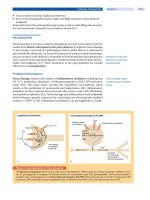
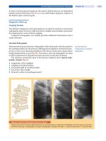
FIGURE 23-5
Occipital paroxysms with fixation off sensitivity of an 11-year-old boy with idiopathic childhood occipital epilepsy. Occipital
paroxysms occur immediately after and as long as fixation and central vision are eliminated by any means (eyes closed, dark-
ness, plus 10 spherical lenses, Ganzfeld stimulation). Under these conditions, even in the presence of light, eye opening does
not inhibit the occipital paroxysms. Conversely, occipital paroxysms are totally inhibited by fixation and central vision. Symbols
of eyes open without glasses indicate conditions in which fixation is possible. Symbols of eyes with glasses indicate conditions
in which central vision and fixation are eliminated. At age 18 years, he is entirely normal and is not receiving medication.
348
III • AGE-RELATED SYNDROMES
Atypical Evolutions of the Benign Focal
Epilepsies of Childhood
Less than 1% of children with rolandic epilepsy have
so-called atypical evolutions (24, 32, 83). These include
the development of severe linguistic, cognitive, or behav-
ioral problems. If such problems develop in a child with
rolandic epilepsy, a sleep EEG should be obtained,
because continuous spike-and-wave during slow-wave
sleep (CSWS) may be present. The Landau-Kleffner syn-
drome is sometimes said to develop from rolandic epi-
lepsy. CSWS may also be seen in children with opercular
status characterized by continuous positive or negative
myoclonias around the mouth or elsewhere in the face
and pseudobulbar problems. Atypical focal epilepsy of
childhood in which other seizure types, including tonic
and atypical absence seizures, occur may also develop in
children with otherwise typical rolandic epilepsy.
There are also case reports of atypical evolutions in
Panayiotopoulos syndrome, including the development of
absences and drop attacks (32, 84, 85) and in idiopathic
childhood occipital epilepsy with cognitive deterioration
and CSWS (86).
Carbamazepine is sometimes implicated in precipi-
tating such atypical evolutions (87, 88).
Other Described Benign Focal
Epilepsies of Childhood
The syndromes discussed previously are the only benign
focal epilepsies of childhood currently recognized by
the ILAE. However, others have been proposed and
are more or less well characterized. They include the
following.
Benign Childhood Seizures with Affective Symptoms (89).
This is reported to have its onset between 2 and 9 years
of age and is characterized by multiple, usually short,
daytime and nighttime seizures in which the predomi-
nant symptom appears to be fear or terror, accompanied
by autonomic disturbances (pallor, sweating, abdominal
pain, and salivation), arrest of speech, and mild impair-
ment of consciousness with automatisms. Interictal EEG
shows sharp and slow wave complexes similar to those
in rolandic epilepsy but located in the frontotemporal
and parietotemporal electrodes. Remission in 1 to 2 years
from onset is expected. This is likely to be an intermedi-
ate phenotype between Panayiotopoulos syndrome and
rolandic epilepsy.
Benign Childhood Epilepsy with Parietal Spikes and Frequent
Giant Somatosensory Evoked Potentials (28, 90). This
putative disorder is mainly defined by its interictal EEG
features reflected in its name. These features are, however,
said to often be associated with a phenotype characterized
by mainly daytime versive seizures, which are infrequent
and have an excellent prognosis.
Benign Childhood Focal Seizures Associated with Frontal
or Midline Spikes (5). Again this putative disorder is
mainly defined by its interictal EEG features. These EEG
features can be seen in children with febrile seizures,
rolandic epilepsy, Panayiotopoulos syndrome, and idio-
pathic childhood occipital epilepsy.
Benign Focal Epilepsy in Infants with Central and Vertex
Spikes and Waves During Sleep (91, 92). Benign focal
epilepsy in infants with central and vertex spikes and
waves during sleep has been recently described as a new
benign syndrome. In terms of age of onset, it is on the
borderline between benign infantile seizures and Panayio-
topoulos syndrome. Age at onset is in the first 2 years of
life with both sexes equally affected. Infants are normal
and all tests other than EEG are normal. Seizures consist
mainly of staring, motor arrest, facial cyanosis, loss of
consciousness, and stiffening of the arms. Clonic con-
vulsions and automatisms are rare. Duration is from 1
to 5 minutes. Seizures are mainly diurnal (but may also
occur during sleep) and may occur in clusters, but are gener-
ally infrequent (1–3 per year). Interictal EEG abnormali-
ties are seen only in non-REM sleep and consist of small,
mostly singular, spikes and waves localized at the vertex
and central electrodes.
There is a strong family history of epilepsy with
benign epilepsies prevailing. The prognosis is excellent
with remission of seizures, normal development, and nor-
malization of the EEG before the age of 4 years.
Benign Focal Seizures of Adolescence (5, 93). This
syndrome of the second decade, and predominantly
occurring in males, features a single seizure or a single
cluster of seizures over a period of up to 36 hours. The
seizures are mainly diurnal, with consciousness initially
preserved. The main manifestations are focal clonic jerk-
ing, usually without a Jacksonian march, and somatosen-
sory symptoms. Secondary GTCS occur in about 50%
of cases. EEG and brain neuroimaging are normal. The
prognosis is excellent and treatment is not required.
References
1. Commission on Classification and Terminology of the International League Against Epi-
lepsy. Proposal for revised classification of epilepsies and epileptic syndromes. Epilepsia
1989; 30:389–399.
2. Geelhoed M, Boerrigter AO, Camfield P, Geerts AT, et al. The accuracy of outcome
prediction models for childhood-onset epilepsy. Epilepsia 2005; 46:1526–1532.
3. Engel J Jr. A proposed diagnostic scheme for people with epileptic seizures and with
epilepsy: Report of the ILAE Task Force on Classification and Terminology. Epilepsia
2001; 42:796–803.
4. Riikonen R. Long-term outcome of patients with West syndrome. Brain Dev 2001;
23:683–687.
349
23 • BENIGN FOCAL EPILEPSIES OF CHILDHOOD
5. Panayiotopoulos CP. Benign childhood focal seizures and related epileptic syndromes. In:
Panayiotopoulos CP, ed. The Epilepsies: Seizures, Syndromes and Management. Oxford:
Bladon Medical Publishing, 2005:223–269.
6. Panayiotopoulos CP. Benign childhood partial epilepsies: benign childhood seizure sus-
ceptibility syndromes [editorial]. J Neurol Neurosurg Psychiatry 1993; 56:2–5.
7. Vadlamudi L, Harvey AS, Connellan MM, Milne RL, et al. Is benign rolandic epilepsy
genetically determined? Ann.Neurol 2004; 56:129–132.
8. Bray PF,.Wiser WC. Evidence for a genetic etiology of temporal-central abnormalities in
focal epilepsy. N Engl J Med 1964; 271:926–933.
9. Heijbel J, Blom S, Rasmuson M. Benign epilepsy of childhood with centrotemporal EEG
foci: a genetic study. Epilepsia 1975; 16:285–293.
10. Neubauer BA, Hahn A, Stephani U, Doose H. Clinical spectrum and genetics of Rolandic
epilepsy. Adv Neurol 2002; 89:475–479.
11. Neubauer BA, Fiedler B, Himmelein B, Kampfer F, et al. Centrotemporal spikes in
families with rolandic epilepsy: linkage to chromosome 15q14. Neurology 1998;
51:1608–1612.
12. Scheffer IE, Berkovic SF. The genetics of human epilepsy. Trends Pharmacol Sci 2003;
24:428–433.
13. Gutierrez-Delicado E, Serratosa JM. Genetics of the epilepsies. Curr Opin Neurol 2004;
17:147–153.
14. Hirose S, Mitsudome A, Okada M, Kaneko S. Genetics of idiopathic epilepsies. Epilepsia
2005; 46 Suppl 1:38–43.
15. Coppola G, Castaldo P, Miraglia DG, Bellini G, et al. A novel KCNQ2 Kϩ channel mutation
in benign neonatal convulsions and centrotemporal spikes. Neurology 2003; 61:131–134.
16. Berkovic SF, Heron SE, Giordano L, Marini C, et al. Benign familial neonatal-infantile sei-
zures: characterization of a new sodium channelopathy. Ann Neurol 2004; 55:550–557.
17. Roll P, Massacrier A, Pereira S, Robaglia-Schlupp A, et al. New human sodium/glucose
cotransporter gene (KST1): identification, characterization, and mutation analysis in
ICCA (infantile convulsions and choreoathetosis) and BFIC (benign familial infantile
convulsions) families. Gene 2002; 285:141–148.
18. Guerrini R, Bonanni P, Nardocci N, Parmeggiani L, et al. Autosomal recessive rolan-
dic epilepsy with paroxysmal exercise-induced dystonia and writer’s cramp: delineation
of the syndrome and gene mapping to chromosome 16p12-11.2. Ann Neurol 1999;
45:344–352.
19. Panayiotopoulos CP. Benign childhood epilepsy with centrotemporal spikes or Rolandic
seizures. In: Panayiotopoulos CP, ed. Benign Childhood Partial Seizures and Related
Epileptic Syndromes. London: John Libbey & Company, 1999:33–100.
20. Dalla Bernardina B, Sgro M, Fejerman N. Epilepsy with centro-temporal spikes and related
syndromes. In: Roger J, Bureau M, Dravet C, Genton P, et al, eds. Epileptic Syndromes in
Infancy, Childhood and Adolescence. 4th ed. Montrouge, France: John Libbey Eurotext,
2005:203–225.
21. Beaussart M, Loiseau P, Roger H. The discovery of “benign rolandic epilepsy.” In:
Berkovic SF, Genton P, Hirsch E, Picard F, eds. Genetics of Focal Epilepsies. London:
John Libbey & Company, 1999:3–6.
22. Lombroso CT. Sylvian seizures and midtemporal spike foci in children. Arch Neurol
1967; 17:52–59.
23. Bouma PA, Bovenkerk AC, Westendorp RG, Brouwer OF. The course of benign partial
epilepsy of childhood with centrotemporal spikes: a meta-analysis. Neurology 1997;
48:430–437.
24. Fejerman N, Caraballo R, Tenembaum SN. Atypical evolutions of benign localization-
related epilepsies in children: are they predictable? Epilepsia 2000; 41:380–390.
25. Gregory DL,.Wong PK. Clinical relevance of a dipole field in rolandic spikes. Epilepsia
1992; 33:36–44.
26. Yoshinaga H, Amano R, Oka E, Ohtahara S. Dipole tracing in childhood epilepsy with
special reference to rolandic epilepsy. Brain Topogr 1992; 4:193–199.
27. De Marco P, Tassinari CA. Extreme somatosensory evoked potential (ESEP): an
EEG sign forecasting the possible occurrence of seizures in children. Epilepsia 1981;
22:569–575.
28. Fonseca LC, Tedrus GM. Somatosensory evoked spikes and epileptic seizures: a study of
385 cases. Clin Electroencephalogr
2000; 31:71–75.
29. Gibbs F A, Gibbs EL. Atlas of electroencephalography. Vol 2: Epilepsy. Reading, MA:
Addison-Wesley, 1952:214–290.
30. Smith JMB, Kellaway P. Central (Rolandic) foci in children: an analysis of 200 cases.
Electroencephalogr Clin Neurophysiol 1964; 17:460–461.
31. Eeg-Olofsson O. The development of the electroencephalogram in normal children
and adolescents from the age of 1 through 21 years. Acta Paediatr Scand Suppl
1970; 208.
32. Kanazawa O. Benign rolandic epilepsy and related epileptic syndromes: electrophysi-
ological studies including magnetoencephalography in ictal and interictal phenomena. In:
Benjamin SM, ed. Trends in Epilepsy Research. New York: Nova Science Publishers,Inc.,
2005. pp 19–54.
33. Minami T, Gondo K, Yamamoto T, Yanai S, et al. Magnetoencephalographic analysis of
rolandic discharges in benign childhood epilepsy. Ann Neurol 1996; 39:326–334.
34. Gelisse P, Corda D, Raybaud C, Dravet C, et al. Abnormal neuroimaging in patients with
benign epilepsy with centrotemporal spikes. Epilepsia 2003; 44:372–378.
35. Gelisse P, Genton P, Raybaud C, Thiry A, et al. Benign childhood epilepsy with centro-
temporal spikes and hippocampal atrophy. Epilepsia 1999; 40:1312–1315.
36. Lundberg S, Eeg-Olofsson O, Raininko R, Eeg-Olofsson KE. Hippocampal asymmetries
and white matter abnormalities on MRI in benign childhood epilepsy with centrotemporal
spikes. Epilepsia 1999; 40:1808–1815.
37. Lundberg S, Weis J, Eeg-Olofsson O, Raininko R. Hippocampal region asymmetry
assessed by 1H-MRS in rolandic epilepsy. Epilepsia 2003; 44:205–210.
38. Peters JM, Camfield CS, Camfield PR. Population study of benign rolandic epilepsy: Is
treatment needed? Neurology 2001; 57:537–539.
39. Loiseau P, Pestre M, Dartigues JF, Commenges D, et al. Long-term prognosis in two forms
of childhood epilepsy: typical absence seizures and epilepsy with rolandic (centrotemporal)
EEG foci. Ann Neurol 1983; 13:642–648.
40. Deonna T, Zesiger P, Davidoff V, Maeder M, et al. Benign partial epilepsy of childhood:
a longitudinal neuropsychological and EEG study of cognitive function. Dev Med Child
Neurol 2000; 42:595–603.
41. Papavasiliou A, Mattheou D, Bazigou H, Kotsalis C, et al. Written language skills in
children with benign childhood epilepsy with centrotemporal spikes. Epilepsy Behav
2005; 6:50–58.
42. Vinayan KP, Biji V, Thomas SV. Educational problems with underlying neuropsychological
impairment are common in children with benign epilepsy of childhood with centrotem-
poral spikes (BECTS). Seizure 2005; 14:207–212.
43. Fonseca LC, Tedrus GM, Tonelotto JM, Antunes TDA, Chiodi MG. [School performance
in children with benign childhood epilepsy with centrotemporal spikes]. Arq Neurop-
siquiatr 2004; 62:459–462.
44. Baglietto MG, Battaglia FM, Nobili L, Tortorelli S, et al. Neuropsychological disorders
related to interictal epileptic discharges during sleep in benign epilepsy of childhood with
centrotemporal or Rolandic spikes. Dev Med Child Neurol 2001; 43:407–412.
45. Yung AW, Park YD, Cohen MJ, Garrison TN. Cognitive and behavioral problems in
children with centrotemporal spikes. Pediatr Neurol 2000; 23:391–395.
46. Croona C, Kihlgren M, Lundberg S, Eeg-Olofsson O, et al. Neuropsychological findings
in children with benign childhood epilepsy with centrotemporal spikes. Dev Med Child
Neurol 1999; 41:813–818.
47. Panayiotopoulos CP. Vomiting as an ictal manifestation of epileptic seizures and syn-
dromes. J Neurol Neurosurg Psychiatr 1988; 51:1448–1451.
48. Ferrie CD, Grunewald RA. Panayiotopoulos syndrome: a common and benign childhood
epilepsy [commentary]. Lancet 2001; 357:821–823.
49. Koutroumanidis M. Panayiotopoulos syndrome: a common benign but underdiagnosed and
unexplored early childhood seizure syndrome [editorial]. BMJ 2002; 324:1228–1229.
50. Panayiotopoulos CP. Panayiotopoulos syndrome: a common and benign childhood epi-
leptic syndrome. London: John Libbey & Company, 2002.
51. Panayiotopoulos CP. Autonomic seizures and autonomic status epilepticus peculiar to
childhood: diagnosis and management. Epilepsy Behav 2004; 5:286–295.
52. Covanis A, Ferrie CD, Koutroumanidis M, Oguni H, et al. Panayiotopoulos syndrome
and Gastaut type idiopathic childhood occipital epilepsy. In: Roger J, Bureau M, Dravet
C, Genton P, et al, eds. Epileptic Syndromes in Infancy, Childhood and Adolescence. 4th
ed, with video. Montrouge, France: John Libbey Eurotext, 2005:227–253.
53. Panayiotopoulos CP. Panayiotopoulos syndrome. In: Panayiotopoulos CP, ed. The Epi-
lepsies: Seizures, Syndromes and Management. Oxford: Bladon Medical Publishing,
2005:235–248.
54. Ferrie C, Caraballo R, Covanis A, Demirbilek V, et al. Panayiotopoulos syndrome: a
consensus view. Dev Med Child Neurol 2006; 48:236–240.
55. Panayiotopoulos CP. Inhibitory effect of central vision on occipital lobe seizures.
Neurology 1981; 31:1330–1333.
56. Panayiotopoulos CP. Benign childhood epilepsy with occipital paroxysms: a 15-year
prospective study. Ann Neurol 1989; 26:51–56.
57. Panayiotopoulos CP. Extraoccipital benign childhood partial seizures with ictal vomiting
and excellent prognosis. J Neurol Neurosurg Psychiatry 1999; 66:82–85.
58. Ferrie CD. Nonconvulsive status epilepticus in the benign focal epilepsies of childhood
with particular reference to autonomic status epilepticus in Panayiotopoulos syndrome.
Epileptic Disord 2005; 7:291–293.
59. Sanders S, Rowlinson S, Manidakis I, Ferrie CD, et al. The contribution of the EEG
technologists in the diagnosis of Panayiotopoulos syndrome (susceptibility to early onset
benign childhood autonomic seizures). Seizure 2004; 13:565–573.
60. Ferrie CD, Beaumanoir A, Guerrini R, Kivity S, et al. Early-onset benign occipital seizure
susceptibility syndrome. Epilepsia
1997; 38:285–293.
61. Oguni H, Hayashi K, Imai K, Hirano Y, et al. Study on the early-onset variant of benign
childhood epilepsy with occipital paroxysms otherwise described as early-onset benign
occipital seizure susceptibility syndrome. Epilepsia 1999; 40:1020–1030.
62. Caraballo R, Cersosimo R, Medina C, Fejerman N. Panayiotopoulos-type benign child-
hood occipital epilepsy: a prospective study. Neurology 2000; 55:1096–1100.
63. Kivity S, Ephraim T, Weitz R, Tamir A. Childhood epilepsy with occipital paroxysms:
clinical variants in 134 patients. Epilepsia 2000; 41:1522–1523.
64. Lada C, Skiadas K, Theodorou V, Covanis A. A study of 43 patients with Panayiotopoulos syn-
drome: a common and benign childhood seizure suceptibility. Epilepsia 2003; 44:81–88.
65. Ohtsu M, Oguni H, Hayashi K, Funatsuka M, et al. EEG in children with early-onset
benign occipital seizure susceptibility syndrome: Panayiotopoulos syndrome. Epilepsia
2003; 44:435–442.
66. Parisi P, Ferri R, Pagani J, Cecili M, et al. Ictal video-polysomnography and EEG spec-
tral analysis in a child with severe Panayiotopoulos syndrome. Epileptic.Disord 2005;
7:333–339.
67. Verrotti A, Salladini C, Trotta D, di Corcia G, et al. Ictal cardiorespiratory arrest in
Panayiotopoulos syndrome. Neurology 2005; 64:1816–1817.
68. Beaumanoir A. Semiology of occipital seizures in infants and children. In: Andermann F,
Beaumanoir A, Mira L, Roger J, et al, eds. Occipital Seizures and Epilepsies in Children.
London: John Libbey and Company, 1993:71–86.
69. Vigevano F, Lispi ML, Ricci S. Early onset benign occipital susceptibility syndrome: video-EEG
documentation of an illustrative case. Clin Neurophysiol 2000; 111 Suppl 2:S81–S86.
70. Demirbilek V, Dervent A. Panayiotopoulos syndrome: video-EEG illustration of a typical
seizure. Epileptic.Disord 2004; 6:121–124.
350
III • AGE-RELATED SYNDROMES
71. Koutroumanidis M, Rowlinson S, Sanders S. Recurrent autonomic status epilepticus in
Panayiotopoulos syndrome: video/EEG studies. Epilepsy Behav 2005; 7:543–547.
72. Kanazawa O, Tohyama J, Akasaka N, Kamimura T. A magnetoencephalographic study
of patients with Panayiotopoulos syndrome. Epilepsia 2005; 46:1106–1113.
73. Sugita K, Kato Y, Sugita K, Kato M, Tanaka Y. Magnetoencephalographic analysis in
children with Panayiotopoulos syndrome. J Child Neurol 2005; 20:616–618.
74. Covanis A, Lada C, Skiadas K. Children with rolandic spikes and ictus emeticus: Rolandic
epilepsy or Panayiotopoulos syndrome? Epileptic Disord 2003; 5:139–143.
75. Gastaut H. A new type of epilepsy: benign partial epilepsy of childhood with occipital
spike-waves. Clin Electroencephalogr 1982; 13:13–22.
76. Gastaut H, Zifkin BG. Benign epilepsy of childhood with occipital spike and wave com-
plexes. In: Andermann F, Lugaresi E, eds. Migraine and Epilepsy. Boston: Butterworths,
1987:47–81.
77. Guerrini R, Dravet C, Genton P, Bureau M, et al. Idiopathic photosensitive occipital lobe
epilepsy. Epilepsia 1995; 36:883–891.
78. Panayiotopoulos CP. Occipital seizures and related epileptic syndromes. In: Panayio-
topoulos CP, ed. Benign Childhood Partial Seizures and Related Epileptic Syndromes.
London: John Libbey & Company, 1999:101–228.
79. Panayiotopoulos CP. Elementary visual hallucinations, blindness, and headache in idio-
pathic occipital epilepsy: differentiation from migraine. J Neurol Neurosurg Psychiatry
1999; 66:536–540.
80. Panayiotopoulos CP. Idiopathic photosensitive occipital lobe epilepsy. In: Panayiotopoulos
CP, ed. The Epilepsies: Seizures, Syndromes and Management. Oxford: Bladon Medical
Publishing, 2005:469–474.
81. Guerrini R, Bonanni P, Parmeggiani A. Idiopathic photosensitive occipital lobe epilepsy.
In: Gilman S, ed. Medlink Neurology. San Diego: Arbor Publishing, 2005.
82. Panayiotopoulos CP. Fixation-off, scotosensitive, and other visual-related epilepsies. Adv
Neurol 1998; 75:139–157.
83. Fejerman N. Atypical evolutions of benign partial epilepsies in children. Int Pediatr 1996;
11:351–356.
84. Caraballo RH, Astorino F, Cersosimo R, Soprano AM, et al. Atypical evolution in child-
hood epilepsy with occipital paroxysms (Panayiotopoulos type). Epileptic Disord 2001;
3:157–162.
85. Ferrie CD, Koutroumanidis M, Rowlinson S, Sanders S, et al. Atypical evolution of
Panayiotopoulos syndrome: a case report [published with video- sequences]. Epileptic
Disord 2002; 4:35–42.
86. Tenembaum S, Deonna T, Fejerman N, Medina C, et al. Continuous spike-waves and
dementia in childhood epilepsy with occipital paroxysms. J Epilepsy 1997; 10:139–145.
87. Corda D, Gelisse P, Genton P, Dravet C, et al. Incidence of drug-induced aggravation in
benign epilepsy with centrotemporal spikes. Epilepsia 2001; 42:754–759.
88. Kikumoto K, Yoshinaga H, Oka M, Ito M, et al. EEG and seizure exacerbation induced
by carbamazepine in Panayiotopoulos syndrome. Epileptic Disord 2006; 8:53–56.
89. Dalla Bernardina B, Colamaria V, Chiamenti C, Capovilla G, et al. Benign partial epilepsy
with affective symptoms (“benign psychomotor epilepsy”). In: Roger J, Bureau M, Dravet
C, Dreifuss FE, et al, eds. Epileptic Syndromes in Infancy, Childhood and Adolescence.
London: John Libbey & Company, 1992: 219–223.
90. Tassinari CA, De Marco P. Benign partial epilepsy with extreme somato-sensory evoked
potentials. In: Roger J, Bureau M, Dravet C, Dreifuss FE, et al, eds. Epileptic Syndromes in
Infancy, Childhood and Adolescence. London: John Libbey & Company, 1992:225–229.
91. Bureau M, Cokar O, Maton B, Genton P, Dravet C. Sleep-related, low voltage Rolandic
and vertex spikes: an EEG marker of benignity in infancy-onset focal epilepsies. Epileptic
Disord 2002; 4:15–22.
92. Capovilla G, Beccaria F, Montagnini A. “Benign focal epilepsy in infancy with vertex spikes
and waves during sleep.” Delineation of the syndrome and recalling as “benign infantile focal
epilepsy with midline spikes and waves during sleep” (BIMSE). Brain Dev 2006; 28:85–91.
93. Loiseau P, Jallon P, Wolf P. Isolated partial seizures of adolescence. In: Roger J, Bureau
M, Dravet C, Genton P, et al, eds. Epileptic Syndromes in Infancy, Childhood and Ado-
lescence
. 4th ed. Montrouge: John Libbey Eurotext, 2005:359–362.
94. Grosso S, Orrica A, Galli L, Di Bartolo R, Sorrentio V, Balestri P. SCN1A mutations
associated with a typical Panayiotopoulos syndrome. Neurology 2007; 69:609–611.
24
351
The Landau-Kleffner
Syndrome and Epilepsy
with Continuous
Spike-Waves during Sleep
he Landau-Kleffner syndrome
(LKS) and epilepsy with continuous
spikewaves during slow-wave sleep
(CSWS) are recognized as specific
epilepsy syndromes by the International League Against
Epilepsy (ILAE) (1–3). They were first classified as epi-
lepsies and syndromes undetermined as to whether they
are focal or generalized (2). They are now classified as an
epileptic encephalopathy, defined as disorders in which
the epileptiform abnormalities may contribute to pro-
gressive dysfunction (3). Other epileptic encephalopathies
are early myoclonic encephalopathy, Ohtahara syndrome,
West syndrome, Dravet syndrome, myoclonic status
in nonprogressive encephalopathies, and the Lennox-
Gastaut syndrome (3). LKS and CSWS are also consid-
ered special syndromes of status epilepticus (4).
Overt clinical seizures are not present in all children
with LKS or CSWS. Both syndromes typically present
with regression in cognitive abilities, either a language
regression, predominantly in LKS (1, 5), or a more global
neuropsychiatric regression in CSWS (5–7), with each dem-
onstrating marked sleep-activated epileptiform activity on
electroencepahlogram (EEG). Patry et al defined the term
“electrical status epilepticus of sleep” (ESES) (6) before
the identification of CSWS by the ILAE. However, ESES
and CSWS are synonymous terms, and “status epilepticus
during sleep” (SES) is also used (8). The strict definition of
James J. Riviello, Jr.
Stavros Hadjiloizou
ESES requires the presence of sleep-activated epileptiform
activity in greater than 85% of slow-wave sleep (6, 7).
Veggiotti and colleagues emphasized the difference between
the EEG pattern of CSWS and the epileptic syndrome of
CSWS (9). Not all patients with a sleep-activated pattern
consistent with ESES have the age-related epileptic syn-
drome of CSWS. We prefer using the term ESES to describe
the EEG, and CSWS to describe the epileptic syndrome.
Regression in intellectual or cognitive abilities, asso-
ciated with behavioral problems, is the hallmark of these
syndromes, and regression may even be the presenting man-
ifestation. In general, cognitive regression should always
raise the suspicion of a sleep-activated epileptic encepha-
lopathy, especially in those with underlying developmental
or neurological disorders. LKS and CSWS may respond
to treatment with the standard antiepileptic drugs (AEDs)
but often require other therapies, such as corticosteroids
(10–15), high dose benzodiazepine (16, 17), and other
immune-modulating therapies such as intravenous immu-
noglobulin (IVIG) (18–21), or the ketogenic diet (22, 23).
EPIDEMIOLOGY
LKS and CSWS are rare syndromes among the pediatric
epilepsy syndromes. In a recent 20-year epidemiologic study
of childhood epilepsy, Kramer and colleagues reported LKS
T
III • AGE-RELATED SYNDROMES
352
and CSWS in 0.2% each, compared with West syndrome
in 9%, myoclonic seizures in 2.2%, and Lennox-Gastaut
syndrome in 1.5% (24). Ohtahara syndrome and myo-
clonic astatic epilepsy also occurred in 0.2% each.
In a review of LKS, Smith and Hoeppner (25) noted
that only 81 cases had been reported between 1956 and
1980, whereas 117 cases were reported between 1980
and 1990. For ESES, 19 cases had been reported between
1971 and 1984 by Tassinari et al and another 25 were
reported in the medical literature (7).
CLINICAL MANIFESTATIONS
The onset of LKS usually occurs in children older than
4 years (26), with a range of 3 to 10 years (27). LKS may
first manifest as an apparent word deafness or a verbal
auditory agnosia. Seizures and behavior disturbances, par-
ticularly attention deficits and hyperactivity, each occur in
approximately two-thirds of children with LKS (5). The
majority of cases are classified as idiopathic, although
any pathologic process affecting the auditory cortex may
cause LKS. Symptomatic cases have been described (see the
section on differential diagnosis), and we have seen symp-
tomatic LKS caused by a left temporal oligodendroglioma,
with clinical improvement noted after resection.
The classical features of LKS are a verbal auditory
agnosia (word deafness) followed by language regression,
seizures, or both in a previously normal child who has
an epileptiform EEG. An important corollary is normal
hearing, because a central disorder cannot be diagnosed
in the presence of peripheral dysfunction. Children with
sleep-activated epileptiform activity without the classic
features of LKS have been referred to as children with
LKS variant (28). These variants include children with
involvement of more anterior language areas with dys-
function characterized by oral-motor apraxia, sialorhea,
seizures, and an abnormal EEG (29), referred to as ante-
rior LKS; children with pervasive developmental disorder
(PDD, autism) with language regression and abnormal
EEGs (30–32); and children with congenital aphasias (33),
also called developmental language disorders, with or
without clinical regression but with epileptiform EEGs,
also referred to as developmental LKS.
The evaluation of LKS should include a baseline
history, physical examination, sleep-deprived EEG, a for-
mal neuropsychological evaluation, neuroimaging, with
magnetic resonance imaging (MRI) preferred, long-term
video-EEG monitoring (LTM), and if needed, dipole analy-
sis, functional neuroimaging with single-photon emis-
sion computed tomography (SPECT), positron emission
tomography (PET), or magnetoencephalography (MEG),
and the frequency-modulated auditory evoked response
(FM-AER). The FM-AER is an evoked response that tests
receptive language function and is usually absent with a
verbal auditory agnosia (34).
The hallmark of CSWS is regression in cognitive
functioning and behavior, but not primarily language, as
occurs in LKS. Although children with CSWS commonly
have seizures, these may not be frequent. Tassinari et al
reported 29 children with CSWS (7); all except 1 child
had seizures, 1 had a single seizure, and 1 had only three
seizures. Eighteen of these children had normal, and 11
had abnormal, psychomotor development before onset.
In the 18 with normal development, all had severe loss
of IQ and behavioral disturbances, including decreased
attention span, hyperactivity; aggression, difficulties with
interaction and inhibition, and two children developed
a psychotic state. In the 11 with abnormal psychomo-
tor development, mental deterioration occurred in all,
3 developed a marked hyperactivity, and 1 showed “mas-
sive regression” including language and a loss of interest
in all activities. Intellectual regression occurs in virtually
all children, although we have seen several in which no
regression has been seen. Attention deficits and hyperac-
tivity occur in the majority, and language disturbances,
aggression, disinhibition, emotional lability, anxiety, and
psychotic behavior may also occur. An expressive apha-
sia occurs, in contrast to LKS, which is characterized by
verbal auditory agnosia (35).
The presence of ESES alone does not diagnose these
specific epileptic syndromes. These are identified by the
combination of the clinical manifestations and EEG find-
ings. Both LKS and CSWS may have the EEG pattern of
ESES; LKS clinically consists predominantly of a language
regression, with a more focal ESES pattern, whereas CSWS
is characterized clinically by a more global neurobehav-
ioral disorder with a more generalized EEG pattern (36,
37). LKS and ESES have been classified as benign epileptic
syndromes; that term refers only to the evolution of the
actual seizures and EEG patterns over time. Given the dev-
astating neuropsychological deficits that occur, we prefer
to consider these malignant epileptic syndromes.
EEG FEATURES (INTERICTAL, ICTAL)
The EEG pattern of ESES may be seen with both syn-
dromes, but not all children with the ESES pattern may
have these specific syndromes. Veggiotti et al emphasized
the difference between the EEG pattern of CSWS and
the epileptic syndrome of CSWS (9). In their series of 32
patients with CSWS, only 10 (34%) had features of the
CSWS syndrome, whereas in the remainder, 4 had LKS, 3
had the acquired opercular syndrome, and 15 had symp-
tomatic epilepsy. Van Hirtum-Das and colleagues identi-
fied 102 children with ESES, using a spike-wave index
greater than 25% (38). In this group, only 18% had LKS.
Although CSWS was named for sleep activation during
slow-wave sleep, this term is misleading because EEG acti-
vation occurs in non-rapid-eye movement (NREM) sleep,
typically starting in drowsiness (8). This is our experience
24 • THE LANDAU-KLEFFNER SYNDROME AND EPILEPSY WITH CONTINUOUS SPIKE-WAVES DURING SLEEP
353
as well. The spike-waves become fragmented during REM
sleep, when focality may be seen, and the spike-wave index
usually decreases below 25% (8). Upon awakening, the
spike-wave frequency dramatically decreases again.
The EEG in LKS shows bilateral, multifocal spikes and
spike-and-wave discharges, occurring usually in the pos-
terior regions, especially the more posterior regions, with
a marked activation during NREM sleep (Figure 24-1).
However, discharges occur in many locations and may
even be generalized. The strict definition of ESES (spike-
wave index greater than 85%) is not absolutely necessary
to diagnose LKS, because the spike-wave index may reach
only 50% (25). The EEG may improve over time, either
spontaneously or with treatment (39, 40).
There is speculation that EEG abnormalities are
more likely present during the actual period of language
regression, with subsequent EEG improvement. This may
have been more likely in the past, before the recognition
of LKS and CSWS. In the first 20 patients diagnosed
with LKS or LKS variant at Children’s Hospital, Boston,
reviewed by Bolanos et al, the EEG became normal in
4 patients within a year (39).
Guilhoto and Morrell reported that when the ESES
pattern was more focal, the LKS with language regression
was the most prominent symptom, whereas when the ESES
pattern was more generalized, the CSWS syndrome with
generalized neurobehavioral dysfunction was the predomi-
nant symptoms (36). Guilhoto and colleagues subsequently
reported 17 children with ESES. Five had LKS and the EEG
showed diffuse activity with accentuation in the centro-
temporal region, whereas the others had widespread dis-
charges (37). Therefore, LKS and CSWS may have similar
clinical and electrographic features. The EEG abnormali-
ties in LKS may be an epiphenomenon (41).
PATHOPHYSIOLOGY
The underlying mechanisms of LKS and CSWS are com-
plex and not yet specifically identified. However, it is
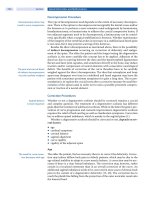
FIGURE 24-1
Electrical status epilepticus of sleep (ESES) electroencephalogram in a child with Landau-Kleffner syndrome.
III • AGE-RELATED SYNDROMES
354
presumed that the neuropsychological deficits are, at least
partially, the result of the epileptiform activity. Landau
and Kleffner (1) suggested that “persistent convulsive
discharges in brain tissue largely concerned with lan-
guage communication result in the functional ablation
of these areas.” Hirsch and colleagues agree with the
hypothesis of a functional ablation (27). Poor daytime
alertness due to sleep fragmentation may contribute to the
neuropsychological deficits (42). Alternatively, the previ-
ous potential interrelations are a hypothesis, and a causal
relation between abnormal interictal discharges and neu-
ropsychological deficits is still controversial (43). A valid
argument is that the dysfunction may represent different
manifestations of the same unknown, possibly genetically
determined, underlying pathogenic mechanism. An argu-
ment against this hypothesis is that the suppression of
discharges with medical or surgical therapy may, at least
partly, reverse these cognitive deficits (44, 45).
Despite the controversy regarding the underlying
pathophysiology of epileptic encephalopathies, the fol-
lowing three crucial questions await answers: (1) what
are the mechanisms involved in the generation of such a
significant, interictal, sleep activation; (2) what are the
mechanisms involved in the cognitive or developmental
regression that accompanies these conditions; and (3)
what is the interrelation between the two, if any?
Although a genetic predisposition was questioned,
there is no strong evidence to support such predilection (46).
The response of the epileptiform discharges to corticoste-
roids raised the question of an autoimmune pathogenesis at
least in a subset of patients including central nervous system
(CNS) vasculitis or demyelination. IgG and IgM antibod-
ies to brain endothelial cells have been identified in these
disorders (28, 47), with higher levels in the patients than
in controls. Brain-derived neurotrophic factor (BDNF),
BDNF autoantibodies, and IgM and IgG antibodies were
elevated in some children with autism and childhood dis-
integrative disorder (CDD). The authors concluded that
these findings suggest a previously unrecognized interaction
between the immune system and BDNF (47). Autoantibod-
ies to rat brain auditory cortex, brainstem, and cerebellum
have been identified in children with LKS (48).
There is increasing evidence that interictal EEG
abnormalities can produce transient cognitive impair-
ment (49–55). Furthermore, benign rolandic epilepsy may
be not so benign, because the interictal discharges may
have a substantial effect on cognitive function (56, 57),
at least for a subset of patients. Additionally, the pres-
ence of continuously abnormal discharges during sleep
may cause disruption of hippocampal function and inter-
fere with the consolidation of memory (58–60). Hence,
the potential impact of the persistent interictal discharges
on brain plasticity is proposed as a mechanism for the
resulting neuropsychological impairment in these children.
More specifically, the occurrence of epileptiform discharges
during a critical time of brain development may result in
defective synaptogenesis and thalamocortical circuit for-
mation. Secondary bilateral synchrony, facilitated by the
corpus callosum with involvement of thalamocortical con-
nections, was hypothesized as a possible mechanism for the
generation of the epileptiform discharges (61–64).
DIAGNOSTIC EVALUATION AND
DIFFERENTIAL DIAGNOSIS
The diagnosis starts by establishing an epileptic disturbance
in the child with regression, usually first with a routine EEG.
All pediatric epilepsy syndromes are classified as symptom-
atic, cryptogenic, or idiopathic. Symptomatic cases exist
for both LKS and CSWS, although symptomatic cases are
more frequent with CSWS. We have seen only one case of
a symptomatic LKS, in a child with a left temporal oligo-
dendroglioma. However, other categories reported include
infectious disorders, such as cysticercosis and toxoplasmo-
sis; inflammatory disorders, such as CNS vasculitis; demy-
elinating disease and acute disseminated encephalomyelitis
(ADEM); congenital brain malformations, such as polymi-
crogyria; and tumors, including temporal lobe astrocyto-
mas and dysembryoplastic neuroepithelial tumors (DNET)
(4, 5). Therefore, neuroimaging is warranted.
Typically in the idiopathic cases, no structural abnor-
malities are seen with routine neuroimaging, although
bilateral volume reduction using an MRI cortical parcel-
lation technique has been reported in the superior tem-
poral gyrus (65) and perisylvian polymicrogyria has been
reported in a single case (66). Functional neuroimaging
has demonstrated temporal dysfunction with SPECT (67,
68), PET (69, 70), or MEG scans (71). These studies are
usually done when a patient has failed treatment and
epilepsy surgery is considered.
The differential diagnosis of a sleep-activated EEG
includes (3): LKS, CSWS, and PDD with regression, con-
genital aphasia or developmental language disorders, or
the epilepsy syndromes benign focal epilepsy with centro-
temporal discharges, benign focal epilepsy with occipital
discharges, atypical benign partial epilepsy of childhood,
the Lennox-Gastaut syndrome, and myoclonic-astatic
epilepsy (Doose syndrome). Language or intellectual
regression associated with behavioral problems in any
of these syndromes may make the differential diagno-
sis difficult and not all pediatric epilepsy syndromes are
readily classified. In our experience, children with PDD
with regression and an epileptiform EEG are the largest
numbers of children referred for evaluation.
Clinical symptoms other than language regression
have been reported with ESES. Hirsch and colleagues sug-
gested that the definition of LKS should be expanded to
include the acquired deterioration of any higher cortical
function in association with sleep-activated paroxysmal
24 • THE LANDAU-KLEFFNER SYNDROME AND EPILEPSY WITH CONTINUOUS SPIKE-WAVES DURING SLEEP
355
features (72) and not limited just to language regression.
Clinical manifestations include epileptic dysgraphia (73),
visual agnosia (74), and an acquired frontal syndrome (75).
We have seen one child with blindness (Figure 24-2) and
another child with a prosopagnosia, with both demon-
strating a more posterior ESES on EEG.
TREATMENT
All children with LKS and CSWS should have a formal
neuropsychological evaluation to guide their educational
program and track developmental changes. Children with
LKS will, especially, require intensive speech and language
therapy. These two syndromes are associated with signifi-
cant neuropsychiatric comorbidities, and treatment for
hyperactivity, attention deficit disorder, mood instabil-
ity, behavior problems, and even an autistic picture may
require referral to a psychopharmacologist and psycholo-
gist. Despite control of seizures and EEG abnormalities,
these children may have significant residual neurologic,
psychological, and psychiatric dysfunction.
LKS and ESES have similar treatment but the specif-
ics are debated. Smith and Hoeppner recommend that the
treatment goal is the complete elimination of epileptiform
activity within 2 years (25). Treatment options include
standard AEDs, corticosteroids (adrenocorticotropic
hormone [ACTH] or prednisone), high-dose benzodiaz-
epines, intravenous immunoglobulins, or multiple subpial
transections (MST). Although AEDs may control seizures,
the language dysfunction may not improve, whereas corti-
costeroid treatment may control seizures and decrease the
epileptiform activity and improve language (10–12). Early
corticosteroid treatment has been considered the treat-
ment of choice for LKS (12). Because relapse may occur,
LKS often requires long-term corticosteroid treatment,
which increases the risk of side effects (15). Despite either
AED or corticosteroid treatment, many children continue
with language dysfunction. Regardless of treatment, 50%
to 80% of children have long-term language or neurobe-
havioral abnormalities (76–78).
Landau and Kleffner reported a positive relationship
between AED treatment and aphasia improvement (1). In
1967, Deuel and Lenn reported a case with a clear rela-
tionship between AED treatment and language improve-
ment (79), and there have been subsequent reports of
improvement with various AEDs. However, the conven-
tional wisdom is that the AEDs control the actual clini-
cal seizures but not the aphasia (11, 12). McKinney and
McGreal reported a better response with steroids (10).
Some children who had not responded to AEDs improved
after steroid therapy (11, 12). They also thought that
the rapidity of the response and the sequelae depend on
the duration and severity of symptoms before treatment,
that initial high doses are more effective, and that brief
treatment is ineffective or leads to a high relapse rate.
Both ACTH and prednisone have been used.
Both carbamazepine and valproate have been widely
used, but there are no data suggesting that any one AED
is better than others. We have seen several cases of chil-
dren treated with carbamazepine for seizures with focal
epileptiform abnormalities on EEG who subsequently
developed language regression with ESES. We prefer
using AEDs with antiepileptogenic properties as first-line
AEDs. The term anticonvulsant refers to suppression of
seizures, whereas antiepileptogenic refers to suppression
of the development of epilepsy or the underlying process
that leads to epilepsy (80). We have historically preferred
valproic acid (VPA) because it has both anticonvulsant
and antiepileptogenic properties, and it may normalize
EEGs. It is well known that carbamazepine may worsen
the generalized epilepsies and may even worsen focal
spike-and-wave discharges and activate the EEG (81–83).
For ESES, VPA, benzodiazepines, and ethosuximide have
been the most successful AEDs (7), and lamotrigine and
levetiracetam have also been used. However, we have
seen seizures worsen with every AED used. In general,
for either LKS or ESES, if AEDs do not work, then high-
dose corticosteroids are used. These may work through
GABAergic effects rather than immune mediation (84).
De Negri and colleagues introduced a high-dose diaz-
epam protocol for electrical status epilepticus (ESE) (85).
They gave a rectal dose of 1 mg/kg with EEG monitor-
ing and continued a dose of 0.5 mg/kg orally for several
weeks in those that responded. They found that those
on chronic benzodiazepine treatment did not respond as
well to this treatment. When a clinical relapse occurred,
this dosing schedule was repeated. In the group of De
Negri and colleagues with ESE, only 1 child had LKS
and 1 had ESES. We modified this high-dose diazepam
protocol, using 1 mg/kg either orally or rectally under
EEG guidance, but then treated all children with a dose of
0.5 mg/kg, orally for 3 to 4 weeks (17). If EEG showed no
improvement, we rapidly tapered the diazepam. If EEG
showed an improvement, we tapered then by 2.5 mg/
month. In our series, every child who initially responded
and then had a rapid diazepam taper had either a clinical
or electrographic regression. We now continue a main-
tenance diazepam dose, usually at a dose of 2.5 to 5 mg,
for 2 years. The best responders to high-dose diazepam
have been children with idiopathic LKS.
Tassinari et al recommend trials with several differ-
ent drugs, and they report that a long-lasting effect has
been achieved with VPA along with clobazam, lorazepam,
and clonazepam (8). Smith and Hoeppner recommend
initial treatment with high-dose VPA, with or without
a benzodiazepine, and, in the absence of response, then
several months of corticosteroid therapy (25). Inutsuka
and colleagues (86) reported their treatment results in
15 children, using the following protocol: (1) VPA at
III • AGE-RELATED SYNDROMES
356
FIGURE 24-2
(A) Electroencephalogram (EEG) in child with blindness and posterior electrical status epilepticus of sleep (ESES). (B) EEG
after high dose diazepam treatment; vision has recovered.
24 • THE LANDAU-KLEFFNER SYNDROME AND EPILEPSY WITH CONTINUOUS SPIKE-WAVES DURING SLEEP
357
levels greater than 100 mg/L, (2) combination of VPA plus
ethosuximide, (3) short cycles of high-dose diazepam, (4)
or intramuscular ACTH. Treatment with short cycles
of ACTH (duration 11 to 43 days) or diazepam (DZP)
(duration for 6 to 7 days) did not achieve long-term remis-
sion, whereas either high-dose VPA alone (n ϭ 7) or in
combination with ethosuximide (n ϭ 3) achieved remis-
sion in 10 children (67%). We retrospectively analyzed
our experience with ESES treatment in 12 children (87).
Only 1 of 12 responded to initial short-term therapy with
VPA. We used prednisone for 6 months in 6 children with
the dose schedule outlined in Table 24-1 (88); 5 of 6 had a
positive response, but 4 of 5 (80%) relapsed and required
another course. Before the elective use of corticosteroids,
immunizations should be up to date.
Alternate treatments including immunoglobulins
and the ketogenic diet have been tried, with case reports
documenting efficacy, but long-term follow-up data are
limited (18–22). MST has been performed in selected
children who failed medical therapy, and it may provide
benefit (62, 89).
COURSE AND PROGNOSIS
In general, the outcome of epilepsy is favorable in both
LKS and CSWS (90), whereas cognitive dysfunction occurs
in the majority (25). The prognosis for LKS has varied,
depending on the series. Mantovani and Landau conducted
a long-term follow-up of the original children reported by
Mantovani and Landau (91). In nine patients, with fol-
low-up that varied from 10 to 28 years, four patients had
full recovery, one had a mild language disability, and four
had moderate disability. Later studies have not reported
as positive an outcome. Bishop did a literature review of
45 children with LKS. The age of onset was related to
the outcome, which was less favorable if onset occurred
before 4 years of age (26). Shinnar and colleagues reported
residual language dysfunction in 88% of children who
had language regression, and most had autism or autis-
tic features (76). Deonna et al reported that only one of
seven adult patients had normal language, with the six
others demonstrating varying degrees of language deficits,
some with complete absence of language (75). In a recent
article on the neuropsychological follow-up of 12 patients,
Soprano et al reported that 9 of 12 had a variable degree
of persistent language deficit (78). Only 50% have been
able to lead a normal life (62, 69).
The prognosis is poor in CSWS (92). In an adult fol-
low-up study of seven patients, only one had active epilepsy,
but only two had been in a normal school setting (93). The
two patients with LKS had a normal IQ, but had language
deficits, whereas the five patients with ESES had global
mental deficiency. Scholtes et al (94) performed a long-
term follow-up of 10 children with ESES, with a good
recovery in only 1 child and a partial recovery in only 4.
There are more residual deficits in CSWS, because this
syndrome is more likely to be symptomatic, compared to
LKS, which is more likely to be idiopathic.
TABLE 24-1
Six-Month Dosing Schedule for Oral Prednisone
2 mg/kg/day for 1 month (maximum dose 60 mg)
1.5 mg/kg/day for 1 month
1 mg/kg/day for 1 month
1 mg/kg every other day for 1 month
0.75 mg/kg every other day for 1 month
0.5 mg/kg/day every other day for 1 month
Note: Immunizations should be up to date before the elective
use of corticosteroids.
References
1. Landau W, Kleffner FR. Syndrome of acquired aphasia with convulsive disorder in chil-
dren. Neurology 1957; 7:523–530.
2. Commission on Classification and Terminology of the International League Against Epi-
lepsy. Proposal for revised classification of epilepsies and epileptic syndromes. Epilepsia
1989; 30:389–399.
3. Engel J Jr. A proposed diagnostic scheme for people with epileptic seizures and with
epilepsy: report of the ILAE Task Force on Classification and Terminology. Epilepsia
2001; 42:796–803.
4. Riviello JJ. Drislane F, eds. Status epilepticus in children. Status epilepticus: a clinical
perspective. Totowa, NJ: Humana Press, 2005:313–338.
5. Hadjiloizou S, Riviello JJ. Epileptic and epileptiform encephalopathies. Neurology.
Omaha, NE: eMedicine.com, Inc., 2006.
6. Patry G, Lyagoubi S, Tassinari A. Subclinical “electrical status epilepticus” induced by
sleep in children. A clinical and electroencephalographic study of six cases. Arch Neurol
1971; 24:242–252.
7. Tassinari CA, Bureau M, Dravet C, Dalla Bernardina B, et al. Epilepsy with continuous
spikes and waves during slow sleep-otherwise described as ESES (epilepsy with electrical
status epilepticus during slow sleep. In: Roger J, Bureau M, Dravet Ch, Dreifuss FE, et al,
eds. Epileptic Syndromes in Infancy, Childhood, and Adolescencence. 2nd ed. London:
John Libbey, 1992:245–256.
8. Tassinari CA, Rubboli G, Volpi L, Meletti S, et al. Encephalopathy with electrical status
epilepticus during slow sleep or ESES syndrome including the acquired aphasia. Clin
Neurophsyiol 2000; 111 Suppl 2:S94–S102.
9. Veggiotti P, Beccaria F, Guerrini R, Capovilla G, et al. Continuous spike-and-wave activity
during slow-wave sleep: syndrome or EEG pattern? Epilepsia 1999; 40:1593–1601.
10. McKinney W, McGreal DA. An aphasic syndrome in children. Can Med J 1974;
110:637–639.
11. Marescaux C, Finck S, Maquet P, Schlumberger E, et al. Landau-Kleffner syndrome: a
pharmacologic study of five cases. Epilepsia 1990; 31:768–777.
12. Lerman P, Lerman-Sagie T, Kivity S. Effect of early corticosteroid therapy for Landau-
Kleffner syndrome. Dev Med Child Neurol 1991; 33:257–260.
13. Tsuru T, Mori M, Mizuguchi M, Momoi MY. Effects of high-dose intravenous cortico-
steroid therapy in Landau-Kleffner syndrome. Pediatr Neurol 2000; 22:145–147.
14. Sinclair DB, Snyder TJ. Corticosteroids for the treatment of Landau-Kleffner syn-
drome and continuous spike-wave discharge during sleep. Pediatr Neurol 2005;
32:300–306.
15. Verhelst H, Boon P, Buyse G, Ceulemans B, et al. Steroids in intractable childhood epilepsy:
clinical experience and review of the literature. Seizure 2005; 14:412–421.
16. De Negri M, Baglietto MG, Battaglia FM. Treatment of electrical status epilepticus by
short diazepam (DZP) cycles after DZP rectal bolus test. Brain Dev 1995; 17:330–333.
17. Riviello JJ, Holder DL, Thiele E, Bourgeois BFD, et al. Treatment of continuous spikes and
waves during slow wave sleep with high dose diazepam. Epilepsia 2001; 42 Suppl 7:56.
18. Fayad MN, Choueiri R, Mikati M. Landau-Kleffner syndrome: consistent response to
repeated intravenous gamma-globulin doses: a case report. Epilepsia 1997; 38:489–494.
19. Mikati MA, Saab R. Successful use of intravenous immunoglobulin as initial monotherapy
in Landau-Kleffner syndrome. Epilepsia 2000; 41:880–886.
20. Mikati MA, Saab R, Fayad MN, Choueiri RN. Efficacy of intravenous immunoglobulin
in Landau-Kleffner syndrome. Pediatr Neurol 2002; 26:298–300.
21. Lagae LG, Silberstein J, Gillis PL, Casaer PJ. Successful use of intravenous immunoglobu-
lins in Landau-Kleffner syndrome. Pediatr Neurol
1998; 18:165–168.
III • AGE-RELATED SYNDROMES
358
22. Prasad AN, Stafstrom CF, Holmes GL. Alternative epilepsy therapies: the ketogenic diet,
immunoglobulins, and steroids. Epilepsia 1996; 37 Suppl 1:S81–S95.
23. Bergqvist AG, Chee CM, Lutchka LM, Brooks-Kayal AR. Treatment of acquired epileptic
aphasia with the ketogenic diet. J Child Neurol 1999; 14:696–701.
24. Kramer U, Nevo Y, Neufeld MY, Fatal A, et al. Epidemiology of epilepsy in childhood:
a cohort of 440 consecutive patients. Pediatr Neurol 1998; 18:46–50.
25. Smith MC, Hoeppner TJ. Epileptic encephalopathy of late childhood: Landau-Kleffner
syndrome and the syndrome of continuous spikes and waves during slow sleep. J Clin
Neurophysiol 2003; 20:462–472.
26. Bishop DVM. Age of onset and outcome in acquired aphasia with convulsive disorder
(Landau-Kleffner syndrome). Dev Med Child Neurol 1985; 27:705–712.
27. Hirsch E, Valenti MP, Rudolf G, Seegmuller C, et al. Landau-Kleffner syndrome is not
an eponymic badge of ignorance. Epilepsy Res 2006; 70 Suppl 1:S239–S247.
28. Connolly AM, Chez MG, Pestronk A, Arnold ST, et al. Serum autoantibodies to brain
in Landau-Kleffner variant, autism, and other neurologic disorders. J Pediatr 1999;
134:607–613.
29. Shafrir Y, Prensky AL. Acquired epileptiform opercular syndrome: a second case report,
review of the literature, and comparison to the Landau-Kleffner syndrome. Epilepsia
1995; 36:1050–1057.
30. Tuchman RF, Rapin I. Regression in pervasive developmental disorders: seizures and
epileptiform electroencephalogram correlates. Pediatrics 1997; 99:560–566.
31. Tuchman RF, Rapin I, Shinnar S. Autistic and dysphasic children. I: Clinical characteristics.
Pediatrics 1991; 88:1211–1218.
32. Tuchman RF, Rapin I, Shinnar S. Autistic and dysphasic children. II: Epilepsy. Pediatrics
1991; 88:1219–1225.
33. Echenne B, Cheminal R, Rivier F, Negre C, et al. Epileptic electroencephalographic abnormali-
ties and developmental dysphasias: a study of 32 patients. Brain Dev 1992; 14:216–225.
34. Stefanatos GA, Foley C, Grover W, Doherty B. Steady-state auditory evoked responses to
pulsed frequency modulations in children. EEG Clin Neurophysiol 1997; 104:31–42.
35. Galanopoulou AS, Bojko A, Lado F, Moshe SL. The spectrum of neuropsychiatric abnormal-
ities associated with electrical status epilepticus in sleep. Brain Dev 2000; 22:279–295.
36. Guilhoto LMFF, Morrell F. Electrophysiological differences bewteen Landau-Kleffner syndrome
and other conditions showing the CSWS electrical pattern. Epilepsia 1994; 35 Suppl 8:126.
37. Guilhoto LM, Machado-Haertel LR, Manreza ML, Diament AJ. Continuous spike wave
activity during sleep. Electroencephalographic and clinical features. Arq Neuropsiquiatr
1997; 55:762–770.
38. Van Hirtum-Das M, Licht EA, Koh S, Wu JY, et al. Children with ESES: Variability in
the syndrome. Epilepsy Res 2006; 7S; S248–S258.
39. Bolanos A, Mikati M, Holmes G, Helmers S, et al. Landau-Kleffner syndrome: clinical
and EEG features. Neurology 1995; 45 Suppl 4:A180.
40. Bolanos A, Urion DK, Helmers SL, Lombroso CT, et al. Serial electroencephalographic changes
in children with Landau-Kleffner syndrome. Epilepsia
1997; 38 Suppl 3:27.pl4:A180.
41. Holmes GL, McKeever M, Saunders Z. Epileptiform activity in aphasia of childhood: an
epiphenomenon? Epilepsia 1981; 22:631–639.
42. Kohrman MH, Carney PR. Sleep-related disorders in neurologic disease during childhood.
Pediatr Neurol 2000; 23:107–113.
43. Ben-Ari Y, Holmes GL. Effects of seizures on developmental processes in the immature
brain. Lancet Neurol 2006; 5:1055–1063.
44. Matsuzaka T, Baba H, Matsuo A, Tsuru A, et al. Developmental assessment-based surgical
intervention for intractable epilepsies in infants and young children. Epilepsia 2001; 42
Suppl 6:9–12.
45. Holmes GL, Lenck-Santini PP. Role of interictal epileptiform abnormalities in cognitive
impairment. Epilepsy Behav 2006; 8:504–515. Epub 2006.
46. Landau WM. Landau-Kleffner syndrome. An eponymic badge of ignorance. Arch Neurol
1992; 49:353.
47. Connolly 2005.
48. Boscolo S, Baldas V, Gobbi G, Giordano L, et al. Anti-brain but not celiac disease anti-
bodies in Landau-Kleffner syndrome and related epilepsies. J Neuroimmunol 2005;
160:228–232. Epub 2004.
49. Shewmon DA, Erwin RJ. The effect of focal interictal spikes on perception and reaction time.
II. Neuroanatomic specificity. Electroencephalogr Clin Neurophysiol 1988; 69:338–352.
50. Shewmon DA, Erwin RJ. Transient impairment of visual perception induced by single
interictal occipital spikes. J Clin Exp Neuropsychol 1989; 11:675–691.
51. Kasteleijn-Nolst Trenite DG, Bakker DJ, Binnie CD. Psychological effects of subclinical
epileptiform EEG discharges. I. Scholastic skills. Epilepsy Res 1988; 2:111–116.
52. Aarts JH, Binnie CD, Smit AM, Wilkins AJ. Selective cognitive impairment during focal
and generalized epileptiform EEG activity. Brain 1984; 107 Pt 1:293–308.
53. Binnie CD, Kasteleijn-Nolst Trenite DG, Smit AM, et al. Interactions of epileptiform
EEG discharges and cognition. Epilepsy Res 1987; 1:239–245.
54. Binnie CD. Significance and management of transitory cognitive impairment due to
subclinical EEG discharges in children. Brain Dev 1993; 15:23–30.
55. Binnie CD. Cognitive impairment during epileptiform discharges: is it ever justifiable to
treat the EEG? Lancet Neurol 2003; 2:725–730.
56. Massa R, de Saint-Martin A, Carcangiu R, Rudolf G, et al. EEG criteria predictive of
complicated evolution in idiopathic rolandic epilepsy. Neurology 2001; 57:1071–1079.
57. Nolan MA, Redoblado MA, Lah S, Sabaz M, et al. Memory function in childhood epilepsy
syndromes. J Paediatr Child Health 2004; 40:20–27.
58. Moruzzi G, Magoun HW. Brain stem reticular formation and activation of the EEG.
J Neuropsychiatry Clin Neurosci 1995; 7:251–267.
59. Lorincz A, Buzsaki G. Two-phase computational model training long-term memories in
the entorhinal-hippocampal region. Ann N Y Acad Sci 2000; 911:83–111.
60. Louie K, Wilson MA. Temporally structured replay of awake hippocampal ensemble
activity during rapid eye movement sleep. Neuron 2001; 29:145–156.
61. Morrell F. Secondary epileptogenesis in man. Arch Neurol 1985; 42:318–335.
62. Morrell F, Whisler WW, Smith MC, Hoeppner TJ, et al. Landau-Kleffner syndrome.
Treatment with subpial intracortical transection. Brain 1995; 118:1529–1546.
63. Kobayashi K, Murakami N, Yoshinaga H, Enoki H, et al. Nonconvulsive status epilep-
ticus with continuous diffuse spike-and-wave discharges during sleep in childhood. Jpn
J Psychiatry Neurol 1988; 42:509–514.
64. Monteiro JP, Roulet-Perez E, Davidoff V, Deonna T. Primary neonatal thalamic haemor-
rhage and epilepsy with continuous spike-wave during sleep: a longitudinal follow-up of
a possible significant relation. Eur J Paediatr Neurol 2001; 5(1):41–47.
65. Takeoka M, Riviello JJ Jr, Duffy FH, Kim F, et al. Bilateral volume reduction of the supe-
rior temporal areas in Landau-Kleffner syndrome. Neurology 2004; 63:1289–1292.
66. Huppke P, Kallenberg K, Gartner J. Perisylvian polymicrogyria in Landau-Kleffner syn-
drome. Neurology 2005; 64:1660.
67. O’Tuama LA, Urion DK, Janicek MJ, Treves ST, et al. Regional cerebral perfusion
in Landau-Kleffner syndrome and related childhood aphasias. J Nucl Med 1992; 33:
1758–1765.
68. Guerreiro MM, Camargo EE, Kato M, Menezes Netto JR, et al. Brain single photon emission
computed tomography imaging in Landau-Kleffner syndrome. Epilepsia 1996; 37:60–67.
69. Maquet P, Hirsch E, Dive D, Salmon E, et al. Cerebral glucose utilization during sleep in
Landau-Kleffner syndrome: a PET study. Epilepsia 1990; 31:778–783.
70. da Silva EA, Chugani DC, Muzik O, Chugani HT. Landau-Kleffner syndrome: meta-
bolic abnormalities in temporal lobe are a common feature. J Child Neurol 1997;
12:489–495.
71. Paetau R, Granstrom M-L, Blomstedt G, Jousmaki V, et al. Magnetoencephalography in
presurgical evaluation of Landau-Kleffner syndrome. Epilepsia
1999; 40:326–335.
72. Hirsch E, Maquet P, Metz-Lutz M-N, Motte J, et al. The eponym “Landau-Kleffner
Syndrome” should not be restricted to childhood-acquired aphasia with epilepsy. In:
Beaumanoir A, Bureau M, Deonna T, Mira L, et al, eds. Continuous Spikes and Slow
Waves During Slow Sleep Electrical Status Epilepticus During Slow Sleep: Acquired
Epileptic Aphasia and Related Conditions. London: John Libbey, 1995:57–62.
73. DuBois CM, Zesiger P, Perez ER, Ingvar MM, Deonna T. Acquired epileptic dysgraphia:
a longitudinal study. Dev Med Child Neurol 2003; 45:807–812.
74. Eriksson K, Kylliainen A, Hirvonen K, Nieminen P, et al. Visual agnosia in a child with
non-lesional occipito-temporal CSWS. Brain Dev 2003; 25:262–267.
75. Deonna T, Davidoff V, Maeder-Ingvar M, Zesiger P, et al. The spectrum of acquired
cognitive disturbances in children with partial epilepsy and continuous spike-waves dur-
ing sleep. A 4-year follow-up case study with prolonged reversible learning arrest and
dysfluency. Eur J Paediatr Neurol 1997; 1:19–2916.
76. Shinnar S, Rapin I, Arnold S, Tuchman RF, et al. Language regression in childhood.
Pediatr Neurol 2001; 24:183–189.
77. Mantovani JF. Autistic regression and Landau-Kleffner syndrome: progress or confusion?
Dev Med Child Neurol 2000; 42:349–353.
78. Soprano AM, Garcia EF, Caraballo R, Fejerman N. Acquired epileptic aphasia: neuro-
psychologic follow-up of 12 patients. Pediatr Neurol 1994; 11:230–235.
79. Deuel RK, Lenn NJ. Treatment of acquired epileptic aphasia. J Pediatr 1977; 90:959–961.
80. Silver JM, Shin C, McNamara JO. Antiepileptogenic effects of conventional anticonvul-
sants in the kindling model of epilepsy. Ann Neurol 1991; 29:356–363.
81. Snead OC 3rd, Hosey LC. Exacerbation of seizures in children by carbamazepine. N
Engl J Med 1985; 313:916–921.
82. Kochen S, Giagante B, Oddo S. Spike-and-wave complexes and seizure exacerbation
caused by carbamazepine. Eur J Neurol 2002; 9:41–47.
83. Gansaeuer M, Alsaadi TM. Carbamazepine-induced seizures: a case report and review
of the literature. Clin Electroencephalogr 2002; 33:174–177.
84. Mtchedlishvili Z, Kapur J. A presynaptic action of the neurosteroid pregnenolone sulfate
on GABAergic synaptic transmission. Mol Pharmacol 2003; 64:857–864.
85. De Negri M, Baglietto MG, Battaglia FM. Treatment of electrical status epilepti-
cus by short diazepam (DZP) cycles after DZP rectal bolus test. Brain Dev 1995;
17:330–333.
86. Inutsuka M, Kobayashi K, Oka M, Hattori J, Ohtsuka Y. Treatment of epilesy with
electrical status epilepticus of sleep and its related disorders. Epilepsy Behav 2006;
28:281–286.
87. Albaradie RS, Bourgeois BFD, Thiele E, Duffy FH, et al. Treatment of continuous spike
and wave during slow wave sleep. Epilepsia 2001; 42 Suppl 7:46–47.
88. Stefanatos GA, Grover W, Geller E. Case study: corticosteroid treatment of language
regression in pervasive developmental disorder. J Am Acad Child Adolesc Psychiatry
1995; 34:1107–1111.
89. Irwin K, Birch V, Lees J, Polkey C, et al. Multiple subpial transection in Landau-Kleffner
syndrome. Dev Med Child Neurol 2001; 43:248–252.
90. Bureau M. Outstanding cases of CSWS and LKS: analysis of the data sheets provided
by the participants. In: Beaumanoir A, Bureau M, Deonna T, Mira L, et al, eds. Con-
tinuous Spikes and Slow Waves During Slow Sleep Electrical Status Epilepticus During
Slow Sleep: Acquired Epileptic Aphasia and Related Conditions. London: John Libbey,
1995:213–216.
91. Mantovani JF, Landau WM. Acquired aphasia with convulsive disorder: course and
prognosis. Neurology 1980; 30:524–529.
92. Morikawa T, Seino M, Watanabe M. Long-term outcome of CSWS syndrome. In: Beau-
manoir A, Bureau M, Deonna T, Mira L, et al, eds. Continuous Spikes and Slow Waves
During Slow Sleep Electrical Status Epilepticus During Slow Sleep: Acquired Epileptic
Aphasia and Related Conditions. London: John Libbey, 1995:27–36.
93. Praline J, Hommet C, Barthez M-A, Brault F, et al. Outcome at adulthood of continuous spike
waves of slow sleep and Landau-Kleffner syndrome. Epilepsia 2003; 44:1434–1440.
94. Scholtes FB, Hendriks MP, Renier WO. Cognitive detrioration and electrical status epi-
lepticus during slow sleep. Brain Dev 2005; 6:167–173.
359
Idiopathic Generalized
Epilepsy of Adolescence
Idiopathic generalized epilepsies
(IGEs) are a distinct group of epi-
lepsies, clearly defined in the 1989
International Classification of Epi-
leptic Syndromes and Epilepsies. This classification estab-
lished an important dichotomy between the idiopathic
epilepsies on the one hand, and the symptomatic or cryp-
togenic epilepsies on the other (1). In prior versions of
this classification, IGEs were referred to as primary gen-
eralized epilepsies, and symptomatic/cryptogenic general-
ized epilepsies were categorized under secondary general-
ized epilepsies. This nomenclature was revised because
the term “secondary generalized” was confusing when
applied to epilepsy, where it meant that the epilepsy was
secondary to some other cause, whereas when applied
to seizures, it means that the generalization is secondary
from an originally focal seizure.
Unfortunately, the term “idiopathic” in the current
classification is also confusing in the context of current
medical terminology, because in every other area of medi-
cine “idiopathic” means “of an unknown cause.” It is
now clearly understood that IGEs are primarily of genetic
origin.
IGEs are age-dependent, meaning that a given geno-
type may express itself differently at different ages. This
chapter will focus on the IGE seen in adolescence.
Reza Behrouz
Selim R. Benbadis
GENETICS & PATHOPHYSIOLOGY
IGEs represent a group of epilepsies best described as
a genetically determined low threshold for generalized
seizures, as opposed to symptomatic or cryptogenic gen-
eralized epilepsies, where there is an underlying anatomic
(pathologic) abnormality. Investigations for microscopic
lesions (e.g., microdysgenesis) in IGE, using structural
imaging, have yielded inconsistent results (2). Functional
imaging, not surprisingly, can show evidence for dif-
fuse dysfunction. Proton magnetic resonance spectros-
copy (MRS) has revealed frontal lobe (3) or thalamic
(4) abnormalities.
Because they have “overlapping” genetic origins, it
is appropriate and practical to view IGEs as a continuum
of genetic conditions exhibiting variable phenotypes.
Recent advances have identified IGEs linked to defects
in genes encoding the subunits for voltage- or ligand-
gated ion channels including sodium, potassium, calcium,
or chloride channels affecting the gamma-aminobutyric
acid (GABA)
A
receptors. Hence, they are categorically
referred to as “ion channelopathies” (5). For the clas-
sic and well-defined forms of IGE—juvenile myoclonic
epilepsy (JME) or childhood absence epilepsy (CAE), a
few mutations, including the KCNQ potassium channels
and the ClC-2 chloride channel, have been implicated (6).
C
25
360
III • AGE-RELATED SYNDROMES
The pathophysiology (e.g., the concept of channelopa-
thies) and genetics of IGE are gradually being elucidated.
Fundamentally, IGEs appear to be “phenotypes of many,
often individually rare, gene defects” (7, 8).
The inheritance of IGEs is complex in the sense
that it does not follow a well-defined Mendelian pattern.
Moreover, phenotypic expression influencing semiology
and frequency of a specific genotype may be quite vari-
able from one individual to another. This is likely due to
different degrees of alteration of neuronal excitability
induced by gene errors. At least for JME, a maternal
inheritance has been proposed and confirmed by some (9).
Nevertheless, it is reasonable to state that the genetic
complexity of IGEs necessitates additional investigation
in order to reach a conclusion regarding their precise
mechanism of inheritance.
EPIDEMIOLOGY AND NATURAL HISTORY
IGEs constitute approximately 15–20% of all epilepsies
(10). They affect all races equally and may have a slight pre-
dilection for women (11). Seizures usually, but not always,
have an onset early in life, from childhood to early adult-
hood. In fact, IGEs are the most frequent group of epilepsies
with an adolescent onset (12). IGEs are generally associated
with low mortality and favorable response rate to treat-
ment. The morbidity from IGE is primarily a function of
how well the condition is treated. As it will be mentioned
further in this chapter, the response rate to antiepileptic
drugs (AEDs) is good. Additionally, 50% of patients with
IGE can outgrow them. A recent intriguing finding is the
association between IGE and type 1 diabetes (13).
CLINICAL FEATURES (OF IGE IN GENERAL)
Overall, there are much more similarities than there
are differences among the various types of IGE. These
epilepsies manifest themselves by the same three seizure
types, have similar electroencephalographic (EEG) find-
ings, are said to “evolve” into one another, and have
overlapping genetic origins (family members may express
different IGE syndromes). In all, IGEs are best viewed as
a spectrum of conditions, and many of them may indeed
represent a single entity with slightly different clinical
phenotypes (14–19). Thus, there are good reasons to
study them as a group. In addition, as will be discussed
subsequently, making a diagnosis of IGE as a group (as
opposed to localization-related epilepsy or a symptom-
atic/cryptogenic generalized epilepsy) has critical impli-
cations for patient care, whereas differentiating among
the specific syndromes is relatively less important. The
IGEs are genetically determined and have no structural or
anatomic cause, and seizures are the only manifestation
of the condition. Age of onset is very important, since
IGE usually (but not always) begin early in life, from
childhood to early adulthood. Neurologic examination
is normal, intelligence is normal, imaging studies are nor-
mal, and EEG is normal other than for the epileptiform
abnormalities (i.e., there is no abnormal slow activity or
evidence for diffuse encephalopathy).
Seizure types in IGE include a combination of general-
ized tonic-clonic (GTC), absence, and myoclonic seizures.
• Absence seizures are clinically characterized by
a brief (5–15 sec) impairment of consciousness,
with few or no other symptoms. More specifically,
patients with IGE have typical absence seizures,
characterized by an abrupt onset and termination
and the classic monomorphic 3-Hz spike-wave com-
plexes (ictally and interictally) with no other EEG
abnormality. By contrast, atypical absence seizures
occur in symptomatic generalized epilepsy of the
Lennox-Gastaut type and are less discrete in time,
longer, and with slower and more polymorphic
spike-wave complexes on EEG. Absence seizures are
the defining and predominant seizure type in CAE
and juvenile absence epilepsy, but they do occur in
the other IGEs.
• Myoclonic seizures consist of sporadic jerks, usually
appendicular and symmetrical, associated electrically
with generalized epileptiform discharges, typically
polyspikes (Figure 25-1). Patients sometimes report
them as an electric shock sensation. Myoclonic sei-
zures are the defining and predominant seizure type
in JME, but they do occur in the other IGEs.
• Generalized tonic-clonic (GTC) seizures are of course
the most common type of seizures. GTC seizures in
the IGEs are by definition “primarily generalized”
GTC seizures. They are typically characterized by a
tonic phase that which is gradually interrupted by
quiescence, thus giving rise to the clonic phase, with
rhythmic spikes and spike-wave complexes decreas-
ing in frequency. Often, primarily generalized GTC
seizures begin with clonic or myoclonic jerks before
the tonic phase, thus in reality being clonic-tonic-
clonic rather than tonic-clonic.
The morning predominance of convulsive seizures
is a general feature in IGE that must be specifically asked
about during history taking.
EEG FEATURES (OF IGE IN GENERAL)
The EEG findings of IGEs are characteristic, and consist
of generalized epileptiform discharges with normal back-
ground activity (i.e., no abnormally slow background and
no superimposed slow activity). Epileptiform discharges
361
25 • IDIOPATHIC GENERALIZED EPILEPSY OF ADOLESCENCE
can take the form of spikes, sharp waves, spike-wave
complexes and polyspikes. Spike-wave complexes can be
viewed as the EEG correlate of absence seizures, and poly-
spikes as the EEG correlate of myoclonic seizures. Just as
patients with IGE have various combinations of the three
seizure types described in the preceding section, they also
have various combinations of generalized epileptiform
abnormalities. Although epileptiform discharges are typi-
cally generalized and symmetric, minor EEG asymmetries
may occasionally be present (20–23) and should not lead
to a diagnosis of partial seizures (24).
• Spikes and sharp waves are of high amplitude, with
aftergoing slow waves disrupting the ongoing back-
ground. Those seen in IGE are generalized and typi-
cally maximal frontally (F3/F4 or Fp1/Fp2).
• The spike-wave complexes seen in IGE are very
monomorphic and have the classic frequency of
3 Hz, but this may vary between 2.5 and 3.5 Hz
and is often faster at the onset of the discharges
(3.5 to 4.5 Hz). The near perfect monomorphism
of the spike-wave complexes is as important as
the 3-Hz frequency in distinguishing this pattern
from the “slow” spike-wave complexes seen in the
symptomatic generalized epilepsies of the Lennox-
Gastaut type. They are generalized and symmetric
with a maximum negativity frontally, almost always
at F3 and F4. The onset and offset are abrupt. The
3-Hz spike-wave complexes are both an interictal
and ictal pattern. The border between the two is
imprecise because the clinical impairment may be
difficult to detect during short discharges. It is gen-
erally stated that a discharge of about 3 seconds
or more is associated with detectable impairment
of awareness, but this depends of how well short
impairments can be detected. The use of a clicker to
test responsiveness can be very useful to document
very brief impairments in awareness. Clinical sei-
zures and the 3-Hz spike-wave complexes discharges
are often precipitated by hyperventilation. The 3-Hz
FIGURE 25-1
Typical generalized polyspike during drowsiness in a 17-year-old adolescent with typical juvenile myoclonic epilepsy. Note the
high amplitude (see scale) and the aftergoing slow wave and suppression. This discharge was asymptomatic.
362
III • AGE-RELATED SYNDROMES
frequency, and also the striking monomorphism of
the spike-wave discharges, distinguish absence epi-
lepsy from other syndromes. Spike-wave complexes
can be faster (i.e., 4–6 Hz), especially in patients
with IGE of adolescence, as opposed to those with
typical CAE.
• Polyspikes (Figure 25-1) are also of high amplitude,
generalized, bilateral, and grossly symmetric, and
followed by significant slowing. Polyspikes are the
electrical correlate of myoclonic seizures, and as
such they are typically seen in IGE syndromes that
include this seizure type, most characteristically
JME. Polyspikes can be repetitive and described as
polyspike-wave complexes.
• Photosensitivity is particularly frequent and, in fact,
quite typical of IGE (25). Photosensitivity can be
purely an EEG phenomenon, where photic stimula-
tion elicits a photoparoxysmal response (Figure 25-2).
It can also be a clinical phenomenon, where photic
stimulation triggers a clinical seizure as well (typically
motor seizures, i.e., myoclonic and GTC seizures).
Just as convulsive seizures tend to predominate in
the morning in IGE, epileptiform discharges on awakening
appear to be typical of IGE (26).
TREATMENT
Fortunately, IGEs respond well to therapy if the correct
AED is administered. In general, 80-90% will become
fully controlled. As mentioned earlier, some (such as typi-
cal childhood absence epilepsy) have a tendency to be
outgrown.
Correct diagnosis of IGEs (at least as a group) is
critical, as the choice of pharmacologic treatment mat-
ters. There is good evidence that not all AEDs are optimal
choices for IGEs, and some may potentially worsen the
condition (27–36, see Table 25-1). In fact, up to 70% of
patients with IGE initially receive an ill-advised AED, which
causes the seizures to increase in frequency or appear intrac-
table (pseudo-intractability) (37). The use of inadequate
AEDs in IGE can also result in absence or myoclonic sta-
tus epilepticus (36). In general, sodium channel blockers,
including phenytoin, carbamazepine, and oxcarbazepine,
and GABAergic molecules, such as gabapentin, viagabatrin,
and tiagabine, are not advisable in patients with IGE. The
exact mechanism for these agents’ lack of therapeutic effect
and occasional harmful effect has not been established.
Valproic acid is the classic drug of choice for the
treatment of IGEs. Both ethosuximide and valproic acid
are very effective against absence seizures. In patients
FIGURE 25-2
Photoparoxysmal response in an 18-year-old girl with generalized tonic clonic seizures. Photic stimulation (bottom channel)
on 2 occasions triggers a brief but clear burst of spike-wave complexes. This discharge was asymptomatic. The patient also had
similar discharges of spike-wave complexes without photic stimulation.
363
25 • IDIOPATHIC GENERALIZED EPILEPSY OF ADOLESCENCE
with both absence and generalized tonic-clonic (GTC)
seizures, valproic acid is the medication of choice. A good
argument can be made to select valproic acid over etho-
suximide even in absence epilepsy, since 50% of patients
with absence will develop GTC seizures. However, since
children with typical (pure) childhood absence epilepsy
rarely develop GTC seizures until the second decade, etho-
suximide can be the drug of choice in young children. The
limiting factors in the use of valproic acid are its potential
long-term side effects, including weight gain, hair loss,
tremor, teratogenicity, and polycystic ovarian disease.
Ethosuximide is generally associated with a favorable side
effect profile. It should be used cautiously with patients
who have blood dyscrasias or abnormal renal function.
The newer AEDs are promising based upon growing
evidence (38), and their place in the treatment of IGE is
evolving. Newer AEDs are typically approved initially
as adjunct for partial epilepsies, and only later are some
of them tested for and approved for IGE. Thus, their use
in IGE may be “off-label” (39–53, Table 25-2). In this
regard, however, it should be pointed out that no AED,
even standard ones like VPA, has specific indications for
epilepsy syndromes, so that the “off-label” terminology
in this context is a little artificial. In the recent years, topi-
ramate, levetiracetam, and lamotrigine have received spe-
cific approval for IGE (Table 25-2). Lastly, levetiracetam
has also recently (August 2006) received specific approval
as “adjunctive therapy in the treatment of myoclonic sei-
zures in JME,” which in practice means JME (with both
GTC and myoclonic seizures), and probably implies effi-
cacy in the IGEs as a group. As usual newer AEDs first
obtain approval as adjunctive therapy. However, no AED
has ever been found to work as adjunctive therapy and
not in monotherapy, and there is no doubt that these
newer AEDs will work in monotherapy.
TABLE 25-1
Published Evidence That Some AEDs Are
Not Effective in IGEs and May Exacerbate
Some Seizure Types
SEIZURE
OR
EPILEPSY
TYPE
AED STUDIED REFERENCE
CBZ IGE Snead and Hosey, 1985 (27)
CBZ, PHT JME Genton et al, 2000 (28)
GBP CAE Trudeau et al, 1996 (29)
TGB IGE Knake et al, 1999 (30)
CBZ Unclear Shields and Saslow, 1983 (31)
CBZ Mixed Horn et al, 1986 (32)
CBZ Unclear Liporace et al, 1994 (33)
CBZ Unclear Talwar et al, 1994 (34)
PHT, CBZ JME Panayiotopoulos et al, 1994 (35)
Multiple IGE Thomas et al, 2006 (36)
IGE: idiopathic generalized epilepsy; JME: juvenile myo-
clonic epilepsy; CAE: childhood absence epilepsy. CBZ: carbam-
azepine, GBP: gabapentin, PHT: phenytoin, TGB: tiagabine.
TABLE 25-2
Published Evidence of the Efficacy of New AEDs in the IGEs
SEIZURE OR EPILEPSY
AED TYPE STUDIED N REFERENCE
LTG Absence 45 Frank et al, 1999 (39)
LTG IGE 26 Beran et al, 1998 (40)
TPM GTC 8 Biton et al, 1999 (41)
LEV Photosensitive 12 Kasteleijn-Nolst-Trenite et al, 1996 (42)
LEV IGE 3 Cohen, 2003 (43)
LTG, ZNS IGE 3 Wallace, 1998 (44)
TPM Absence 5 Cross 2002 (45)
LTG JME 63 Morris et al, 2004 (46)
ZNS JME 15 Kothare et al, 2004 (47)
LEV JME 35 Labate et al, 2006 (48)
LEV IGE 19 Di Bonaventura et al, 2005 (49)
LEV IGE 55 Krauss et al, 2003 (50)
LEV IGE 25 Kumar and Smith, 2004 (51)
LEV IGE 8 Rocamora et al, 2006 (52)
TPM JME 22 Biton et al, 2005 (53)
LTG
1
GTCS 117 Biton et al. 2005 (74)
LEV
1
GTCS 80 Berkovic et al. 2007 (75)
LEV: levetiracetam; LTG: lamotrigine; TPM: topiramate; ZNS: zonisamide.
1
Double-blind placebo-controlled studies.
364
III • AGE-RELATED SYNDROMES
INDIVIDUAL SYNDROMES
Juvenile myoclonic epilepsy, juvenile absence epilepsy, and
epilepsy with generalized tonic-clonic seizures (GTCS) on
awakening are the three syndromes of idiopathic gener-
alized epilepsy of adolescent onset currently included in
the ILAE classification. Childhood absence epilepsy is
discussed in Chapter 22.
Juvenile Myoclonic Epilepsy (JME)
JME is one of the most common forms of epilepsy seen in
the adolescent population, and is one of the best-defined
syndromes. Although there is some controversy, JME
appears to represent a unique phenotype among IGEs
(54). The genetic abnormality in JME has been linked
to a gene locus on chromosome 6 (55). In about 80%
of cases, it has an age onset of between 12 and 18 years
(mean 14.6 years) (56). Seizures occur predominantly in
the morning, just upon awakening, in a cluster fashion,
and are highly sensitive to sleep deprivation, stress, fatigue,
or alcohol. Clusters of myoclonic seizures typically herald
a GTC seizure. In fact, this pattern of clonic-tonic-clonic
rather than tonic-clonic seizures is quite typical of JME, or
at least IGE. Because JME begins as sporadic myoclonic
seizures before the patient develops GTC seizures, it is
often underdiagnosed and requires careful history taking,
specifically inquiring about morning jerks.
Photosensitivity is very common in JME and is
found in 30% of patients, the highest rate of all epilep-
sies (25). The characteristic EEG pattern in JME consists
of discharges of high-amplitude generalized symmetric
and synchronous 4–6 Hz polyspike-wave complexes.
Probably the most unique feature of JME among
IGEs is that it is not outgrown and requires lifelong treat-
ment, as the rate of recurrence is very high after discon-
tinuation of medication, even after a long remission.
Juvenile Absence Epilepsy (JAE)
The individualization of JAE as a separate syndrome is
relatively recent (1, 57, 58), because the clinical bound-
aries of JAE are rather imprecise. JAE falls somewhere
in-between childhood absence epilepsy and JME on
the spectrum of IGE, and it can have features of both.
Most cases begin near or after puberty, typically between
10 and 17 years of age (59).
Absence seizures are similar though, less “pure” than
those of CAE. They are clinically characterized by a brief
(5–15 seconds) impairment of consciousness with abrupt
onset and termination represented electrographically with
a classic pattern of 3-Hz spike-wave complexes. During
these episodes, the patient often displays automatisms
manifesting as lip-smacking, eye blinks, or repetitive head
movements. The spells have an abrupt onset (without
warning) and termination, a feature distinguishing them
from complex partial seizures. In general, these seizures
can be reliably provoked by having the child hyperven-
tilate for 3 to 5 minutes.
“Other” IGE or IGE “NOS” or Adult-Onset IGE
In addition to the preceding well-defined syndromes, many
patients with IGE, perhaps the majority (37), either have
features that do not meet criteria for a specific syndrome or
have hybrid features. Seizures typically begin in young adult-
hood, and neurologic examination is normal. A positive
family history is not uncommon. Seizures may predominate
in the morning, may be photosensitive, and are sensitive
to sleep deprivation. The 1989 ILAE epilepsy classification
individualized “epilepsy with grand-mal seizures on awak-
ening” and “epilepsies with specific modes of precipitation,”
but these entities are less well defined and too restrictive.
In that scheme, most adults with IGE would not fit into
any syndrome. Thus the evolving classification of IGEs
uses more inclusive terms such as “IGE with GTC seizures
only” (60). Other terms for the same condition include the
“adult-onset IGE” (61) and IGE with pure grand mal (62).
This common syndrome is typically characterized by GTC
only, and likely the same as GTC “on awakening” in which
seizures are not limited to the morning. When other seizure
types exist in these IGEs, the seizures can clinically include
mixed features, giving rise to confusing names. For example,
seizures with features of both absence and myoclonic jerks
can be referred to as “myoclonic absence.” Examples of
questionable syndromes include some reflex epilepsies, such
as photosensitive GTC seizures (63), epilepsy with GTC
seizures on awakening (1), epilepsy induced by thinking and
spatial tasks (64, 65), or adult myoclonic epilepsy (66). In
fact, many such variants can be described as, or hypothetized
to be, individual syndromes, but it is more practical to view
them as variations within the continuum of IGE (60).
Similarly, the EEG patterns can show mixtures of
generalized epileptiform discharges of any type (spikes,
sharp waves, spike-wave complexes, and polyspikes)
and photosensitivity, but can vary to become a hybrid
of spike-wave complexes, polyspikes, polyspike-wave
complexes, and spikes (21).
Thus, in many patients (especially adolescents and
young adults), pigeonholing the IGE syndrome is not
necessary and may be futile, but it is critical to diagnose
the patient correctly as having an IGE rather then a local-
ization-related epilepsy, since this has crucial therapeutic
and prognostic implications.
MEDICALLY INTRACTABLE IGE
The entity of “medically intractable” IGE is not well rec-
ognized and has received little attention because the vast
365
25 • IDIOPATHIC GENERALIZED EPILEPSY OF ADOLESCENCE
majority of patients with IGE are fully controlled with
AEDs. In addition, most patients with IGE that are not
controlled are not truly intractable but instead have been
treated with inadequate AEDs (37, 67, 68). Nevertheless,
epilepsy centers encounter patients with clear IGE who
are refractory to medications. For these patients, there is
some preliminary evidence that vagus nerve stimulation
(VNS) is a good option. VNS may have comparable effi-
cacy against IGE as it does in localization-related epilepsy
(69, 70), but this (like newer AEDs) is off-label use. One
study in an animal model of absence epilepsy found no
benefit of VNS (71). Another option in truly intractable
IGE may the ketogenic diet, but no data are available to
support this.
Secondary bilateral synchrony, in which frontal
lobe epilepsy masquerades as IGE, should be considered
when seizures appear refractory, but in the absence of a
structural lesion or other clear evidence for focality, this
should not be pursued surgically (24).
CONCLUSION: THE IMPORTANCE OF
MAKING THE DIAGNOSIS OF IGE
Making a diagnosis of IGE is a critical step in the man-
agement of patients with seizures. Making a diagnosis
of the specific type of IGE is often not possible, usually
controversial, and is not necessarily important, but a
diagnosis of IGE (as a group) is critical. First, the cause
is not “unknown.” Rather, it is a genetically determined
low threshold for seizures. This is easier for patients and
families to accept. Second, not all AEDs are equal for
IGE. Missing the diagnosis of IGE results in inadequate
drug treatment and poor seizure control (37). Third, a
diagnosis of IGE has important (usually optimistic) prog-
nostic implications.
Unfortunately, it is often taught that adults with
epilepsy should be assumed to have partial (focal) epi-
lepsy. Staring spells are loosely labeled “complex par-
tial” seizures, and GTCs are assumed to be secondarily
generalized (61). This assumption should be avoided, as
it will be wrong at least 25% of the time in adults, and
even more so in adolescents. A significant proportion
of IGEs begin beyond childhood and adolescence: 28%
after age 20 (61), and 35% after age 18 (72). And in a
study of 300 patients with new-onset seizures (a mean
age of 31), a quarter turned out to have an IGE (73). The
EEG, when abnormal, is extremely helpful and clearly
points to IGE. Occasionally the diagnosis will require
video-EEG monitoring. In practice, young adults with
rare GTCs often have normal EEGs, and it may be dif-
ficult to determine whether they have IGE or a focal
(e.g., frontal lobe) epilepsy. In this situation, one should
keep an open mind and not assume partial epilepsy. In a
patient with infrequent GTC seizures and a normal (not
helpful) EEG, the diagnosis should not be assumed to be
localization-related epilepsy. Instead, it is prudent to keep
an open mind, using a diagnosis such as “epilepsy with
GTC seizures, type (IGE vs focal) uncertain,” and to use
broad-spectrum AEDs.
References
1. Commission on Classification and Terminology of the International League Against
Epilepsy: Proposal for revised classification of epilepsy and epileptic syndromes. Epilepsia
1989; 30:389–399.
2. Opeskin K, Kalnins RM, Halliday G, Cartwright H, et al. Idiopathic generalized epilepsy:
lack of significant microdysgenesis. Neurology 2000; 55:1101–1106.
3. Simister RJ, McLean MA, Barker GJ, Duncan JS. Proton MRS reveals frontal lobe metabo-
lite abnormalities in idiopathic generalized epilepsy. Neurology 2003; 61:897–902.
4. Bernasconi A, Bernasconi N, Natsume J, Antel SB, et al. Magnetic resonance spectros-
copy and imaging of the thalamus in idiopathic generalized epilepsy. Brain 2003; 126
(Pt 11):2447–2454.
5. Hirose S, Mitsudome A, Okada M, et al. Genetics of idiopathic epilepsies. Epilepsia 2005;
46(Suppl 1):38–43.
6. Lerche H, Weber YG, Jurkat-Rott K, et al. Ion channel defects in idiopathic epilepsies.
Curr Pharm Des 2005; 11(21):2737–2752.
7. Noebels JL. Exploring new gene discoveries in idiopathic generalized epilepsy. Epilepsia
2003; 44 Suppl 2:16–21.
8. Anderson E, Berkovic S, Dulac O, et al. ILAE Genetics Commission conference report:
molecular analysis of complex genetic epilepsies. Epilepsia 2002; 43:1262–1267. Erratum:
Epilepsia 2002; 43:1600–1602.
9. Pal DK, Durner M, Klotz I, Dicker E, et al. Complex inheritance and parent-of-origin
effect in juvenile myoclonic epilepsy. Brain Dev 2006; 28(2):92–98.
10. Jallon P, Latour P. Epidemiology of idiopathic generalized epilepsies. Epilepsia 2005;
46(Suppl 9):10–14.
11. Christensen J, Kjeldsen MJ, Andersen H, et al. Gender differences in epilepsy. Epilepsia
2005; 46:956–960.
12. Wheless JW, Kim HL. Adolescent seizures and epilepsy syndromes. Epilepsia 2002;
43(Suppl 3):33–52.
13. McCorry D, Nicolson A, Smith D, Marson A, et al. An association between type 1 diabetes
and idiopathic generalized epilepsy. Ann Neurol 2006; 59:204–206.
14. Benbadis SR, Lüders HO. Generalized epilepsies. Neurology 1996; 46:1194–1195.
15. Berkovic SF, Andermann F, Andermann E, et al. Concepts of absence epilepsies: discrete
syndromes or biological continuum. Neurology 1987; 37:993–1000.
16. Janz D. Juvenile myoclonic epilepsy. Cleve Clin J Med 1989; 56 (Suppl 1):S23–S33.
17. Reutens DC, Berkovic SF. Idiopathic generalized epilepsy of adolescence: are the syn-
dromes clinically distinct? Neurology 1995; 45:1469–1476.
18. Briellmann RS, Torn-Broers Y, Berkovic SF. Idiopathic generalized epilepsies: do sporadic
and familial cases differ? Epilepsia 2001; 42:1399–1402.
19. Loiseau P, Duché B, Pedespan JM. Absence epilepsies. Epilepsia 1995; 36:1182–1187.
20. Lancman ME, Asconapé JJ, Penry JK. Clinical and EEG asymmetries in juvenile myoclonic
epilepsy. Epilepsia 1994; 35:302–306.
21. Yenjun S, Harvey AS, Marini C, Newton MR, et al. EEG in adult-onset idiopathic gen-
eralized epilepsy. Epilepsia 2003; 44:252–256.
22. Lombroso CT. Consistent EEG focalities detected in subjects with primary generalized
epilepsies monitored for two decades. Epilepsia 1997; 38:797–812.
23. Usui N, Kotagal P, Matsumoto R, Kellinghaus C, et al. Focal semiologic and electroen-
cephalographic features in patients with juvenile myoclonic epilepsy. Epilepsia 2005;
46:1668–1676.
24. Benbadis SR. Observations on the misdiagnosis of generalized epilepsy as partial epilepsy:
causes and consequences. Seizure 1999; 8:140–145.
25. Wolf D, Goosses R. Relationship of photosensitivity to epileptic syndromes. J Neurol
Neurosurg Psychiatry 1986; 49:1386–1391.
26. Fittipaldi F, Curra A, Fusco L, Ruggieri S, et al. EEG discharges on awakening: a marker
of idiopathic generalized epilepsy.
Neurology 2001; 56:123–126.
27. Snead OC 3rd, Hosey LC. Exacerbation of seizures in children by carbamazepine. N Engl
J Med 1985; 313(15):916–921.
28. Genton P, Gelisse P, Thomas P, Dravet C. Do carbamazepine and phenytoin aggravate
juvenile myoclonic epilepsy? Neurology 2000; 55(8):1106–1109.
29. Trudeau V, Myers S, LaMoreaux L, Anhut H, et al. Gabapentin in naive childhood absence
epilepsy: results from two double-blind, placebo-controlled, multicenter studies. J Child
Neurol 1996; 11(6):470–475.
30. Knake S, Hamer HM, Schomburg U, Oertel WH, et al. Tiagabine-induced absence status
in idiopathic generalized epilepsy. Seizure 1999; 8:314–317.
31. Shields WD, Saslow E. Myoclonic, atonic, and absence seizures following institution of
carbamazepine therapy in children. Neurology 1983; 33:1487–1489.
32. Horn CS, Ater SB, Hurst DL. Carbamazepine-exacerbated epilepsy in children and ado-
lescents. Pediatr Neurol 1986; 2:340–345.
366
III • AGE-RELATED SYNDROMES
33. Liporace JD, Sperling MR, Dichter MA. Absence seizures and carbamazepine in adults.
Epilepsia 1994; 35:1026–1028.
34. Talwar D, Arora MS, Sher PK. EEG changes and seizure exacerbation in young children
treated with carbamazepine. Epilepsia 1994; 35:1154–1159.
35. Panayiotopoulos CP, Obeid T, Tahan AR. Juvenile myoclonic epilepsy: a 5-year prospec-
tive study. Epilepsia 1994; 35:285–296.
36. Thomas P, Valton L, Genton P. Absence and myoclonic status epilepticus precipitated by
antiepileptic drugs in idiopathic generalized epilepsy. Brain 2006; 129(Pt 5):1281–1292.
37. Benbadis SR, Tatum WO, Gieron M. Idiopathic generalized epilepsy and choice of anti-
epileptic drugs. Neurology 2003; 61:1793–1795.
38. Bergey GK. Evidence-based treatment of idiopathic generalized epilepsies with new anti-
epileptic drugs. Epilepsia 2005; 46 Suppl 9:161–168.
39. Frank LM, Enlow T, Holmes GL, et al. Lamictal (lamotrigine) monotherapy for typical
absence seizures in children. Epilepsia 1999; 40:973–979.
40. Beran RG, Berkovic SF, Dunagan FM, et al. Double-blind, placebo-controlled, cross-
over study of lamotrigine in treatment-resistant generalised epilepsy. Epilepsia 1998;
39:1329–1333.
41. Biton V, Montouris GD, Ritter F, et al. A randomized, placebo-controlled study of
topiramate in primary generalized tonic-clonic seizures. Topiramate YTC Study Group.
Neurology 1999; 52:1330–1337. Erratum: 1999; 53:1162.
42. Kasteleijn-Nolst Trenite DG, Marescaux C, Stodieck S, Edelbroek PM, et al. Photo-
sensitive epilepsy: a model to study the effects of antiepileptic drugs. Evaluation of the
piracetam analogue, levetiracetam. Epilepsy Res 1996; 25:225–230.
43. Cohen J. Levetiracetam monotherapy for primary generalised epilepsy. Seizure 2003;
12:150–153.
44. Wallace SJ. Myoclonus and epilepsy in childhood: a review of treatment with valproate,
ethosuximide, lamotrigine and zonisamide. Epilepsy Res 1998; 29:147–154.
45. Cross JH. Topiramate monotherapy for childhood absence seizures: an open label pilot
study. Seizure 2002; 11:406–410.
46. Morris GL, Hammer AE, Kustra RP, Messenheimer JA. Lamotrigine for patients with
juvenile myoclonic epilepsy following prior treatment with valproate: results of an open-
label study. Epilepsy Behav 2004; 5:509–512.
47. Kothare SV, Valencia I, Khurana DS, Hardison H, et al. Efficacy and tolerability of
zonisamide in juvenile myoclonic epilepsy. Epileptic Disord 2004; 6:267–270.
48. Labate A, Colosimo E, Gambardella A, Leggio U, et al. Levetiracetam in patients with
generalised epilepsy and myoclonic seizures: an open label study. Seizure 2006; 15:
214–218.
49. Di Bonaventura C, Fattouch J, Mari F, Egeo G, et al. Clinical experience with levetiracetam
in idiopathic generalized epilepsy according to different syndrome subtypes. Epileptic
Disord 2005; 7:231–235.
50. Krauss GL, Betts T, Abou-Khalil B, et al. Levetiracetam treatment of idiopathic generalised
epilepsy. Seizure 2003; 12:617–620.
51. Kumar SP, Smith PE. Levetiracetam as add-on therapy in generalised epilepsies.
Seizure
2004; 13:475–477.
52. Rocamora R, Wagner K, Schulze-Bonhage A. Levetiracetam reduces frequency and dura-
tion of epileptic activity in patients with refractory primary generalized epilepsy. Seizure
2006; 15:428–433.
53. Biton V, Bourgeois BF, YTC/YTCE Study Investigators. Topiramate in patients with
juvenile myoclonic epilepsy. Arch Neurol 2005; 62:1705–1708.
54. Martinez-Juarez IE, Alonso ME, Medina MT, et al. Juvenile myoclonic epilepsy subsyn-
dromes: family studies and long-term follow up. Brain 2006; 129:1269–1280.
55. Greenberg DA, Durner M, Resor S, Rosenbaum D, et al. The genetics of idiopathic gen-
eralized epilepsies of adolescent onset: differences between juvenile myoclonic epilepsy
and epilepsy with random grand mal and with awakening grand mal. Neurology 1995;
45:942–946.
56. Panayiotopoulos CP, Obeid T, Tahan AR. Juvenile myoclonic epilepsy: a 5-year prospec-
tive study. Epilepsia 1994; 35:258–296.
57. Obeid T. Clinical and genetic aspects of juvenile absence epilepsy. J Neurol 1994; 241:
487–491.
58. Wolf P, Inoue Y. Therapeutic response of absence seizures in patients of an epilepsy clinic
for adolescents and adults. J Neurol 1984; 231:225–229.
59. Wolf P. Juvenile absence epilepsy. In: Roger J, Bureau M, Dravet C, et al, eds. Epilep-
tic Syndromes in Infancy, Childhood and Adolescents. 2nd ed. London: John Libbey,
1992:307–312.
60. Engel J. A proposed diagnostic scheme for people with epileptic seizures and with epi-
lepsy: report of the ILAE Task Force on Classification and Terminology. Epilepsia 2001;
42:796–803.
61. Marini C, King MA, Archer JS, et al. Idiopathic generalised epilepsy of adult onset: clinical
syndromes and genetics. J Neurol Neurosurg Psychiatry 2003; 74:192–196.
62. Unterberger I, Trinka E, Luef G, et al. Idiopathic generalized epilepsies with pure grand
mal: clinical data and genetics. Epilepsy Res 2001; 44:19–25.
63. Zifkin BG, Kasteleijn-Nolst Trenite D. Reflex epilepsy and reflex seizures of the visual
system: a clinical review. Epileptic Disord 2002:129–36.
64. Andermann F, Zifkin B, Andermann E. Epilepsy induced by thinking and spatial tasks.
Adv Neurol 1998; 75:263–272.
65. Goossens LAZ, Andermann F, Andermann E, et al. Reflex seizures induced by calcula-
tion, card or board games, and spatial tasks: a review of 25 patients and delineation of
the epileptic syndrome. Neurology 1990; 40:1171–1176.
66. Gilliam F, Steinhoff BJ, Bittermann HJ, Kuzniecky R, et al. Adult myoclonic epi-
lepsy: a distinct syndrome of idiopathic generalized epilepsy. Neurology 2000; 55:
1030–1033.
67. Genton P, Gelisse P, Thomas P, Dravet C. Do carbamazepine and phenytoin aggravate
juvenile myoclonic epilepsy? Neurology 2000; 55:1106–1109.
68. Perucca E. The management of refractory idiopathic epilepsies. Epilepsia 2001; 42(Suppl 3):
31–35.
69. Holmes MD, Silbergeld DL, Drouhard D, Wilensky AJ, et al. Effect of vagus nerve
stimulation on adults with pharmacoresistant generalized epilepsy syndromes.
Seizure
2004; 13:340–345.
70. Ng M, Devinsky O. Vagus nerve stimulation for refractory idiopathic generalised epilepsy.
Seizure 2004; 13:176–178.
71. Dedeurwaerdere S, Vonck K, Van Hese P, Wadman W, et al. The acute and chronic effect
of vagus nerve stimulation in genetic absence epilepsy rats from Strasbourg (GAERS).
Epilepsia 2005; 46 Suppl 5:94–97.
72. Gastaut H. Individualisation des épilepsies dites “bénignes” ou “fonctionnelles” aux
différents âges de la vie. Appréciation des variations correspondantes de la prédisposition
épileptiques à ces âges. Rev EEG Neurophysiol 1981; 11:346–366.
73. King MA, Newton MR, Jackson GD, et al. Epileptology of the first-seizure presenta-
tion: a clinical, electroencephalographic, and magnetic resonance imaging study of 30
consecutive patients. Lancet 1998; 352(9133):1007–1011.
74. Biton V, Sackellares JC, Vuong A, Hammer AE, Barrett PS, Messenheimer JA. Double-
blind, placebo controlled study of lamotrigine in primary generalized tonic-clonic seizures.
Neurology 2005; 65:1737–43.
75. Berkovic SF, Knowlton RC, Leroy RF, Schiemann J, Falter U; on behalf of the
Levetiracetam N01057 Study Group. Placebo-controlled study of levetiracetam in
idiopathic generalized epilepsy. Neurology 2007 Jul 11; (Neurology, doi:10.1212/01.
wnl.0000268699.34614.d3).
367
Progressive Myoclonus
Epilepsies
he syndrome of progressive myoc-
lonus epilepsy (PME) consists of
myoclonic seizures, tonic-clonic
seizures, and progressive neurologic
dysfunction, particularly ataxia and dementia. Onset may
be at any age but is usually in late childhood or adoles-
cence. Myoclonus in PME is typically fragmentary and
multifocal and often is precipitated by posture, action, or
external stimuli such as light, sound, or touch. It is par-
ticularly apparent in facial and distal limb musculature.
Bilateral massive myoclonic jerks, which tend to involve
proximal limb muscles, may also occur (1, 2).
In its fully developed form with florid, unremitting
myoclonic seizures and progressive neurologic deteriora-
tion, diagnosis of the PME syndrome can hardly be missed.
Diagnosis may be more difficult in the early stages, and
confusion with more benign epilepsies is common. There
are a large number of causes of the PME syndrome; most
are due to specific genetic disorders, which can now be
accurately diagnosed in life. Spectacular advances in the
molecular genetics of these disorders have occurred in
the last few years (Table 26-1).
Diagnosis of the specific type of PME is challeng-
ing, as most individual clinicians have limited experience
with these rare disorders. The main causes of PME are
described in this chapter, which is largely derived from
Samuel F. Berkovic
another publication (3). Description of the rarer forms
can be found elsewhere (1–3).
UNVERRICHT-LUNDBORG DISEASE
Unverricht-Lundborg disease is the prototypic cause of
PME (4, 5). No storage material is present, but there is
neuronal loss and gliosis, particularly affecting the cer-
ebellum, medial thalamus, and spinal cord (6).
Clinical Features
Clinical onset is with myoclonus or tonic-clonic seizures
between the ages of 8 and 13 years (mean 10, range 6–16).
The myoclonus usually is quite severe and may be
precipitated by movement, stress, or sensory stimuli.
Repetitive morning myoclonus is also typical, frequently
building up and culminating in a major tonic-clonic sei-
zure (7, 8). Seizures may be difficult to control, but pro-
gression in terms of ataxia and dementia is mild and late.
The clinical course is variable, and there may be consider-
able intrafamily variation in the severity of the seizures.
Some patients are relatively mildly affected and survive
to old age. Others have a more fulminant course, with
death within a few years of onset; this outcome seems
T
26
368
III • AGE-RELATED SYNDROMES
to be rare now and may have been due to unrecognized
deleterious effects of phenytoin (9, 10).
The electroencephalogram (EEG) background may
show some diffuse theta that increases over years as well
as some frontal beta activity. Epileptic activity consists
of 3–5 Hz spike-wave or multiple spike-wave activity
with the maximum field being anterior. Sporadic focal
spikes, particularly in the occipital region, may be seen
but are usually not prominent. Photosensitivity typically
is marked. The spike-wave activity is diminished during
non–rapid-eye-movement (non-REM) sleep (8, 11).
Genetics
Unverricht-Lundborg disease is an autosomal recessive
condition (12) initially recognized as a geographic cluster
in Finland and eastern Sweden (hence the name “Baltic
myoclonus”). An erroneous but frequently held view is
that this disorder is confined to the Baltic region. Clusters
of a phenotypically identical disorder, the so-called “Med-
iterranean myoclonus,” occur in southern Europe and
North Africa (13). It is also found sporadically worldwide
in Caucasians, blacks, and Japanese (9, 14, 15).
The disorder was linked to the long arm of chromo-
some 21 in Finnish cases in 1991 (16), and the gene for cys-
tatin B was identified as the responsible gene in 1996 (17).
The clinical prediction that similar cases seen outside the
Baltic region have the same condition was confirmed by
showing the identification of mutations in the cystatin B
gene (CSTB) in families from around the world. The com-
monest mutation, responsible for about 90% of abnormal
alleles, is an unstable expansion of a dodecamer repeat
in the 5Ј untranslated promoter region. The remaining
mutations are missense mutations (18–21).
Diagnosis
Unverricht-Lundborg disease is recognized clinically
by its characteristic age of onset and clinical pattern,
with an absence of other clinical or pathologic features.
Diagnosis is confirmed by molecular genetic study of
the cystatin B gene.
MYOCLONUS EPILEPSY WITH
RAGGED RED FIBERS
The syndrome of myoclonus epilepsy with ragged red
fibers (MERRF) has emerged as one of the most common
causes of PME. It may be familial or sporadic, and its
clinical features and severity are extremely variable.
Clinical Features
Myoclonus epilepsy with ragged red fibers was first
described in cases with a florid clinical myopathy and
myoclonus epilepsy (22, 23). It is now clear that the clini-
cal spectrum of MERRF is extremely broad. It should
be suspected in a wide variety of situations, even when
clinical and pathologic evidence of myopathy are absent
(24). Symptoms may begin at any age, and there may
be marked intrafamily variation in the age of onset and
clinical severity (24, 25). The clinical features include
myoclonus, tonic-clonic seizures, dementia, and ataxia,
with less common findings of myopathy, neuropathy,
deafness, and optic atrophy. Some cases show striking
axial lipomas. Occasional patients or families have focal
neurologic events, and there is an overlap with the syn-
drome of mitochondrial encephalomyopathy, lactic acido-
sis, and strokelike episodes (MELAS), in which strokelike
TABLE 26-1
Molecular Genetics of Major Progressive Myoclonus Epilepsies
SPECIFIC DISORDER LINKAGE GENE PRODUCT
Unverricht-Lundborg disease 21q22 Cystatin B
Myoclonus epilepsy with ragged red fibers mtDNA tRNA
Lys
Lafora disease 6q24 EPM2A (Laforin)
6p22 EPM2B (NHLRC1)
Neuronal ceroid lipofuscinoses
Late infantile 11p15 Tripeptidyl peptid I
(CLN2)
Finnish late infantile 13q CLN 5
Late-infantile variant 15q21 CLN6
Turkish late infantile 8p23 CLN8
Juvenile 16p12 CLN3
Adult ? ?
Sialidosis type I 6p21 Neuraminidase
Sialidosis type II 20q13 Protective protein/ cathepsin A
TABLE 26-1
Molecular Genetics of Major Progressive Myoclonus Epilepsies
SPECIFIC DISORDER LINKAGE GENE PRODUCT
Unverricht-Lundborg disease 21q22 Cystatin B
Myoclonus epilepsy with ragged red fibers mtDNA tRNA
Lys
Lafora disease 6q24 EPM2A (Laforin)
6p22 EPM2B (NHLRC1)
Neuronal ceroid lipofuscinoses
Late infantile 11p15 Tripeptidyl pepti-
dase I (CLN2)
Finnish late infantile 13q CLN 5
Late-infantile variant 15q21 CLN6
Turkish late infantile 8p23 CLN8
Juvenile 16p12 CLN3
Adult ? ?
Sialidosis type I 6p21 Neuraminidase
Sialidosis type II 20q13 Protective protein/
cathepsin A
369
26 • PROGRESSIVE MYOCLONUS EPILEPSIES
episodes, frequently preceded by migrainous headaches
with vomiting, are characteristic.
The EEG shows slowly progressive background
slowing, paralleling the degree of clinical deteriora-
tion. There are generalized spike-and-wave discharges
at 2–5 Hz or multiple spike-and-wave discharges. Spo-
radic occipital spikes and sharp waves may be seen.
Prominent photosensitivity may occur. Non-REM sleep
is disorganized, and spike-and-wave discharges are
diminished (11, 26).
Genetics
Virtually all familial cases of MERRF are transmitted
through the maternal line and are examples of mitochon-
drial inheritance (25). The peculiarities of mitochondrial
inheritance provide an explanation for the wide pheno-
typic variability in patients with MERRF and the extraor-
dinary intrafamily variation.
A single base substitution at nucleotide pair 8344
of mitochondrial DNA, causing an A-to-G substitution
in the tRNA
Lys
gene, occurs in many familial cases of
MERRF (27). The fact that this mutation affects tRNA
rather than a gene for a respiratory enzyme probably
explains the heterogeneous results for respiratory
enzyme assays reported in MERRF. This tRNA
Lys
mutation has been confirmed in numerous laborato-
ries around the world and appears to underlie most
but not all familial cases and some sporadic examples
of MERRF. Other rare identified molecular causes of
MERRF are mutations at nucleotides 8356 and 8363
in the same tRNA
Lys
(28, 29) and mutations in tRNA
Ser
(30), but in some cases no molecular defect has been
found. Recently, autosomal recessive mutations in
the nuclear encoded mitochondrial gene polymerase
gamma (POLG) have been identified in some MERRF
cases (31).
Diagnosis
Diagnosis can usually be suspected clinically but may
be difficult to confirm with laboratory markers. The
clinical clues to the diagnosis include deafness, optic
atrophy, myopathy, lipomas, intrafamily variation in
age of onset and severity, and a pattern of inheritance
compatible with maternal transmission. Serum lactate,
ragged red fibers, and respiratory enzyme activities
in muscle can all be normal in patients known to be
affected (e.g., family members of proven cases). Mag-
netic resonance spectroscopy (MRS) of muscle may
show elevated levels of inorganic phosphate and a
decrease of the phosphocreatine:inorganic phosphate
concentration ratio (32). When present, molecular
defects in mitochondrial DNA can be detected in
peripheral blood or muscle (33, 34).
LAFORA DISEASE
Lafora disease is characterized by the presence of Lafora
bodies, which are polyglucosan inclusions found in neu-
rons and in a variety of other sites, including the heart,
skeletal muscle, liver, and sweat gland duct cells (35, 36).
Clinical Features
Onset of Lafora disease is between the ages of 10 and
18 years, with a mean age of onset of 14 years. Clinical
features are myoclonus, tonic-clonic seizures, and relent-
less cognitive decline. Focal seizures, particularly that arise
from the occipital regions, occur in approximately half the
patients. Recognition of Lafora disease in its fully developed
form is not difficult. At the onset, however, the disorder may
resemble a typical benign adolescent generalized epilepsy
with no evidence of cognitive decline. It also may present
as a dementing illness with relatively infrequent seizures, or
it may mimic a nonspecific secondary generalized epilepsy
because myoclonus is not obvious (37, 38). The prognosis of
Lafora disease is dismal, with death occurring 2 to 10 years
after onset and the mean age of death being 20 years.
The clinical picture, including the relatively narrow
age range of onset and relentlessly progressive course
to death within 2 to 10 years of onset, is constant in all
reports with the exception of a few cases. These cases,
sometimes erroneously labeled “type Lundborg,” had
symptoms beginning in late adolescence or early adult
life with a milder protracted course. They may represent
a genetic subtype of Lafora disease separate from the
classic form (39, 40).
At onset the EEG background is well organized,
and there are multiple spike-and-wave discharges that
are increased by intermittent photic stimulation. Erratic
myoclonus is seen without EEG correlation. Spike-and-
wave discharges are not accentuated during sleep. Over
the next few months to years, the background deterio-
rates, the physiologic elements of sleep become disrupted,
and only REM sleep can be identified. Multifocal, par-
ticularly posterior, epileptiform abnormalities appear in
addition to the generalized bursts, and in the terminal
phase of the illness the EEG is quite disorganized (41).
Genetics
Lafora disease is an autosomal recessive condition. The
largest series have been reported from southern Europe
(41), but it is found worldwide, apparently without a
marked racial or ethnic predilection. Approximately
90% of cases have mutations in the gene EPM2A, which
encodes a dual phosphatase known as laforin (42, 43),
or in EPM2B (also called NHLRC1), which codes for
an E3 ubiquitin ligase known as malin (44, 45). There is
evidence for a third, as yet unknown, locus (46).