Báo cáo y học: "Bone Morphogenetic Protein (BMP)-4 and BMP-7 regulate differentially Transforming Growth Factor (TGF)-β1 in normal human lung fibroblasts (NHLF)" pdf
Bạn đang xem bản rút gọn của tài liệu. Xem và tải ngay bản đầy đủ của tài liệu tại đây (1.54 MB, 11 trang )
Pegorier et al. Respiratory Research 2010, 11:85
/>
Open Access
RESEARCH
Bone Morphogenetic Protein (BMP)-4 and BMP-7
regulate differentially Transforming Growth Factor
(TGF)-β1 in normal human lung fibroblasts (NHLF)
Research
Sophie Pegorier, Gaynor A Campbell, A Barry Kay and Clare M Lloyd*
Abstract
Background: Airway remodelling is thought to be under the control of a complex group of molecules belonging to
the Transforming Growth Factor (TGF)-superfamily. The Bone Morphogenetic Proteins (BMPs) belong to this family and
have been shown to regulate fibrosis in kidney and liver diseases. However, the role of BMPs in lung remodelling
remains unclear. BMPs may regulate tissue remodelling in asthma by controlling TGF-β-induced profibrotic functions in
lung fibroblasts.
Methods: Cell cultures were exposed to TGF-β1 alone or in the presence of BMP-4 or BMP-7; control cultures were
exposed to medium only. Cell proliferation was assessed by quantification of the incorporation of [3H]-thymidine. The
expression of the mRNA encoding collagen type I and IV, tenascin C and fibronectin in normal human lung fibroblasts
(NHLF) was determined by real-time quantitative PCR and the main results were confirmed by ELISA. Cell differentiation
was determined by the analysis of the expression of α-smooth muscle actin (α-SMA) by western blot and
immunohistochemistry. The effect on matrix metalloproteinase (MMP) activity was assessed by zymography.
Results: We have demonstrated TGF-β1 induced upregulation of mRNAs encoding the extracellular matrix proteins,
tenascin C, fibronectin and collagen type I and IV when compared to unstimulated NHLF, and confirmed these results
at the protein level. BMP-4, but not BMP-7, reduced TGF-β1-induced extracellular matrix protein production. TGF-β1
induced an increase in the activity of the pro-form of MMP-2 which was inhibited by BMP-7 but not BMP-4. Both BMP-4
and BMP-7 downregulated TGF-β1-induced MMP-13 release compared to untreated and TGF-β1-treated cells. TGF-β1
also induced a myofibroblast-like transformation which was partially inhibited by BMP-7 but not BMP-4.
Conclusions: Our study suggests that some regulatory properties of BMP-7 may be tissue or cell type specific and
unveil a potential regulatory role for BMP-4 in the regulation of lung fibroblast function.
Background
Asthma is a chronic inflammatory disorder of the airways
characterized by structural changes of the airway wall,
collectively named remodelling. Airway remodelling is
characterized by subepithelial fibrosis, with thickening of
the subepithelial basement membrane, fibroblast and
myofibroblast accumulation, increased expression of
fibrogenic growth factors, and augmented extracellular
matrix (ECM) deposition in the subepithelial areas of the
proximal airways [1-3]. Other features of airway remodel* Correspondence:
1
Leukocyte Biology Section, MRC and Asthma UK Centre in Allergic
Mechanisms of Asthma, National Heart and Lung Institute, Faculty of Medicine,
Imperial College London, London, UK
ling include an increase in airway smooth muscle (ASM)
mass caused by hypertrophy and hyperplasia, goblet cell
hyperplasia, and angiogenesis [1-3]. Resident lung fibroblasts and myofibroblasts are the primary source of ECM
proteins which are released under the influence of growth
factors such as Transforming Growth Factor (TGF)-β
superfamily members [4,5].
The TGF-β superfamily of ligands comprises more than
35 members in mammals, including TGF-β1-3, activins
and Bone Morphogenetic Proteins (BMPs), which are the
largest subgroup of structurally and functionally related
proteins of this family [6]. TGF-β contributes to airway
remodelling in asthma via induction of a multitude of
responses in lung resident cells. These include apoptosis
Full list of author information is available at the end of the article
© 2010 Pegorier et al; licensee BioMed Central Ltd. This is an Open Access article distributed under the terms of the Creative Commons
Attribution License ( which permits unrestricted use, distribution, and reproduction in
any medium, provided the original work is properly cited.
Pegorier et al. Respiratory Research 2010, 11:85
/>
of epithelial cells, dysregulation of epithelial cell adhesion
properties leading to damage of the epithelial cell layer
[7], and enhancement of goblet cell proliferation and
mucus hyper-secretion [5,8]. TGF-β also induces differentiation of fibroblasts into myofibroblasts and their subsequent proliferation, as well as collagen and other ECM
protein production including tenascin-C (Tn-C) and
fibronectin by these cells [9-11]. Tn-C is a purported
marker of reactivation of the epithelial-mesenchymal
trophic unit (EMTU) in asthma. Transient increase of
Tn-C in the asthmatic airway following allergen challenge
has been identified [12], and increased production of
fibronectin by myofibroblasts may promote epithelialmesenchymal transition in-vivo [13]. TGF-β also
enhances proliferation of ASM cells and contributes to
increased ASM mass [14,15]. Anti-TGF-β treatment has
been found to prevent these airway remodelling changes
in a murine model of chronic allergen challenge model
[8,16].
The BMPs are a large class of multifunctional growth
factors and are a major developmental signalling pathway
critical for embryogenesis and tissue generation in organs
such as the kidney and lung [17]. However, they are also
essential during postnatal life, and regulate cell proliferation, differentiation, apoptosis, angiogenesis, and secretion of ECM components [17,18]. BMP-7 is thought to
have inhibitory effects since it is able to counteract TGFβ1-induced fibrotic effects in vitro and to reverse established fibrosis in organs as diverse as the kidney, heart
and colon [19-26]. However, these antifibrotic effects may
be tissue and indeed cell specific since BMP-7 has no
effect in a bleomycin-induced lung fibrosis model or on
skin fibrosis [27], and does not reverse TGF-β1-induced
epithelial-to-mesenchymal transition in human renal
proximal tubule epithelial cells [28]. In contrast, little is
known about the role of BMP-4 in vitro or in vivo in lung
remodelling although previous studies have shown that
BMP-4 inhibits proliferation and promotes myocyte differentiation of lung fibroblasts [29,30]. We recently demonstrated for the first time the presence of BMP-4 and
BMP-7 as well as their receptors in the airways of adult
asthmatics [31]. In this study, BMP receptor expression
was down-regulated in asthmatic airways compared to
healthy controls which may impede repair responses,
although allergen provocation increased expression of
BMP-7, activated BMP signalling and increased receptor
expression in the asthmatic airways, all of which may
contribute to repair [31]. The cellular targets and regulatory mechanisms activated by the BMPs remain to be
determined and nothing is known about their function in
the adult lung.
We hypothesised that BMP-4 and BMP-7 may regulate
airway remodelling by inhibiting TGF-β1 effects in lung
Page 2 of 11
fibroblasts. Our results indicate that BMP-4, but not
BMP-7, inhibits TGF-β1 induced cell proliferation of normal human lung fibroblasts (NHLF) and also blocks the
production of ECM proteins by these cells. Both BMP-4
and BMP-7 inhibited the differentiation of fibroblasts
into myofibroblasts and blocked the release of matrix
metalloproteinase (MMP)-13, whereas only BMP-7 was
able to inhibit TGF-β1-induced MMP-2 activity. In conclusion, BMP-4 acts as a potent negative regulator of
TGF-β1 whereas BMP-7 is only partially effective in our
in vitro model of fibroblast activation.
Methods
Normal human lung fibroblast culture and stimulation
Primary adult human lung fibroblasts obtained from
healthy, non-smoking donors, (NHLF, Lonza Rockland
Inc, Rockland, ME, USA) were seeded in 12-well plastic
culture dishes (Sigma-Aldrich, Gillingham, Dorset, UK)
and grown at 37°C in a humidified 5% CO2 atmosphere in
fibroblast growth medium (FGM, Lonza Rockland Inc,
Rockland, ME, USA) supplemented with 0.5 ml recombinant human fibroblast growth factor-B, 0.5 ml insulin, 0.5
ml gentamicin sulphate amphotericin-B and 2% foetal
bovine serum (FBS). Once they reached 80% confluence,
NHLF were stimulated for 24 h, 48 h and 72 h with either
5 ng/ml TGF-β1 or 100 ng/ml human recombinant BMP4 or BMP-7 (R&D Systems Europe Ltd., Abingdon, UK).
Cells were also stimulated with 5 ng/ml TGF-β1 in combination with either 100 ng/ml BMP-4 or BMP-7. Those
concentrations are based on previously published data
obtained in other cell types [24,32]
Assessment of NHLF viability and proliferation
The effect of TGF-β1 and BMPs on NHLF viability was
determined by colorimetric MTT based assay (Cell Proliferation Kit I [MTT]; Roche Diagnostics Ltd, West Sussex, UK) according to the manufacturer's instructions.
Briefly, NHLF were seeded in 96-well plates (SigmaAldrich, Dorset, UK) and stimulated as described above
for 24, 48, and 72 h in FGM with or without 2% FBS. Cells
were labelled by 4 h incubation in MTT labelling agent at
37°C and then solubilisation solution was added overnight. The plates were read on a Microplate reader photometer at 600-nm wavelength. Three independent
experiments were conducted. For proliferation experiments, fibroblasts were stimulated as above for 36 h with
addition of [3H]-thymidine (1 μCi/ml) for the final 6 h of
incubation. Incorporation of [3H]-thymidine was terminated by washing the cells twice with PBS. Cells were
then lysed with 0.1 N NaOH, and radioactivity (degradation/minute) measured by a scintillation counter and
used as an index of DNA synthesis and fibroblast proliferation, five independent experiments were conducted.
Pegorier et al. Respiratory Research 2010, 11:85
/>
RNA isolation and reverse transcription
Confluent NHLF that had been stimulated for 24 h were
recovered in 350 μl lysis buffer RLT contained in the
RNeasy Mini Kit (Qiagen, West Sussex, UK) supplemented with 1% 2-βmercaptoethanol (Sigma-Aldrich,
Gillingham, Dorset, UK) and then stored at -80°C. Total
RNA was isolated using this same kit according to manufacturer's instructions. Reverse transcription was performed for 2 h at 37°C using Moloney murine leukemia
virus reverse transcriptase (Promega UK, Southampton,
UK) and 1 μg total RNA in 50 μl volume.
Real-time quantitative PCR
Real-time quantitative PCR was performed using the
SYBRGreen JumpStart Taq Ready Mix detection kit
(Sigma-Aldrich, Gillingham, Dorset, UK). In all assays,
cDNA was amplified using a standardized program (2
min JumpStart Taq Polymerase activation step at 94°C; 40
cycles of 30 s at 94°C and 1 min at 60°C). All assays were
performed in a volume of 20 μl, and primers were used at
a final concentration of 0.33 μM. Reactions were conducted using the PCR ABI 7500 apparatus (Applied Biosystems, Warrington, UK). For a more accurate and
reliable normalization of the results, the intensity of gene
expression was normalized to the geometrical mean of
the levels of transcripts encoding the 3 most stable
housekeeping genes: ubiquitin-C (UBC), succinate dehydrogenase (SDHA), and ribosomal protein 13a (RPL13a)
[33]. Normalization and calculation were assessed using
the GeNorm method [33]. Primers were designed using
Primer Express 2 Software (Applied Biosystems, Warrington, UK) and were synthesized by Invitrogen Life
Technologies Ltd. (Paisley, UK). Primer sequences and
basal gene expression in unstimulated NHLF are
described in Table 1.
Page 3 of 11
Determination of total soluble collagen, tenascin C and
fibronectin in cell supernatant
The levels of total soluble collagen, tenascin C and
fibronectin were assessed in supernatants from NHLF
stimulated for 48 h, and 72 h with TGF-β1 and BMP-4 or
BMP-7 as described. Soluble collagen was measured by
Sircol assay (Biocolor Ltd., County Antrim, UK) and tenascin C and fibronectin by ELISA (Human Tenascin-C
Large kit from Immuno-Biological Laboratories, Gunma,
Japan and Fibronectin ELISA reagent kit from Technoclone Ltd., Surrey, UK). The threshold of detection was
2.5 μg/ml for total soluble collagen, 0.38 ng/ml for tenascin C and 250 ng/ml for fibronectin.
MMP activation and production
MMP-1 and MMP-2 activation was quantified by gelatin
zymography. Proteins of cell supernatants were separated
on a 10% acrylamide/0.1% gelatin gel (Invitrogen Life
Technologies Ltd., Paisley, UK). After electrophoresis, the
gel was washed twice for 30 min in a buffer containing
2.7% Triton X-100 at room temperature and incubated
for 48 h in 50 mM Tris-base, 40 mM HCl, 200 mM NaCl,
5 mM CaCl2, 0.02% Brij 35, at 37°C. The gels were then
stained with Coomassie brilliant blue and analysed.
Bands were quantified by densitometry with ImageJ software. Levels of MMP-13 were quantified in supernatants
from NHLF stimulated for 72 h by ELISA (Collagenase-3
ELISA Kit from Merck Chemicals Ltd. Nottingham, UK).
The threshold of detection was 32 pg/ml.
αSMA immunostaining
To determine whether BMPs can counteract TGF-β1induced myofibroblast formation, NHLF were grown on
chamber slides (ICN, Basingstoke, U.K) for 3 days until
~70% confluent and cells were stimulated as described
above for 72 h, washed with PBS and fixed with 4% para-
Table 1: Real-time primer sequences and basal levels of transcript expression in normal human lung fibroblasts
GenBank Identifier
Gene
Forward Primer
Reverse Primer
Basal Ct
NM_001105
ALK-2
CGGGAGATGACCTGTAAGACCCCG
GGGCCGTGATGTTCCTGTTAC
25.00 ± 0.70
NM_004329
ALK-3
CAGAAACCTATTTGTTCATCATTTCTCG
ATCCCAGTGCCATGAAGCATAC
21.97 ± 0.82
NM_001203
ALK-6
CGAATGGGGTGTAGGTCTTTATTACATTCG
CCCATTCCTCATCAAAGAAGATCA
26.50 ± 0.93
NM_001204
BMPRII
CGGTTTCCACCTCATTCATTTAACCG
ACAGAGACTGATGCCAAAGCAAT
24.93 ± 0.42
NM_000088
COL1a1
CTTTGCATTCATCTCTCAAACTTAGTTTT
CCCCGCATGGGTCTTCA
19.03 ± 0.69
NM_001845
COL4a1
CTAATCACAAACTGAATGACTTGACTTCA
AAATGGCCCGAATGTGCTTA
19.87 ± 0.95
X02761
Fibronectin
TGGACCAGAGATCTTGGATGTTC
CGCCTAAAACCATGTTCCTCAA
21.70 ± 0.79
X56160
Tenascin C
GGTCCACACCTGGGCATTT
TTGCTGAATCAAACAACAAAACAGA
17.00 ± 0.92
NM_001613
αSMA
CCGACCGAATGCAGAAGGA
ACAGAGTATTTGCGCTCCGAA
20.60 ± 0.10
NM_021009
UBC
CACTTGGTCCTGCGCTTGA
TTTTTTGGGAATGCAACAACTTT
17.50 ± 1.35
NM_012423
RPL13A
CCTGGAGGAGAAGAGGAAAGAGA
TTGAGGACCTCTGTGTATTTGTCAA
19.65 ± 0.31
NM_004168
SDHA
TGTGTCCATGTCATAACTGTCTTCATA
AAGAATGAAGCAAGGGACAAAGG
19.00 ± 0.91
Pegorier et al. Respiratory Research 2010, 11:85
/>
formaldehyde. Following permeabilization in PBS containing 0.1% saponin, endogenous peroxidases were
removed by 45 min incubation in peroxidase blocking
solution (DAKO, Glostrup, Denmark) and avidin and biotin were blocked using the avidin/biotin blocking kit
(Vector Laboratories Inc., Burlingame, UK). The slides
were then stained with a rabbit polyclonal anti-SMA antibody (Ab) diluted in PBS containing 0.1% saponin and
10% normal human serum for 1 h at room temperature (2
μg/ml, Abcam, Cambridge, UK). After washes in PBS,
slides were incubated with a biotinylated goat anti-rabbit
Ab (6.5 μg/ml; Stratech Scientific Unit, Newmarket Suffolk, UK) for 45 min at room temperature. A third layer of
soluble complexes of StreptABComplex/HRP (DAKO,
Glostrup, Denmark) was incubated for an additional 30
min and developed with peroxidase substrate kit DAB
(Vector Laboratories Inc., Burlingame, California, USA).
Fibroblasts were counterstained with Harris' hematoxylin
(VWR, Leicestershire, UK) and mounted in faramount
aqueous mounting medium (DAKO, Glostrup, Denmark). Images were acquired using a Leica TCS SP confocal microscope (Heidelberg, Germany). Substitution of
the primary Ab with an irrelevant isotype-matched Ab of
the same species was used as a negative control.
Western blotting
Confluent NHLF were stimulated as before then harvested using RIPA buffer (Invitrogen) following the manufacturer's instructions. Protein concentration was
determined using the BCA protein assay (Pierce), against
a bovine serum albumin standard curve.
15 μg protein samples were separated on 10% Bis-Tris
gels in MOPS SDS Running Buffer (Invitrogen), transferred to polyvinylidene difluoride membrane (Bio-Rad)
and probed with a rabbit polyclonal anti-α-SMA Ab (1/
1000 dilution; AbCam). Immunoblots were then incubated with peroxidase-conjugated goat anti-rabbit IgG
(1/2000 dilution, DakoCytomation) and developed using
the ECL + Western blotting detection system (Amersham). Blots were stripped and re-probed with a mouse
monoclonal anti-vimentin antibody (1/2000 dilution,
Sigma), to ensure equal protein loading.
Page 4 of 11
measured by the dual luciferase assay system (Promega
UK, Southampton, UK) according to manufacturer's
instruction using a TopCount.NXT microplate luminescence counter (PerkinElmer Life, Milano, Italy). Firefly
luciferase activity was normalized by the activity of the
Renilla luciferase under the control of thymidine kinase
promoter of phRL-TK. Results are given as relative light
units. MFB-F11 cells (mouse fibroblasts isolated from
Tgfb1-/- mice stably transfected with TGF-β responsive
Smad-binding elements coupled to a secreted alkaline
phosphatase reporter gene, SBE-SEAP plasmid [34]) were
seeded at 4 × 10⁴ cells/well in 96-well plates. After 4 h in
DMEM containing 10% FBS, cells were incubated with
TGF-β1 and/or BMP-4 and BMP-7 as described for 24 h
in 100 μl of serum free DMEM. All the conditions were
tested in duplicate. SEAP activity was measured in 10 μl
culture supernatant using Great EscAPe SEAP Reporter
System 3 (Clontech Laboratories, Inc., California, USA)
according to the manufacturer's instructions with a
microplate luminescence counter.
Statistical analysis
Data were analyzed using Prism 4.0 for Windows (GraphPad Software Inc.) using Friedman test and Wilcoxon
post test. The results are expressed as means ± SEM for
the indicated number of experiments. The Spearman
rank-order method was assessed to determine correlations between the different molecules studied.
Results
BMP receptor expression in NHLF
In order to confirm the ability of NHLF to respond to the
BMPs, we determined the basal expression of mRNA
encoding the BMP receptors. Unstimulated adult NHLF
expressed the BMP type I receptors Activin receptor-like
kinase (ALK)-2, ALK-3 and ALK-6 as well as the type II
receptor, BMPRII, at the mRNA level as shown in Table 1.
The transcripts encoding ALK-2, ALK-3 and ALK-6 were
not modulated (Figures 1A, B and 1C) whereas mRNA
for BMPRII was significantly up-regulated by TGF-β1,
BMP-4 and BMP-7 (Figure. 1D).
Transfection and promoter assays
TGF-β superfamily members do not affect NHLF viability
and proliferation
The connective tissue growth factor (CTGF) promoter(pCT-sb, 2 μg) Luciferase plasmid and Renilla luciferase
control reporter vector (phRL-TK, 5 ng) were transfected
into NHLF, seeded in 6-well plates, with PrimeFect I
DNA Transfection Reagent (1:10 dilution, Lonza Rockland Inc, Rockland, ME, USA) diluted in serum free
FGM. Transfection medium was changed after 24 h to
0.2% FBS containing 5 ng/ml TGF-β1 alone, or 100 ng/ml
BMP-4 or BMP-7 alone or 5 ng/ml TGF-β1 and 100 ng/
ml BMP-4 or BMP-7. After 24 h, luciferase activity was
Cell viability was determined by MTT assay to verify that
the concentrations of TGF-β1 and BMPs used were not
toxic to NHLF. None of the conditions tested affected viability of NHLF in FGM media with or without 2% FBS
(data not shown). Fibroblast and myofibroblast proliferation and accumulation in the sub-epithelial area is a feature of lung remodelling. Therefore, we determined the
effect of TGF-β family members on proliferation of
NHLF. TGF-β1, BMP-4 and BMP-7 had no effect on cell
proliferation as compared to untreated-cells. However,
Pegorier et al. Respiratory Research 2010, 11:85
/>
1.5
T
h
e
i
m
a
g
e
1.0
c
a
n
n
o
t
0.5
i
m
a
g
e
b
e
c
a
n
n
o
t
d
i
s
p
l
a
y
e
0.0
T
h
e
b
e
T
h
e
i
m
a
g
e
c
a
n
n
o
t
*
2.0
†
1.5
T
h
e
1.0
i
m
a
g
e
c
a
n
n
o
t
0.5
b
e
d
i
s
p
l
a
y
e
d
.
0.0
D
C
5
4
3
T
h
e
i
m
a
g
e
2
1
0
c
a
n
n
o
t
T
h
e
i
m
a
g
e
b
e
d
i
s
p
l
a
y
e
Unstimulated cells
T
h
e
i
m
a
g
e
c
TGF-β1
4
*
3
2
9000
8000
7000
6000
5000
4000
3000
2000
1000
0
-
+
-
+
-
+
BMP-4 (100 ng/ml) -
-
+ +
-
-
BMP-7 (100 ng/ml) -
-
-
+
+
TGF-β1 (5 ng/ml)
*
*
1
0
BMP-4
dpm
2.0
Relative BMPRII mRNA level
Relative ALK-2 mRNA level
2.5
Relative ALK-3 mRNA level
B
A
Relative ALK-6 mRNA level
Page 5 of 11
BMP-7
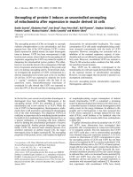
Figure 1 Effect of TGF-β superfamily members on BMP type I and
type II receptor transcript levels. NHLF were stimulated with 5 ng/
ml TGF-β1 or 100 ng/ml BMP-4 or BMP-7 for 24 h. Cells were harvested,
RNA extracted and reverse transcribed, and a real-time quantitative
PCR for ALK-2 (A), ALK-3 (B), ALK-6 (C), and BMPRII (D) was performed. Results are expressed as the ratio of each transcript relative to the geometric mean of mRNA expression of the housekeeping genes UBC,
SDHA, and RPL13a. Data are mean ± SD of five independent experiments. *, p < 0.05, as compared to unstimulated cells.
the addition of BMP-4, but not BMP-7, to TGF-β1-stimulated NHLF led to a significant decrease in cell proliferation as compared to either untreated or TGF-β1stimulated cells (Figure 2).
BMP-4, but not BMP-7, downregulates TGF-β1-induced
ECM protein expression
There is extensive published literature describing TGFβ1-driven ECM production in the airways as well as the
contribution of fibroblasts to the thickness of the subbasement membrane, however the role of BMPs in this
phenomenon is not yet described in the lung. Incubation
of NHLF for 24 h in the presence of 5 ng/ml TGF-β1 significantly up-regulated the expression of mRNAs encoding collagen types I and IV (10- and 9-fold increase,
respectively, Figures 3A and 3B). The increase in mRNA
transcripts correlated with increased synthesis and
release of total soluble collagen measured in cell supernatants (Figure 3C). Transcripts for tenascin C and
fibronectin were also upregulated by TGF-β1 (11- and
2.5-fold increase, respectively, Figures 4A and 4C). This
-
Figure 2 Simultaneous incubation of NHLF with TGF-β1 and
BMP-4 inhibits cell proliferation. [3H]thymidine incorporation in
NHLF in response to tissue culture media with 2% FBS in the presence
of 5 ng/ml TGF-β1 or 100 ng/ml BMP-4 or BMP-7 alone or with TGF-β1
in the presence of BMP-4 or BMP-7 for 36 h. [3H]thymidine was added
for the last 6 h of incubation. Data are mean ± SD of five independent
experiments. *, p < 0.05, as compared to unstimulated cells and †, p <
0.05, as compared to TGF-β1-stimulated cells.
increase was reflected at the protein level (18- and 1.7fold increase, Figures 4B and 4D, respectively), as determined by specific ELISA. In contrast, BMP-4 and BMP-7
(100 ng/ml) did not affect expression of the transcripts
encoding collagen type I or IV (Figures 3A and 3B), or
fibronectin (Figure 4C). However, a moderate but significant induction of the mRNA for tenascin C was measured after incubation of NHLF with both BMP-4 and
BMP-7 (Figure 4A). BMP-4 inhibited the TGF-β1induced increase in the level of the transcripts encoding
collagen type I and IV (Figures 3A and 3B), tenascin and
fibronectin (Figures 4A and 4C). A similar effect was
observed at the protein level with a 50% decrease in total
soluble collagen synthesis (Figure 3C), inhibition of the
release of tenascin C and fibronectin (30% and 20%,
respectively, Figures 4B and 4D). In contrast, BMP-7 did
not modify the TGF-β1-induced up-regulation of the
transcripts and proteins examined except for a significant
suppression of the expression of mRNA for tenascin C
(Figure 4A) but this result was not confirmed at the protein level (Figure 4B).
TGF-β family members modulate collagenase and
gelatinase activities and expression
The ECM accumulation observed in the asthmatic lung
can result from an increase in ECM protein production
Pegorier et al. Respiratory Research 2010, 11:85
/>
A
C
B
15
10.0
*†
7.5
5.0
2.5
mRNA level
*
0.0
*
*
30
µg/ml total
soluble collagen
*
Relative COL4a1
mRNA level
12.5
Relative COL1a1
Page 6 of 11
10
*†
5
0
*
20
†
10
0
-
+
-
+
-
+
TGF-β1 (5 ng/ml)
-
+
-
+
-
+
TGF-β1 (5 ng/ml)
-
+
-
+
-
BMP-4 (100 ng/ml) -
-
+
+
-
-
BMP-4 (100 ng/ml) -
-
+
+
-
-
BMP-4 (100 ng/ml) -
-
+
+
-
-
BMP-7 (100 ng/ml) -
-
-
-
+
+
BMP-7 (100 ng/ml) -
-
-
-
+
+
BMP-7 (100 ng/ml) -
-
-
-
+
+
TGF-β1 (5 ng/ml)
+
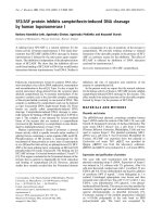
Figure 3 TGF-β1-induced collagen expression in NHLF is downregulated by BMP-4. NHLF were stimulated with 5 ng/ml TGF-β1 or 100 ng/ml
BMP-4 or BMP-7 alone, or with TGF-β1 in the presence of BMP-4 or BMP-7 for 24 h (A and B) or 72 h (C). Cells were harvested, RNA was extracted, reverse
transcribed, and a real-time quantitative PCR for collagen type I alpha 1 chain (COL1a1, A) and collagen type IV alpha 1 chain (COL4a1, B) was performed. Results are expressed as the ratio of each transcript relative to the geometric mean of mRNA expression of the housekeeping genes UBC, SDHA, and RPL13a. Total soluble collagen release was quantified in the cell supernatants by Sircol assay (C). Data are mean ± SD of five independent
experiments. *, p < 0.05, as compared to unstimulated cells and †, p < 0.05, as compared to TGF-β1-stimulated cells.
and/or a deregulation in proMMP activities, the activation of these proenzymes being a critical step that leads to
ECM breakdown. NHLF were stimulated for 72 h with
either TGF-β1, BMP-4 or BMP-7 or TGF-β1 in combination with BMP-4 or BMP-7, and MMP activity in the cell
supernatants was detected on gelatine gels by zymography. Both TGF-β1 and BMP-4 led to a moderate but significant increase in the gelatinolytic activity of the proforms of MMP-1 (57 and 52 kDa, Figure 5A) and MMP-2
(72 kDa, Figure 5B) whereas the activity of the active
forms was not modulated (47 and 42 kDa for MMP-1 and
67 kDa for MMP-2). BMP-7 itself did not alter the expression of MMP-1 or MMP-2 but its addition to TGF-β1stimulated cells led to a significant down-regulation in
the activity of the pro-MMP-2 as compared to cells stimulated with TGF-β1 alone (Figure 5B). MMP-9 activity
was not detected, regardless of the stimulation conditions. MMP-13 release from NHLF was decreased in the
presence of BMP-4 and BMP-7 compared to untreatedor TGF-β1-stimulated cells (Figure 5C). The inhibition of
MMP-13 release was of similar magnitude when the
BMPs were incubated in the presence of TGF-β. Increasing the concentration of BMPs to 1 μg/ml did not result in
further MMP-13 reductions (data not shown).
TGF-β1-induced fibroblast differentiation is partially
inhibited by BMP-7
Fibroblast differentiation into myofibroblasts is crucial in
tissue remodelling, wound healing, and various fibrotic
disorders in the lung and the contribution of TGF-β to
this phenomenon in vitro is well documented [5,11,35].
Here we characterized the effect of BMP-4 and BMP-7 on
the induction of a myofibroblast-like phenotype in nor-
mal lung fibroblasts exposed to TGF-β1. In culture,
NHLF basally expressed low levels of αSMA as demonstrated by immunohistochemistry (first panel, Figure 6A).
Stimulation with TGF-β1 led to a discernable increase in
α-SMA+ cell number (Figure 6B). Western blot of NHLF
cell lysates confirmed our observations. Incubation with
BMP-4 also led to an increase in the number of αSMA+
cells, whereas BMP-7 alone had no effect (Figure 6A and
6B). BMP-4 did not affect TGF-β1 driven α-SMA expression. In contrast, BMP-7 significantly inhibited TGF-β1
induced differentiation (Figure 6A and 6B).
BMPs do not affect TGF-β1-induced CTGF promoter and
Smad-Binding Element reporter gene activities
In order to determine the mechanism by which BMPs
counteract TGF-β1 effects, activity assays were performed on the CTGF promoter (pCT-sp) transfected in
NHLF and TGF-β responsive Smad-binding elements
(SBE) reporter gene in the MFB-F11 cell line. TGF-β1
increased luciferase activity in the pCT-sp 6-fold, indicative of CTGF promoter activity (Figure 7A) and SEAP
activity in the SBE-SEAP reporter 37-fold (Figure 7B) and
this response to TGF-β was not inhibited by either BMP4 or BMP-7. BMP-4 moderately increased pCT-sp activity (3.6-fold induction, Figure 7A) demonstrating that
BMP-4 partially acts via increasing CTGF promoter
activity. In contrast, the BMPs had no direct effect on the
SBE-SEAP reporter, indicating that they are not able to
inhibit binding of phosphorylated Smads (downstream
signalling molecules of TGF-β1) to the Smad-Binding
Element present on many genes regulated by TGF family
members.
Pegorier et al. Respiratory Research 2010, 11:85
/>
Page 7 of 11
A
B
600
*
15
*†
10
5
*
*†
*
ng/ml tenascin
mRNA level
Relative tenascin
20
*
500
*
400
*†
300
200
100
0
0
TGF-β1 (5 ng/ml)
-
+
-
+
-
+
TGF-β1 (5 ng/ml)
-
+
-
+
-
+
BMP-4 (100 ng/ml)
-
-
+
+
-
-
BMP-4 (100 ng/ml)
-
-
+
+
-
-
BMP-7 (100 ng/ml)
-
-
-
-
+
+
BMP-7 (100 ng/ml)
-
-
-
-
+
+
mRNA level
Relative fibronectin
3
*
*
2
*†
1
ng/ml fibronectin
D
C
*
600
500
*†
*
400
300
200
100
0
0
TGF-β1 (5 ng/ml)
-
+
-
+
-
+
TGF-β1 (5 ng/ml)
-
+
-
+
-
+
BMP-4 (100 ng/ml)
-
-
+
+
-
-
BMP-4 (100 ng/ml)
-
-
+
+
-
-
BMP-7 (100 ng/ml)
-
-
-
-
+
+
BMP-7 (100 ng/ml)
-
-
-
-
+
+
Figure 4 TGF-β1-induced ECM protein expression in NHLF is down-regulated by BMP-4. NHLF were stimulated with 5 ng/ml TGF-β1 or 100 ng/
ml BMP-4 or BMP-7 alone or with TGF-β1 in the presence of BMP-4 or BMP-7 for 24 h (A and B) or 48 h (C and D). Cells were harvested, RNA was extracted, reverse transcribed, and a real-time quantitative PCR for tenascin C (A) and fibronectin (C) was performed. Results are expressed as the ratio of
each transcript relative to the geometric mean of mRNA expression of the housekeeping genes UBC, SDHA, and RPL13a. Tenascin C and fibronectin
protein were quantified in the cell supernatants by specific ELISAs (B and D, respectively). Data are mean ± SD of five independent experiments. *, p
< 0.05, as compared to unstimulated cells and †, p < 0.05, as compared to TGF-β1-stimulated cells.
Discussion
In the current study, we determined the ability of two
Bone Morphogenetic Proteins, BMP-4 and BMP-7, to
modulate the profibrotic effects of TGF-β1 on NHLF. We
found that BMP-4 and BMP-7 are able to regulate the
synthesis and production of ECM proteins, MMPs and αSMA in primary lung fibroblasts. BMP-4 inhibits TGFβ1-induced cell proliferation and ECM protein release.
Both BMP-4 and BMP-7 decreased MMP-13 release in
TGF-β1-stimulated cells. In contrast, only BMP-7 inhibited myofibroblast differentiation and activation of
MMP-2 induced by TGF-β1. We have also shown that
TGF-β1 can act directly on the BMP pathways by increasing expression of the mRNA encoding ALK-6 and
BMPRII.
The ECM is known to be involved in a variety of cellular processes, including morphogenesis, lung remodelling, and modifications in cell shape that occur during
differentiation of a number of lung structural cells [5,36].
As a result, changes in the composition of the ECM can
profoundly affect the behaviour of cells and lead to airway
remodelling in lung fibrotic diseases, including asthma.
The increase in ECM deposition results from either
increased production or decreased breakdown of matrix
Pegorier et al. Respiratory Research 2010, 11:85
/>
Page 8 of 11
Pro-form MMP-1
Pro-form MMP-2
B
600
*
500
*
*
*
400
300
200
100
0
57/52 pro-MMP-1
47/42 active MMP-1
BMP-4 (100 ng/ml) BMP-7 (100 ng/ml) pg MMP-13 /ml supernatant
*
400
*
†
300
200
100
0
72kDa pro-MMP-2
67kDa active MMP-2
TGF-β1 (5 ng/ml)
C
Relative density of
gelatinolytic bands
Relative density of
gelatinolytic bands
A
+
-
+
+
+
-
+
-
TGF-β1 (5 ng/ml)
-
+
-
+
-
+
BMP-4 (100 ng/ml) -
-
+
+
-
-
-
-
-
+
+
BMP-7 (100 ng/ml) -
-
-
-
+
+
50
40
30
*†
20
*
*
10
*†
0
-
+
-
+
-
+
BMP-4 (100 ng/ml) -
-
+
+
-
-
BMP-7 (100 ng/ml) -
-
-
-
+
+
TGF-β1 (5 ng/ml)
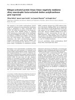
Figure 5 Effect of TGF-β superfamily members on MMP activity and expression level. NHLF were stimulated with 5 ng/ml TGF-β1 or 100 ng/ml
BMP-4 or BMP-7 alone or with TGF-β1 in the presence of BMP-4 or BMP-7 for 72 h. Cell supernatants were collected to perform zymography (A and B)
and ELISA (C). Representative gelatin zymograms and related graphic plot of the bands obtained in zymographs for the pro-forms of MMP-1 (A) and
MMP-2 (B) were performed. Gelatinolytic activity of the pro- and active forms of MMP-1 (57/52 and 47/42 kDa) and pro- and active forms of MMP-2
(72 and 67 kDa) are indicated. MMP-13 release was quantified in the cell supernatants by specific ELISA (C). Data are mean ± SD of five independent
experiments. *, p < 0.05, as compared to unstimulated cells and †, p < 0.05, as compared to TGF-β1-stimulated cells.
products. Deregulation of the proteolytic-antiproteolytic
network and inappropriate secretion of various MMPs by
stimulated lung structural cells is thought to be involved
in the pathophysiology of asthma [37]. The contribution
of TGF-β1 to ECM accumulation, and to fibroblast differentiation and proliferation has been widely reported
[5,35,38,39]. Its action is mainly driven by activation of
CTGF, resulting in stimulation of fibroblast proliferation,
myofibroblast differentiation and collagen synthesis
[40,41]. In this study, we confirmed the ability of TGF-β1
to induce production of the ECM proteins collagen types
I and IV, fibronectin and tenascin C, and to induce myofi-
broblastic differentiation. However, we did not observe
TGF-β1-induced fibroblast proliferation as previously
reported by some groups [9,42,43] but those data might
be considered controversial since the effect of TGF-β1 on
fibroblast proliferation is dependent on its concentration
[44]. The increased expression of αSMA correlates with
the release of collagen and activation of MMP-1, the
major enzyme involved in degradation of native collagen,
which is in accordance with the data showing that myofibroblasts are the major source of collagen type I in the
lung [45]. Finally we confirmed the ability of TGF-β1 to
activate both the CTGF promoter and Smad-binding ele-
Pegorier et al. Respiratory Research 2010, 11:85
/>
A
- TGF-β1
Page 9 of 11
+ TGF-β1
Figure 7 TGF-β1-induced CTGF promoter and SBE-SEAP reporter
activities are not modulated by the BMPs. (A) The CTGF promoter
pCT-sb was transiently transfected into NHLF, cells were then treated
with 5 ng/ml TGF-β1 or 100 ng/ml BMP-4 or BMP-7 or with TGF-β1 in
the presence of BMP-4 or BMP-7 in FGM containing 0.2% FBS. All assays
were performed with 150000 cells/well in 2 ml total volume in 6-well
plates and luciferase activity was measured after 24 h induction in 50
μl cell pellet. (B) MFB-F11 cells stably transfected with SBE-SEAP were
stimulated with 5 ng/ml TGF-β1 or 100 ng/ml BMP-4 or BMP-7 or with
TGF-β1 in the presence of BMP-4 or BMP-7 in serum-free DMEM. All assays were performed with 40000 cells/well in 100 μl total volume in 96well plates and SEAP activity was measured after 24 h induction in 10
μl supernatant. Data are mean ± SD of five independent experiments.
*, p < 0.05, as compared with unstimulated cells.
B
42 kDa
TGF-β1 (5 ng/ml)
-
+
-
-
+
BMP-4 (100 ng/ml)
-
-
+
-
+
-
BMP-7 (100 ng/ml)
-
-
-
+
-
+
+
Figure 6 TGF-β1-induced myofibroblast like phenotype in NHLF
is partially inhibited by BMP-7. NHLF were stimulated with 5 ng/ml
TGF-β1 or 100 ng/ml BMP-4 or BMP-7 or with TGF-β1 in the presence
of BMP-4 or BMP-7 for 72 h. Representative panel of α-SMA expression
was obtained by immunohistochemistry (A) and western blot of cell
lysates for α-SMA is shown in (B). Data are representative of five independent experiments.
ments (SBE) contained in the promoter region of more
than 500 target genes responding to TGF-β1 [34].
In most models and cell types, BMP-7 opposes TGFβ1-mediated ECM protein production in vivo and in vitro
[19-26]. BMP-7 regulates the ECM breakdown in human
chondrocytes by downregulating MMP-13 [46]. Nevertheless, two recent studies have shown that BMP-7 fails to
inhibit TGF-β mediated fibrosis in the lung, skin and
renal tubular epithelial cells [27,28]. In our model, BMP-7
did not counteract the increase in ECM proteins induced
by TGF-β1. However, we have shown for the first time in
lung fibroblasts that BMP-7 reduces not only the basal
fibroblast-related expression of MMP-13 but also the
induced expression of this protein following stimulation
by TGF-β1. MMP-13, an interstitial collagenase, is the
principal enzyme involved in the initiation of collagen
breakdown. MMP-2 can serve as an activator of other
MMPs, namely MMP-13 [47]. Thus, the downregulation
of TGF-β1-induced MMP-2 activity by BMP-7 is in
accordance with the inhibition shown for MMP-13.
BMP-7 could contribute to a reduction in airway remodelling by inhibiting some MMPs without affecting ECM
protein release. BMP-7 was also able to counteract TGFβ1-induced fibroblast differentiation. This potential regulatory function of BMP-7 confirms its ability to contribute to resolution of lung remodelling since increased
numbers of myofibroblasts and fibroblast differentiation
are major features of airway remodelling.
The role of BMP-4 in degradation and remodelling of
the ECM remains unclear, particularly in the lung. In fact,
little is known about the properties of BMP-4 either in
vivo or in vitro in the lung or other tissues. A regulatory
effect of BMP-4 on MMP-13 release in human adipocytes
has been reported [48] as well as an inhibition of cell proliferation and an upregulation of αSMA expression in foetal lung fibroblasts [30], but nothing is known of its
effects on adult lung fibroblasts. Here, we demonstrate
for the first time that BMP-4 is able to counteract the
increase in ECM protein release induced by TGF-β1 in
NHLF. We also reported that BMP-4 not only reduces the
basal fibroblast-related expression of MMP-13 but also its
expression induced by TGF-β1. The contribution of
BMP-4 to the reduction of airway remodelling could
result from a direct modulation of the production of
ECM proteins as well as MMP-13. In our study, BMP-4
Pegorier et al. Respiratory Research 2010, 11:85
/>
had no direct effect on fibroblast proliferation. This is in
contrast to the study of Jeffery et al. which reported inhibition of fibroblast proliferation but their study was performed on foetal fibroblasts which possess a higher
intrinsic capacity for self-renewal than adult cells. The
differential response of NHLF to BMP-4 and BMP-7 may
also be a function of the signalling pathways utilized or,
alternatively, the regulation of different transcriptional
repressors or activators. It is likely that BMP-4 and BMP7 act via different pathways to regulate ECM accumulation. BMP-7 selectively binds to receptors distinct from
those of BMP-4: BMP-4 binds and activates ALK-3 and
ALK-6 whereas BMP-7 preferentially binds to ALK-2 and
ALK-6 [49-51]. Furthermore, the actions of the BMPs, at
least BMP-7, may be tissue or cell type specific since the
inhibitory effects of BMP-7 on remodelling are less pronounced in the lung than other tissues.
Conclusions
Evidence from animal models suggests that airway
remodelling in asthma may be prevented or reversed
using agents which target TGF-β [8,52]. Therefore, modulation of TGF-β or its activity represents a potential
therapeutic target for asthma and other fibrotic diseases.
We were the first to report dysregulation of BMP and
BMPR expression in asthma [31]. Others have shown an
up-regulation of Gremlin, an inhibitor of BMP-4 signaling pathways, in idiopathic pulmonary fibrosis and have
suggested that this increased expression of Gremlin may
be a key event in the persistence of myofibroblasts in the
lung interstitium [53]. Taken together, these data lend
weight to the argument that BMP-4 plays a crucial role in
the regulation of lung fibroblasts in disease. Our current
study has determined that BMP-7 can also exert some
functional effects on TGF-β1-driven profibrotic processes in normal lung fibroblasts. These BMPs appear to
be attractive targets for therapeutic intervention in asthmatic disease although the blockade of TGF-β1 by only
one of these molecules may not be sufficient to totally
inhibit activity. A better understanding of how BMPs act
in vitro on lung structural cells and in vivo in animal
models of asthma could potentially lead to the amelioration of airway remodelling and consequently a decrease
of asthma symptoms.
Competing interests
The authors declare that they have no competing interests.
Authors' contributions
SP carried out the majority of experimental work and drafted the manuscript.
GAC carried out the western blotting. ABK participated in the design and coordination of the study. CML conceived of the study, participated in its design
and coordination and helped to draft the manuscript. All authors read and
approved the final manuscript.
Page 10 of 11
Acknowledgements
This work was funded by Wellcome Trust grant number PC3292 and the
Asthma UK grant number P16033.
Author Details
Leukocyte Biology Section, MRC and Asthma UK Centre in Allergic Mechanisms
of Asthma, National Heart and Lung Institute, Faculty of Medicine, Imperial
College London, London, UK
Received: 20 August 2009 Accepted: 23 June 2010
Published: 23 June 2010
© 2010 Pegorier et al; licenseedistributed under the terms of the Creative Commons Attribution License ( which permits unrestricted use, distribution, and reproduction in any medium, provided the original work is properly cited.
This is an Open Access from: />Respiratory is available article BioMed Central Ltd.
article Research 2010, 11:85
References
1. Bousquet J, Jeffery PK, Busse WW, Johnson M, Vignola AM: Asthma. From
bronchoconstriction to airways inflammation and remodeling. Am J
Respir Crit Care Med 2000, 161(5):1720-1745.
2. James AL, Wenzel S: Clinical relevance of airway remodelling in airway
diseases. Eur Respir J 2007, 30(1):134-155.
3. Lloyd CM, Robinson DS: Allergen-induced airway remodelling. Eur
Respir J 2007, 29(5):1020-1032.
4. Munz B, Hubner G, Tretter Y, Alzheimer C, Werner S: A novel role of activin
in inflammation and repair. J Endocrinol 1999, 161(2):187-193.
5. Makinde T, Murphy RF, Agrawal DK: The regulatory role of TGF-beta in
airway remodeling in asthma. Immunol Cell Biol 2007, 85(5):348-356.
6. Chen D, Zhao M, Harris SE, Mi Z: Signal transduction and biological
functions of bone morphogenetic proteins. Front Biosci 2004,
9:349-358.
7. Szefler SJ: Airway remodeling: therapeutic target or not? Am J Respir Crit
Care Med 2005, 171(7):672-673.
8. McMillan SJ, Xanthou G, Lloyd CM: Manipulation of allergen-induced
airway remodeling by treatment with anti-TGF-beta antibody: effect
on the Smad signaling pathway. J Immunol 2005, 174(9):5774-5780.
9. Khalil N, Xu YD, O'Connor R, Duronio V: Proliferation of pulmonary
interstitial fibroblasts is mediated by transforming growth factorbeta1-induced release of extracellular fibroblast growth factor-2 and
phosphorylation of p38 MAPK and JNK. J Biol Chem 2005,
280(52):43000-43009.
10. Jinnin M, Ihn H, Asano Y, Yamane K, Trojanowska M, Tamaki K: Tenascin-C
upregulation by transforming growth factor-beta in human dermal
fibroblasts involves Smad3, Sp1, and Ets1. Oncogene 2004,
23(9):1656-1667.
11. Evans RA, Tian YC, Steadman R, Phillips AO: TGF-beta1-mediated
fibroblast-myofibroblast terminal differentiation-the role of Smad
proteins. Exp Cell Res 2003, 282(2):90-100.
12. Phipps S, Benyahia F, Ou TT, Barkans J, Robinson DS, Kay AB: Acute
allergen-induced airway remodeling in atopic asthma. Am J Respir Cell
Mol Biol 2004, 31(6):626-632.
13. Camara J, Jarai G: Epithelial-mesenchymal transition in primary human
bronchial epithelial cells is SMad-dependent and enhanced by
fibronectin and TNF-alpha. Fibrogenesis & Tissue Repair 2010, 3(2):.
14. Schmidt-Weber CB, Blaser K: Regulation and role of transforming
growth factor-beta in immune tolerance induction and inflammation.
Curr Opin Immunol 2004, 16(6):709-716.
15. Chen G, Khalil N: TGF-beta1 increases proliferation of airway smooth
muscle cells by phosphorylation of map kinases. Respir Res 2006, 7:2.
16. Alcorn JF, Rinaldi LM, Jaffe EF, van Loon M, Bates JH, Janssen-Heininger
YM, Irvin CG: Transforming growth factor-beta1 suppresses airway
hyperresponsiveness in allergic airway disease. Am J Respir Crit Care
Med 2007, 176(10):974-982.
17. Chen D, Zhao M, Mundy GR: Bone morphogenetic proteins. Growth
Factors 2004, 22(4):233-241.
18. Reddi AH: Bone morphogenetic proteins: an unconventional approach
to isolation of first mammalian morphogens. Cytokine Growth Factor Rev
1997, 8(1):11-20.
19. Gonzalez EA, Lund RJ, Martin KJ, McCartney JE, Tondravi MM, Sampath TK,
Hruska KA: Treatment of a murine model of high-turnover renal
osteodystrophy by exogenous BMP-7. Kidney Int 2002, 61(4):1322-1331.
20. Izumi N, Mizuguchi S, Inagaki Y, Saika S, Kawada N, Nakajima Y, Inoue K,
Suehiro S, Friedman SL, Ikeda K: BMP-7 opposes TGF-beta1-mediated
collagen induction in mouse pulmonary myofibroblasts through Id2.
Am J Physiol Lung Cell Mol Physiol 2006, 290(1):L120-126.
Pegorier et al. Respiratory Research 2010, 11:85
/>
21. Klahr S: The bone morphogenetic proteins (BMPs). Their role in renal
fibrosis and renal function. J Nephrol 2003, 16(2):179-185.
22. Klahr S, Morrissey J: Obstructive nephropathy and renal fibrosis: The
role of bone morphogenic protein-7 and hepatocyte growth factor.
Kidney Int Suppl 2003:S105-112.
23. Maric I, Poljak L, Zoricic S, Bobinac D, Bosukonda D, Sampath KT, Vukicevic
S: Bone morphogenetic protein-7 reduces the severity of colon tissue
damage and accelerates the healing of inflammatory bowel disease in
rats. J Cell Physiol 2003, 196(2):258-264.
24. Zeisberg M, Hanai J, Sugimoto H, Mammoto T, Charytan D, Strutz F, Kalluri
R: BMP-7 counteracts TGF-beta1-induced epithelial-to-mesenchymal
transition and reverses chronic renal injury. Nat Med 2003, 9(7):964-968.
25. Zeisberg M, Bottiglio C, Kumar N, Maeshima Y, Strutz F, Muller GA, Kalluri
R: Bone morphogenic protein-7 inhibits progression of chronic renal
fibrosis associated with two genetic mouse models. Am J Physiol Renal
Physiol 2003, 285(6):F1060-1067.
26. Zeisberg M, Shah AA, Kalluri R: Bone morphogenic protein-7 induces
mesenchymal to epithelial transition in adult renal fibroblasts and
facilitates regeneration of injured kidney. J Biol Chem 2005,
280(9):8094-8100.
27. Murray LA, Hackett TL, Warner SM, Shaheen F, Argentieri RL, Dudas P,
Farrell FX, Knight DA: BMP-7 does not protect against bleomycininduced lung or skin fibrosis. PLoS ONE 2008, 3(12):e4039.
28. Dudas PL, Argentieri RL, Farrell FX: BMP-7 fails to attenuate TGF-{beta}1induced epithelial-to-mesenchymal transition in human proximal
tubule epithelial cells. Nephrol Dial Transplant 2008.
29. Shannon JM, Hyatt BA: Epithelial-mesenchymal interactions in the
developing lung. Annu Rev Physiol 2004, 66:625-645.
30. Jeffery TK, Upton PD, Trembath RC, Morrell NW: BMP4 inhibits
proliferation and promotes myocyte differentiation of lung fibroblasts
via Smad1 and JNK pathways. Am J Physiol Lung Cell Mol Physiol 2005,
288(2):L370-378.
31. Kariyawasam HH, Xanthou G, Barkans J, Aizen M, Kay AB, Robinson DS:
Basal Expression of Bone Morphogenetic Protein (BMP) Receptor is
Reduced in Mild Asthma. Am J Respir Crit Care Med 2008,
177(10):1074-81.
32. Gould SE, Day M, Jones SS, Dorai H: BMP-7 regulates chemokine,
cytokine, and hemodynamic gene expression in proximal tubule cells.
Kidney Int 2002, 61(1):51-60.
33. Vandesompele J, De Preter K, Pattyn F, Poppe B, Van Roy N, De Paepe A,
Speleman F: Accurate normalization of real-time quantitative RT-PCR
data by geometric averaging of multiple internal control genes.
Genome Biol 2002, 3(7):RESEARCH0034.
34. Tesseur I, Zou K, Berber E, Zhang H, Wyss-Coray T: Highly sensitive and
specific bioassay for measuring bioactive TGF-beta. BMC Cell Biol 2006,
7:15.
35. Desmouliere A, Geinoz A, Gabbiani F, Gabbiani G: Transforming growth
factor-beta 1 induces alpha-smooth muscle actin expression in
granulation tissue myofibroblasts and in quiescent and growing
cultured fibroblasts. J Cell Biol 1993, 122(1):103-111.
36. Yamauchi K, Inoue H: Airway remodeling in asthma and irreversible
airflow limitation-ECM deposition in airway and possible therapy for
remodeling. Allergol Int 2007, 56(4):321-329.
37. Gueders MM, Foidart JM, Noel A, Cataldo DD: Matrix metalloproteinases
(MMPs) and tissue inhibitors of MMPs in the respiratory tract: potential
implications in asthma and other lung diseases. Eur J Pharmacol 2006,
533(1-3):133-144.
38. Chambers RC, Leoni P, Kaminski N, Laurent GJ, Heller RA: Global
expression profiling of fibroblast responses to transforming growth
factor-beta1 reveals the induction of inhibitor of differentiation-1 and
provides evidence of smooth muscle cell phenotypic switching. Am J
Pathol 2003, 162(2):533-546.
39. Kurosaka H, Kurosaka D, Kato K, Mashima Y, Tanaka Y: Transforming
growth factor-beta 1 promotes contraction of collagen gel by bovine
corneal fibroblasts through differentiation of myofibroblasts. Invest
Ophthalmol Vis Sci 1998, 39(5):699-704.
40. Grotendorst GR: Connective tissue growth factor: a mediator of TGFbeta action on fibroblasts. Cytokine Growth Factor Rev 1997,
8(3):171-179.
41. Burgess JK: Connective tissue growth factor: a role in airway
remodelling in asthma? Clin Exp Pharmacol Physiol 2005, 32(11):988-994.
Page 11 of 11
42. Pelaia G, Gallelli L, D'Agostino B, Vatrella A, Cuda G, Fratto D, Renda T,
Galderisi U, Piegari E, Crimi N, Rossi F, Caputi M, Costanzo FS, Vancheri C,
Maselli R, Marsico SA: Effects of TGF-beta and glucocorticoids on map
kinase phosphorylation, IL-6/IL-11 secretion and cell proliferation in
primary cultures of human lung fibroblasts. J Cell Physiol 2007,
210(2):489-497.
43. Bhowmick NA, Chytil A, Plieth D, Gorska AE, Dumont N, Shappell S,
Washington MK, Neilson EG, Moses HL: TGF-beta signaling in fibroblasts
modulates the oncogenic potential of adjacent epithelia. Science 2004,
303(5659):848-851.
44. Duvernelle C, Freund V, Frossard N: Transforming growth factor-beta
and its role in asthma. Pulm Pharmacol Ther 2003, 16(4):181-196.
45. Phan SH: The myofibroblast in pulmonary fibrosis. Chest 2002, 122(6
Suppl):286S-289S.
46. Im HJ, Pacione C, Chubinskaya S, Van Wijnen AJ, Sun Y, Loeser RF:
Inhibitory effects of insulin-like growth factor-1 and osteogenic
protein-1 on fibronectin fragment- and interleukin-1beta-stimulated
matrix metalloproteinase-13 expression in human chondrocytes. J Biol
Chem 2003, 278(28):25386-25394.
47. Li H, Simon H, Bocan TM, Peterson JT: MMP/TIMP expression in
spontaneously hypertensive heart failure rats: the effect of ACE- and
MMP-inhibition. Cardiovasc Res 2000, 46(2):298-306.
48. Otto TC, Bowers RR, Lane MD: BMP-4 treatment of C3H10T1/2 stem cells
blocks expression of MMP-3 and MMP-13. Biochem Biophys Res
Commun 2007, 353(4):1097-1104.
49. ten Dijke P, Yamashita H, Sampath TK, Reddi AH, Estevez M, Riddle DL,
Ichijo H, Heldin CH, Miyazono K: Identification of type I receptors for
osteogenic protein-1 and bone morphogenetic protein-4. J Biol Chem
1994, 269(25):16985-16988.
50. Rosenzweig BL, Imamura T, Okadome T, Cox GN, Yamashita H, ten Dijke P,
Heldin CH, Miyazono K: Cloning and characterization of a human type II
receptor for bone morphogenetic proteins. Proc Natl Acad Sci USA 1995,
92(17):7632-7636.
51. Macias-Silva M, Hoodless PA, Tang SJ, Buchwald M, Wrana JL: Specific
activation of Smad1 signaling pathways by the BMP7 type I receptor,
ALK2. J Biol Chem 1998, 273(40):25628-25636.
52. Le AV, Cho JY, Miller M, McElwain S, Golgotiu K, Broide DH: Inhibition of
allergen-induced airway remodeling in Smad 3-deficient mice. J
Immunol 2007, 178(11):7310-7316.
53. Koli K, Myllarniemi M, Vuorinen K, Salmenkivi K, Ryynanen MJ, Kinnula VL,
Keski-Oja J: Bone morphogenetic protein-4 inhibitor gremlin is
overexpressed in idiopathic pulmonary fibrosis. Am J Pathol 2006,
169(1):61-71.
doi: 10.1186/1465-9921-11-85
Cite this article as: Pegorier et al., Bone Morphogenetic Protein (BMP)-4 and
BMP-7 regulate differentially Transforming Growth Factor (TGF)-?1 in normal
human lung fibroblasts (NHLF) Respiratory Research 2010, 11:85