Báo cáo y học: "Alterations in the muscle-to-capillary interface in patients with different degrees of chronic obstructive pulmonary disease" pps
Bạn đang xem bản rút gọn của tài liệu. Xem và tải ngay bản đầy đủ của tài liệu tại đây (635.95 KB, 7 trang )
Eliason et al. Respiratory Research 2010, 11:97
/>Open Access
RESEARCH
© 2010 Eliason et al; licensee BioMed Central Ltd. This is an Open Access article distributed under the terms of the Creative Commons
Attribution License ( which permits unrestricted use, distribution, and reproduction in
any medium, provided the original work is properly cited.
Research
Alterations in the muscle-to-capillary interface in
patients with different degrees of chronic
obstructive pulmonary disease
Gabriella Eliason*
1
, Samy M Abdel-Halim
1,2
, Karin Piehl-Aulin
1,3
and Fawzi Kadi
1
Abstract
Background: It is hypothesized that decreased capillarization of limb skeletal muscle is implicated in the decreased
exercise tolerance in COPD patients. We have recently demonstrated decreased number of capillaries per muscle fibre
(CAF) but no changes in CAF in relation to fibre area (CAFA), which is based on the diffusion distance between the
capillary and muscle fibre. The aim of the current study is to investigate the muscle-to-capillary interface which is an
important factor involved in oxygen supply to the muscle that has previously been suggested to be a more sensitive
marker for changes in the capillary bed compared to CAF and CAFA.
Methods: 23 COPD patients and 12 age-matched healthy subjects participated in the study. Muscle-to-capillary
interface was assessed in muscle biopsies from the tibialis anterior muscle using the following parameters:
1) The capillary-to-fibre ratio (C:F
i
) which is defined as the sum of the fractional contributions of all capillary contacts
around the fibre
2) The ratio between C:F
i
and the fibre perimeter (CFPE-index)
3) The ratio between length of capillary and fibre perimeter (LC/PF) which is also referred to as the index of tortuosity.
Exercise capacity was determined using the 6-min walking test.
Results: A positive correlation was found between CFPE-index and ascending disease severity with CFPE-index for
type I fibres being significantly lower in patients with moderate and severe COPD. Furthermore, a positive correlation
was observed between exercise capacity and CFPE-index for both type I and type IIa fibres.
Conclusion: It can be concluded that the muscle-to-capillary interface is disturbed in the tibialis anterior muscle in
patients with COPD and that interface is strongly correlated to increased disease severity and to decreased exercise
capacity in this patient group.
Introduction
COPD (chronic obstructive pulmonary disease) is a dis-
ease characterized by irreversible airflow obstruction [1].
A significant number of patients with COPD develop
skeletal muscle wasting and decreased exercise capacity
[2-5]. Previous studies have demonstrated the occurrence
of a shift towards fatigue-susceptible anaerobic glycolytic
muscle properties relative to the aerobic oxidative muscle
properties [6-9]. We have previously shown that changes
in fibre type composition occur in the later stages of
COPD while exercise capacity is decreased already in
mild and moderate COPD, indicating that other factors
also may influence decreased exercise capacity in these
patients [10]. Exercise tolerance is partly dependent on
the oxidative capacity of the skeletal muscle and an
important limiting factor for exercise capacity in COPD
is the oxygen supply to the muscle [11]. The oxidative
metabolism in skeletal muscle is dependent on the mito-
chondrial volume density and activity and on the capil-
lary supply. Therefore alterations in muscle capillary
network or mitochondria can cause decreased exercise
tolerance in COPD. Indeed, a previous study has reported
lower mitochondrial volume density but no changes in
* Correspondence:
1
School of Medical Sciences, Örebro University, Örebro, Sweden
Full list of author information is available at the end of the article
Eliason et al. Respiratory Research 2010, 11:97
/>Page 2 of 7
mitochondrial respiratory function in patients with
COPD [12]. Furthermore, three previous studies have
suggested a decreased number of capillaries/muscle fibre
(CAF) in patients with COPD [8,9,13]. However, it is
important to highlight the fact that the ratio between the
number of capillaries and the area of muscle fibres
(CAFA) is not significantly altered in COPD patients
[8,9,13]. This finding can be explained by a reduction in
fibre area as previously shown in COPD patients [10].
This is also in line with a previous study [9] indicating a
parallel reduction in the number of capillaries around the
fibre and the area of the muscle fibre in COPD patients.
Therefore based on the capillary parameter CAFA, mus-
cle capillarization is not decreased in COPD patients.
The capillary supply is usually assessed by counting the
number of capillaries around each fibre (CAF) or by com-
puting the ratio between CAF and the area of the muscle
fibre (CAFA). CAFA is a parameter based on the diffu-
sion distance between the capillary and the centre of the
fibre. Capillary parameters essentially determining the
diffusion distance may not detect actual disturbances in
muscle capillarization. It has previously been suggested
that the muscle-to-capillary interface is an important fac-
tor involved in oxygen supply to the muscle [14-19] and
may thereby be used as a more sensitive marker for
changes in the capillary bed compared to CAF and CAFA
[15,16]. To asses muscle-to-capillary interface precise ste-
reological procedures such as the capillary-to-fibre
perimeter ratio have been used [20]. Some stereological
methods cannot be used in human studies since muscle
samples need to be perfused and fixed in order not to col-
lapse. However, capillary-to-fibre ratio for an individual
fibre (C:F
i
) can be assessed by determining the number of
capillaries around the fibre and the sharing factor (SF) for
each fibre and thereafter calculating the sum of the frac-
tional contributions of each capillary contact. Thereafter
the capillary-to-fibre perimeter exchange index (CFPE-
index) can be calculated as the quotient C:F
i
and fibre
perimeter [19]. As CFPE-index has been shown to be cor-
related to precise stereological methods it can be used to
assess muscle fibre-to-capillary interface in human stud-
ies [15,19].
Measuring the percentage of fibre perimeter in contact
with the capillary wall (index of tortuosity (LC/PF))
which is based on the length of capillaries, the number of
capillaries and the perimeter of the fibre is another sensi-
tive method for assessment of muscle-to-capillary inter-
face which also takes in account the capillary geometry
[21]. To our knowledge CFPE-index and LC/PF have not
been evaluated in COPD and the question of whether
muscle-to-capillary interface is altered in this patient
group remains unknown.
Given earlier reports suggesting disturbed limb skeletal
muscle capillarization in COPD patients [8,13] the cur-
rent study aim was to examine the muscle-to-capillary
interface in different stages of COPD and its correlation
with the degree of airflow obstruction and exercise
capacity.
Materials and methods
Study population
Twenty-three COPD patients (10 males and 13 females)
mean age 62.0 ± 6.6 years, were recruited from the
Department of Respiratory Medicine at Örebro Univer-
sity Hospital (Table 1). The patients were selected in a
stable condition and were not suffering from any respira-
tory tract infections or exacerbations of their disease four
weeks prior to sampling date. Exclusion criteria were
Table 1: Anthropometry and exercise capacity in 23 COPD patients and 12 age-matched healthy subjects.
Healthy
Subjects
(n = 12)
Mild COPD (n = 8) Moderate COPD
(n = 9)
Severe
COPD
(n = 6)
P-value
Age (years) 61.9 ± 7.9 60.8 ± 7.5 61.1 ± 3.8 64.0 ± 8.7 ns
Height (cm) 170.2 ± 10.4 169.5 ± 10.4 170.5 ± 8.2 167.7 ± 6.3 ns
Weight (kg) 77.1 ± 12.4 78.4 ± 21.0 82.4 ± 22.0 66.4 ± 11.3 ns
BMI (kg/m
2
)
26.6 ± 3.6 27.0 ± 5.4 28.0 ± 5.5 23.6 ± 3.9 ns
FEV
1.0
(% of expected) 115 ± 12 86 ± 6 48 ± 8* 26 ± 3* < 0.001
PaO
2
(kPa) 11.1 ± 1.3 10.5 ± 1.5 9.5 ± 1.1 9.2 ± 1.1* 0,01
PaCO
2
(kPa) 5.2 ± 0.3 4.8 ± 0.3 5.0 ± 0.6 5.6 ± 0.7* 0.02
Distance walked in 6 min (m) 502 ± 52 422 ± 43 336 ± 46* 248 ± 91* < 0.001
Data are presented as mean ± SD.
* Significant difference compared to healthy subjects.
n = number of test subjects; BMI = body mass index; FEV
1.0
= forced expiratory volume in one second; PaO2 = partial arterial pressure for
oxygen; PaCO2 = partial arterial pressure for carbon dioxide; ns = not significant.
Eliason et al. Respiratory Research 2010, 11:97
/>Page 3 of 7
malignancy, cardiac failure and severe endocrine-,
hepatic- or renal disorder. Based on the severity of airflow
obstruction the patients were divided into three sub-
groups based on the "Global Initiative for Chronic
Obstructive Lung Disease (GOLD)" criteria [1]. Eight
patients (four males and four females) had mild COPD
(forced expiratory volume in 1 s (FEV
1.0
) > 80% of pre-
dicted), nine patients (two males and seven females) had
moderate COPD (FEV
1.0
30-80% of predicted) and six
patients (four males and two females) had severe COPD
(FEV
1.0
< 30% of predicted). Twelve age-matched, healthy,
non-smoking subjects (n = 12, 6 male, 6 female) were
recruited as a control group (Table 1).
Written informed consent was obtained from all sub-
jects before their participation in the study, which was
approved by the Ethics Board of Uppsala University, Swe-
den (dnr 2004:M-355).
Pulmonary function tests
All patients and age-matched healthy subjects underwent
a spirometry with reversibility test to determine FEV
1.0
with the highest value from at least three technically
acceptable assessments being used.
Blood samples
All participants were sampled for arterial blood gases
from the radial artery at rest. The samples were analysed
for partial arterial pressure for oxygen (PaO
2
) and carbon
dioxide (PaCO
2
).
Exercise capacity test
Exercise capacity was determined using a 6 min walking
test performed on a 25 meter "court" as previously
reported [10] (Table 1).
Muscle samples
Muscle biopsies were obtained from the bulk of the tibia-
lis anterior muscle, which is an important postural mus-
cle active daily for long periods and involved in balance
control and foot stability during walking [22], under local
anaesthesia (Xylocaine
®
2%) as previously described
[10,13,23]. The biopsies were frozen in isopentane cooled
to its freezing point in liquid nitrogen and stored in -80°C
until analyses were performed.
Immunohistochemistry
Serial transverse sections, 5 μm thick, were cut at -22°C
using a microtome (Leica CM1850, Leica Microsystems,
Germany) and mounted on glass slides. Muscle fibre
composition was determined by immunohistochemical
staining using the monoclonal antibodies N2.261 and
A4.951 (Developmental Studies Hybridoma Bank, Uni-
versity of Iowa) [24] as previously described [10,13]
(Table 2). Fibres of type I, type IIa, type IIx, type IIx-a and
type IIa-b were determined. Fibre area and fibre perime-
ter for type I and type IIa fibres were determined on four
to ten randomly selected areas (table 2). For the visualiza-
tion of capillaries the monoclonal antibody CD31 (Dako,
Glostrup, Denmark; MO823) was used [13,16]. For visu-
alization of the fibre cytoplasm the histological staining
using eosin was applied. CD 31 has been used for the
identification of capillaries in several studies and a com-
parison between CD 31 staining and α-amylase-PAS for
identifying capillaries showed that the use of both meth-
ods results in a similar number of capillaries counted by
the observer and that CD31 staining allows an easier
visualization of capillaries [16]. Sequential estimation
analyses indicate that 50 fibres from one biopsy are suffi-
cient to characterise capillary parameters [25]. In the
present study capillaries in contact with oxidative type I
fibres and glycolytic type IIa fibres were analysed from
pictures taken with a magnitude of ×20 obtained from
four to ten randomly selected cross-sectional areas corre-
sponding to a mean of 80 fibres from each biopsy.
In a previous study we have reported the number of
capillaries around a single muscle fibre (CAF) and the
ratio between CAF and the fibre area (CAFA) [13] (table
2). The capillary parameters measured in transverse sec-
tions of the muscle biopsies in the present study were:
1) The capillary-to-fibre ratio (C:F
i
), which was calcu-
lated by determining the number of capillaries around for
the fibre in question followed by determination of the
sharing factor (SF) for each capillary and thereafter tak-
ing the sum of the fractional contributions of all capillary
contacts around the fibre [19].
2) The quotient between C:F
i
and the fibre perimeter,
i.e. the CFPE-index [19]
3) The ratio between length of capillary and fibre
perimeter (LC/PF) which represents the percent of mus-
cle fibre perimeter in contact with capillary wall. LC/PF is
also referred to as the index of tortuosity [15,16].
Statistic analysis
Statistics were performed using Statistix
®
8 (Analytic Soft-
ware, Tallahassee).
All data are presented as mean ± standard deviation.
For comparison between groups the Kruskal-Wallis one
way ANOVA test was used. When significance was found
the Kruskal-Wallis all pairwise comparison post-hoc test
was applied. Relationships between variables were stud-
ied using Spearmans rank correlation test. p < 0.05 was
considered to be significant.
Results
The following parameters associated with muscle-to-cap-
illary interface were assessed in the COPD population:
the capillary-to-fibre ratio (C:F
i
), the capillary-to-fibre
Eliason et al. Respiratory Research 2010, 11:97
/>Page 4 of 7
perimeter exchange index (CFPE-index) and the index of
tortuosity (LC/PF).
The C:F
i
for type I fibres was significantly lower (p =
0.007) in the groups with moderate and severe COPD
compared with the age-matched healthy subjects and the
C:F
i
for type IIa fibres was significantly lower (p = 0.002)
in the group with severe COPD compared to the age-
matched healthy subjects, indicating that each capillary is
shared by more muscle fibres in these patient groups
(Table 3). We also found that CFPE-index for type I fibres
was significantly lower (p = 0.002) in the groups with
moderate and severe COPD compared with the age-
matched healthy subjects (Table 3).
There were no significant differences in LC/PF between
the different groups (Table 3). However, the length of
capillaries in contact with type IIa fibres (LC type IIa) was
significantly lower (p = 0.03) in the group with moderate
COPD compared to healthy subjects.
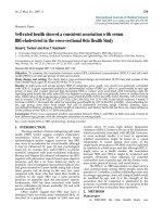
A positive correlation was seen between the degree of
airflow obstruction expressed as percent of predicted
FEV
1.0
and CFPE-index for both type I fibres (r = 0.61, p <
0.001) and type IIa fibres (r = 0.37, p = 0.04) (Fig 1) likely
indicating that each capillary is shared by more muscle
fibres when airflow obstruction increases.
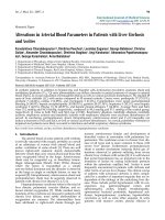
A positive correlation was observed between exercise
capacity, expressed as distance walked in six minutes, and
CFPE-index for both type I fibres (r = 0.67, p < 0.001) and
type IIa fibres (r = 0.40, p = 0.02) (Fig 2) indicating a par-
allel reduction in exercise capacity and muscle capillar-
ization. Exercise capacity, expressed as distance walked in
six minutes, was also found to correlate positively to PaO
2
(r = 0.57, p < 0.001) (Fig 3).
Discussion
Previous studies have suggested alterations in the capil-
lary bed of skeletal muscle in COPD patients. Extending
these findings the current study provides first evidence of
a disturbed muscle-to-capillary interface expressed as
CFPE-index in COPD. Additionally, we offer evidence for
a positive correlation between the degree of muscle capil-
larization, degree of airflow obstruction and exercise
capacity in COPD patients.
The presence of an adequate capillarization is essential
for maintenance of adequate oxygen supply required for
normal muscle function. Recently, we have demonstrated
an increased proportion of type IIa fibres and a decreased
proportion of type I fibres in the tibialis anterior muscle
of patients with COPD [10]. This is in line with previous
studies [3,7-9] and together with previous findings of a
decrease in oxidative enzyme activities in COPD [6,7] our
findings indicate the occurrence of a shift towards a more
glycolytic profile in the limb muscle of COPD patients.
Furthermore, the number of capillaries around a single
muscle fibre (CAF) was decreased in patients with COPD
compared to healthy subjects, indicating decreased mus-
cle capillarization in COPD [13]. However, CAF in rela-
Table 2: Fibre type distribution, fibre area, fibre perimeter, CAF and CAFA for type I and type IIa fibres.
Healthy subjects (n = 12) Mild
COPD
(n = 8)
Moderate COPD
(n = 9)
Severe
COPD
(n = 6)
P-value
Proportion type I fibres (%) 78.3 ± 8.7 70.2 ± 11.4 74.5 ± 11.9 59.4 ± 9.5* 0.009
Proportion type IIa fibres (%) 20.2 ± 8.9 24.6 ± 10.6 22.6 ± 11.4 40.2 ± 7.6* 0.02
Proportion type IIx fibres (%) 0 1.0 ± 1.6 0.6 ± 1.1 1.2 ± 2.1 ns
Proportion type IIx-a fibres (%) 0 0.2 ± 0.3 0.3 ± 0.6 0.5 ± 0.7 ns
Proportion type I-IIa fibres (%) 1.5 ± 1.4 4.0 ± 6.4 2.0 ± 2.3 1.6 ± 1.4 ns
Perimeter type I fibres 330 ± 38 332 ± 60 295 ± 40 320 ± 87 ns
Perimeter type IIa fibres 380 ± 48 348 ± 56 274 ± 53* 303 ± 81 0,003
Area type I fibres (μm
2
)
7143 ± 1508 7315 ± 2471 5736 ± 1497 6557 ± 2901 ns
Area type IIa fibres (μm
2
)
9262 ± 2215 7711 ± 2427 4880 ± 1970* 5418 ± 2232 0,004
CAF type I fibres (μm) 6,8 ± 1,1 6,8 ± 2,6 5,2 ± 0,9* 5,1 ± 1,2* 0,006
CAF type IIa fibres (μm) 6,7 ± 1,9 6,3 ± 2,4 4,4 ± 0,8* 4,5 ± 1,0* 0,002
CAFA type I fibres 1,0 ± 0,1 1,1 ± 0,4 1,0 ± 0,3 0,9 ± 0,2 ns
CAFA type IIa fibres 0,8 ± 0,3 1,0 ± 0,3 1,1 ± 0,3 1,0 ± 0,3 ns
Data are presented as mean ± SD.
*Significant difference compared to healthy subjects.
n = number of test subjects; CAF = number of capillaries around a single fibre; CAFA = the ratio between CAF and the area of the muscle fibre;
ns = not significant
Eliason et al. Respiratory Research 2010, 11:97
/>Page 5 of 7
tion to fibre area (CAFA) did not differ between healthy
subjects and patients with COPD [13]. Capillary parame-
ters essentially determining the diffusion distance (capil-
lary density or CAFA) may, thus, reflect the reduction in
fibre area in COPD patients [9,10] while disturbed capil-
larization may still be undetected. The current study has
investigated muscle to capillary interface in COPD
patients as this parameter is involved in oxygen supply to
the muscle and has been suggested to be a sensitive
marker for changes in the capillary network of limb mus-
cle [14-19]. Our results demonstrate a positive relation-
ship between the degree of airflow obstruction and
CFPE-index for both type I and type IIa fibres, indicating
that muscle capillarization decreases with increased dis-
ease severity. Furthermore, the CFPE-index for type I
fibres, but not for type IIa fibres, was significantly
reduced in patients with moderate and severe COPD
compared to healthy subjects. A larger capillary network
is associated with oxidative type I fibres compared to gly-
colytic type IIa fibres, which may explain why alterations
in CFPE-index are more evident in the type I fibres. Inter-
estingly, we found no differences in LC/PF between
COPD patients and healthy subjects. A main difference
between CFPE-index and LC/PF is that the calculation of
Table 3: Muscle-capillary interface parameters for type I and type IIa fibres.
Healthy
subjects
(n = 12)
Mild
COPD
(n = 8)
Moderate
COPD
(n = 9)
Severe
COPD
(n = 6)
p value
C:F
i
type I 2.7 ± 0.5 2.7 ± 1.3 2.0 ± 0.4* 2.0 ± 0.5* 0.007
C:F
i
type IIa 2.5 ± 0.5 2.5 ± 1.1 1.6 ± 0.4* 1.7 ± 0.4 0.002
CFPE type I 8.3 ± 1.1 8.1 ± 3.0 6.8 ± 0.9* 6.2 ± 0.6 * 0.002
CFPE type IIa 6.6 ± 0.9 7.3 ± 2.6 5.9 ± 0.8 5.8 ± 1.1 ns
LC type I 72.4 ± 8.4 77.0 ± 25.0 58.8 ± 14.6 67.4 ± 29.0 ns
LC type IIa 64.8 ± 19.5 64.7 ± 17.8 45.2 ± 10.6 57.4 ± 25.8* 0.03
LC/PF type I 21.8 ± 1.4 22.3 ± 5.6 20.0 ± 4.7 20.4 ±4.1 ns
LC/PF type IIa 17.5 ± 2.5 19.0 ± 4.1 16.3 ± 2.2 18.2 ± 2.5 ns
Data are presented as mean ± SD.
*Significantly lower compared to healthy subjects.
n = number of test subjects; C:F
i
= the sum of the fractional contributions of all capillary contacts around the fibre, i.e. the individual capillary-
to-fibre ratio; CFPE = quotient between C:F
i
and the fibre perimeter; LC = length of capillaries in contact with muscle fibre; LC/PF = ratio
between LC and fibre perimeter; ns = not significant.
Figure 1 Relationship between degree of airflow obstruction ex-
pressed as percent of predicted FEV
1.0
and CFPE-index for type I
and type IIa fibres; "black circle" = type I fibres ( = regression
line for type CFPE-index for type I fibres, r = 0.61, p < 0.001), "grey
square"= type IIa fibres ( = regression line for CFPE-index for
type IIa fibres, r = 0.37, p = 0.04); FEV
1,0
= forced expiratory vol-
ume in one second; CFPE-index = quotient between individual
capillary-to-fibre ratio and fibre perimeter.
Figure 2 Relationship between distance walked in six minutes
and CFPE-index for type I and type IIa fibres; "black circle" = type
I fibres ( = regression line for CFPE-index for type I fibres, r =
0.67, p < 0.001), "grey square"= type IIa fibres ( = regression line
for CFPE-index for type IIa fibres, r = 0.40, p = 0.02); CFPE-index =
quotient between individual capillary-to-fibre ratio and fibre pe-
rimeter.
Eliason et al. Respiratory Research 2010, 11:97
/>Page 6 of 7
CFPE-index relies on the measurement of the capillary-
to-fibre ratio (C:F
i
, i.e. the sum of the fractional contribu-
tions of all capillary contacts around the fibre). We sug-
gest that the C:F
i
is the capillary variable mainly affected
in COPD patients compared to healthy subjects i.e. each
capillary is shared by more muscle fibres. The specific
alterations of this variable may be due to the fact that it is
sensitive to alterations in the two-dimensional capillary-
fibre geometrical arrangement [19]. We speculate that
the presence of hypoxia in COPD leads to decreased oxy-
gen delivery to the muscle fibres which may lead to rear-
rangements of the capillary-fibre geometry. Still, the
mechanisms behind these rearrangements are not known
and further studies are needed to confirm these specula-
tions. However, as the CFPE-index is considered a sensi-
tive marker for changes in muscle-to-capillary interface
[16], we conclude that the interface is disturbed in the
tibialis anterior muscle of COPD patients. As it has previ-
ously been reported that muscle-to-capillary interface
measured as capillary-to-fibre surface ratio is regulated
as a function of the fibre mitochondrial volume per
length of fibre [26] another plausible explanation to our
findings may be a reduced mitochondrial volume of the
muscle fibre. This explanation would be in line with a
recent study showing a decrease in mitochondrial volume
in patients with COPD [12]. However, further studies on
the relationship between capillarization and mitochon-
drial volume density in COPD are needed to confirm this
hypothesis.
Recently, we have demonstrated that decreased exercise
capacity, as determined by the 6-min walking test, was
strongly correlated with increased severity of COPD (p >
0.001) [10]. Here, we extend these findings and show that
CFPE-index is also correlated to the degree of airflow
obstruction as determined by spirometry and to exercise
capacity determined by the 6-min walking test. In the
context of motor unit recruitment during different mus-
cular activities it is known that low intensity muscle activ-
ities mainly recruit low threshold slow, oxidative type I
fibres, while the high threshold more glycolytic type IIa
fibres are mainly recruited during high speed, high-force
generating muscle activities. As walking is considered a
low intensity activity the 6-min walking test recruits type
I fibres to a larger extent than type II fibres [27]. As dis-
turbance of the muscle-to-capillary interface is more pro-
nounced for type I fibres the correlation between exercise
capacity and CFPE-index is also stronger for type I fibres
than for type IIa fibres. This strongly suggests a contrib-
uting role for decreased muscle capillarization and subse-
quently impaired oxygen delivery in the development of
reduced exercise capacity in COPD. Indeed, a correlation
between reduced muscle oxygen supply and reduction in
exercise capacity in COPD has previously been suggested
[11]. However, the finding of a strong correlation between
exercise capacity and partial arterial pressure for oxygen
indicates that other factors such as lung disease also are
limiting factors for exercise capacity in COPD.
The mechanisms mediating decreased muscle capillar-
ization in COPD are unknown. However, it is known that
skeletal muscle adapts to physiological stimuli such as
exercise and environmental factors such as hypoxia by
changes in microvascularization [28,29]. As it has previ-
ously been suggested, one plausible explanation to the
decreased capillarization may be the lower physical activ-
ity levels in COPD patients [30]. Another explanation to
the findings in the present study may be the presence of
hypoxia in COPD. This hypothesis is strengthened by a
recent study where we have demonstrated an overexpres-
sion of the von Hippel-Lindau tumor suppressor protein
(pVHL) in the tibialis anterior muscle of patients with
COPD [13]. Increased pVHL may have an adverse effect
on tissue capillarization as it impairs transduction of
hypoxic-angiogenetic transcription factors including vas-
cular endothelial growth factor (VEGF) [13,31]. Indeed,
evidence for attenuation of VEGF gene expression during
long term exposure to hypoxia has previously been dem-
onstrated in skeletal muscle of rats [32]. Additionally, pre-
vious studies examining the effect of hypoxia on skeletal
muscle have suggested that short term exposure to
hypoxic conditions leads to an increase in capillaries/
muscle fibre which is explained by a reduction in muscle
fibre area and not by capillary neoformation [29,33,34].
During chronic exposure to hypoxia an actual reduction
in muscle capillarity has been reported [29,35]. Taken
together, these findings indicate that the presence of a
hypoxic state may account for decreased skeletal muscle
capillarization in COPD. However, further studies are
needed to confirm this theory.
In conclusion, the present study provides evidence of a
positive correlation between decreased muscle-to-capil-
Figure 3 Relationship between exercise capacity expressed as
distance walked in six minutes and partial oxygen pressure
(PaO
2
), r = 0.57, p < 0.001.
Eliason et al. Respiratory Research 2010, 11:97
/>Page 7 of 7
lary interface and increased disease severity in COPD.
Furthermore, a positive correlation is demonstrated
between decreased muscle-to-capillary interface and
decreased exercise capacity in patients with COPD. Exer-
cise is known to have a positive effect on muscle-to-capil-
lary interface [16], which highlights the need to develop
rehabilitation strategies to promote capillarization,
improve oxygen delivery and consequently improve exer-
cise capacity in COPD patients.
Competing interests
The authors declare that they have no competing interests.
Authors' contributions
GE carried out the exercise capacity test and the immunohistochemistry, par-
ticipated in the design of the study, performed the statistical analysis and
drafted the manuscript. SA-H participated in the design of the study as well as
patient recruitment. KP-A performed the muscle biopsy sampling and partici-
pated in the design and coordination of the study. FK conceived the study and
helped on the draft of the manuscript. All authors read and approved the final
manuscript.
Author Details
1
School of Medical Sciences, Örebro University, Örebro, Sweden,
2
Department
of Medical Sciences, Respiratory Medicine and Allergology, Uppsala University,
Uppsala, Sweden and
3
Department of Rheumatology, Danderyds hospital,
Stockholm, Sweden
References
1. Rabe KF, Hurd S, Anzueto A, Barnes PJ, Buist SA, Calverley P, Fukuchi Y,
Jenkins C, Rodriguez-Roisin R, van Weel C, Zielinski J: Global strategy for
the diagnosis, management, and prevention of chronic obstructive
pulmonary disease: GOLD executive summary. Am J Respir Crit Care Med
2007, 176:532-555.
2. Decramer M, De Benedetto F, Del Ponte A, Marinari S: Systemic effects of
COPD. Respir Med 2005, 99(Suppl B):S3-10.
3. Jagoe RT, Engelen MP: Muscle wasting and changes in muscle protein
metabolism in chronic obstructive pulmonary disease. Eur Respir J
Suppl 2003, 46:52s-63s.
4. Mador MJ, Bozkanat E: Skeletal muscle dysfunction in chronic
obstructive pulmonary disease. Respir Res 2001, 2:216-224.
5. Wouters EF: Nutrition and metabolism in COPD. Chest 2000,
117:274S-280S.
6. Maltais F, LeBlanc P, Whittom F, Simard C, Marquis K, Belanger M, Breton
MJ, Jobin J: Oxidative enzyme activities of the vastus lateralis muscle
and the functional status in patients with COPD. Thorax 2000,
55:848-853.
7. Gosker HR, van Mameren H, van Dijk PJ, Engelen MP, van der Vusse GJ,
Wouters EF, Schols AM: Skeletal muscle fibre-type shifting and
metabolic profile in patients with chronic obstructive pulmonary
disease. Eur Respir J 2002, 19:617-625.
8. Jobin J, Maltais F, Doyon JF, LeBlanc P, Simard PM, Simard AA, Simard C:
Chronic obstructive pulmonary disease: capillarity and fiber-type
characteristics of skeletal muscle. J Cardiopulm Rehabil 1998,
18:432-437.
9. Whittom F, Jobin J, Simard PM, Leblanc P, Simard C, Bernard S, Belleau R,
Maltais F: Histochemical and morphological characteristics of the
vastus lateralis muscle in patients with chronic obstructive pulmonary
disease. Med Sci Sports Exerc 1998, 30:1467-1474.
10. Eliason G, Abdel-Halim S, Kadi F, Piehl-Aulin K: Physical performance and
muscular characteristics in different stages of COPD. Scan J Med Sci
Sports 2008 in press.
11. Aliverti A, Macklem PT:
How and why exercise is impaired in COPD.
Respiration 2001, 68:229-239.
12. Picard M, Godin R, Sinnreich M, Baril J, Bourbeau J, Perrault H, Taivassalo T,
Burelle Y: The mitochondrial phenotype of peripheral muscle in chronic
obstructive pulmonary disease: disuse or dysfunction? Am J Respir Crit
Care Med 2008, 178:1040-1047.
13. Jatta K, Eliason G, Portela-Gomes GM, Grimelius LP, Caro O, Nilholm L,
Sirsjo A, Piehl-Aulin K, Abdel-Halim SM: Overexpression of von Hippel-
Lindau (VHL) in skeletal muscles of patients with chronic obstructive
pulmonary disease (COPD). J Clin Pathol 2009, 62:70-6.
14. Charles M, Charifi N, Verney J, Pichot V, Feasson L, Costes F, Denis C: Effect
of endurance training on muscle microvascular filtration capacity and
vascular bed morphometry in the elderly. Acta Physiol (Oxf) 2006,
187:399-406.
15. Hepple RT, Mathieu-Costello O: Estimating the size of the capillary-to-
fiber interface in skeletal muscle: a comparison of methods. J Appl
Physiol 2001, 91:2150-2156.
16. Charifi N, Kadi F, Feasson L, Costes F, Geyssant A, Denis C: Enhancement
of microvessel tortuosity in the vastus lateralis muscle of old men in
response to endurance training. J Physiol 2004, 554:559-569.
17. Gayeski TE, Honig CR: O2 gradients from sarcolemma to cell interior in
red muscle at maximal VO2. Am J Physiol 1986, 251:H789-799.
18. Honig CR, Connett RJ, Gayeski TE: O2 transport and its interaction with
metabolism; a systems view of aerobic capacity. Med Sci Sports Exerc
1992, 24:47-53.
19. Hepple RT: A new measurement of tissue capillarity: the capillary-to-
fibre perimeter exchange index. Can J Appl Physiol 1997, 22:11-22.
20. Mathieu-Costello O, Ellis CG, Potter RF, MacDonald IC, Groom AC: Muscle
capillary-to-fiber perimeter ratio: morphometry. Am J Physiol 1991,
261:H1617-1625.
21. Charifi N, Kadi F, Feasson L, Denis C: Effects of endurance training on
satellite cell frequency in skeletal muscle of old men. Muscle Nerve
2003, 28:87-92.
22. Louwerens JW, van Linge B, de Klerk LW, Mulder PG, Snijders CJ: Peroneus
longus and tibialis anterior muscle activity in the stance phase. A
quantified electromyographic study of 10 controls and 25 patients
with chronic ankle instability. Acta Orthop Scand 1995, 66:517-523.
23. Henriksson KG: "Semi-open" muscle biopsy technique. A simple
outpatient procedure. Acta Neurol Scand 1979, 59:317-323.
24. Kadi F, Hagg G, Hakansson R, Holmner S, Butler-Browne GS, Thornell LE:
Structural changes in male trapezius muscle with work-related
myalgia. Acta Neuropathol (Berl) 1998, 95:352-360.
25. Porter MM, Koolage CW, Lexell J: Biopsy sampling requirements for the
estimation of muscle capillarization. Muscle Nerve 2002, 26:546-548.
26. Mathieu-Costello O: Comparative aspects of muscle capillary supply.
Annu Rev Physiol 1993, 55:503-525.
27. Åstrand P-O RK, Dahl H, Stromme S: Textbook of Work physiology,
physiological bases of exercise fourth edition. Leeds: Human Kinetics; 2003.
28. Gustafsson T: Exercise and Angiogenic Growth Factors in Human
Skeletal Muscle. Karolinska Institutet; 2005.
29. Hoppeler H, Vogt M: Muscle tissue adaptations to hypoxia. J Exp Biol
2001, 204:3133-3139.
30. Pitta F, Troosters T, Spruit MA, Probst VS, Decramer M, Gosselink R:
Characteristics of physical activities in daily life in chronic obstructive
pulmonary disease. Am J Respir Crit Care Med 2005, 171:972-977.
31. Kondo K, Kaelin WG: The von Hippel-Lindau tumor suppressor gene.
Exp Cell Res 2001, 264:117-125.
32. Olfert IM, Breen EC, Mathieu-Costello O, Wagner PD: Chronic hypoxia
attenuates resting and exercise-induced VEGF, flt-1, and flk-1 mRNA
levels in skeletal muscle. J Appl Physiol 2001, 90:1532-1538.
33. Hoppeler H, Desplanches D: Muscle structural modifications in hypoxia.
Int J Sports Med 1992, 13(Suppl 1):S166-168.
34. Mathieu-Costello O: Muscle adaptation to altitude: tissue capillarity and
capacity for aerobic metabolism. High Alt Med Biol 2001, 2:413-425.
35. Desplanches D, Hoppeler H, Tuscher L, Mayet MH, Spielvogel H, Ferretti G,
Kayser B, Leuenberger M, Grunenfelder A, Favier R: Muscle tissue
adaptations of high-altitude natives to training in chronic hypoxia or
acute normoxia. J Appl Physiol 1996, 81:1946-1951.
doi: 10.1186/1465-9921-11-97
Cite this article as: Eliason et al., Alterations in the muscle-to-capillary inter-
face in patients with different degrees of chronic obstructive pulmonary dis-
ease Respiratory Research 2010, 11:97
Received: 18 September 2009 Accepted: 15 July 2010
Published: 15 July 2010
This article is available from: 2010 Eliason et al; licensee BioMed Central Ltd. This is an Open Access article distributed under the terms of the Creative Commons Attribution License ( ), which permits unrestricted use, distribution, and reproduction in any medium, provided the original work is properly cited.Respiratory Research 2010, 11:97